Article

Characterization and Biotechnological Potential of Two Native Marine Microalgae Isolated from the Tunisian Coast

1
Laboratoire de Biotechnologies des Plantes Appliquées à l’Amélioration des Cultures, Faculté des Sciences, Université de Sfax, Sfax 3038, Tunisia
2
Laboratoire de Génie Enzymatique et Microbiologie, Equipe de Biotechnologie des Algues, Ecole Nationale d’Ingénieurs de Sfax, Université de Sfax, Sfax 3038, Tunisia
3
Department of Cell Biology and Histology, University of Murcia, 30100 Murcia, Spain
4
CNRS, SIGMA Clermont, Institut Pascal, Université Clermont Auvergne, F-63000 Clermont-Ferrand, France
*
Authors to whom correspondence should be addressed.
Submission received: 26 April 2021 / Revised: 31 May 2021 / Accepted: 1 June 2021 / Published: 7 June 2021
(This article belongs to the Special Issue Underutilized Natural Sources in Food and Dietary Supplements)

Abstract

:

Featured Application

Newly isolated microalgae strains with potential applications in different fields, especially fish feeds and wastewater bioremediation.

Abstract

Microalgae are attracting considerable interest worldwide. In the present study, two native microalgae strains isolated from Tunisian cost were identified as Chlamydomonas sp. and Navicula sp. We characterized their pigment and protein contents, as well as their carbohydrate and lipid productivity. The predominant fatty acids were found to be α-linolenic acid (C18:3n-3) and palmitoleic acid (C16:1n-7) for Chlamydomonas sp. and Navicula sp. strains respectively. Microalgae methanol extracts showed important in vitro antibacterial activity against all tested gram negative bacteria. Antioxidant activities of methanol extracts were investigated by determining radical scavenging activity according to DPPH (2,2-diphenyl-1-picrylhydrazyl) and ABTS (2,2′-azino-bis(3-ethylbenzothiazoline-6-sulfonic acid)) methods. MTT (3-[4,5-dimethylthiazol-2-yl]-2,5 diphenyl tetrazolium bromide) assay showed that the newly isolated microalgae were not toxic and have potential application in the fish feeds domain as a supplement in fish meal. Also, the biosorption of cadmium from aqueous solutions by microalgae living cells was evaluated. Large removal capacities were obtained with values ranging from 89.8% to 99.38%. These results are very promising as a starting point for a potential application of these new isolates for in situ bioremediation of heavy metals contaminating aqueous systems.

1. Introduction

Microalgae comprise a vast group of both marine and freshwater habitats organisms. Based on DNA sequence data, they have been classified into ten major phyta which are Glaucophyta, Euglenophyta, Cryptophyta, Haptophyta, Dinophyta, Heterocontophyta (including diatoms), Rhodophyta, Chlorophyta, and the prokaryotic Cyanophyta (cyanobacteria) [1]. In recent years, a focus has been shifted towards these organisms due to their diverse metabolic contents. Indeed, they synthesize a large number of bioactive compounds, including pigments, sterols, polyphenols, fatty acids, proteins, vitamins, alkaloids, and sulfated polysaccharides with various chemical structures and biological activities [2,3]. These biomolecules are useful for microalgal-based feed and food, nutraceutical, pharmaceutical, and cosmetic industries through their antioxidant, antifungal, antibacterial, antiviral, antienzymatic, anti-cancer, or anti-inflammatory activities [4,5,6,7]. Several studies have reported the therapeutic properties of Spirulina and Nostoc species and their ability to scavenge superoxide and hydroxyl radicals and inhibit lipid peroxidation [8,9]. Besides, due to their rich nutritional properties, some Chlorophycae and diatoms genera have been widely used as dietary supplements for both humans and animals [10,11].
Nowadays, there is a focus on using microalgae in renewable energy sources as well as environmental applications. Several studies reported the conversion of microalgae biomass into biofuels [12,13]. Also, microalgae environmental applications are mainly represented in CO2 sequestration and wastewater treatment [14]. Recent studies have investigated microalgae capacities for the final polishing of municipal wastewater, or the direct treatment of a variety of industrial wastewaters [14,15]. They demonstrate that several microalgae species can uptake heavy metals with high efficiency [16]. Moreover, multiple metal-biosorption mechanisms have been reported that differ according to the species used, biomass kind, and adopted procedure [15]. These include ion exchange, chelation, complexation, and microprecipitation [16]. The goal of the present work was to isolate, identify, and characterize native microalgae strains from Tunisian coasts to study their biotechnological potentialities for future industrial-scale valorization in biotechnological fields.

2. Materials and Methods

2.1. Isolation of Axenic Uniclonal Cultures

Seawater samples were collected from the Tunisian Coast of Ksour-Essef (governorate of Mahdia) (Latitude: 35°23′32.85′′ N; Longitude: 11°2′57.1704′′ E) in February 2018. They were pre-filtered through a 60 μm pore size membrane then the flow-through fractions were transferred into sterile flasks containing f/2-medium [17]. Inoculated cultures were grown in continuous aeration and illumination of 2000 lux, 25 ± 1 °C until visible growth appeared in flasks. Afterwards, an aliquot of each flask was spread over a sterilized agar plate, incubated at 28 °C and continuously illuminated at a light intensity of 2000 lux until visible growth appeared in the plate. A single colony was transferred into sterile liquid f/2-medium aseptically. After 72 h, cultures were examined using inverted microscopy (Motic microscope AE2000, Barcelona, Spain) at 40× magnification. The pure cultures were obtained by performing the micromanipulation method after serial dilutions [18]. Cultures were maintained in the f/2 medium, at 25 ± 1 °C with continuous aeration and an illumination of 2000 lux.

2.2. DNA Extraction, PCR Amplification, Sequencing and Phylogenetic Analysis

The genomic DNA was extracted using phenol/chloroform extraction standard method followed by ethanol precipitation [19]. The 18S rRNA encoding gene of each isolated strain was amplified by PCR using the EukA—EukB primer pair [20] according to the procedure reported by Ben Amor et al. [21]. The PCR products obtained were purified from agarose gel using MiniElute Gel Extraction Kit (Qiagen S. A. Courtaboeuf, France) by following the instruction manual. The purified products were sequenced by Sanger sequencing using 3500 Series Genetic Analyzer by Thermo Fisher Scientific, USA. The sequences obtained were compared with the sequences available in GenBank using the BLAST server from the NCBI website (www.ncbi.nlm.nih.gov/BLAST, 26 June 2020).

2.3. Pigments Contents

Pigments contents were determined spectrophotometrically, in ethanol extracts, as previously detailed by Fazeli et al. [22]. Briefly, the cell density of microalgae cultures were adjusted to 5 × 10⁹ cell/L, then 2 mL of each culture were centrifuged at 5000× g for 10 min. The pellet was suspended in ethanol 100% and sonicated at 65 °C for 30 min. After sonication, the solution was centrifuged at 5,000× g for 10 min then the absorbance was measured at 666, 653, and 470 nm using a UV/VIS spectrophotometer (Pharo 300; Merck, NJ, USA). Chlorophyll a, Chlorophyll b, and carotenoids amount were calculated according to the formula reported by Lichtenthaler [23]. Pigments contents were expressed in mg/L of microalgae culture. All assays were set up in triplicates.

2.4. Proteins Contents

Freeze-dried samples (50 mg) were incubated at 4 °C for 12 h with 4 mL ultra-pure water then ground with a mortar and pestle. Homogenates were collected and centrifuged at 15,000× g for 20 min at 4 °C to recover the supernatants, which were transferred to fresh centrifuge tubes and kept at 4 °C. The pellets were re-extracted with 1 mL of NaOH (0.1 M) under the same conditions. Collected supernatants from both extractions were mixed and proteins were precipitated using cold trichloroacetic acid as detailed by Barbarino and Lourenço [24]. Precipitated proteins were suspended in 0.5 mL NaOH (1 M) and quantified with the Bradford method [25]. Absorbance was recorded at 595 nm using a UV/VIS spectrophotometer (Pharo 300; Merck, NJ, USA). Samples were calibrated against the bovine serum albumin (Sigma) curve. Assays were made in triplicate.

2.5. Carbohydrates Contents

The carbohydrate content (poly- and oligo-) of the studied microalgae was assessed by the phenol sulfuric-acid colorimetric method described by Dubois et al. [26]. In brief, fresh microalgae biomass was hydrolyzed using 1 mL phenol 5% (w/v) and 5 mL concentrate sulfuric-acid. The mixture was vortexed and incubated for 10 min at room temperature. The absorbance was measured at 490 nm with a UV/VIS spectrophotometer (Pharo 300; Merck, NJ, USA). The concentration of carbohydrates was determined based on the dextrose standard curve. Three replicates were made for each assay.

2.6. Lipids Contents and Fatty Acid Profiles

Total lipids were prepared according to the method reported by Fendri et al. [27]. Briefly, microalgae fresh biomass (2 g) was homogenized with 20 mL of chloroform/methanol (2:1, v/v) using a tissue disrupter (IKA ULTRA-TURRAX T 25 digital; IKA-WERKE, Staufen, Germany). Non-lipid impurities were removed by washing with KCl (0.88%, w/v). Lipid weight was determined gravimetrically after evaporating the solvent under a stream of nitrogen and overnight vacuum drying. Fatty acid methyl esters (FAME) were prepared by acid-catalyzed trans-esterification of total lipids using 2 mL of 1% sulfuric acid (v/v) in methanol, at 50 °C for 24 h. Methyl esters were extracted twice in 5 mL of hexane–diethyl ether (1:1, v/v) after neutralization with 2 mL of 2% KHCO3, dried under a stream of nitrogen, then dissolved in 1 mL of iso-hexane. The obtained FAME were analyzed through gas-liquid chromatography in an SPTM 2560 flexible fused silica capillary column (length 100 mm, internal diameter 0.25 mm, film thickness of 0.20 mm SUPELCO) in a Hewlett–Packard 5890 gas chromatograph. The oven temperature program was set according to the following. The initial temperature was 140 °C, then it increased at a rate of 3 °C/min to 230 °C, followed by 2 °C/min to reach 240 °C and then it was held there for 12 min. The injector and flame ionization detector were set at 260 °C. Helium was used as the carrier gas at a column pressure of 300 kPa. Peaks were identified by comparing their retention times with appropriate FAME standards from the Sigma Chemical Company (St. Louis, MO, USA). Data for the individual components are expressed as a percentage of total content. Each extraction experiment was performed in triplicate, and each extract was independently analyzed three times.

2.7. Determination of Phenols Contents

Phenolic content was determined according to Folin–Ciocalteu method using a gallic acid standard (10–200 mg/mL). The absorbance was measured at 720 nm [28,29]. Phenols contents were expressed in mg/mL of microalgae culture using an initial cell density of 5 × 10⁹ cell/L. All measurements were performed as independent triplicates.

2.8. Crude Extracts Preparation

Microalgae biomass (1 g) was collected from 15-day-old cultures by centrifugation at 5000× g for 15 min. Then, it was extracted twice using 15 mL methanol, with shaking for 20 min at the room temperature. The obtained extracts were dried in a rotary evaporator at 40 °C and under reduced pressure, then stored at −20 °C for further studies.

2.9. DPPH Free-Radical Scavenging Assay

The 2, 2 diphenyl-1-picrylhydrazyl (DPPH) tests were performed as previously detailed by Yan et al. [30]. An aliquot (1 mL) of algal extracts dissolved in dimethylsulfoxide (DMSO) (100 µg/mL) was mixed with DPPH (0.02%, w/v) in methanol. After incubation for 1 h in the dark at 25 °C, the absorbance was measured at 517 nm using a UV/VIS spectrophotometer (Pharo 300; Merck, NJ, USA). Ascorbic acid was used as positive controls. The percentage of inhibition was calculated according to the following formula:
% DPPH radical scavenging = [(𝐴control − 𝐴test)/𝐴control] × 100
where 𝐴control  =  Absorbance of DPPH and 𝐴test  =  Absorbance of sample (extract/ascorbic acid). All tests were carried out in triplicates.

2.10. ABTS Radical Scavenging Assay

ABTS tests were carried out according to the method described by Arnao et al. [31]. Briefly, 50 μL of microalgae extracts dissolved in DMSO (100 µg/mL) were added to 950 μL of cation ABTS+ and the absorbance was measured at 734 nm using a UV/VIS spectrophotometer (Pharo 300; Merck, NJ, USA). Ascorbic acid was used as positive control. The antioxidative activity was calculated using the formula reported by Re et al. [32].
%ABTS radical scavenging = [(𝐴control − 𝐴test)/𝐴control] × 100
where 𝐴control = absorbance of ABTS and 𝐴test = absorbance of sample (extract/ascorbic acid).
The ascorbic acid equivalent was computed by extrapolating the results with the standard pattern of decay using four different concentrations of ascorbic acid [33]. All analyses were performed in triplicates.

2.11. Antioxidant Enzyme Assays

The catalase activity was determined following the method proposed by Aebi [34]. The assay mixture consisted of 30 mM 30% H2O2 and 10 µL microalgae enzyme extract. Absorbance was immediately determined at 240 nm using a UV/VIS spectrophotometer (Pharo 300; Merck, NJ, USA). The rate of H2O2 decomposition is directly proportional to the catalase activity. One unit of catalase activity is defined as the amount of enzyme required to decompose 1 µM H2O2. The enzyme activity was expressed as U/mg protein.
Peroxidase activity was assayed according to established protocol [35]. Briefly, the reaction mixture consisted of 100 µL guaiacol (34 mM), 100 µL H2O2 (100 mM) and 100 µL of microalgae enzyme extract in phosphate buffer (100 mM, pH 7). The rate of formation of oxidized guaiacol was followed spectrophotometrically at 470 nm using a UV/VIS spectrophotometer (Pharo 300; Merck, NJ, USA). Enzyme activity was calculated using the extinction coefficient of 26.6 mM−1 cm−1. All measurements were made in triplicates.

2.12. Antibacterial Activities

The Gram-negative bacteria Vibrio harveyi, Vibrio Anguillarum, Photobacterium damselae, Aeromonas salmonicida, Shewanella putrefaciens (Pdp11), and Tenacibaculum mesophyllum were used as test organisms [36]. Antibacterial tests were performed in 96-well-flat-bottomed plates (Nunc, Thermo Fisher Scientific, United States), following the method of Stevens et al. [37] with slight modification. 20 µL of test extracts were added to 20 µL of the previously cultured bacteria (log phase) adjusted to 108 c.f.u/mL. After incubation for 5 h at 25 °C, 25 µL of MTT (1 mg/mL) were added to each well, and incubated for 10 min at 25 °C to allow the formation of formazan. Then, the plates were centrifuged at 2000× g for 10 min. The supernatants were removed and the precipitates were dissolved in 200 µL DMSO. Dissolved formazan absorbance was measured at 570 nm. Bacteria plus sterile PBS or methanol served as negative controls. The antibacterial activities were expressed as a percentage of non-viable bacteria, calculated as the difference between absorbance of surviving bacteria compared to the absorbance of bacteria from positive control (100%). Control and treatments were performed in triplicate.

2.13. Cytotoxicity and Antitumor Assay

Cytotoxicity effects of microalgae methanol extracts were evaluated against SAF-1 (ECACC nº 00122301) and PLHC1 (ATCC® CRL2406™) cell lines, using the standard MTT method, as previously described by Espinosa Ruiz et al. [38]. The tested concentration of each microalgae extract was 0.1 mg/mL. Cells incubated with medium with methanol served as a negative control. All tests were performed in five replicates.

2.14. Cadmium Removal Capacity

The capacity of both studied strains to remove metal was determined in batch assays, by culturing microalgal biomass (0.02 g/L) in f/2 medium, containing cadmium at initial concentrations of 0.5, 2.5, and 5 mg/L, under the previously mentioned conditions. After incubation for 5 days and 10 days, culture samples were collected, centrifuged, and residual cadmium concentration (in the supernatant) was determined via atomic absorption spectrophotometry (BMG LABTECH) according to the method described by Travieso et al. [39]. Then, percentages of removed cadmium were calculated. Experiments were run in triplicate. Blank controls (containing f/2 medium plus cadmium) and negative controls (containing f/2 medium and microalgae biomass) were also carried out.

2.15. Statistical Analysis

Statistical differences between antioxidant assay results were performed by Student’s t-test, n = 3, using SPSS 17.0 software (SPSS, Inc., Chicago, IL, USA). One-way ANOVA followed by Duncan multiple range tests were used to compare antibacterial activities, cytotoxicity assays, and cadmium removal capacities results. Data were considered significant when at least p was < 0.05.

3. Results

3.1. Isolation and Identification of the Microalgae Strains

Sea water samples were taken from Ksour-Essef (governorate of Mahdia) and two microalgae strains were isolated (MAa1 and MAn1). Different selective nutrient media were used; f/2 medium provided the optimal culturing conditions. Using the EukA and EukB universal primer pairs, the amplified products of the 18S rDNA genomic region, of approximately 1.8 kb for both isolated microalgae strains were sequenced. Based on sequence comparison against DNA sequences already available in GenBank database, the isolated strains, MAa1 and MAn1, were found to be affiliated to Chlamydomonas and Navicula genus, respectively, with more than 98% similarity. These results demonstrated that MAa1 and MAn1 belong to the Chlorophyceae and Bacillariophyceae classes, respectively.

3.2. Biochemical Composition

The biochemical composition of each native microalgae isolate was evaluated. The whole algal biomass compositions of newly isolated strains, MAa1 and MAn1, were summarized in Table 1.
Proteins and carbohydrates were the most abundant compounds for both studied strains, which could be of interest for several biotechnological applications. As shown in Table 1, Chlamydomonas sp. MAa1 strain accumulated a significantly higher quantity of proteins (47.22%), lipids (25.55%), chlorophyll (1.5 mg/L), and phenolic compounds (40.5 mg/mL) than Navicula sp. MAn1 strain. Besides, an important amount of carbohydrates (50.83%) has been found in the algal biomass of Navicula sp. MAn1 strain.
Fatty acid profiles were determined for each isolate via fatty acid methyl esters analysis (Table 2).
Eighteen and twenty fatty acids have been identified in the present study for Chlamydomonas sp. MAa1 and Navicula sp. MAn1 isolates, respectively. Chlamydomonas sp. MAa1 strain, showed the highest portion of polyunsaturated fatty acids (PUFAs) 44.35% with 7.92% of n-6 PUFA and 36.43% of n-3 PUFA. Regarding MAn1 isolate, belonging to Navicula genus, MUFAs was the major portion of the total fatty acids (50.88%) with a dominance of palmitoleic acid (C16:1n-7) (43.48%).

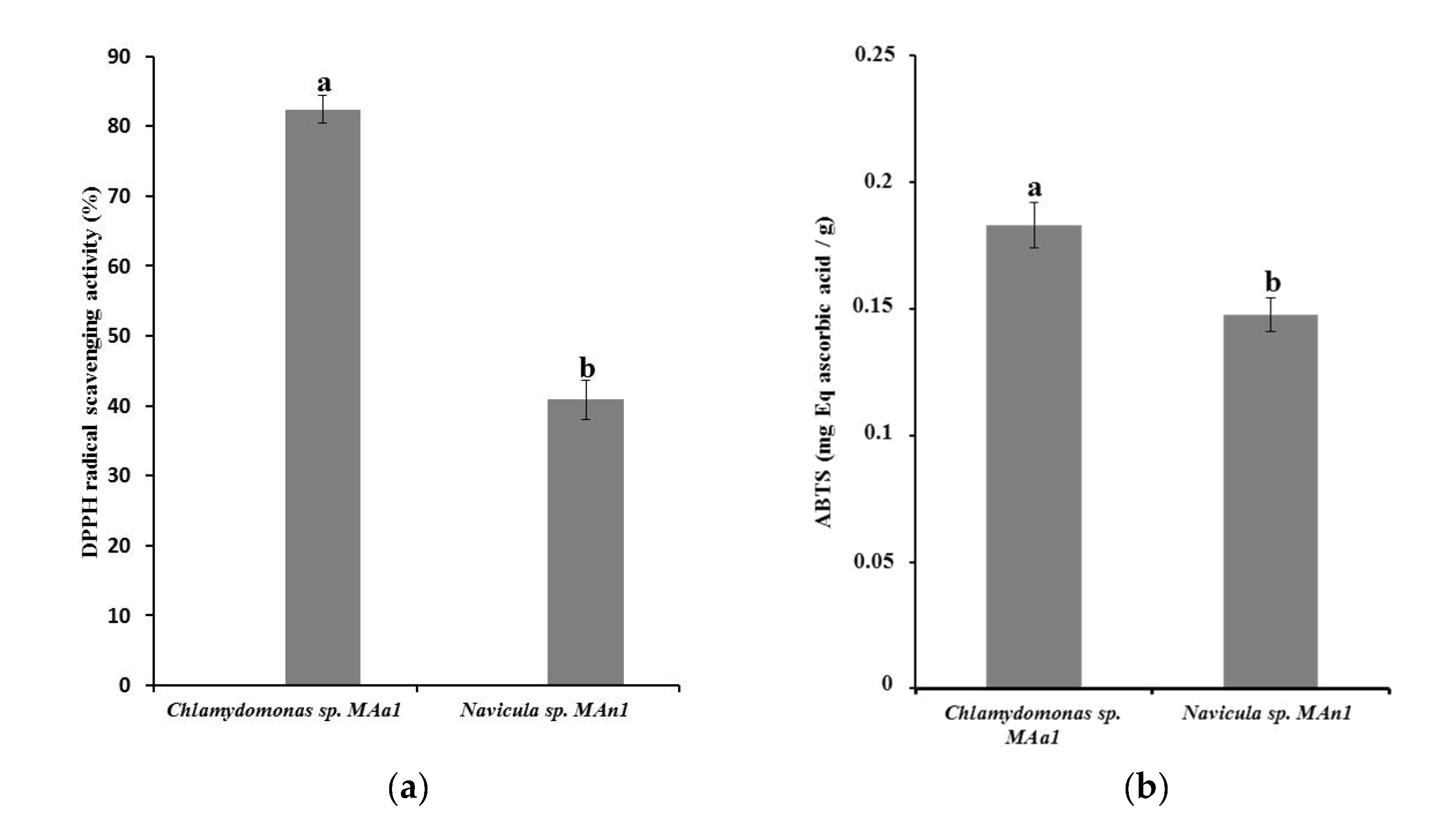
3.3. Antioxidant Activity

Antioxidant activities of Chlamydomonas sp. MAa1 and Navicula sp. Man1 isolates methanol extracts were performed by two spectrophotometric methods: the 2,2-di(4-tert-octylphenyl)-1-picrylhydrazyl (DPPH) assay and the 2,2′-azinobis-(3-ethylbenzothiazoline-6-sulfonic acid (ABTS) assay (Figure 1a,b).
The results obtained showed that the green microalgae isolate Chlamydomonas sp. MAa1 exhibited a higher antioxidant activity compared to Navicula sp. Man1 strain using both tests.
The enzymatic antioxidants from Chlamydomonas sp. MAa1 and Navicula sp. Man1 isolates were investigated (Figure 2).
The obtained peroxidase activities were 1.0625 U/mg of proteins and 0.8 U/mg of proteins for Chlamydomonas sp. MAa1 and Navicula sp. MAn1 isolates, respectively. Also, 4101.27 U/mg of protein of catalase activity was recorded for Chlamydomonas sp. MAa1 and 3281.01 U/mg of protein for Navicula sp. MAn1.

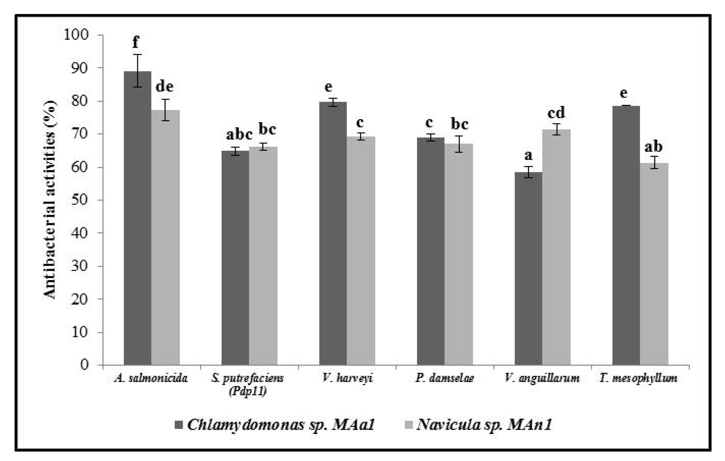
3.4. Antibacterial Activity

Antimicrobial activities of methanol extracts from both isolates were determined by MTT method against six Gram negative pathogenic bacteria strains for fish aquaculture: V. harveyi, V. anguillarum, P. damselae, A. salmonicida, S. putrefaciens (Pdp11), and T. mesophyllum (Figure 3).
Methanol extracts of Chlamydomonas sp. MAa1 and Navicula sp. MAn1 exhibited interesting antibacterial activities against tested pathogenic bacteria strains. The obtained inhibition percentages were greater than 60% against all tested Gram negative pathogenic bacteria. In fact, methanol extracts of both microalgae isolates showed comparable antibacterial activities against P. damselae and S. putrefaciens (Pdp11) with inhibition percentages of around 65%. Navicula sp. MAn1 extract was the most active against V. anguillarum. Interestingly, inhibition percentages greater than 80% were obtained against V. harveyi, A. salmonicida, and T. mesophyllum using Chlamydomonas sp. MAa1 extract.

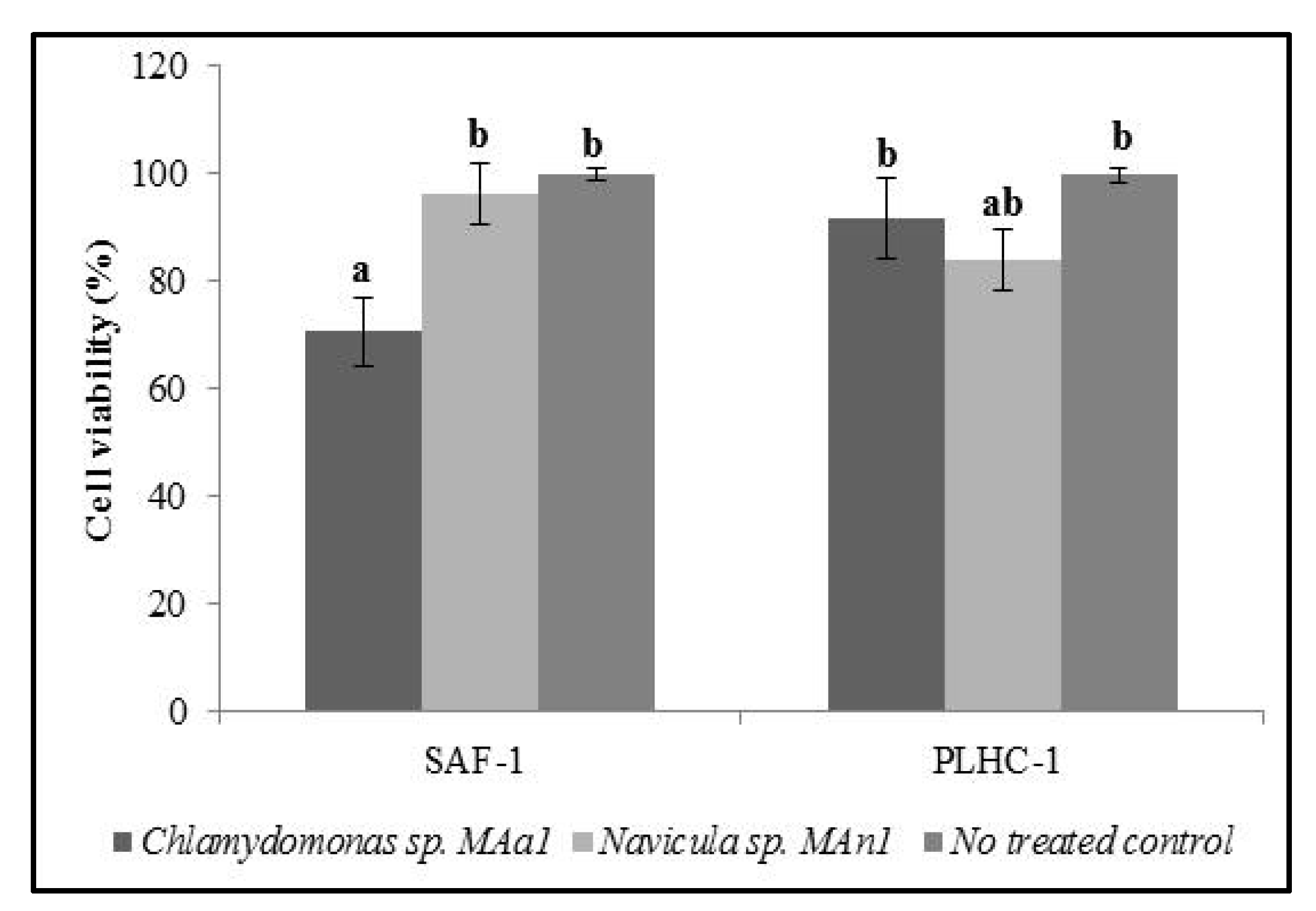
3.5. Cytotoxic Assay

Cytotoxic effect Chlamydomonas sp. MAa1 and Navicula sp. MAn1 methanol extracts were tested against SAF-1 and PLHC-1 cells cultures using MTT colorimetric assay (Figure 4).
The obtained results showed that Navicula sp. MAn1 extract has no significant effect on cell viability of SAF-1 tested cell lines (96%). However, it caused a slight decrease in cell viability (83%) of PLHC-1 tumor cell lines. Regarding Chlamydomonas sp. MAa1 extract, the obtained cell viabilities were 70.4% and 91.4% using SAF-1 and PLHC-1 cell lines, respectively.

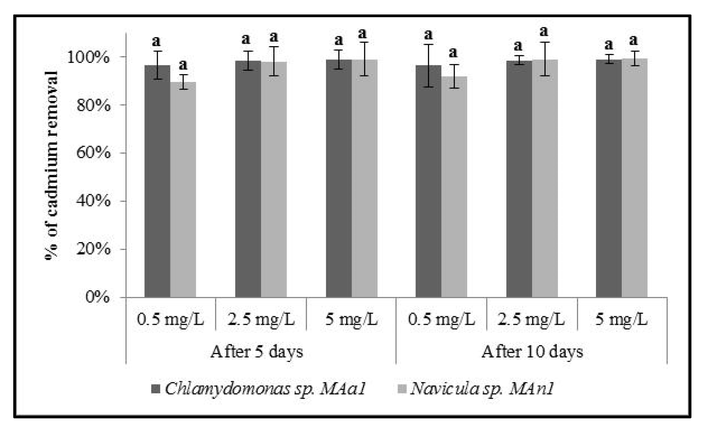
3.6. Capacity of Microalgae to Remove Heavy Metals

The capacity of Chlamydomonas sp. MAa1 and Navicula sp. MAn1 strains to remove cadmium were tested (Figure 5).
Both tested strains exhibited high removal efficiencies of cadmium from water at all tested concentrations. In fact, the obtained percentages of removed cadmium vary between 89.8% and 99.38%.

4. Discussion

The bio-prospecting of microalgae from local biotopes has been considered an efficient alternative to identify species of different genera that exhibit a wide of biotechnological activities [40,41]. In this context, two microalgae strains, MAa1 and MAn1, were isolated from local marine habitat and identified as microalgae belonging to Chlamydomonas and Navicula genera of Chlamydomonadaceae and Naviculaceae families using the nuclear 18S rDNA gene as a universal DNA barcode marker [42]. Microalgae biomasses were characterized and a genus-dependent biochemical composition was observed, as previously reported [43]. The obtained composition of the green microalgae isolate Chlamydomonas sp. MAa1 was in agreement with that reported by Behl et al. [44], with a remarkable abundance of proteins of about 47.22%, while Navicula sp. MAn1 strain biomass was richer in organic compounds especially carbohydrates (50.83%) than previously studied Navicula isolates [45,46]. The growth pattern and biochemical composition of microalga cells are closely related to biotic and abiotic culture parameters such as cultivation conditions and nutrient media composition and concentrations [44].
Both newly isolated strains exhibited moderate lipid contents. However, they were found to exhibit important proportions of mono-unsaturated and polyunsaturated fatty acids; 69.65% and 82.02% for Chlamydomonas sp. MAa1 and Navicula sp. MAn1, respectively. Fatty acid profiles have been considered as chemotaxonomical characteristics, since a comparatively higher homology of fatty acid profiles is always found within the strains with closer phylogenic relationships [47]. Chlamydomonas sp. MAa1 strain is particularly rich in monounsaturated especially oleic acid (C18:1n-9), vaccenic acid (C18:1n-7), and polyunsaturated α-linolenic acid (C18:3n-3) accounting for approximately 12.5%, 10.5%, and 30.08% of the total Chlamydomonas sp. MAa1 oil, respectively. The main abundant fatty acid in Navicula sp. MAn1 isolate is palmitoleic acid (C16:1n-7) representing 43.48% of the total fatty acids. The obtained results agree with multiple previously-published reports showing that almost all diatoms contained high proportions of (C16: 0) and (C16: 1n-7) fatty acids [48]. Unsaturated fatty acid production should be of great importance in different fields such as special human or animal dietary, nutraceuticals, and/or cosmeceuticals given the functional properties of PUFAs and their health benefits [44]. In fact, PUFAs, especially n-3 PUFAs, have many potential applications including disease prevention and treatment [49].
Under oxidizing conditions, microalgae cells produce various antioxidants [50]. There are several reports on the evaluation of the antioxidant activity of many genera belonging to Cyanobacteria [51,52], Chlorophycae [53,54,55] and some diatoms [56]. The antioxidant and antibacterial properties investigation of newly isolated strains showed high potential of in vitro activities. In fact, methanolic extracts from both studied strains exhibited interesting antibacterial activities against six Gram negative pathogenic bacteria strains. These results seem to be in perfect agreement with the data of biochemical compositions since a significant relationship between total antioxidant activity and total phenolic compounds has been reported [57,58]. It is worth noting that microalgae methanolic extracts seem to be particularly rich in flavonoids, and that terpenes and carbohydrate were present in moderate amount and alkaloids were also found at trace levels in [59,60]. Previously, a correlation between metabolites like flavonoids, terpenes and carbohydrates presence and antibacterial activities was established [59,60].
Additionally, important antimicrobial activities of both microalgae extracts were obtained against tested Gram- bacteria pathogen for vertebrates and invertebrate marine animals, especially fish, bivalves, and crustaceans. Those activities could be due to the presence of bioactive compounds that may include isoprenoids, polyketides, polyunsaturated fatty acids, and alkaloids which may affect the growth and metabolism of bacteria [61,62]. It has been reported that antibacterial activities of microalgae extracts, against both Gram+ and Gram- bacteria, were attributed to eicosapentaenoic acid and palmitoleic acid, particularly produced by diatoms [63,64]. Interestingly, no antibacterial activities of Chlamydomonas or Navicula species against aquatic animal pathogen have been previously reported. However, antibacterial activities were described for Chlamydomonas reinhardtii and Navicula sp. against human pathogenic bacteria such us Staphylococcus aureus, Escherichia coli, Salmonella typhi, and Bacillus sp. [65,66]. Darah and Lim [67] reported the mechanisms of action involved in the bacterial killing process. Among them are the interactions of the antimicrobial compounds with the cell membrane and intracellular accumulation through the cell wall and membrane permeability [68].
To ensure algae biomass safety for potential feed supplement applications, the toxicity of microalgae extracts was studied against fish cell lines. Hepatocytes, such as PLHC1 cells, are good models for studying toxicity since the liver is the primary site for drug metabolism and biotransformation [69]. According to Abdillahi et al. [70], treated concentrations of Chlamydomonas sp. MAa1 and Navicula sp. MAn1 methanol extracts are considered non-toxic since the cell viability is greater than 70%. In recent years, many studies have reported the in vivo immunostimulant effect of different microalgae such as Nannochloropsis gaditana, Tetraselmis chuii, and Phaeodactylum tricornutum on fish production [71,72].
Microalgae have been gaining attention in the treatment of wastewater, especially those contaminated by heavy metal. Interestingly, the newly isolated strains showed large cadmium removal capacities, with values ranging from 89.8% to 99.38%. Previously, rapid cadmium bioremoval by diatom cells has been observed in several other studies using cadmium concentration lower than that tested in the present study. In fact, Navicula incerta showed linear removal of cadmium using initial ion metal concentrations of 5 mg/L [73]. Cherifi et al. [74] also reported a cadmium removal capacity of Navicula subminuscula in aqueous solution containing 8 mg/L cadmium.
Chlamydomonas genus has a great potential in metal remediation [75]. Mera et al. [76] reported that Chlamydomonas moewusii remove up to 8 mg/L of cadmium. Also, Chlamydomonas reinhardtii remove up to 42.6 mg/L 67.4 mg/L of cadmium using fresh cells and lyophilized biomass, respectively [77,78]. Several other microalgae have shown an important ability to remove cadmium ions, especially Spirulina platensis and Chlorella vulgaris [79,80,81]. It is worth noting that the initial concentration of metal ions, contact time, pH, temperature, biosorbent concentration, and agitation rate are key factors affecting biosorption efficiency [82,83].

5. Conclusions

Two microalgae strains MAa1 and MAn1 were isolated from seawater of Tunisia costs and molecularly identified as Chlamydomonas sp. and Navicula sp., respectively. Both strains were initially characterized and exhibited significant heterogeneity in key parameters, such as carbohydrate productivity, lipid productivity, and fatty acids composition. Investigation of their biological activities showed that the newly isolated strains exhibited interesting antioxidant and antibacterial activities, particularly Chlamydomonas sp. MAa1. Chlamydomonas sp. MAa1, and Navicula sp. MAn1 biomass safety was verified against fish cell lines which support their potential application in the fish feeds domain. Furthermore, both isolated strains were capable of removing cadmium from aqueous solution, opening the horizon towards their application in the wastewater treatment domain.

Author Contributions

Conceptualization, J.E., P.M., S.A., I.F.: methodology, J.B.M., I.F., M.Á.E.; Formal analysis, J.B.M., J.E., M.D., Supervision, I.F.; Investigation, J.B.M., M.D.; I.F., J.E. Writing original draft, J.B.M., I.F.; writing review and editing, J.B.M., S.A., I.F., M.Á.E., J.E., P.M. All authors have read and agreed to the published version of the manuscript.

Funding

This research was funded by the TUNISIAN MINISTRY OF HIGHER EDUCATION AND SCIENTIFIC RESEARCH. The funders had no role in study design, data collection and analysis, decision to publish and preparation of the manuscript.

Institutional Review Board Statement

Not applicable.

Informed Consent Statement

Not applicable.

Acknowledgments

This work was supported by grants from the Tunisian Ministry of Higher Education and Scientific Research.

Conflicts of Interest

The authors declare no conflict of interest.

References

  1. Groendahl, S.; Kahlert, M.; Fink, P. The best of both worlds: A combined approach for analysing microalgal diversity via metabar coding and morphology-based methods. PLoS ONE 2017, 12, e0172808. [Google Scholar] [CrossRef] [PubMed]
  2. Dammak, M.; Haase, S.M.; Miladi, R.; Ben Amor, F.; Barkallah, M.; Gosset, D.; Pichon, C.; Huchzermeyer, B.; Fendri, I.; Denis, M.; et al. Enhanced lipid and biomass production by a newly isolated and identified marine microalga. Lipids Health Dis. 2016, 15, 209. [Google Scholar] [CrossRef] [Green Version]
  3. Nehmé, R.; Atieh, C.; Fayad, S.; Claude, B.; Chartier, A.; Tannoury, M.; Elleuch, F.; Abdelkafi, S.; Pichon, C.; Morin, P. Microalgae amino acid extraction and analysis at nanomolar level using electroporation and capillary electrophoresis with laser-induced fluorescence detection. J. Sep. Sci. 2016, 40, 558–566. [Google Scholar] [CrossRef] [PubMed]
  4. Ben Hlima, H.; Bohli, T.; Kraiem, M.; Ouederni, A.; Mellouli, L.; Michaud, P.; Abdelkafi, S.; Smaoui, S. Combined effect of Spirulina platensis and Punica granatum peel extacts: Phytochemical content and antiphytophatogenic activity. Appl. Sci. 2019, 9, 5475. [Google Scholar] [CrossRef] [Green Version]
  5. Ben Hlima, H.; Dammak, M.; Karkouch, N.; Hentati, F.; Laroche, C.; Michaud, P.; Fendri, I.; Abdelkafi, S. Optimal cultivation towards enhanced biomass and floridean starch production by Porphyridium marinum. Int. J. Biol. Macromol. 2019, 129, 152–161. [Google Scholar] [CrossRef]
  6. Elleuch, F.; Baril, P.; Barkallah, M.; Perche, F.; Abdelkafi, S.; Fendri, I.; Pichon, C. Deciphering the biological activities of Dunaliella sp. aqueous extract from stressed conditions on breast cancer: From in vitro to in vivo investigations. Int. J. Mol. Sci. 2020, 21, 1719. [Google Scholar] [CrossRef] [PubMed] [Green Version]
  7. Elleuch, J.; Hadj Kacem, F.; Amor, F.B.; Hadrich, B.; Michaud, P.; Fendri, I.; Abdelkafi, S. Extracellular neutral protease from Arthrospira platensis: Production, optimization and partial characterization. Int. J. Biol. Macromol. 2021, 167, 1491–1498. [Google Scholar] [CrossRef]
  8. Yasin, D.; Zafaryab, M.; Ansari, S.; Ahmad, N.; Khana, N.F.; Zaki, A.; Rizvi, M.M.A.; Fatma, T. Evaluation of antioxidant and anti-proliferative efficacy of Nostoc muscorum NCCU-442. Biocatal. Agric. Biotechnol. 2019, 17, 284–293. [Google Scholar] [CrossRef]
  9. Santos, D.S.; Lauria, P.S.S.; Evangelista, A.F.; Azeredo, F.J.; Costa, J.A.V.; Soares, M.B.P.; Druzian, J.I.; Villarreal, C.F. Beyond inflammation: Centrally mediated antinociceptive properties of Spirulina platensis LEB-18 biomass via the opioid system. J. Funct. Foods 2020, 72, 104083. [Google Scholar] [CrossRef]
  10. Barkallah, M.; Ben Slima, A.; Elleuch, F.; Fendri, I.; Pichon, C.; Abdelkafi, S.; Baril, P. Protective role of Spirulina platensis against bifenthrin-induced reprotoxicity in adult male mice by reversing expression of altered histological, biochemical, and molecular markers including microRNAs. Biomolecules 2020, 10, 7539. [Google Scholar] [CrossRef] [PubMed]
  11. Torres-Tiji, Y.; Fields, F.J.; Mayfield, S.P. Microalgae as a future food source. Biotechnol. Adv. 2020, 41, 107536. [Google Scholar] [CrossRef]
  12. Bošnjaković, M.; Sinaga, N. The perspective of large-scale production of algae biodiesel. Appl. Sci. 2020, 10, 8181. [Google Scholar] [CrossRef]
  13. Fattah, I.M.R.; Noraini, M.Y.; Mofijur, M.; Silitonga, A.S.; Badruddin, I.A.; Khan, T.M.Y.; Ong, H.C.; Mahlia, T.M.I. Lipid extraction maximization and enzymatic synthesis of biodiesel from microalgae. App. Sci. 2020, 10, 6103. [Google Scholar] [CrossRef]
  14. Gupta, S.; Pawar, S.B.; Pandey, R.A. Current practices and challenges in using microalgae for treatment of nutrient rich wastewater from agro-based industries. Sci. Total Environ. 2019, 687, 1107–1126. [Google Scholar] [CrossRef] [PubMed]
  15. Benedetti, M.; Vecchi, V.; Barera, S.; Dall’Osto, L. Biomass from microalgae: The potential of domestication towards sustainable biofactories. Microb. Cell Factories 2018, 17, 173. [Google Scholar] [CrossRef] [Green Version]
  16. Elleuch, J.; Barkallah, M.; Smith, K.F.; Ben Neila, I.; Fendri, I.; Abdelkafi, S. PCR assay for the simultaneous identification and enumeration of multiple Karenia species. Environ. Sci. Poll. Res. 2020, 27, 36889–36899. [Google Scholar] [CrossRef]
  17. Ben Amor, F.; Elleuch, F.; Ben Hlima, H.; Garnier, M.; Saint-Jean, B.; Barkallah, M.; Pichon, C.; Abdelkafi, S.; Fendri, I. Proteomic analysis of the chlorophyta Dunaliella new strain AL-1 revealed global changes of metabolism during high carotenoid production. Mar. Drugs 2017, 15, 293. [Google Scholar] [CrossRef] [PubMed] [Green Version]
  18. Dammak, M.; Hadrich, B.; Barkallah, M.; Hentati, F.; Ben Hlima, H.; Pichon, C.; Denis, M.; Fendri, I.; Michaud, P.; Abdelkafi, S. Modelling Tetraselmis sp. growth-kinetics and optimizing bioactive-compound production through environmental conditions. Bioresour. Technol. 2018, 249, 510–518. [Google Scholar] [CrossRef]
  19. Wittkopp, T.M. Isolation of genomic DNA from Chlamydomonas reinhardtii. Bio-Protocol 2018, e2838. [Google Scholar] [CrossRef]
  20. Chtourou, H.; Dahmen, I.; Hassairi, I.; Abdelkafi, S.; Sayadi, S.; Dhouib, A. Dunaliella sp. a wild algal strain isolated from the Sfax-Tunisia solar evaporating salt-ponds, a high potential for biofuel production purposes. J. Biobased Mater. Bioenergy 2014, 8, 27–34. [Google Scholar] [CrossRef]
  21. Ben Amor, F.; Barkallah, M.; Elleuch, F.; Karkouch, N.; Dammak, M.; Baréa, B.; Villeneuve, P.; Abdelkafi, S.; Fendri, I. Cyanobacteria as source of marine bioactive compounds: Molecular specific detection based on Δ9 desaturase gene. Int. J. Biol. Macromol. 2017, 105, 1440–1445. [Google Scholar] [CrossRef]
  22. Fazeli, M.R.; Tofighi, H.; Samadi, N.; Jamalifar, H.; Fazeli, A. Carotenoids ccumulation by Dunaliella tertiolecta (Lake Urmia isolate) and Dunaliella salina (CCAP 19⁄18 & WT) under stress conditions. DARU J. Pharm. Sci. 2006, 14, 146–150. [Google Scholar]
  23. Lichtenthaler, H.K. Chlorophylls and carotenoids: Pigments of photosynthetic biomembranes. Methods Enzymol. 1987, 148, 350–382. [Google Scholar]
  24. Barbarino, E.; Lourenço, S.O. An evaluation of methods for extraction and quantification of protein from marine macro-and microalgae. J. Appl. Phycol. 2005, 17, 447–460. [Google Scholar] [CrossRef]
  25. Elleuch, J.; Jaoua, S.; Ginibre, C.; Chandre, F.; Tounsi, S.; Zghal, R.Z. Toxin stability improvement and toxicity increase against dipteran and lepidopteran larvae of Bacillus thuringiensis crystal protein Cry2Aa Pest. Manag. Sci. 2016, 72, 2240–2246. [Google Scholar] [CrossRef]
  26. Dubois, M.; Gilles, K.A.; Hamilton, J.K.; Rebers, P.A.; Smith, F. Colorimetric method for determination of sugars and related substances. Anal. Chem. 1956, 28, 350–356. [Google Scholar] [CrossRef]
  27. Fendri, I.; Chaari, A.; Dhouib, A.; Jlassi, B.; Abousalham, A.; Carrière, F.; Sayadi, S.; Abdelkafi, S. Isolation, identification and characterization of a new lipolytic Pseudomonas sp., strain AHD-1, from Tunisian soil. Environ. Technol. 2010, 31, 87–95. [Google Scholar] [CrossRef] [PubMed]
  28. Singleton, V.L.; Orthofer, R.; Lamuela-Raventos, R.M. Analysis of total phenols and other oxidation substrates and antioxidant by means of Folin-Ciocalteu reagent. Methods Enzymol. 1999, 299, 152–178. [Google Scholar] [CrossRef]
  29. Abdelkafi, S.; Labat, M.; Ben Ali Gam, Z.; Lorquin, J.; Sayadi, S. Optimized conditions for the synthesis of vanillic acid under hypersaline conditions by Halomonas elongata DSM 2581T resting cells. World J. Microbiol. Biotechnol. 2018, 24, 675–680. [Google Scholar] [CrossRef]
  30. Yan, X.; Nagata, T.; Fan, X. Antioxidative activities in some common seaweeds. Plant Foods Hum. Nutr. 1998, 52, 253–262. [Google Scholar] [CrossRef]
  31. Arnao, M.B.; Cano, A.; Acosta, M. Methods to measure the antioxidant activity in plant material. A comparative discussion. Free Radic. Res. 1999, 31, 89–96. [Google Scholar] [CrossRef] [PubMed]
  32. Re, R.; Pellegrini, N.; Proteggente, A.; Pannala, A.; Yang, M.; Rice-Evans, C. Antioxidant activity applying an improved ABTS radical cation decolorization assay. Free Radic. Biol. Med. 1999, 26, 1231–1237. [Google Scholar] [CrossRef]
  33. Scott, S.L.; Chen, W.J.; Bakac, A.; Espenson, J.H. Spectroscopic parameters, electrode potentials, acid ionisation constants and electron exchange rates of the 2,2′-azinobis(3-ethylbenzothiazoline-6-sulfonate) radicals and ions. J. Phys. Chem. 1993, 97, 6710–6714. [Google Scholar] [CrossRef]
  34. Aebi, H. Catalase in vitro. Methods. Enzymol. 1984, 105, 121–126. [Google Scholar] [CrossRef]
  35. Singh, S.; Verma, A.; Dubey, V.K. Effectivity of anti-oxidative enzymatic system on diminishing the oxidative stress induced by aluminium in chickpea (Cicer arietinum L.) seedlings. Braz. J. Plant Physiol. 2012, 24, 47–54. [Google Scholar] [CrossRef]
  36. Ben Hsouna, A.; Ben Halima, N.; Abdelkafi, S.; Hamdi, N. Essential oil from Artemisia phaeolepis: Chemical composition and antimicrobial activities. J. Oleo Sci. 2013, 62, 973–980. [Google Scholar] [CrossRef] [PubMed] [Green Version]
  37. Stevens, M.G.; Kehrli, M.E., Jr.; Canning, P.C. A colorimetric assay for quantitating bovine neutrophil bactericidal activity. Vet. Immunol. Immunopathol. 1991, 28, 45–56. [Google Scholar] [CrossRef]
  38. Espinosa Ruiz, C.; Manuguerra, S.; Cuesta, A.; Santulli, A.; Messina, C.M. Oxidative stress, induced by sub-lethal doses of BDE 209, promotes energy management and cell cycle modulation in the marine fish cell line SAF-1. Int. J. Environ. Res. Public Health 2019, 16, 474. [Google Scholar] [CrossRef] [Green Version]
  39. Travieso, L.; Canizares, R.O.; Borja, R.; Benitez, F.; Dominguez, A.R.; Valiente, V. Heavy metal removal by microalgae. Bull. Environ. Contam. Toxicol. 1999, 62, 144–151. [Google Scholar] [CrossRef]
  40. Pradhan, J.; Das, S.; Das, B.K. Antibacterial activity of freshwater microalgae: A review. Afr. J. Pharm. Pharmacol. 2014, 8, 809–818. [Google Scholar] [CrossRef]
  41. Kroumov, A.D.; Scheufele, F.B.; Trigueros, D.E.G.; Modenes, A.N.; Zaharieva, M.; Najdenski, H. Algal Green Chemistry; Elsevier: Amsterdam, The Netherlands, 2017; pp. 201–241. [Google Scholar]
  42. Beszteri, B.; Acs, E.; Makk, J.; Kovács, G.; Márialigeti, K.; Kiss, K.T. Phylogeny of six naviculoid diatoms based on 18S rDNA sequences. Int. J. Syst. Evol. Microbiol. 2001, 51, 1581–1586. [Google Scholar] [CrossRef] [PubMed] [Green Version]
  43. Olsson, J.; Toth, G.B.; Albers, E. Biochemical composition of red, green and brown seaweeds on the Swedish west coast. J. Appl. Phycol. 2020, 32, 3305–3317. [Google Scholar] [CrossRef]
  44. Behl, K.; SeshaCharan, P.; Joshi, M.; Sharma, M.; Mathur, A.; Kareya, M.S.; Jutur, P.P.; Bhatnagar, A.; Nigam, S. Multifaceted applications of isolated microalgae Chlamydomonas sp. TRC-1 in wastewater remediation, lipid production and bioelectricity generation. Bioresour. Technol. 2020, 304, 122993. [Google Scholar] [CrossRef]
  45. Fimbres-Olivarria, D.; López-Elias, J.A.; Martínez-Córdova, L.R.; Carvajal-Millan, E.; Enriquez-Ocana, F.; Valdéz-Holguin, E.; Miranda-Baeza, A. Growth and biochemical composition of Navicula sp. cultivated at two light intensities and three wavelengths. Isr. J. Aquac. 2015, 67, 1155. [Google Scholar] [CrossRef]
  46. Brown, M.; Jeffrey, S.; Volkman, J.; Dunstan, G. Nutritional properties of microalgae for mariculture. Aquaculture 1997, 151, 315–331. [Google Scholar] [CrossRef]
  47. Yang, H.-L.; Lu, C.-K.; Chen, S.-F.; Chen, Y.-M.; Chen, Y.-M. Isolation and characterization of taiwanese heterotrophic microalgae: Screening of strains for docosahexaenoic acid (DHA) production. Mar. Biotechnol. 2010, 12, 173–185. [Google Scholar] [CrossRef] [PubMed]
  48. Liang, Y.; Mai, K.; Sun, S. Total lipid and fatty acid composition of eight strains of marine diatoms. Chin. J. Oceanol. Limnol. 2000, 18, 345–349. [Google Scholar] [CrossRef]
  49. Balic, A.; Vlasic, D.; Zuzul, K.; Marinovic, B.; Mokos, Z.B. Omega-3 versus Omega-6 polyunsaturated fatty acids in the prevention and treatment of inflammatory skin diseases. Int. J. Mol. Sci. 2020, 21, 741. [Google Scholar] [CrossRef] [Green Version]
  50. Matsukawa, A.; Hogaboam, C.M.; Lukacs, N.W.; Lincoln, P.M.; Strieter, R.M.; Kunkel, S.L. Endogenous MCP-1 influences systemic cytokine balance in a murine model of acute septic peritonitis. Exp. Mol. Pathol. 2000, 68, 77–84. [Google Scholar] [CrossRef]
  51. Jaime, L.; Mendiola, J.A.; Herrero, M.; Soler-Rivas, C.; Santoyo, S.; Senorans, F.; Cifuentes, A. Ibanez, E. Separation and characterization of antioxidants from Spirulina platensis microalga combining pressurized liquid extraction, TLC, and HPLC-DAD. J. Sep. Sci. 2005, 28, 2111–2119. [Google Scholar] [CrossRef] [Green Version]
  52. Li, H.-B.; Cheng, K.-W.; Wong, C.-C.; Fan, K.-W.; Chen, F.; Jiang, Y. Evaluation of antioxidant capacity and total phenolic content of different fractions of selected microalgae. Food. Chem. 2007, 102, 771–776. [Google Scholar] [CrossRef]
  53. Wu, L.; Ho, J.A.; Shieh, M.-C.; Lu, I.-W. Antioxidant and antiproliferative activities of Spirulina and Chlorella water extracts. J. Agric. Food Chem. 2005, 53, 4207–4212. [Google Scholar] [CrossRef] [PubMed]
  54. Herrero, M.; Jaime, L.; Martín-Álvarez, P.J.; Cifuentes, A.; Ibañez, E. Optimization of the extraction of antioxidants from Dunaliella salina microalga by pressurized liquids. J. Agric. Food Chem. 2006, 54, 5597–5603. [Google Scholar] [CrossRef]
  55. Rao, A.R.; Sarada, R.; Baskaran, V.; Ravishankar, G.A. Antioxidant activity of Botryococcus braunii extract elucidated in vitro models. J. Agric. Food. Chem. 2006, 54, 4593–4599. [Google Scholar] [CrossRef] [PubMed]
  56. Karthikeyan, P.; Manimaran, K.; Sampathkumar, P.; Jaikumar, M.; Robin, R.S.; Saravanakumar, C.; Sureshkuma, C. In vitro antioxidant activity of marine diatoms. OSR J. Environ. Sci. Toxicol. Food. Technol. 2013, 5, 32–37. [Google Scholar] [CrossRef]
  57. Jimenez-Escrig, A.; Jimenez-Jimenez, I.; Pulido, R.; Saura-Calixto, F. Antioxidant activity of fresh and processed edible seaweeds. J. Sci. Food Agric. 2001, 81, 530–534. [Google Scholar] [CrossRef]
  58. Safafar, H.; van Wagenen, J.; Moller, P.; Jacobsen, C. Carotenoids, phenoliccompounds and tocopherols contribute to the antioxidative properties of some microalgae species grown on industrial wastewater. Mar. Drugs 2015, 13, 7339–7356. [Google Scholar] [CrossRef] [Green Version]
  59. Dineshkumar, R.; Narendran, R.; Jayasingam, P.; Sampathkumar, P. Cultivation and chemical composition of microalgae Chlorella vulgaris and its antibacterial activity against human pathogens. J. Aquac. Mar. Biol. 2017, 5, 00119. [Google Scholar] [CrossRef]
  60. Little, S.M.; Senhorinho, G.N.A.; Saleh, M.; Basiliko, N.; Scott, J.A. Antibacterial compounds in green microalgae from extremeenvironments: A review. Algae 2021, 36, 61–72. [Google Scholar] [CrossRef]
  61. Costa, M.; Costa-Rodrigues, J.; Fernandes, M.H.; Barros, P.; Vasconcelos, V.; Martins, R. Marine cyanobacteria compounds with anticancer properties: A review on the implication of apoptosis. Mar. Drugs 2012, 10, 2181–2207. [Google Scholar] [CrossRef] [Green Version]
  62. Gupta, V.; Ratha, S.K.; Sood, A.; Chaudhary, V.; Prasanna, R. New insights into the biodiversity and applications of cyanobacteria (blue-green algae): Prospects and challenges. Algal. Res. 2013, 2, 79–97. [Google Scholar] [CrossRef]
  63. Ward, O.P.; Singh, A. Omega-3/6 fatty acids: Alternative sources of production. Process. Biochem. 2005, 40, 3627–3652. [Google Scholar] [CrossRef]
  64. Smith, V.J.; Desbois, A.P.; Dyrynda, E.A. Conventional and unconventional antimicrobials from fish, marine invertebrates and micro-algae. Mar. Drugs 2010, 8, 1213–1262. [Google Scholar] [CrossRef] [PubMed] [Green Version]
  65. Okunowo, W.O.; Oyedeji, A.O.; Ilesanmi, J.A.; Afolabi, L.O.; Umunnakwe, I.E. Antimicrobial, antioxidant potential and chemical composition of the methanolic extracts of Spirogyra setiformis and Navicula species. J. Sci. Res. Dev. 2018, 2, 15–20. [Google Scholar]
  66. Amaro, H.M.; Guedes, A.C.; Malcata, F.X. Antimicrobial activities of microalgae: An invited review. In Science against Microbial pathogens: Communicating Current Research and Technological Advances; Méndez-Vilas, A., Ed.; Formatex: Badajoz, Spain, 2011; pp. 1272–1280. [Google Scholar]
  67. Darah, I.; Lim, S.H.; Ninthianantham, K. Effects of methanolic extract of Wedelia chinensis Osbeck (Asteraceae) leaves against pathogenic bacteria with emphasize on Bacillus cereus. Indian J. Pharm. Sci. 2013, 75, 533–539. [Google Scholar] [CrossRef] [PubMed]
  68. Abu-Shanab, B.; Adwan, G.; Abu-Safiya, D.; Adwan, K.; Abu-Shanab, M. Antibacterial activity of Rhus coriaria. L. extracts growing in Palestine. J. Islamic Univ. Gaza 2005, 13, 147–153. [Google Scholar]
  69. Lauritano, C.; Andersen, J.H.; Hansen, E.; Albrigtsen, M.; Escalera, L.; Esposito, F.; Helland, K.; Hanssen, K.; Romano, G.; Ianora, A. Bioactivity screening of microalgae for antioxidant, anti-inflammatory, anticancer, anti-diabetes, and antibacterial activities. Front. Mar. Sci. 2016, 3, 68. [Google Scholar] [CrossRef] [Green Version]
  70. Abdillahi, H.S.; Verschaeve, L.; Finnie, J.F.; Van Staden, J. Mutagenicity, antimutagenicity and cytotoxicity evaluation of south African Podocarpus species. J. Ethnopharmacol. 2012, 139, 728–738. [Google Scholar] [CrossRef]
  71. Cerezuela, R.; Guardiola, F.A.; Meseguer, J.; Esteban, M.A. Enrichment of gilthead seabream (Sparusaurata L. ) diet with microalgae: Effects on the immune system. Fish. Physiol. Biochem. 2012, 38, 1729–1739. [Google Scholar] [CrossRef] [PubMed]
  72. Yadav, G.; Meena, D.K.; Sahoo, A.K.; Das, B.K.; Sen, R. Effective valorization of microalgal biomass for the production of nutritional fish-feed supplements. J. Clean. Prod. 2020, 243, 118697. [Google Scholar] [CrossRef]
  73. Al-Quraishi, D.O.; Al-Mayaly, I.K. Biosorption of Ccadmium, lead and nickel in their aqueous solution by Nitzschia palea and Navicula incerta. J. Eng. Appl. Sci. 2019, 14, 9114–9120. [Google Scholar]
  74. Cherifi, O.; Sbihi, K.; Bertrand, M.; Cherifi, K. The removal of metals (Cd, Cu and Zn) from the Tensift river using the diatom Navicula subminuscula Manguin: A laboratory study. Int. J. Adv. Res. Biol. Sci. 2016, 3, 177–187. [Google Scholar] [CrossRef]
  75. Kumar, K.S.; Dahms, H.U.; Won, E.J.; Lee, J.S.; Shin, K.H. Microalgae—A promising tool for heavy metal remediation. Ecotoxicol. Environ. Saf. 2015, 113, 329–352. [Google Scholar] [CrossRef]
  76. Mera, R.; Torres, E.; Abalde, J. Sulphate, more than a nutrient, protects the microalga Chlamydomonas moewusii from cadmium toxicity. Aquat. Toxicol. 2014, 148, 92–103. [Google Scholar] [CrossRef] [Green Version]
  77. Adhiya, J.; Cai, X.; Sayre, R.T.; Traina, S.J. Binding of aqueous cadmium by the lyophilized biomass of Chlamydomonas reinhardtii. Colloid Surf. A-Physicochem. Eng. Asp. 2002, 210, 1–11. [Google Scholar] [CrossRef]
  78. Tüzün, İ.; Bayramoğlu, G.; Yalçin, E.; Başaran, G.; Çelik, G.; Arica, M.Y. Equilibrium and kinetic studies on biosorption of Hg(II), Cd(II) and Pb(II) ions onto microalgae Chlamydomonas reinhardtii. J. Environ. Manag. 2005, 77, 85–92. [Google Scholar] [CrossRef]
  79. Maeda, S.; Sakaguchi, T. Introduction to Applied Phycology; Academic Publishing: Amsterdam, The Netherlands, 1990; pp. 109–136. [Google Scholar]
  80. Rangsayatorn, N.; Upatham, E.S.; Kruatrachue, M.; Pokethitiyook, P.; Lanza, G.R. Phytoremediation potential of Spirulina (Arthrospira) platensis: Biosorption and toxicity studies of cadmium. Environ. Pollut. 2002, 119, 45–53. [Google Scholar] [CrossRef]
  81. Solisio, C.; Lodi, A.; Soletto, D.; Converti, A. Cadmium biosorption on Spirulina platensis biomass. Bioresour. Technol. 2008, 99, 5933–5937. [Google Scholar] [CrossRef]
  82. Elleuch, J.; Ben Amor, F.; Chaaben, Z.; Frikha, F.; Michaud, P.; Fendri, I.; Abdelkafi, S. Zinc biosorption by Dunaliella sp. AL-1: Mechanism and effects on cell metabolism. Sci. Total Environ. 2021, 773, 145024. [Google Scholar] [CrossRef]
  83. Salam, K.A. Towards sustainable development of microalgal biosorption for treating effluents containing heavy metals. Biofuel Res. J. 2019, 6, 948–961. [Google Scholar] [CrossRef]
Figure 1. Non enzymatic antioxidant activities of Chlamydomonas sp. MAa1 and Navicula sp. MAn1 methanol extracts. (a) DPPH scavenging activities; (b) ABTS scavenging activities. Data show means ± SD (n = 3). Statistical significance was determined by a Student’s t test; significant difference (p < 0.05) is indicated by different lowercase letters.
Applsci 11 05295 g001
Figure 2. Enzymatic antioxidant activities of Chlamydomonas sp. MAa1 and Navicula sp. MAn1 methanol extracts. (a) Peroxidase activities; (b) catalase activities. Data show means ± SD (n = 3). Statistical significance was determined by a Student’s t test; significant difference (p < 0.05) is indicated by different lowercase letters.
Applsci 11 05295 g002
Figure 3. Antibacterial activities of Chlamydomonas sp. MAa1 and Navicula sp. MAn1 methanol extracts against V. harveyi, V. anguillarum, P. damselae, A. salmonicida, S. putrefaciens (Pdp11) and T. mesophyllum. Data show means ± SD (n = 3). Statistical significance was determined by one-way ANOVA followed by Duncan multiple range test; significant difference (p < 0.05) is indicated by different lowercase letters.
Applsci 11 05295 g003
Figure 4. Cytotoxic activities of Chlamydomonas sp. MAa1 and Navicula sp. MAn1 methanol extracts against SAF-1 and PLHC-1 cell lines. Data show means ± SD (n = 5). Statistical significance was determined by one-way ANOVA followed by Duncan multiple range test; significant difference (p < 0.05) is indicated by different lowercase letters.
Applsci 11 05295 g004
Figure 5. Capacities of Chlamydomonas sp. MAa1 and Navicula sp. MAn1 to remove cadmium from aqueous solutions. Data show means ± SD (n = 3). Statistical significance was determined by one-way ANOVA followed by Duncan multiple range test; no significant difference (p < 0.05) is indicated by the same lowercase letters.
Applsci 11 05295 g005
Table 1. Microalgae biomass composition.
Chlamydomonas sp. MAa1Navicula sp. MAn1
Proteins (%)47.22 ± 0.1934.16 ± 0.05
Lipids (%)25.55 ± 0.215.00 ± 0.1
Carbohydrates (%)27.23 ± 0.0550.83 ± 0.2
Chlorophyll (mg/L)1.5 ± 0.30.95 ± 0.05
Phenols (mg/mL)40.5 ± 0.1511.32 ± 0.05
Carotenoid (mg/L)3.81 ± 0.34.72 ± 0.15
Table 2. Fatty acid compositional profiles of lipid extracts from microalgae isolates.
Fatty Acid (Relative Percentage)
Chlamydomonas Sp. MAa1Navicula Sp. MAn1
C14:0 Myristic acid0.694.96
C15:0Pentadecylicacid0.131.62
C16:0 Palmitic acid23.8710.80
C18:0 Stearic acid4.980.45
C20:0 Arachidic acid0.270.00
C22:0 Behenic acid0.410.15
Total saturated (%)30.3517.98
C16:1n-7 Palmitoleic acid1.1443.48
C18:1n-9 Oleic acid12.505.04
C18:1n-7 Vaccenic acid10.452.07
C20:1n-9 Gondoic acid1.000.14
C22:1n-9 Erucic acid0.000.08
C24:1n-9 Nervonic acid0.210.07
Total monounsaturated (%)25.3050.88
C18:2n-6 Linoleic acid5.822.91
C18:3n-6 γ-linoleic acid0.331.46
C20:3n-6 Dihomo-γ-linoleic acid0.000.56
C20:4n-6 Arachidonic acid1.415.64
C22:2n-6 Docosadonic acid0.360.14
C22:4n-6 Adrenic acid0.000.14
Total n-6 PUFA (%)7.9210.85
C18:3n-3 α-linolenic acid30.080.00
C18:4n-3 Stearidonic acid0.000.05
C20:3n-3 Dihomo-α-linolenic acid0.200.00
C20:5n-3 Eicosapentaenoic acid6.1519.99
C22:6n-3 Docosahexanoic acid0.000.25
Total n-3 PUFA (%)36.4320.29
Total PUFAs (%)44.3531.14
n-3/n-64.591.87
Publisher’s Note: MDPI stays neutral with regard to jurisdictional claims in published maps and institutional affiliations.

