Article

Fermented Antler Improves Endurance during Exercise Performance by Increasing Mitochondrial Biogenesis and Muscle Strength in Mice

R&BD Center, hy Co., Ltd., 22, Giheungdanji-ro 24beon-gil, Giheung-gu, Yongin-si 17086, Korea
*
Author to whom correspondence should be addressed.
These authors contributed equally to this work.
Submission received: 11 May 2021 / Revised: 3 June 2021 / Accepted: 7 June 2021 / Published: 10 June 2021
(This article belongs to the Special Issue Functional Foods in Disease Prevention and Health Promotion)

Abstract

:
In this study, we investigated whether antler fermented with lactic acid bacteria (LAB) increases mitochondrial biogenesis and muscle strength in vitro and in vivo. LAB from a strain library were grown in antler extract agar at the Yakult Central Research Institute of Korea. Isolated LAB, named Lactobacillus curvatus HY7602, were used to ferment antlers. Analysis of the effects of fermented antler (FA) revealed that it enhanced the insulin-like growth factor 1 (IGF-I), signaling pathway and mitochondrial metabolic activity in mouse skeletal myotube (C2C12) cells. Next, we evaluated the effect of non-fermented antler (NFA) and FA on exercise performance in C57BL/6J mice. The results showed that HY7602-FA increased treadmill exercise capacity and forced swimming endurance. Furthermore, blood markers associated with muscle fatigue, endurance, and energy supply (e.g., alanine aminotransferase, lactate dehydrogenase, creatinine, creatine kinase, and lactate) in the FA-intake group were lower than in the NFA-intake group. In addition, the expression index of genes associated with muscle protein synthesis, and with mitochondrial energy production and supply, in muscle tissue was remarkably higher in the FA group than in the control and NFA groups. Taken together, these results suggested that HY7602-FA may be an effective functional food and health supplement.

1. Introduction

Antlers, used medicinally in Korea and China for over 2000 years, have been studied extensively. Pharmacologically active ingredients identified in antlers include gangliosides, pantocrin (70% ethanol extract), amino acids, calcium phosphate, calcium carbonate, collagens, phospholipids, chondroitin, glucosamines, and hyaluronic acid [1,2,3].
From the top to the bottom of an antler, the amount of collagen, mineral (including ash), and calcium (all indicators of chemical composition) increases, while the amount of functional material (proteins, fats, glycosaminoglycans, sialic acids, and uronic acid) decreases [4].
For use as a herbal medicine, antlers are immersed in alcohol and then minced, which inactivates some of the key ingredients. To resolve this, fermented antler (FA) is produced by various fermentation processes, which increases the yield of effective ingredients, reduces or removes pesticide residues, increases the absorption rate, and increases the number of beneficial intestinal microorganisms [5,6,7].
The human gut microbiota affects host environment by regulating several processes, including nutrient absorption, inflammation, immune control, and anabolic balance [8]. As interest in the gut microbiota increases, a connection with muscle strength improvement has emerged. Lactic acid bacteria (LAB) play a central role in the intestinal flora. Previous studies show that LAB have anti-obesity effects [9] and improve cholesterol levels [10], as well as showing anti-inflammatory [11], antioxidant [12], and anti-cancer activity [13]. Indeed, studies show that the anti-inflammatory, anti-reactive oxygen species (ROS), and mitochondrial regulatory properties of LAB are associated with prevention of sarcopenia [14,15,16]. LAB ferment food nutrients to produce short-chain fatty acids, which can be used as an energy source by muscle and liver cells; the resulting stabilization of blood sugar levels can improve endurance [17]. LAB also produces vitamins B12 and folic acid, which affect metabolic activity related to energy production [18], as well as promoting uptake of amino acids by muscle tissue, inhibiting fatigue caused by lactic acid production, and promoting muscle protein synthesis [19].
Skeletal muscle, which makes up approximately 50% of the total body weight, is responsible mainly for generating force (which translates to movement) and heat [20]. The most important factor that determines muscle function is muscle mass, which is maintained by the balance between protein synthesis and decomposition. Currently, there are no treatments that effectively prevent onset of muscle weakness or fatigue.
Antlers (which are regenerated annually) contain high contents of growth factors such as insulin-like growth factor (IGF), making them an ideal research model for skeletal growth and bone differentiation. Indeed, studies on FA and muscle strength have been conducted [21,22,23]; most of the previous studies have only seen behavioral experiments of exercise performance ability with antlers before fermentation. Even in studies proceeding fermentation, the analysis of the mechanism of action is inadequate. Furthermore, little is known about whether fermenting antlers with various LAB affects biological activity.
Here, we show that products derived from LAB-fermented antlers increase mitochondrial biogenesis, muscle strength, and endurance in mice both in vitro and in vivo via the IGF-1/AKT/mTOR pathway and AMPK signal pathway.

2. Materials and Methods

2.1. Antler Samples

The antler used for the experiments was derived from raw materials collected from Cervus elaphus Linné that met the standards of the herbal medicine standard (The Korean Herbal Pharmacopoeia, Ministry of Food and Drug Safety, Korea). The middle/lower zones of hot air-dried antlers were pulverized with a grinder, frozen, and stored at −80 °C.

2.2. Screening of LAB

The experiment was conducted by screening 577 species of LAB from a strain library owned by Yakult, Korea. First, to prepare an antler extract, 300 mL of water (1:30, g/v) was added to 10 g of antler and refluxed for 3 h at 95 °C. The primary screen of fermented antler LAB was conducted by streaking each of the 577 lactic acid bacteria strains on antler agar medium (80 mL antler extract/20 mL of distilled water/4 g of agar). LAB colonies that grew on the agar were inoculated into Lactobacillus culture broth (1 × 10⁷–108 CFU/mL) and cultured in the antler extract at 37 °C for 24 h. The culture broth (Fermented antler) was filtered through Whatman No. 4 paper (Whatman plc, Maidstone, UK).

2.3. Identification of Fermented LAB

LAB able to ferment antler were identified by morphological and molecular methods. For morphological identification, LAB were inoculated into De Man, Rogosa and Sharpe (MRS) media (BD Difco, Sparks, MD, USA) agar plates and cultured at 37 °C for 24–48 h. Morphological characteristics (macro-/microscopic appearance of colonies) were examined. For molecular identification, the LAB were confirmed by DNA sequence analysis of the 16S rRNA gene.

2.4. Cell Culture and Treatments

C2C12 (a mouse myoblast cell line) was purchased from ATCC (Manassas, VA, USA) and cultured as described by the ATCC. Briefly, cells were cultured in Dulbecco’s modified Eagle’s medium (DMEM, Gibco, El Paso, TX, USA) containing 10% fetal bovine serum (Gibco, El Paso, TX, USA) and 1% antibiotic–antimycotic (AA, Gibco, El Paso, TX, USA) in an atmosphere of 5% CO2 at 37 °C. When cells reached 90% confluence, the medium was changed to a differentiation medium (DMEM supplemented with 2% horse serum (Gibco)) to induce myotube. Cells were reseeded into 6-well plates (for real-time PCR) at a density of 2 × 10⁶ cells/mL. Finally, cells were treated for 24 h with non-fermented antler (NFA), HY7602-fermented antler (FA), or creatine (all at 1 mg/mL).

2.5. Preparation of RNA for RT-PCR

Total cellular RNA (or total RNA extracted from the extensor digitorum longus (EDL) muscle; (100 mg of tissue)) was amplified using the Qiagen RNA Prep Kit (Qiagen, Valencia, CA, USA) under the following conditions: 95 °C for 10 min, followed by 40 cycles at 95 °C for 15 s and 60 °C for 1 min. RT-PCR was performed using primers purchased from Applied Biosystems (Foster City, CA, USA). The amplified products were analyzed using the QuantStudio 6 real-time polymerase chain reaction (RT-PCR) program (Thermo Fisher 150 Scientific, Waltham, MA, USA). Briefly, each RNA sample (2 mg) was reverse-transcribed using murine leukemia virus reverse transcriptase, 1 mM dNTPs, and 0.5 µg/µL oligo (12–18 dT). The resulting cDNA was used as a template in RT-PCR reactions to detect expression of mRNA encoding Igf-1, phosphoinositide 3-kinases (Pi3ks), protein kinase B (Akt), mammalian target of rapamycin (Mtor), ribosomal S6 protein kinase (S6K-alpha-1, also known as P70s6k), AMP-activated protein kinase alpha 1 catalytic subunit (Ampk), proliferator-activated receptor-gamma coactivator 1-alpha (Ppargc1a), and sirtuin 1 (Sirt1). Glyceraldehyde-3-phosphate dehydrogenase (Gapdh) was used as an internal standard. TaqMan probes (Applied Biosystems) were also used to detect the following genes: IGF-1 (Igf1, Mm00439560_m1), phosphoinositide 3-kinase (Pik3cg, Mm00445038_m1), protein kinase B (Ikbkap, Mm00550658_m1), mammalian target of rapamycin (Mtor, Mm00444968_m1), ribosomal S6 protein kinase (Rps6ka1, Mm00436395_m1), AMP-activated protein kinase alpha 1 catalytic subunit (Prkaa1, Mm01296700_m1), proliferator-activated receptor-gamma coactivator 1-alpha (Pgc, Mm00482488_m1), sirtuin 1 (Gtf2ird1, Mm01195467_m1), and glyceraldehyde-3-phosphate dehydrogenase (Gapdh, Mm99999915_g1).

2.6. Animals, Diet, and Experimental Design

Male C57BL/6J mice (aged 4 weeks) were purchased from DooYeol Biotech (Seoul, Korea). All mice were housed individually in separate cages in an animal room maintained at a constant temperature and humidity (22 ± 1 °C, 55 ± 10% humidity) under a 12 h light/dark cycle. During the first week of acclimation, mice were fed an AIN-93G diet containing 200 g casein, 100 g sucrose, 397.486 g cornstarch, 132 g dyetrose, 3 g L-cystine, 50 g cellulose, 0.014 g t-Butylhydroquinone, 35 g mineral mix, 10 g vitamin mix, 2.5 g choline bitartrate, and 70 g of soybean oil/kg diet. During the 4 weeks treatment period, the exercise control group (n = 9) was fed an AIN-93G diet, whereas the test groups (n = 9 mice per group) were fed an AIN-93G diet supplemented with NFA (250 mg/kg/day), HY7602-FA (250 mg/kg/day), or creatine (75 mg/kg/day; positive control). Food intake and body weight were measured weekly. Mice were sacrificed at the end of the 4-week experimental period, at which time the muscles of the hind limbs were harvested and weighed. All studies were conducted according to the hy Co., Ltd. animal experiment guide and approved by Institutional Animal Care and Use Committee of the hy Co., Ltd. (IACUC approval number AEC-2020-00004-Y).

2.7. Treadmill Exercise Performance Test and Grip Strength Measurement

A treadmill exercise performance test was conducted using a variable speed-belt treadmill (JD-A-09, Jeong Do B&P, Seoul, Korea) enclosed in a Plexiglas chamber along with a stimulating device (a shock grid attached to the rear of the belt). Mice were treadmill-tested to measure indices that define exercise endurance capacity. An exhaustive exercise performance test began at 14 m/min for 2–3 min, with an 11° incline. The speed was then increased gradually to 21 m/min and maintained until exhaustion. Exhaustion was defined as the inability to run for 7–10 s. The grip strength of the forelimbs was measured using a grip strength meter equipped with a pull bar, with the mouse gently pulling its tail until the bar was released (Bioseb, Vitrolles Cedex, France). Each mouse was tested three times in a row to obtain a peak value.

2.8. Swimming Endurance

Shortly before the mice were sacrificed, a swimming endurance test was performed. Swimming capacity was performed in an acrylic plastic pool (24 cm × 14 cm × 14 cm) filled with water to a depth of 25 cm and heated to 25 ± 1 °C. Swimming until exhaustion (defined as the inability to swim to the surface to breathe within 5 s) was used as the index for forced swimming capacity. After mice reached exhaustion, they were sacrificed immediately under deep CO2 anesthesia.

2.9. Blood Analysis

Blood samples were collected by cardiac puncture and centrifuged at 1000× g for 15 min at 4 °C, and the plasma was stored at −80 °C until assayed. Plasma levels of alanine aminotransferase (ALT), lactate dehydrogenase (LDH), creatine kinase (CK), lactate, and creatinine were measured using an automated analyzer (Hitachi 7020, Hitachi, Japan).

2.10. Statistical Analyses

All data were collected in MS Excel (version 2016; Microsoft, Redmond, WA, USA). Data are representative results of the means ± SD of three independent experiments. The differences in the relative levels were evaluated using unpaired Student’s t-tests in SPSS software (version 26; IBM, Somers, NY, USA), and those with p-values < 0.05 (*) or < 0.01 (**) were considered statistically significant.

3. Results

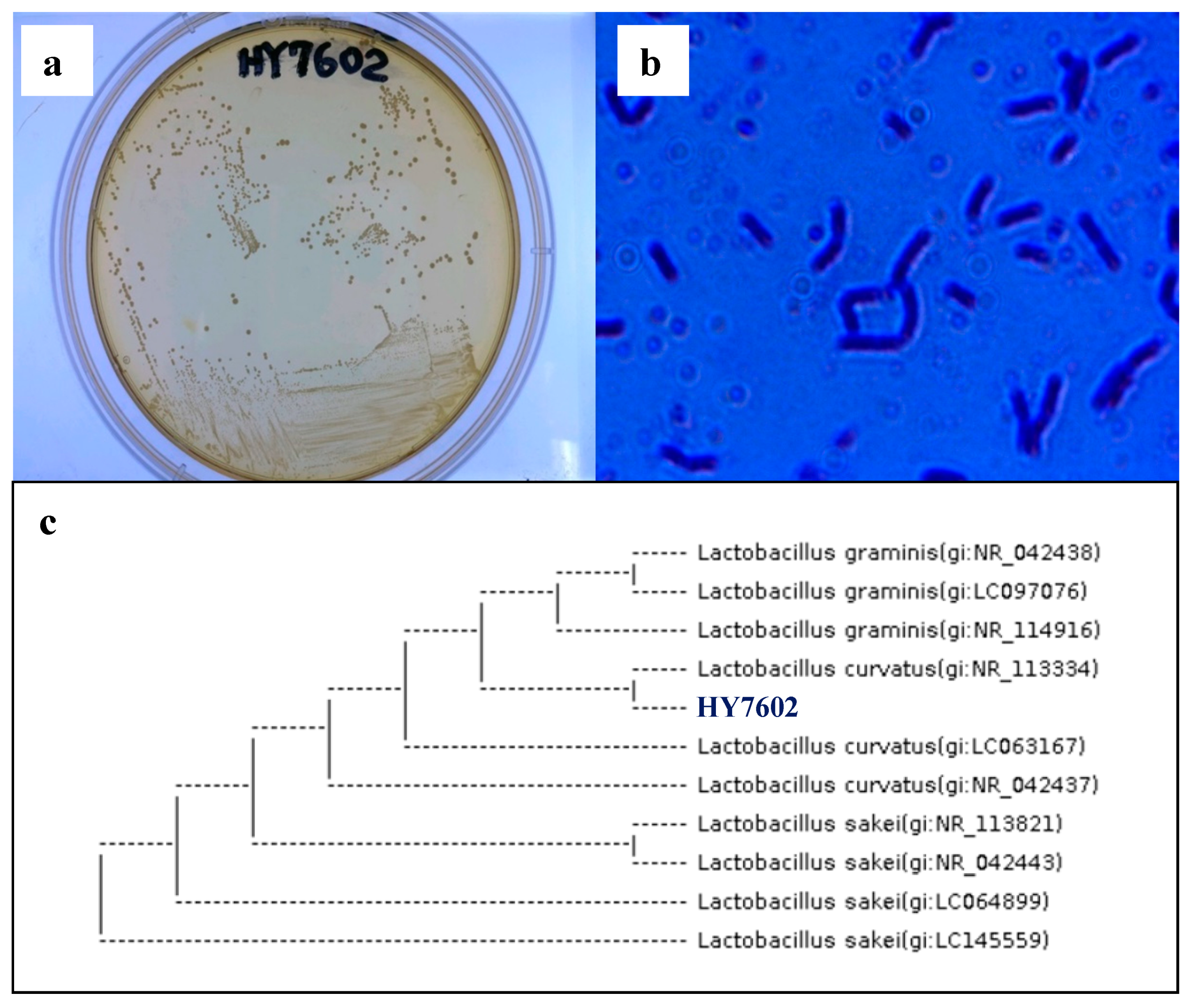
3.1. Screening of Antler-Fermenting LAB

The first screen of 577 types of LAB identified 12 types that grew on antler agar medium. Each was inoculated into the antler extract (1 × 10⁸ CFU/mL) and cultured at 37 °C. After incubation for 36 h, the number of viable cells in the antler extract was counted. Strain No. 4 showed the highest number of viable cells.

3.2. Identification of Deer Antler-Fermenting LAB

Morphological and molecular identification of LAB with antler-fermenting activity revealed that the colonies had an appearance typical of LAB. Microscopic evaluation confirmed that culture HY7602 formed disc-shaped colonies, and that individual cells were rod shaped (Figure 1a,b). These results showed that the cultured bacteria belonged to the genus Lactobacillus. Four types of primer were used for accurate species classification. The gene sequences of the cultured Lactobacillus spp. strains were more than 99% identical to those of Lactobacillus curvatus strains in the NCBI database, a finding confirmed by phylogenetic analysis (Figure 1c). Based on these results, the deer antler-fermenting LAB isolate was named Lactobacillus curvatus HY7602.

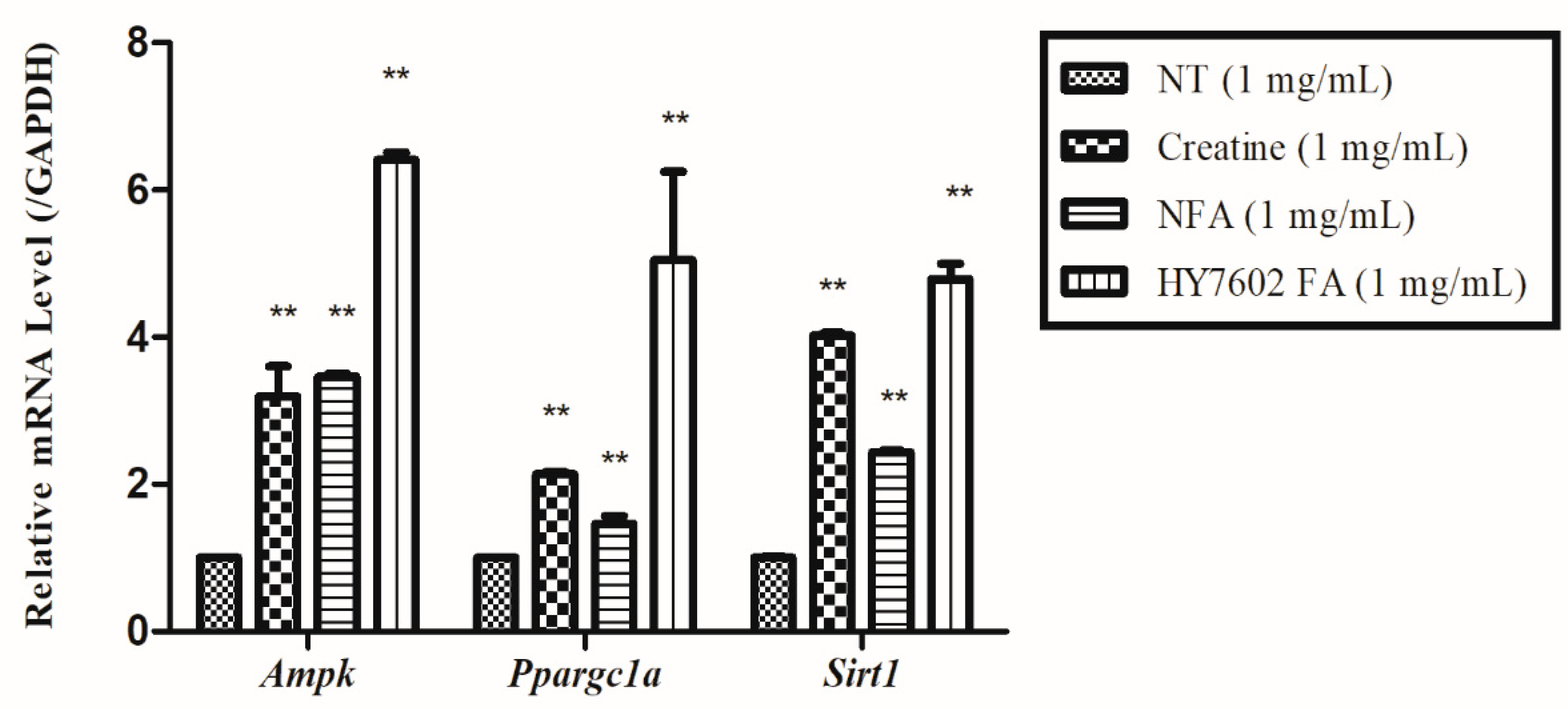
3.3. Effects of FA on Expression of Genes Related to Muscle Strength and Mitochondrial Energy Metabolism in C2C12 Cells

Expression of Igf-1, Pi3k, Akt, Mtor, and P70s6k genes was evaluated by RT-PCR. The results showed that expression of Igf-1, Pi3k, Akt, Mtor, and P70s6k in cells treated with FA was greater than that in the no treatment (NT) control, creatine, or NFA groups (Figure 2). In addition, expression of Ampk, Sirt1, and Ppargc1a in the FA-treated group was higher than that in the NT-, creatine-, and NFA-treated groups (Figure 3).

3.4. HY7602–FA Increases Exercise Endurance

The treadmill exercise tests were conducted at 1-week intervals throughout the 4-week feeding period. The results showed that total exercise duration and distance traveled by mice receiving the HY7602-FA diet were significantly greater than those of mice receiving NFA or creatine (Figure 4a,b). HY7602-FA intake resulted in a significant increase in muscle endurance when compared with NFA (p < 0.05). In addition, lactic acid buildup in the HY7602-FA group decreased, which may increase muscle endurance (data not shown). Similar results were observed in the forced swimming test. The time to exhaustion in all antler-treated groups was higher than that in the normal diet (exercise control) group (Figure 4c); however, it was remarkably higher in the HY7602-FA group than in the NFA group (p < 0.01). There was no significant difference in body weight or food intake across the groups during the 4-week feeding period (Figure 4d,e).

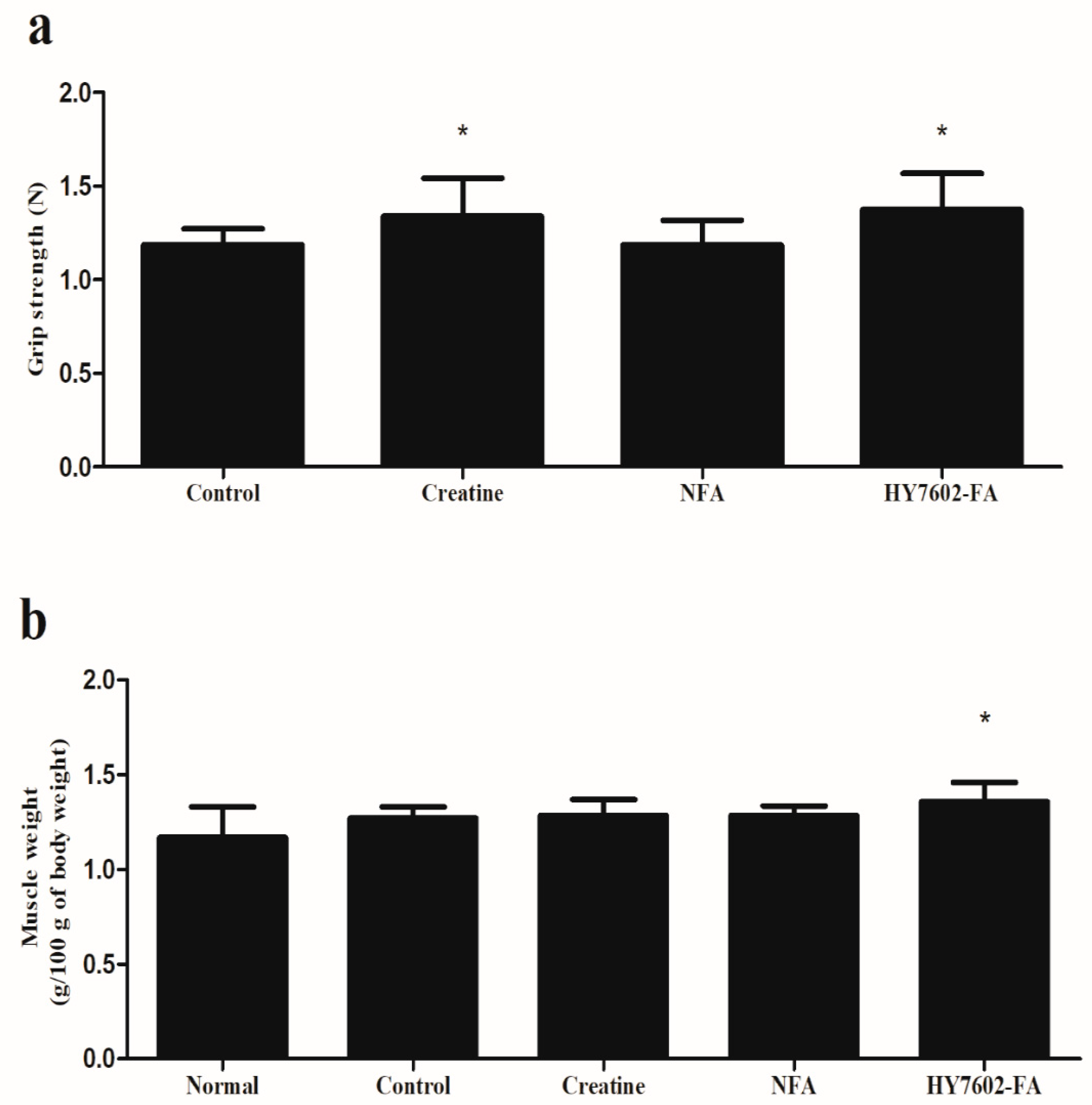
3.5. HY7602–FA Improves Muscle Hypertrophy and Strength

To analyze the effect of HY7602-FA on exercise capacity, muscle grip strength and hindlimb skeletal muscle weight were measured. Muscle strength was measured weekly throughout the 4-week feeding period. The results showed that muscle strength in all groups was greater than that in the negative control group (non-exercise group).
As shown in Figure 5a, grip strength in the control, NFA, creatine, and HY7602-FA groups was 1.19 ± 0.09, 1.35 ± 0.18, 1.18 ± 0.13, and 1.37 ± 0.19 N respectively, demonstrating that HY7602-FA increased muscle strength (p < 0.05). Importantly, there was a significant difference in muscle strength between the HY7602-FA and NFA groups.
Next, to further investigate the effects of HY7602-FA on skeletal muscle, we measured the weight of skeletal muscle in the hindlimbs. The ratio of skeletal muscle weight/body weight, expressed as the relative number for the soleus, EDL, and gastrocnemius muscles, was significantly higher in HY7602-FA fed mice than in the exercise control group (p < 0.05) (Figure 5b).
Overall, these data suggest that HY7602-FA improves grip strength by inducing muscle hypertrophy.

3.6. HY7602-FA Improves Biomarkers of Muscle Endurance, Energy Supply, and Muscle Fatigue after Exercise

At the end of the experiment, we performed a swimming endurance capacity test, after which mice were sacrificed and blood was taken for blood analysis. Table 1 shows the effects of HY7602-FA on serum ALT, LDH, and CK levels, all of which are markers of muscle and liver damage, after the swimming endurance capacity test. Serum ALT, LDH, and CK levels in the HY7602-FA group were significantly lower than in the exercise control group, indicating that HY7602-FA reduces liver and muscle damage during intense exercise (p < 0.05). Moreover, serum markers of muscle endurance, energy supply, and fatigue (creatinine and lactate) were significantly lower in the HY7602-FA group than in the exercise control group (p < 0.05). Blood analysis also indicated that serum levels of ALT, LDH, CK, lactate, and creatinine in the HY7602-FA group were lower than those in the NFA-fed group. These results suggest that HY7602-FA effectively increases muscle endurance and energy supply, and reduces fatigue.

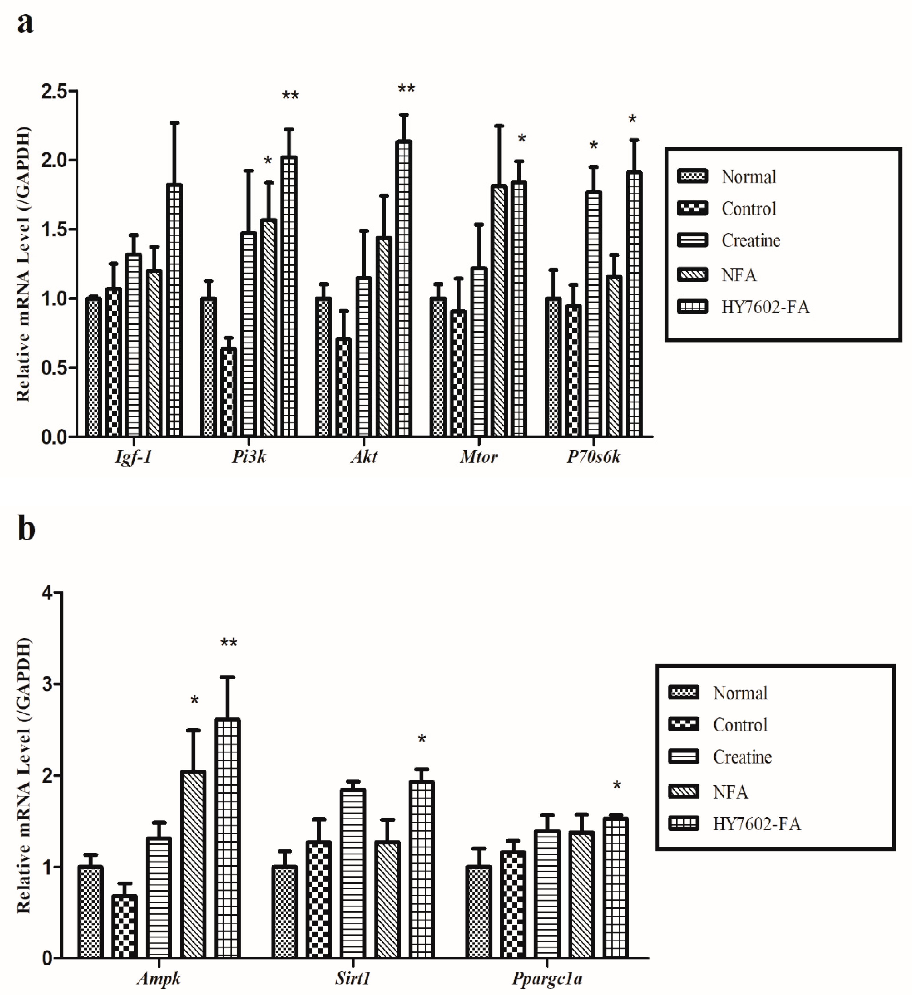
3.7. HY7602-FA Activates Muscle Protein Synthesis, Mitochondrial Biogenesis, and Exercise Signaling-Related mRNA Expression

To analyze the molecular mechanisms underlying the effects of FA in mice, we examined expression of genes encoding Igf-1, Pi3k, Akt, Mtor, and P70s6k, which play roles in muscle protein synthesis, by RT-PCR. As shown in Figure 6a, expression of these genes in the HY7602 FA group was higher than that in the exercise control, positive control (creatine), and NFA groups. In particular, expression of all factors (except for Igf-1) in the HY7602-FA group was significantly higher than that in the exercise control group (p < 0.05).
We also examined expression of Ampk, Sirt1, and Ppargc1a, which are essential for mitochondrial energy supply and metabolism, in response to intense exercise within the same muscle tissue (Figure 6b). As with the IGF-1 signaling pathway, HY7602-FA increased the expression of genes associated with mitochondrial biogenesis and exercise-related signaling when compared with that in the exercise control, positive control (creatine), and NFA-treated groups (p < 0.05). Overall, the data suggest that HY7602-FA increases exercise performance by inducing muscle hypertrophy and by increasing the efficiency of mitochondrial energy metabolism and supply.

4. Discussion

Natural products are being re-evaluated as the demand for traditional medicines increases. Antler is a popular traditional medicine in Korea, China and South East Asia. However, concerns about potential side effects are discussed/reported rarely. Previous studies suggest that fermented and non-fermented antler are safe for in vitro and in vivo use [24,25,26]. Deer antlers contain the highest concentration of medicinal agents in the tip. Previous studies show that Bacillus spp. or Cordyceps militaris increase the amount of uronic acid and sialic acid (ganglioside) [27], which increase the capacity of the musculoskeletal system and improve exercise performance [28].
Based on the results of previous studies, we fermented the middle/lower antlers with various LAB candidates and then studied the effects of one LAB strain on exercise performance.
There are many ways to improve exercise performance. One is to activate the IGF-1 pathway. IGF-1 is contained within antlers. When the IGF-1 pathway is activated, exercise performance is improved [29]. Muscle hypertrophy inducers trigger protein synthesis by phosphorylating proteins downstream of the PI3K/AKT pathway in muscle cells. Among them, mTOR is recognized as a major growth signaling factor that integrates various growth signals in cells [30]. Activation of mTOR can increase muscle mass by inducing muscle protein synthesis via p70s6k, confirming that activation of the IGF-1 pathway increases muscle mass and exercise performance [31].
Here, we showed that HY7602-FA increases IGF-1 levels in mouse cells. IGF-1 signaling was also identified in EDL muscle tissue from mice. The IGF-1 signaling pathway in the FA-treated group was activated to a greater extent than that in the exercise control, positive control (creatine), and NFA groups. This suggests that FA activates pathways associated with protein synthesis, thereby improving muscle strength and inducing hypertrophy.
Another way to improve exercise performance is to increase muscle mass by increasing the number of mitochondria, a process called mitochondrial biogenesis. When the number of mitochondria increases, fatty acid oxidation and ATP production increase [32]. Therefore, mitochondrial biogenesis increases energy production and improves exercise performance. Mitochondrial biogenesis is regulated by a coactivator called PGC-1α (peroxisome proliferator-activated receptor-gamma coactivator 1 alpha), and the activity of PGC-1α is regulated by the AMPK pathway and SIRT1 (sirtuin 1) [33].
We confirmed in mouse cells that FA increased muscle protein synthesis and expression of genes associated with mitochondrial energy metabolism. Therefore, we conducted animal experiments to verify these in vitro observations.
Expression of AMPK/SIRT1/PGC-1α (genes related with mitochondrial energy metabolism) increased significantly in the HY7602-FA group compared with the exercise control, positive control (creatine), and NFA groups. The forced swimming and treadmill exercise tests showed that HY7602-FA significantly improved muscle endurance, even at relatively low concentrations. Furthermore, mice fed HY7602-FA were able to exercise for longer and showed decreased levels of serum markers of fatigue. Serum ALT, LDH, CK, lactate, and creatinine are indicators of accumulated fatigue stress resulting from exercise [34]. Under anaerobic conditions, the level of lactic acid in the blood is a product of carbohydrate conversion, which is the main source of energy for short-term intensive exercise. Accumulation of lactic acid in the blood causes fatigue; rapid removal aids faster recovery [35]. FA mitigated increases in fatigue and stress indices caused by intense exercise.

5. Conclusions

In summary, our data indicate that FA can regulate the IGF-1/AKT/mTOR and AMPK signaling system to increase mitochondrial energy metabolism, thereby increasing muscle strength and improving exercise performance/endurance both in vitro and in vivo. The effects of FA were confirmed in mouse experiments using grip strength, forced swimming and treadmill exercise test. Furthermore, the activity of FA was higher when compared to unfermented antlers.
It can be concluded that lactic acid bacteria with anti-inflammatory, anti-cholesterol, and immunomodulatory activities, etc., increase the efficacy of antlers through fermentation. In future studies, we plan to investigate the functional ingredients that are in-creased by fermentation and the difference in fermentation depending on the part of the antler.
Further studies are needed to prove the same effect of FA on the human body. How-ever, our study indicates the potential of FA as a natural health/functional material that could help improve muscle strength and endurance, which can develop to a natural healing agent for enhancing age-related muscle atrophy.

Author Contributions

Conceptualization, S.J., S.-H.K., W.J., J.R., J.-J.S. and J.-L.L.; methodology, S.J. and S.-H.K.; software, S.J. and S.-H.K.; validation, S.J. and S.-H.K.; formal analysis, S.J. and S.-H.K.; investigation, S.J., S.-H.K., W.J. and J.R.; writing—original draft preparation, S.J. and S.-H.K.; writing—review and editing, K.H.; visualization, S.J. and S.-H.K. All authors have read and agreed to the published version of the manuscript.

Funding

This research received no external funding.

Institutional Review Board Statement

Animal experiments were reviewed and approved by the Institutional Animal Care and Use Committee of hy Co., Ltd (IACUC approval number AEC-2020-00004-Y).

Informed Consent Statement

Not applicable.

Data Availability Statement

The data presented in this study are available in the article.

Conflicts of Interest

The authors declare no conflict of interest.

References

  1. Hong, N.D.; Won, D.H.; Kim, N.J.; Chang, S.Y.; Youn, W.-G.; Kim, H.-S. Studies on the Analysis of Constituents of Deer Horn (I)-Assay of Trace Elements and TLC Pattern Analysis of Gangliosides. Korean Deer J. 1991, 14, 12–21. [Google Scholar]
  2. Ivankina, N.F.; Isay, S.V.; Busarova, N.G.; Mischenko, T.Y. Prostaglandin-like activity, fatty acid and phospholipid composition of sika deer (Cervus nippon) antlers at different growth stages. Comp. Biochem. Physiol. 1993, 106, 159–162. [Google Scholar] [CrossRef]
  3. Ha, H.; Yoon, S.-H. Analytical studies of constituents of antlers. J. Korean Soc. Food Sci. Nutr. 1996, 25, 279–282. [Google Scholar]
  4. Sunwoo, H.H.; Nakano, T.; Hudson, R.J.; Sim, J.S. Chemical composition of antlers from wapiti (Cervus elaphus). J. Agric. Food Chem. 1995, 43, 2846–2849. [Google Scholar] [CrossRef]
  5. Yoo, Y.S.; Kim, Y.S.; Jhon, G.-J.; Park, J. Separation of gangliosides using cyclodextrin in capillary zone electrophoresis. J. Chromatogr. A 1993, 652, 431–439. [Google Scholar] [PubMed]
  6. Pette, D.; Staron, R.S. Myosin isoforms, muscle fiber types, and transitions. Microsc. Res. Tech. 2000, 50, 500–509. [Google Scholar] [CrossRef]
  7. Zong, H.; Ren, J.M.; Young, L.H.; Pypaert, M.; Mu, J.; Birnbaum, M.J.; Shulman, G.I. AMP kinase is required for mitochondrial biogenesis in skeletal muscle in response to chronic energy deprivation. Proc. Natl. Acad. Sci. USA 2002, 99, 15983–15987. [Google Scholar] [CrossRef] [PubMed] [Green Version]
  8. Ticinesi, A.; Nouvenne, A.; Cerundolo, N.; Catania, P.; Prati, B.; Tana, C.; Meschi, T. Gut microbiota, muscle mass and function in aging: A focus on physical frailty and sarcopenia. Nutrients 2019, 11, 1633. [Google Scholar] [CrossRef] [Green Version]
  9. Sivamaruthi, B.S.; Kesika, P.; Suganthy, N.; Chaiyasut, C. A review on role of microbiome in obesity and antiobesity properties of probiotic supplements. Biomed Res. Int. 2019, 2019, 3291367. [Google Scholar] [CrossRef]
  10. Huang, W.-C.; Chen, Y.-M.; Kan, N.-W.; Ho, C.-S.; Wei, L.; Chan, C.-H.; Huang, H.-Y.; Huang, C.-C. Hypolipidemic effects and safety of Lactobacillus reuteri 263 in a hamster model of hyperlipidemia. Nutrients 2015, 7, 3767–3782. [Google Scholar] [CrossRef] [Green Version]
  11. Sichetti, M.; De Marco, S.; Pagiotti, R.; Traina, G.; Pietrella, D. Anti-inflammatory effect of multistrain probiotic formulation (L. rhamnosus, B. lactis, and B. longum). Nutrition 2018, 53, 95–102. [Google Scholar] [CrossRef]
  12. Pourramezan, Z.; Kermanshahi, R.K.; Oloomi, M.; Aliahmadi, A.; Rezadoost, H. In vitro study of antioxidant and antibacterial activities of Lactobacillus probiotic spp. Folia Microbiol. 2018, 63, 31–42. [Google Scholar] [CrossRef]
  13. Tiptiri-Kourpeti, A.; Spyridopoulou, K.; Santarmaki, V.; Aindelis, G.; Tompoulidou, E.; Lamprianidou, E.E.; Saxami, G.; Ypsilantis, P.; Lampri, E.S.; Simopoulos, C.; et al. Lactobacillus casei exerts anti-proliferative effects accompanied by apoptotic cell death and up-regulation of TRAIL in colon carcinoma cells. PLoS ONE 2016, 11, e0147960. [Google Scholar] [CrossRef]
  14. Oliveira, M.; Bosco, N.; Perruisseau, G.; Nicolas, J.; Segura-Roggero, I.; Duboux, S.; Briand, M.; Blum, S.; Benyacoub, J. Lactobacillus paracasei reduces intestinal inflammation in adoptive transfer mouse model of experimental colitis. Clin. Dev. Immunol. 2011, 2011, 807483. [Google Scholar] [CrossRef] [Green Version]
  15. Chen, L.-H.; Chen, Y.-H.; Cheng, K.-C.; Chien, T.-Y.; Chan, C.-H.; Tsao, S.-P.; Huang, H.-Y. Antiobesity effect of Lactobacillus reuteri 263 associated with energy metabolism remodeling of white adipose tissue in high-energy-diet-fed rats. J. Nutr. Biochem. 2018, 54, 87–94. [Google Scholar] [CrossRef]
  16. Grosicki, G.J.; Fielding, R.A.; Lustgarten, M.S. Gut microbiota contribute to age-related changes in skeletal muscle size, composition, and function: Biological basis for a gut-muscle axis. Calcif. Tissue Int. 2018, 102, 433–442. [Google Scholar] [CrossRef] [PubMed] [Green Version]
  17. Walsh, M.E.; Bhattacharya, A.; Sataranatarajan, K.; Qaisar, R.; Sloane, L.; Rahman, M.M.; Kinter, M.; Van Remmen, H. The histone deacetylase inhibitor butyrate improves metabolism and reduces muscle atrophy during aging. Aging Cell 2015, 14, 957–970. [Google Scholar] [CrossRef]
  18. LeBlanc, J.G.; Milani, C.; De Giori, G.S.; Sesma, F.; Van Sinderen, D.; Ventura, M. Bacteria as vitamin suppliers to their host: A gut microbiota perspective. Curr. Opin. Biotechnol. 2013, 24, 160–168. [Google Scholar] [CrossRef] [PubMed]
  19. Stanley, D.; Denman, S.E.; Hughes, R.J.; Geier, M.S.; Crowley, T.M.; Chen, H.; Haring, V.R.; Moore, R.J. Intestinal microbiota associated with differential feed conversion efficiency in chickens. Appl. Microbiol. Biotechnol. 2012, 96, 1361–1369. [Google Scholar] [CrossRef] [PubMed]
  20. Kunkel, S.D.; Suneja, M.; Ebert, S.M.; Bongers, K.S.; Fox, D.K.; Malmberg, S.E.; Alipour, F.; Shields, R.K.; Adams, C.M. mRNA expression signatures of human skeletal muscle atrophy identify a natural compound that increases muscle mass. Cell Metab. 2011, 13, 627–638. [Google Scholar] [CrossRef] [Green Version]
  21. Nader, G.A. Molecular determinants of skeletal muscle mass: Getting the “AKT” together. Int. J. Biochem. Cell Biol. 2005, 37, 1985–1996. [Google Scholar] [CrossRef] [PubMed]
  22. Jang, S.; Park, E.D.; Suh, H.J.; Lee, S.H.; Kim, J.S.; Park, Y. Enhancement of exercise endurance capacity by fermented deer antler in BALB/c mice. Biosci. Biotechnol. Biochem. 2014, 78, 1716–1722. [Google Scholar] [CrossRef] [Green Version]
  23. Huang, W.-C.; Huang, C.-C.; Chuang, H.-L.; Chiu, C.-C.; Chen, W.-C.; Hsu, M.-C. Cornu cervi pantotrichum supplementation improves physiological adaptions during intensive endurance training. J. Vet. Med. Sci. 2017, 79, 674–682. [Google Scholar] [CrossRef] [Green Version]
  24. Kim, M.-K.; Jung, E.-Y.; Lee, H.-S.; Shin, K.-S.; Kim, Y.-K.; Ra, K.-S.; Park, C.-S.; Woo, M.-J.; Lee, S.-H.; Kim, J.-S.; et al. Isolation of strain for the preparation of the fermented antler and its physiological activities. J. Korean Soc. Food Sci. Nutr. 2009, 38, 1237–1242. [Google Scholar] [CrossRef]
  25. Wu, F.; Li, H.; Jin, L.; Li, X.; Ma, Y.; You, J.; Li, S.; Xu, Y. Deer antler base as a traditional Chinese medicine: A review of its traditional uses, chemistry and pharmacology. J. Ethnopharmacol. 2013, 145, 403–415. [Google Scholar] [CrossRef] [PubMed]
  26. Suh, J.H.; Seo, H.W.; Han, B.C.; Kyung, J.S.; So, S.H. 13-Week repeated oral dose toxicity study on mixture of Korean red ginseng and deer antler extract in Sprague-Dawley rats. Toxicol. Res. 2019, 36, 1–12. [Google Scholar] [CrossRef]
  27. Kim, D.-H.; Han, S.-B.; Park, J.-S.; Han, M.-J. Fermentation of antler and its biological activity. Korean J. Pharmacogn. 1994, 25, 233–237. [Google Scholar]
  28. Chen, J.-C.; Hsiang, C.-Y.; Lin, Y.-C.; Ho, T.-Y. Deer Antler Extract Improves Fatigue Effect through Altering the Expression of Genes Related to Muscle Strength in Skeletal Muscle of Mice. Evid. Based Complement. Alternat. Med. 2014, 2014, 540580. [Google Scholar] [CrossRef] [PubMed]
  29. Han, G.-H.; Kwon, H.-T.; Son, H.-J.; Chai, J.-H.; Shan, Z.X.; Kim, H.-J.; Kim, C.-K. Change of mTOR, p70S6K, 4E-BP1, and AMPK protein expressions with a intensive endurance exercise in rats. Korean Soc. Sports Sci. 2011, 20, 1551–1561. [Google Scholar]
  30. Hornberger, T.A. Mechanotransduction and the regulation of mTORC1 signaling in skeletal muscle. Int. J. Biochem. Cell Biol. 2011, 43, 1267–1276. [Google Scholar] [CrossRef] [Green Version]
  31. Glass, D.J. Signalling pathways that mediate skeletal muscle hypertrophy and atrophy. Nat. Cell Biol. 2003, 5, 87–90. [Google Scholar] [CrossRef] [PubMed]
  32. Lin, J.; Handschin, C.; Spiegelman, B.M. Metabolic control through the PGC-1 family of transcription coactivators. Cell Metab. 2005, 1, 361–370. [Google Scholar] [CrossRef] [PubMed] [Green Version]
  33. Gureev, A.P.; Shaforostova, E.A.; Popov, V.N. Regulation of mitochondrial biogenesis as a way for active longevity: Interaction between the Nrf2 and PGC-1α signaling pathways. Front Genet. 2019, 10, 435. [Google Scholar] [CrossRef] [PubMed] [Green Version]
  34. Kimura, Y.; Sumiyoshi, M. Effects of various Eleutherococcus senticosus cortex on swimming time, natural killer activity and corticosterone level in forced swimming stressed mice. J. Ethnopharmacol. 2004, 95, 447–453. [Google Scholar] [CrossRef]
  35. Cairns, S.P. Lactic acid and exercise performance: Culprit or friend? Sports Med. 2006, 36, 279–291. [Google Scholar] [CrossRef] [PubMed]
Figure 1. Morphological characteristics and phylogenetic tree depicting the taxonomic position of Lactobacillus curvatus strains based on 16S rRNA sequences. The lactic acid bacteria HY7602 were inoculated onto MRS plates, incubated at 37 °C for 36 h, and examined under a microscope. (a) HY7602 colonies. (b) HY7602 cells (4000X). (c) The 16S rRNA gene sequence of HY7602 was used to identify its respective phylogenetic position. A phylogenetic tree was constructed using a neighbor-joining method. The “T” after the collection number indicates the strain type of the species.
Applsci 11 05386 g001
Figure 2. Effects of fermented antler (FA) on expression of IGF-1/AKT pathway-related genes (Igf-1, Pi3k, Akt, Mtor, P70s6k) in mouse skeletal muscle myotubes. FA induced expression of genes related to the IGF-1/AKT/mTOR pathway. To examine expression of Igf-1, Pi3k, Akt, Mtor, and P70s6k, total RNA was prepared from C2C12 cells incubated for 24 h with NFA (1 mg/mL), HY7602-FA (1 mg/mL), or creatine (1 mg/mL). Igf-1, Pi3k, Akt, Mtor, and P70s6k were detected by using the RT-PCR program. Data are expressed as the mean ± SD of three determinations. Asterisks indicate significant differences: * p < 0.05 and ** p < 0.01 vs. NT. Akt, protein kinase B; Igf-1, insulin-like growth factor 1; Mtor, mammalian target of rapamycin; NT, no treatment; P70s6k, phosphorylated 70-kDa ribosomal S6 kinase; Pi3k, phosphoinositide 3-kinases.
Applsci 11 05386 g002
Figure 3. Intestinal effects of fermented antler (FA) on AMPK signal pathway-related genes (Ampk, Ppargc1a, Sirt1) associated with mitochondrial activity in skeletal myotubes. FA upregulates the expression of mRNA encoding Ampk, which is associated with mitochondrial activity. To check Ampk, Ppargc1a, and Sirt1 expression, total RNA was prepared from C2C12 cells incubated for 24 h with NFA (1 mg/mL), HY7602-FA (1 mg/mL), or creatine (1 mg/mL). Ampk, Ppargc1a, and Sirt1 were detected by RT-PCR. Data are expressed as the mean ± SD of three determinations. Asterisks indicate significant differences: ** p < 0.01 vs. NT. Ampk, AMP-activated protein kinase; NT, no treatment; Ppargc1a, peroxisome proliferator-activated receptor-gamma coactivator 1 alpha; Sirt1, sirtuin 1.
Applsci 11 05386 g003
Figure 4. Effect of HY7602-FA on exercise endurance. Exercise capacity was measured based on total exercise time (a), running distance on a treadmill (b), or total swimming time (c). Data are expressed as the mean ± SD (n = 9 mice per group), and asterisks indicate significant differences: * p < 0.05 and ** p < 0.01 vs. the control group. Changes in (d) body weight and (e) food intake over 4 weeks. NFA, non-fermented antler; FA, fermented antler.
Applsci 11 05386 g004
Figure 5. Effect of HY7602-FA on grip strength and skeletal muscle mass. Forelimb grip strength was measured using a grip strength meter equipped with a T-shaped pull bar. Each mouse performed three consecutive tests to obtain the peak value (a). Ratio of skeletal muscle mass (soleus, EDL, and gastrocnemius muscles)/bodyweight increases after HY7602-FA intake (b). Data are expressed as the mean ± SD (n = 9 mice per group), and asterisks indicate significant differences: * p < 0.05 and ** p < 0.01 vs. the control group. NFA, non-fermented antler; FA, fermented antler.
Applsci 11 05386 g005
Figure 6. Effect of HY7602-FA on expression of mRNA encoding genes related to muscle protein synthesis, mitochondrial biogenesis, and exercise signaling pathways in skeletal muscle tissue. (a) Expression of Igf-1, Pi3k, Akt, Mtor, and P70s6k (related to muscle protein synthesis) in mouse skeletal muscle tissue was detected by RT-PCR. (b) Expression of Ampk, Sirt1, and Ppargc1a, which are known to be important for mitochondrial energy supply and metabolism in mouse skeletal muscle tissue, were detected by RT-PCR. All data represent the mean ± SD (n = 9 mice per group); asterisks indicate significant differences: * p < 0.05 and ** p < 0.01 vs. control group. NFA, non-fermented antler; FA, fermented antler.
Applsci 11 05386 g006
Table 1. Blood biomarkers in HY7602-FA-intake mice after the swimming endurance capacity test.
NormalControlCreatineNFAHY7602-FA
ALT (U/L)18.00 ± 2.719.57 ± 2.919.29 ± 2.016.38 ± 3.215.57 ± 4.6 *
LDH (U/L)376.14 ± 73.1262.43 ± 101.5273.00 ± 129.8258.38 ± 117.9145.71 ± 23.5 *
Creatine kinase (mg/dL)51.71 ± 21.9132.71 ± 71.9102.57 ± 39.672.25 ± 34.655.43 ± 22.9 *
Lactate (mg/dL)69.77 ± 5.678.13 ± 10.462.84 ± 16.461.23 ± 5.748.90 ± 13.7 *
Creatinine (mg/dL)0.32 ± 0.080.35 ± 0.10.44 ± 0.090.29 ± 0.040.26 ± 0.03 *
The data are presented as the mean ± SD (n = 9 mice per group). * p < 0.05 vs. exercise control. ALT, alanine aminotransferase; LDH, lactate dehydrogenase.
Publisher’s Note: MDPI stays neutral with regard to jurisdictional claims in published maps and institutional affiliations.

