Article

Determination of the Vertical Dimension and the Position of the Occlusal Plane in a Removable Prosthesis Using Cephalometric Analysis and Golden Proportion

1
Department of Biomedical and Dental Sciences and Morphological and Functional Imaging, University of Messina, 98100 Messina, Italy
2
Department of Medicine and Surgery, School of Medicine, University of Insubria, 21100 Varese, Italy
3
Independent Researcher, 95100 Catania, Italy
4
Multidisciplinary Department of Medical-Surgical and Odontostomatological Specialties, University of Campania “Luigi Vanvitelli”, 80121 Naples, Italy
5
Department of Dental Therapy, Faculty of Dental Medicine, University of Medicine, 1005 Tirana, Albania
6
Department of Biomedical, Surgical and Dental Sciences, Section of Orthodontics, University of Milan, 20100 Milan, Italy
*
Authors to whom correspondence should be addressed.
Submission received: 5 July 2021 / Revised: 24 July 2021 / Accepted: 27 July 2021 / Published: 28 July 2021

Abstract

:
The aim of this study is to demonstrate the use and the effectiveness of cephalometry and golden proportions analysis of the face in planning prosthetic treatments in totally edentulous patients. In order to apply this method, latero-lateral and posterior-anterior X-rays must be performed in addition to the common procedure. Two main concerns for totally edentulous patients are the establishment of the vertical dimension and the new position of the occlusal plane. The divine proportion analysis was carried out by the use of a golden divider. The prosthetic protocol was divided into three steps and a case was selected for better understanding. Referring to the golden relations, if the distance from the chin to the wing of the nose is 1.0, the distance from the nose to eye is 0.618. This proportion is useful and effective in determining the correct prosthetic vertical dimension. The incisal margin of the lower incisor must be positioned between Point A (A) and protuberance menti (Pm) according to the gold ratio 0.618 of the total height A-Pm. Posteriorly the occlusal plane must be placed 2 mm below the divine occlusal plane (traced from the incisal margin of lower incisors to Xi point). A prosthesis made in accordance with cephalometric parameters and divine proportions of the face helps to improve the patient’s aesthetics, function and social personality.

1. Introduction

In prosthodontics, the evaluation and determination of the correct vertical dimension of the maxillofacial complex and the determination of a valid occlusal plane are two factors and indispensable characteristics for prosthetic success. They contribute to the physical, psychological and social health of the patient, as they are essential for chewing, swallowing, phonation, tissue health, aesthetics and individual personality.
Prosthetists usually divide the patient profile into three equal parts in order to evaluate the vertical dimension. The vertical relation, called the vertical dimension, is a convenient term used in referring to the nose and chin relations which are affected by the distance between the jaws [1].
Specifically, the Glossary of Prosthodontic Term defines the rest position as “the postural relation of the mandible to the maxillae when the patient is resting comfortably in the upright position and the condyles are in a neutral unstrained position in the glenoid fossae” [2]. Therefore, it is influenced by neuromuscular, dental (presence or absence of dental elements) and psychological factors. In fact, the rest position may be assumed voluntarily and is constantly assumed subconsciously [1].
On the other hand, the occlusion vertical dimension is defined “the length of the face when the teeth (occlusal rims, central-bearing point, or any other stop) are in contact and the mandible is in centric relation or the teeth are in centric relation” [3].
Another determining prosthetic factor is the construction of a correct occlusal plane.
On cephalometric radiography, the occlusal plane (OP) is a two-dimensional representation of a three-dimensional entity. The original occlusal plane (OOP), as defined by Downs, is a line connecting the point bisecting the first molar cusp height and the point bisecting the incisal overbite [4]. While, the functional occlusal plane (FOP) is obtained by connecting the intercuspation of first premolars and first molars [5,6].
Ricketts recognized that the position of both lower and upper incisors is influenced by normal and abnormal functions of the lips and the tongue. For this reason, he suggests using the buccal occlusal plane that he defines the true buccal occlusal plane (TBOP). It is a line through the bisection of the buccal cusps of premolars and first molars or through the deciduous buccal occlusion [7].
From the prosthetics functional point of view, the occlusal table is a milling surface, strategically placed so that the tongue on the lingual side and the buccinator muscle on the buccal side are able to position the food bolus onto it and hold it there while mastication takes place. Faulty orientation of the occlusal plane in fixed or removable prostheses jeopardizes this interaction between the tongue and buccinator muscle and results at one extreme in food collection in the sulcus, and at the other extreme in biting the cheek or tongue [8].
There is not a universally accepted method for the determination of the occlusal plane and vertical dimension in edentulous patients; there are many factors to consider and the subjectivity and variability of the patients’ characteristics and dental and skeletal patterns takes over. So when a clinician has to select the best method to use, the criteria to be considered are accuracy and repeatability of the measurement, adaptability of the technique, type and complexity of the equipment needed and the time required to plan and obtain the measurements [9,10].
It is also important to underline that the final objective of an oral rehabilitation is the stability of occlusion [11] and, consequently, the equilibrium of the kinetic chain of the body [12,13,14].
The aim of our study is to demonstrate the efficacy of cephalometry and golden proportions applied to the face in planning prosthetic treatment in totally edentulous patients.

2. Materials and Methods

2.1. Search Strategy and Study Selection

A search in electronic databases, Medline (PubMed) and Google Scholar, was carried out including papers from 1938 to 2020. The search included only English-language articles published in dental journals. The following keywords were combined ‘cephalometric analysis’ AND ‘complete prosthesis’, ‘vertical dimension’, ‘occlusal plane’, ‘golden proportion’.

2.2. Target Questions

The questions processed the following guidelines, according to PICO (Patient, Intervention, Comparison, Outcome):
How to obtain a functional and aesthetic mobile prosthesis?
How to choose the correct vertical dimension for the patient and the correct occlusal plane of the prosthesis?
To explain the method to plan the prosthetic design and understand the results, it is necessary to analyze and explain the anatomical points and reference planes used.
Soft Tissue references used in the search for the vertical dimension using the golden proportions of the face included:
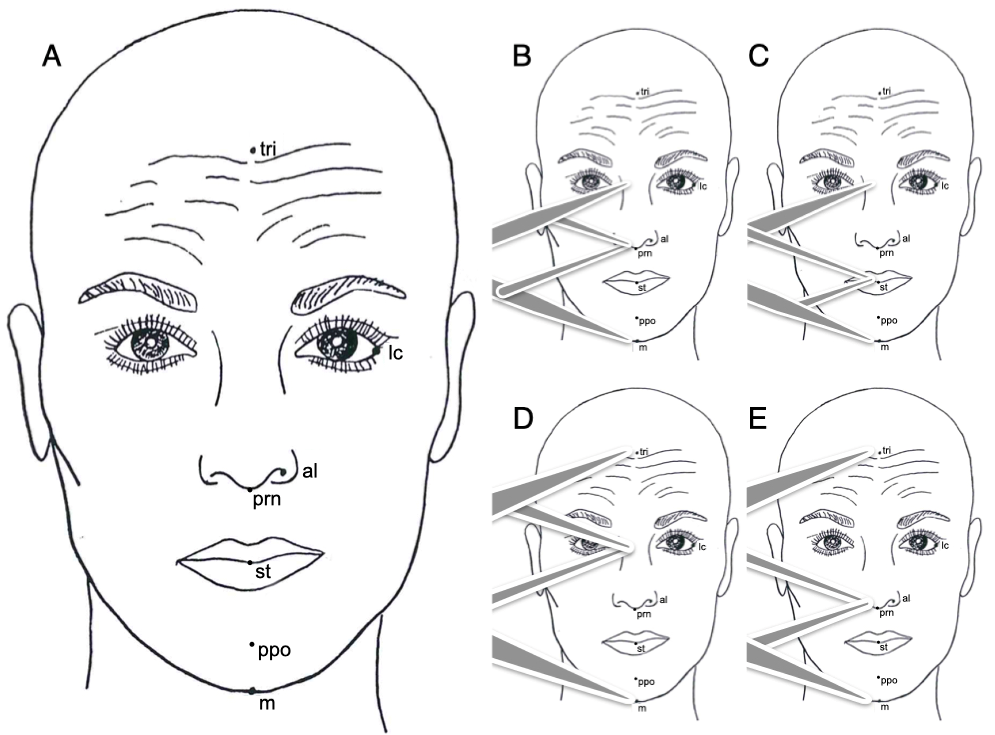
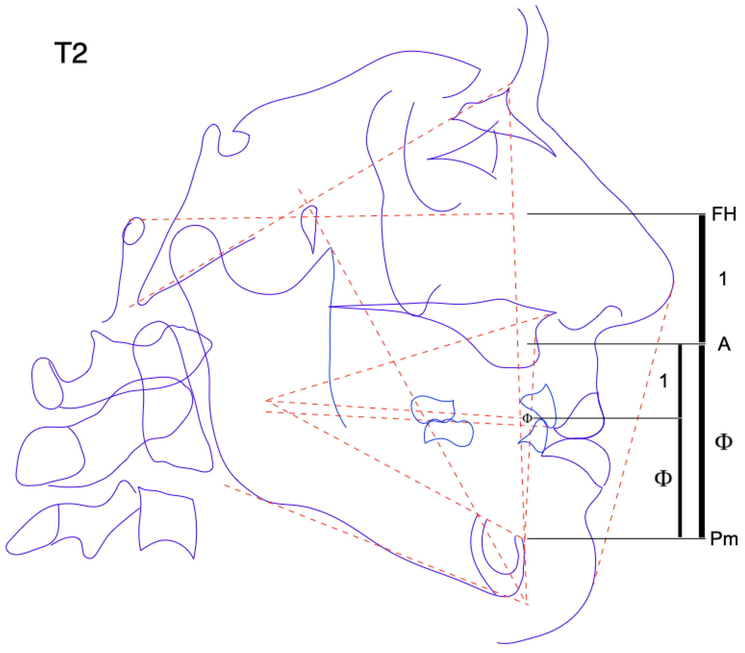
  • Trichion (tri): is the point on the hairline in the midline of the forehead.
  • Lateral canthus of the eyes (lc): is the point at the outer commissure of the eye fissure.
  • Ala of the nose (al): the most lateral point on alar contour.
  • Pronasalis (prn): the most anterior point of the tip of the nose.
  • Stomion (st): the contact point of the upper and lower lips in the mid-sagittal line when the mouth is closed.
  • Propogonion (ppo): the most anterior point of the chin profile.
  • Menton (m): the lowest point in the midline on the lower border of the chin.
The cranial, maxillary and mandibular references to determine the prosthetic occlusal plane using the latero-lateral X-ray are:
Cranial reference:
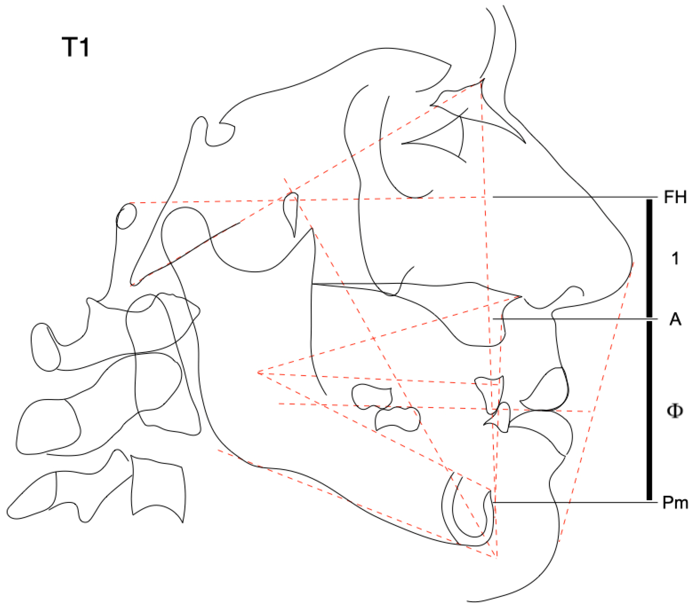
  • Basion (Ba): located at the anterior border of the foramen magnun.
  • Nasion (N): selected at the frontal margin of the fronto-nasal suture.
  • Porion (P): selected at the center of the superior border of the ear canal.
  • Orbitale (O): located at the lower rim of the orbit.
  • Pterygoid point (Pt): selected at the lower border of the Foramen Rotundum at the origin of Pterygoid buttress.
  • Center cranium (Cc): is the intersection between Frankfort Plane and Basion-Nasion Plane.
Maxillary reference:
  • Anterior Nasal Spine (ANS): is selected on the most forward point of maxillae bones.
  • Point A (A): selected at the deepest curvature of the contour below the anterior nasal spine.
Mandibular reference:
  • Pogonion (Po): defined as the most anterior point on the outline of the chin.
  • Protuberance menti (Pm): selected where the symphyseal cortical plate ends and where the supramental contour starts to recede into the alveolar process.
  • Gnathion (Gn): selected cephalometrically and constructed by the intersection of the Facial Plane with the Mandibular Plane.
  • Menton (M): is the lowest point on the inferior border of the symphysis.
  • Xi: is selected as a centroid reference for the ramus.
  • Subgonion (Sgo): is selected at the lower border of the angle of the mandible.
Teeth:
  • Upper incisor (A1): representative position of the mean of the upper central incisors traced with the Ricketts template.
  • Lower incisor (B1): representative position of the mean of the lower central incisors traced with the Ricketts template.
  • Lower incisor divine point (Φ): selected in golden proportion between point A and Pm. The height from incisal edge to Pm should be 0.618.
  • Upper first molar (A6): selected as the bisection of the two sides. Traced with the Ricketts template.
  • Lower first molar (B6): selected as the mesial margin of the lower first molar for anterior reference and vertically as the bisection of the two sides and of the first molar occlusion. Traced with the Ricketts template.
  • Lower second molar (B7): selected as the mesial margin of the lower second molar for anterior reference and vertically as the bisection of the two sides and of the second molar occlusion. Traced with the Ricketts template.
Reference employed in frontal perspective to determine the prosthetic occlusal plane:
  • Zygomatic arch point (Za): Zygomatic arch center.
  • B6L: lower left first molar
  • B6R: lower right first molar
Lateral cranial planes:
  • Basion-Nasion plane: Ba-N
  • Frankfort Plane: P-O
  • True Buccal Occlusal Plane (TBOP): line through the bisection of the buccal cusp of premolars and first molar.
  • Divine Occlusal Plane (DOP): traced from Xi to Lower Incisor Divine point (Φ).
Planes and lines for the Face:
  • Facial plane: N-Po
  • Mandibular plane: Sgo-M
  • Facial axis: Cc-Gn
  • Corpus axis: Xi-Pm
  • Org line: Xi-ANS
  • Denture plane: A-Po
Soft tissue:
  • Esthetic line: prn-ppo
Linear and angular cephalometric measurements.
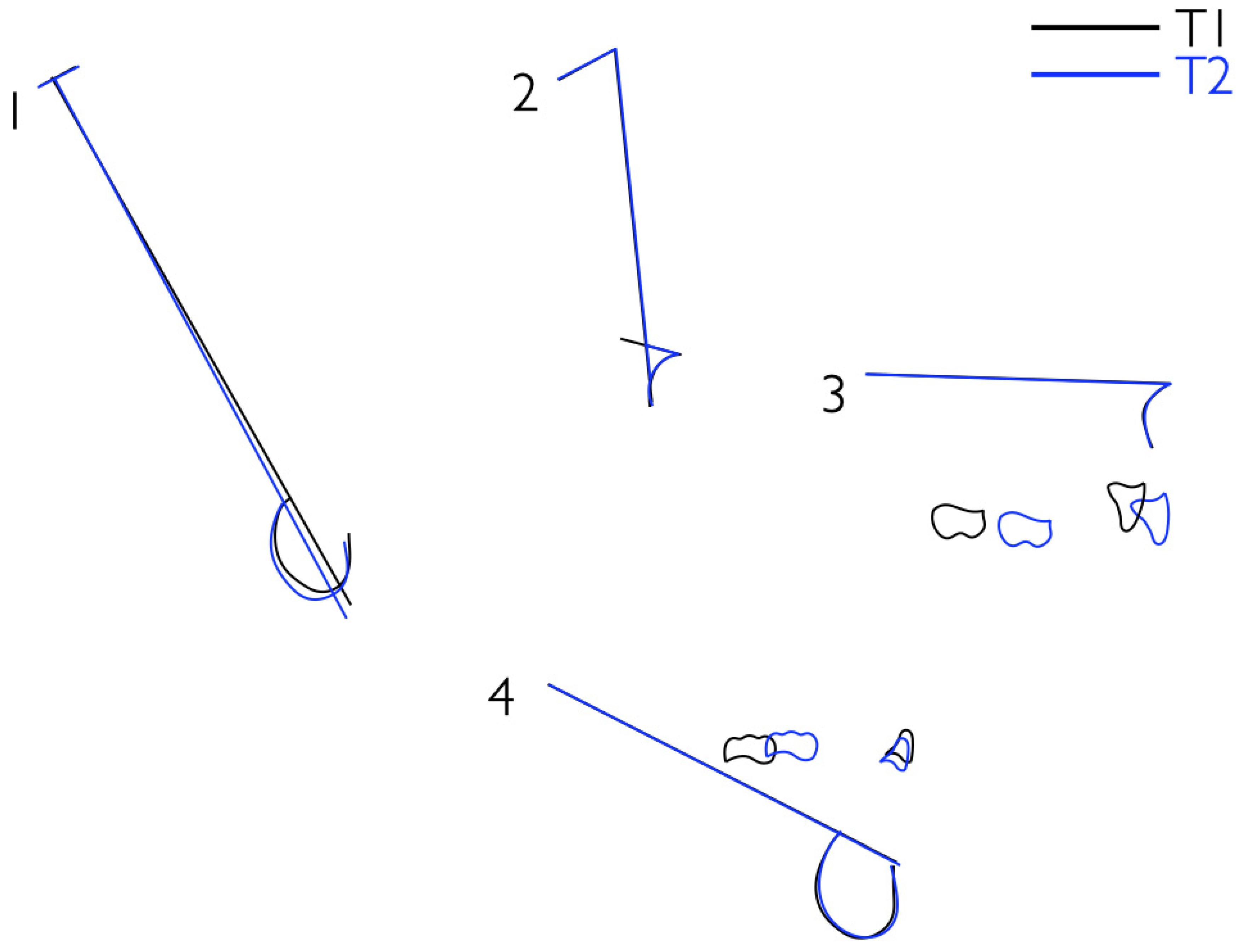
  • Central facial direction (Facial axis-BaNa plane) (degrees);
  • Lower facial height (Corpus axis-Org line) (degrees);
  • Incisor overjet (mm);
  • Incisor overbite (mm);
  • Horizontal B1 position (B1-denture plane) (mm);
  • Lower lip protrusion (lower lip to E line) (mm)
Frontal reference planes:
  • Zygomatic plane (ZA-ZA): represents a frontal Frankfort plane.
  • Frontal occlusal plane (B6L to B6R): a bisection of the molar occlusion connection represents a fronto-occlusal plane.

3. Results

The prosthetic protocol proposed was divided into three steps. In order to better explain these procedures a totally edentulous patient wearing an old incongruous full denture was selected.

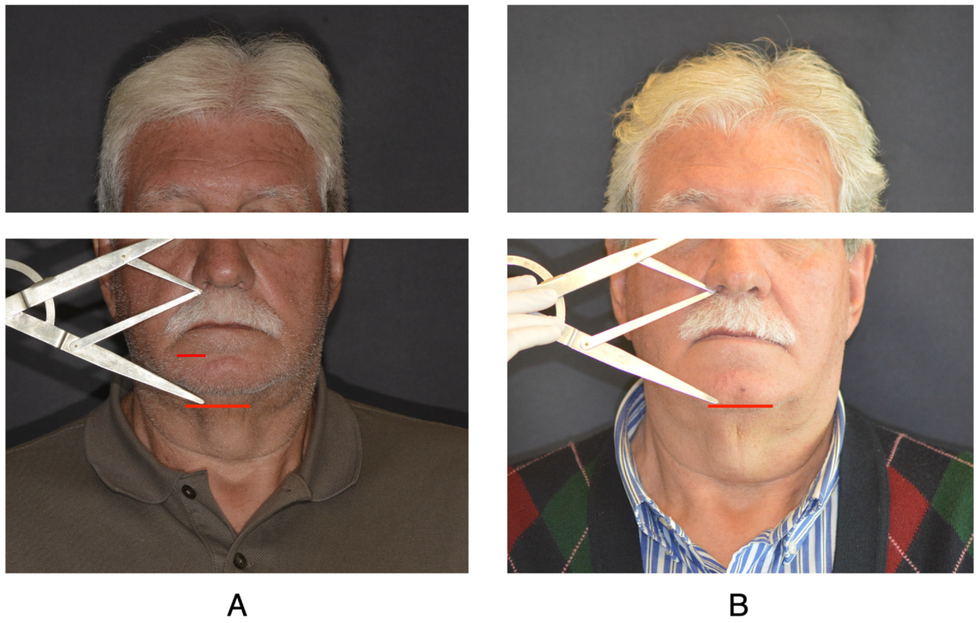
3.1. Step 1: Determination of the Vertical Dimension

The starting point was analyzing the patient’s esthetics and studying the proportions of the face (Figure 1). The frontal and lateral divine proportion analysis could be performed on the frontal and lateral photographs or directly on the patient, using a golden divider (Figure 2). This could also be used for morphological analysis of the teeth and the skeleton. It is based on the “Golden Section” also called the “Divine Proportion”. Upon widening the divider, the short side and a longer side are measured off proportionally as the divider is extended. The longer side is 1.618 times the shorter side and the shorter side is 0.618 the length of the longer. In turn, the longer side is 0.618 the length of the total outer measurement [15]. The points used for the search of the correct soft tissue vertical dimension were tri, lc, al, st and m. Referring to the golden relations described by Ricketts, if the distance from tri to lc is 1.0, the distance from lc to al is 0.618 (Figure 3A) [16].

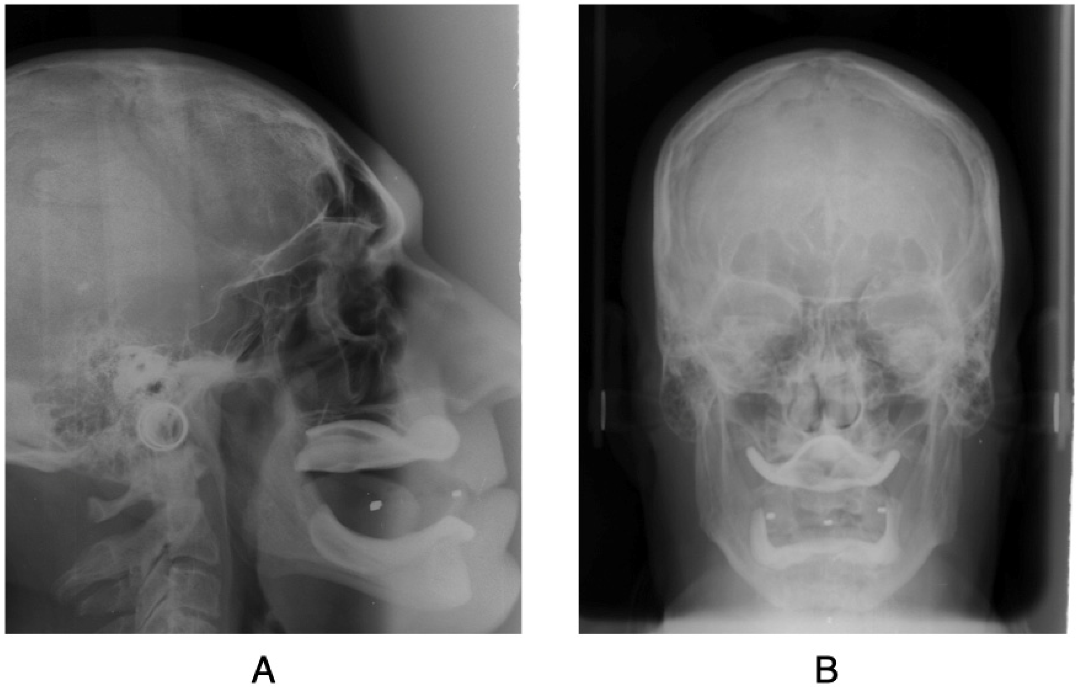
3.2. Step 2: Cephalometric Skeletal Verification of the Vertical Dimension and Determination of the Divine Occlusal Plane

The lateral cephalometric skeletal vertical dimension is represented by the golden ratio between Frankfort plane, Point A and Pm: if the distance from Frankfort plane to Point A is 1.0, the distance from Point A to Pm should be 1618.
Once the soft tissue vertical dimension was measured, we made two occlusal wax registration plates that in occlusion respected the golden ratio above described (Figure 3B).
In the lower occlusal wax registration plate were arranged three radiopaque landmarks, one positioned at the level of the lower incisors and other two for each first lower molar. Hence a latero-lateral and a postero-anterior teleradiograph were performed on the patient.
From lateral cephalogram it was possible to evaluate the position of the occlusal plane (Figure 4).
The sagittal orientation of the cephalometric occlusal plane was established by searching for the lower Incisor Divine point (Φ) and Xi point in order to trace the divine occlusal plane (DOP). B1 must be placed vertically on the DOP, sagitally 1.5 mm forward of the A-Po plane.
B6 must be positioned 2 mm below the DOP vertically, and sagitally 23.5 mm backward to B1.
Is important to underline that when the lower first molar was positioned as above stated, the distance between Xi-OP measured the at the center of the ramus was about 4 mm.
Frontally the occlusal plane must be positioned parallel to the zygomatic plane [16].
If a discrepancy is revealed from the cephalometry, it is possible to modify the position of radiopaque landmarks on the lower occlusal wax registration plate to evaluate the new position of the occlusal plane and to take both lateral and frontal headfilms in order to check the changes (Figure 5, Figure 6 and Figure 7).

3.3. Step 3: Phonetic Test

The last step was the phonetic test. Phonetics is a cardinal factor contributing to the success of the dental prosthesis. The primary concern in phonetics is the changes in the air flow when it passes through the oral cavity. The tongue is the principal articulator of the consonants and changes position and shape for the pronunciation of each of the vowels. In pronouncing each consonant, the tongue contacts a specific part of the teeth, alveolar ridge or hard palate. These structures are covered or replaced by the denture, and the dentist must know where the tongue contacts them so that they may he appropriately restored in the prosthesis [17,18].
The word “Boston” was largely employed by Ricketts in order to estimate the rest position of the mandible to construct the “biotemplate” and it was found useful and repeatable [19,20]. The M phoneme was used by us to evaluate the vertical resting dimension chosen. When the patient was in the resting position, there was a space between the arches evaluated on average between 2 and 4 mm, which is never completely occupied by the teeth (“free space”) [21,22].
The S phoneme was used to evaluate freeway space. During the phonation of this syllable the upper and lower teeth reach the maximum degree of adjacency. The use of this phoneme is the most practical method to determine the clinical acceptability of the vertical dimension [23,24,25].
F and V phonemes were used by us for the evaluation of incisal length and profile. During the pronunciation of these, the upper incisal crest and lower lip must gently contrast [26,27].
According to Spear, another aid for phonetically assessing the incisal length of the upper teeth is the prolonged diction of the vowel “E” [28].
Comparing the patient’s old full dentures with the new one made according to the described method, differences emerged in the vertical dimension and the inclination of the occlusal plane. The lower face height (ANS-Xi-Pm) varied from 41.7° to 45.4°, approaching the ideal value according to Ricketts (45° ± 3°). The occlusal plane in the new dentures became less steep with the incisal margin positioned on Φ and the occlusal surface of the first lower molar placed 2 mm below the DOP. In addition, the distance of the lower incisor (B1) from the A-Po plane changed from 3 mm to 1.5 mm in the new prosthesis. The negative overjet of 4 mm (class III ratio) between A1 and B1 of the old dentures became +4 mm in the new prosthesis.

4. Discussion

In 1931 Hofrath, a prosthodontist, considered cephalometry a useful diagnostic tool.
Unlike Broadbent, whose goal was to use this technique to measure changes due to cranio-facial growth, Hofrath’s idea was to evaluate the results of prosthetic reconstruction using cephalometric measurements.
Although it has been mostly applied over the years to the orthodontic branch, cephalometry has proven to be, and still is, useful in providing information for the diagnosis and planning of a valid personalized prosthetic treatment for the edentulous patient [29,30,31].
Bassi et al. believe that the use of cephalomentric criteria determines the fabrication of prostheses with a lower vertical dimension and with an anterorotation of the occlusal plane that does not coincide with the Camper plane [32].
However, there are conflicting opinions in the literature. In fact, many comparative studies have demonstrated that the use of clinical criteria alone determines a variable occlusal plane quite different from the real occlusal plane, according to the craniofacial typologies [33,34]. Then a more valid method is necessary such as latero-lateral teleradiography, which can define the sagittal orientation of the occlusal plane and allow the determination of the individual radius of curvature.
In fact, this method can be used to restore the spatial position of lost structures, such as teeth, and restore the correct orientation of the occlusal plane and the correct vertical dimension. This is possible by identifying predictable relationships between the dental elements and other cranial reference points not subject to post extractive changes [35].
Different cephalometric methods are reported in the literature to analyze the cranial and tissue structures of edentulous patients undergoing prosthetic treatment.
As early as 1956, Ricketts identified the importance of the cephalometric role in prosthetics. From cephalometrics, information has been derived to classify the anatomic relationships and to determine the physiologic conditions of the structures of the head and neck. More important, however, cephalometrics has revealed the changes induced by treatment and served as a record to evaluate the ultimate success or failure of treatment objectives years later [36]. Chaconas et al. also believe that the use of skeletal and dental criteria and the analysis of the facial profile can provide all the necessary information for the prosthetic treatment. The main criteria they examined were the maxillo-mandibular relationship with maxillary and mandibular depth angles, the patient’s facial type, the correct vertical dimension using the lower face height measurement with subsequent phonetic and aesthetic tests, positioning of the occlusal plane at 3.5 mm below the lip embrasure anteriorly and passing through the center of the ramus (Xi point) posteriorly, positioning the lower incisal edge 1 mm in front of the APO line and evaluation of aesthetic harmony through the E-line [29].
Despite the variability and validity of the different methods described in the literature, the aim of current study was to present the technique used and applied in daily clinical practice for the realization of a reliable and effective mobile prosthesis for the totally edentulous patient. For the identification of the vertical dimension the golden divider made by Ricketts and published in 1980 was used. It is a tool used to analyze the morphology of teeth, skeleton and soft tissue of the face. lt is based on the “Golden Section” also called the “Divine Proportion”. Upon widening the divider, the short side and longer side are measured off proportionally as the divider is extended. The longer side is 1.618 times the shorter side and the shorter side is 0.618 the length of the longer. In turn, the longer side is 0.618 the length of the total outer measurement. The golden relation (1:1.618) is called phi and given the Greek symbol Φ. The use of this instrument applies to esthetic values because so many relations found to be beautiful to the human eye or comforting and pleasing to the human psyche follow these proportions [15]. The height of the face is calculated from trichion (upper part of the wrinkled forehead) to the chin (soft tissue chin) and is divided in height from eye to chin, which is 1.68 times the distance from eye to trichion. If the distance between lateral singing and trichion is taken as a unit of 1.0, the eye-chin distance respects the golden ratio. On the contrary, from the chin upwards, if the distance from the chin to the wing of the nose is 1.0, the distance from the nose to trichion is 1.6. If the distance from the wing edge to the upper lip is 1.0, the distance from the chin is 1.6, as well as the distance from the eye. This shows that in a beautiful face there are three very close areas: forehead-eye, eye-mouth and nose-chin. In lateral vision, three aurei rectangles similar to the previous areas, such as trichion-eyes, eye-mouth, and nose-mouth, are formed from profile evaluation. Analyzing the horizontal portion between the nose and tragus, a golden section falls in the lateral chant of the eye. Considering that the vertical distance between the mouth and eyes is 1,6, the chant-border distance of the nose must be 1.0 [7].
In order to identify the correct occlusal plane, the divine occlusal plane of Ricketts has been used. The height from incisal edge to Pm should be 0.618, the total height from A to Pm. That point connected to Xi becomes a reference line for planning orthodontics in the long term. The lower incisor tip should be on the line, and the lower molar 2 mm below for the natural occlusal curve [16]. The posterior level of the occlusal plane is important for mandibular function and the health of the temporo-mandibular structures [29].

5. Conclusions

Rehabilitation with a total denture takes on great importance both functionally and aesthetically when rehabilitating edentulous patients. Determining vertical dimension, especially in patients who have long lost their teeth, is more critical than it may seem. The use of the golden ratio and lateral and frontal cephalometry allows reproducible results by improving the aesthetic condition of the patient, the skeletal and tissue relationships, which also improves the function of chewing and swallowing, avoiding muscle fatigue and protecting the TMJ.

Author Contributions

Conceptualization, S.S. and S.B.; methodology, S.B.; software, F.M.; validation, P.A.Z., S.B.; investigation, L.F.; writing—original draft preparation, S.B.; writing—review and editing, G.C.; visualization, C.S.; supervision, M.C. and A.M.; project administration, A.C. All authors have read and agreed to the published version of the manuscript.

Funding

This research received no external funding.

Institutional Review Board Statement

Not applicable.

Informed Consent Statement

The study was conducted according to the guidelines of the Declaration of Helsinki and approved by the Institutional Review Board (or Ethics Committee) according to the Journal’s standards.

Data Availability Statement

Data are available on request to corresponding author.

Conflicts of Interest

The authors declare no conflict of interest.

References

  1. Niswonger, M. Obtaining the Vertical Relation in Edentulous Cases That Existed Prior to Extraction. J. Am. Dent. Assoc. Dent. Cosm. 1938, 25, 1842–1847. [Google Scholar] [CrossRef]
  2. Available online: https://www.academyofprosthodontics.org/ (accessed on 10 July 2021).
  3. Glossary of prosthodontic terms. J. Prosthet Dent. The Academy of Prosthodontics Foundation 2017, 117, e1–e105.
  4. William, B.; Downs, D.D.S. Variations in facial relationships: Their significance in treatment and prognosis. Am. J. Orthod. 1948, 34, 812–840. [Google Scholar]
  5. Braun, S.; Kim, K.; Tomazic, T.; Legan, H.L. The relationship of the glenoid fossa to the functional occlusal plane. Am. J. Orthod. Dentofac. Orthop. 2000, 118, 658–661. [Google Scholar] [CrossRef]
  6. Thayer, T.A. Effects of functional versus bisected occlusal planes on the Wits appraisal. Am. J. Orthod. Dentofac. Orthop. 1990, 97, 422–426. [Google Scholar] [CrossRef]
  7. Robert, M. Provocation and Perceptions in Cranio-Facial Orthopedics, 1st ed.; Rocky Mountain Orthodontics: Denver, CO, USA, 1989. [Google Scholar]
  8. Monteith, B.D. A cephalometric method to determine the angulation of the occlusal plane in edentulous patients. J. Prosthet. Dent. 1985, 54, 81–87. [Google Scholar] [CrossRef]
  9. Bhat, G. Reliability of determining vertical dimension of occlusion in complete denture: A clinical study. J. Prosthet. Dent. 2006, 6, 38. [Google Scholar] [CrossRef]
  10. Chirico, V.; Lacquaniti, A.; Vinci, S.; Piraino, B.; Manti, S.; Marseglia, L.; Salpietro, A.; Gitto, E.; Arrigo, T.; Salpietro, C.; et al. High-mobility group box 1 in allergic and non-allergic upper airway inflammation. J. Biol. Regul. Homeost. Agents 2015, 29, 55–57. [Google Scholar]
  11. Sambataro, S.; Cervino, G.; Fiorillo, L.; Cicciù, M. Upper First Premolar Positioning Evaluation for the Stability of the Dental Occlusion: Anatomical Considerations. J. Craniofacial Surg. 2018, 29, 1366–1369. [Google Scholar] [CrossRef]
  12. Militi, A.; Cicciù, M.; Sambataro, S.; Bocchieri, S.; Cervino, G.; De Stefano, R.; Fiorillo, L. Dental occlusion and sport performance. Minerva Stomatol. 2020, 69. [Google Scholar] [CrossRef]
  13. Sambataro, S.; Bocchieri, S.; Cervino, G.; La Bruna, R.; Cicciù, A.; Innorta, M.; Torrisi, B.; Cicciù, M. Correlations between Malocclusion and Postural Anomalies in Children with Mixed Dentition. J. Funct. Morphol. Kinesiol. 2019, 4, 45. [Google Scholar] [CrossRef] [PubMed] [Green Version]
  14. Sambataro, S.; Cervino, G.; Bocchieri, S.; La Bruna, R.; Cicciù, M. TMJ Dysfunctions Systemic Implications and Postural Assessments: A Review of Recent Literature. J. Funct. Morphol. Kinesiol. 2019, 4, 58. [Google Scholar] [CrossRef] [PubMed] [Green Version]
  15. Ricketts, R.M. The golden divider. J. Clin. Orthod. 1981, 15, 752–759. [Google Scholar]
  16. Robert, M.R. Progressive Cephalometrics Paradigma 2000; American Institute for Bioprogressive Education (AIBE): Scottsdale, AZ, USA, 1996. [Google Scholar]
  17. Landa, J.S. Phonetic consideration in denture prosthesis. J. Prosthet. Dent. 1961, 11, 214–223. [Google Scholar]
  18. Allen, L.R. Improved phonetics in denture construction. J. Prosthet. Dent. 1958, 8, 753. [Google Scholar] [CrossRef]
  19. Grummons, D. Smile because of Dr Robert Murray Ricketts. Am. J. Orthod. Dentofac. Orthop. 2015, 148, 368–373. [Google Scholar] [CrossRef]
  20. Ricketts, R.M. Advanced Orthodontic Syllabus. In Proceedings of the Advanced Orthodontic Seminar Institure for Bioprogressive Education, Scottsdale, AZ, USA, 1960. [Google Scholar]
  21. Chiche, J.G.; Pinault, A. Esthetics of Anterior Fixed Prosthodontics; Quintessence Pub Co.: Berlin, Germany, 1994. [Google Scholar]
  22. Calandriello, M.C.; Ricci, G. Parodontologia, Cides Odonto 1986 Italy.
  23. Silness, J. Fixed prosthodontics and periodontal health. Dent. Clin. N. Am. 1980, 24, 317–329. [Google Scholar]
  24. Jois, K.C. Altering Gingival Levels: The Restorative Connection. J. Prosthet. Dent. 1994, 6, 3–7. [Google Scholar]
  25. Garber, D.A.; Salama, M.A. The aesthetic smile: Diagnosis and treatment. Periodontology 1996, 11, 18–28. [Google Scholar] [CrossRef]
  26. Nevins, M.; Skurow, H.M. The intracrevicular restorative margin, the biologic width, and the maintenance of the gingival margin. Int. J. Periodontics Restor. Dent. 1984, 4, 30–49. [Google Scholar]
  27. Stacchi, C.; Lombardi, T.; Cusimano, P.; Berton, F.; Lauritano, F.; Cervino, G.; Di Lenarda, R.; Cicciù, M. Bone Scrapers Versus Piezoelectric Surgery in the Lateral Antrostomy for Sinus Floor Elevation. J. Craniofacial Surg. 2017, 28, 1191–1196. [Google Scholar] [CrossRef] [PubMed]
  28. Lang, N.P.; Kiel, R.A.; Anderhalden, K. Clinical and microbiological effects of subgingival restorations with overhanging or clinically perfect margins. J. Clin. Periodontol. 1983, 10, 563–578. [Google Scholar] [CrossRef] [PubMed]
  29. Chaconas, S.J.; Gonidis, D. A cephalometric technique for prosthodontic diagnosis and treatment planning. J. Prosthet. Dent. 1986, 56, 567–574. [Google Scholar] [CrossRef]
  30. Hofrath, H. Die Bedeutung der Röntgenfern- und Abstandsaufnahme für die Diagnostik der Kieferanomalien. J. Orofac. Orthop. Fortschr. der Kieferorthopädie 1931, 1, 232–258. [Google Scholar] [CrossRef]
  31. Broadbent, B.H. A new x-ray technique and its application to orthodontia. Angle Orthod. 1931, 1, 45–66. [Google Scholar]
  32. Bassi, F.; Deregibus, A.; Previgliano, V.; Bracco, P.; Preti, G. Evaluation of the utility of cephalometric parameters in constructing complete denture. Part I: Placement of posterior teeth. J. Oral. Rehabil. 2001, 28, 234–238. [Google Scholar] [CrossRef]
  33. Génin, G.; Helfer, M.; Chevalley, F. Conception et orientation de la surface occlusale en prothèse amovible complète [In French]. Stratégie Prothétique. 2012, 12, 109–120. [Google Scholar]
  34. Nissan, J.; Barnea, E.; Zeltzer, C.; Cardash, H.S. Relationship between occlusal plane determinants and craniofacial structures. J. Oral Rehabil. 2003, 30, 587–591. [Google Scholar] [CrossRef]
  35. Lo Giudice, G.; Lo Giudice, R.; Matarese, G.; Isola, G.; Cicciù, M.; Terranova, A.; Palaia, G.; Romeo, U. Evaluation of magnification systems in restorative dentistry. An in-vitro study. Dental Cadmos 2015, 83, 296–305. [Google Scholar] [CrossRef]
  36. Robert, M.; Ricketts, D.D.S. The role of cephalometrics in prosthetic diagnosis. J. Prosthet. Dent. 1956, 6, 488–503. [Google Scholar]
Figure 1. (A) Anatomical landmarks for frontal analysis of divine proportions. (tri = Trichion, lc = Lateral canthus of the eyes, al = Ala of nose, prn = Pronasalis, st = Stomion, ppo = Propogonion, m = menton). (B) Golden relationship 1–1618 from the eye to the nose to the chin. (C) Without extending the divider, notice that the eye to the mouth to the chin is in an inverse golden proportion 1618–1. (D) Golden proportion 1–1618 from trichion to the alar rim to the chin. (E) Without extending the divider, a proportion of 1618–1 is seen from the trichion to the eye to the chin.
Applsci 11 06948 g001
Figure 2. The golden divider expands while maintaining the divine proportion.
Applsci 11 06948 g002
Figure 3. Variation of the vertical dimension of the patient according to the golden proportions. (A). Reduced vertical dimension with the old full denture worn. (B) Vertical dimension corrected according to divine proportions with the occlusal wax registration plates.
Applsci 11 06948 g003
Figure 4. Latero-lateral (A) and postero-antero (B) rx with occlusal wax registration plates in which are positioned three radiopaque landmarks, one at level of the lower incisors (B1) and two posteriorly, one for each first lower molar (B6).
Applsci 11 06948 g004
Figure 5. Cephalometric tracing of the patient examined before treatment (T1). The patient wore the old full dentures. The occlusal plane is altered. Note the wrong position of B1 below the DOP (Divine Occlusal Plane) and the inverted overjet of −4 mm (class III ratio) between A1 (upper incisor) and B1 (lower incisor). As consequence of the abnormal occlusal plane the distance between DOP and Xi is 9 mm.
Applsci 11 06948 g005
Figure 6. Cephalometric tracing of the patient examined after treatment (T2). The occlusal plane was corrected according to the divine proportions. The overjet is +2 mm and the distance between DOP (Divine Occlusal Plane) and Xi is 4 mm.
Applsci 11 06948 g006
Figure 7. Superimposition analysis of the lateral cephalometric tracings before and after the treatment. (1) The mandible rotates posteriorly 2°, improving the vertical dimension. (2) The maxilla is in the same position. (3) A1 (upper incisor) was moved forward and downward and A6 (upper molar) moved forward and downward. (4) B1 (lower incisor) was moved downward and the B6 was moved forward and upward according to the DOP (Divine Occlusal Plane).
Applsci 11 06948 g007
Publisher’s Note: MDPI stays neutral with regard to jurisdictional claims in published maps and institutional affiliations.

