Article

Influence of Pre-Etched Area and Functional Monomers on the Enamel Bond Strength of Orthodontic Adhesive Pastes

1
Department of Orthodontics, Nihon University School of Dentistry, Tokyo 101 8310, Japan
2
Division of Clinical Research, Dental Research Center, Nihon University School of Dentistry, Tokyo 101 8310, Japan
3
Department of Oral Structural and Functional Biology, Nihon University Graduate School of Dentistry, Tokyo 101 8310, Japan
4
Department of Operative Dentistry, Nihon University School of Dentistry, Tokyo 101 8310, Japan
5
Division of Biomaterials Science, Dental Research Center, Nihon University School of Dentistry, Tokyo 101 8310, Japan
*
Author to whom correspondence should be addressed.
Submission received: 18 August 2021 / Revised: 3 September 2021 / Accepted: 3 September 2021 / Published: 6 September 2021
(This article belongs to the Special Issue Current Techniques and Materials in Dentistry)

Abstract

:
This study was performed to investigate the influence of pre-etching area and functional monomers in orthodontic adhesive pastes on enamel bond strength. Bovine enamel was partially pre-etched with phosphoric acid for 30 s over areas with a diameter of 1.0, 2.0 or 3.0 mm, and metal brackets were then bonded with or without functional monomers in the orthodontic adhesive paste. For the baseline groups, the whole adherent area was pre-etched. The shear bond strength (SBS) and adhesive remnant index (ARI) were determined. The adhesive paste/enamel interfaces were observed by scanning electron microscopy (SEM). Although the adhesive paste with functional monomers showed higher SBS than the functional monomer-free adhesive paste in all groups, there were no significant differences in SBS between them regardless of the pre-etched area. The SBS increased with increasing pre-etched area in both orthodontic adhesive pastes. In SEM images of adhesive paste/enamel interfaces, although adhesive with functional monomers showed excellent adaptation, the functional monomer-free adhesive paste showed gap formation at the interface. These findings suggested that the pre-etching area greatly influenced bond strength, regardless of the presence or absence of the functional monomer in the orthodontic adhesive paste.

1. Introduction

In recent years, the number of bonding steps has decreased not only for direct resin composite restorations, but also for direct bonding techniques in orthodontic treatment. Simplified bonding procedures are very user-friendly and reduce technique sensitivity [1]. Single-step self-etching adhesive systems combine the functions of etching, priming and bonding into a single bottle [2], which contains acidic functional monomers that simultaneously decalcify and prime the tooth substrate. Functional monomers based on phosphoric or carboxylic acid ester exhibit strong bond strength with enamel and dentin [3]. The chemical bond between hydroxyapatite (HAp) and functional monomers plays an important role in long-lasting bond performance, in conjunction with micromechanical interlocking.
Functional monomers have been utilised in other resin-based materials, such as self-adhesive resin cements, self-adhesive flowable restorative materials and orthodontic adhesive pastes. These materials do not require etching, priming and bonding procedures, as the self-adherent materials can simply be applied to the tooth substrate. However, there are adhesion concerns, due to low elastic modulus and gap formation at marginal regions, about using these materials on enamel without any pre-treatment, because they have weaker etching capabilities than previous adhesive systems [4,5]. Therefore, selective etching is recommended for retention of restorations. In orthodontic treatment using bonding brackets, phosphoric acid pre-etching is the standard bonding procedure. On the other hand, when the pre-etched area extends beyond the area covered with resin bonding agents, there are concerns about the risk of caries, progression of enamel deficiency and staining during and after orthodontic treatment [6].
Bonding orthodontic brackets does not necessarily require extremely strong adhesive, because the brackets are physically removed after treatment. A previous study reported that moderate bond strength (6–8 MPa) may be necessary to maintain orthodontic brackets [7], but a bond strength of 13.7 MPa or more may lead to cracking of the enamel when debonding the brackets [8]. Therefore, pre-etching a limited area of enamel, in combination with the use of an adhesive containing functional monomers, may be helpful to reduce the risk of over-etching while ensuring sufficient retention of brackets. However, little information is available about the enamel bonding effectiveness of such techniques.
This study was performed to investigate the influence of the pre-etched area and functional monomers in orthodontic adhesive pastes on enamel bond strength and the ultrastructure at the adhesive paste/enamel interface. The null hypotheses were as follows: the enamel bond strength of different orthodontic adhesive pastes would not differ according to the presence or absence of functional monomers, and the enamel bond strength would not differ according to the pre-etched area. Therefore, in order to compare their adhesive effects, adhesion strength was examined by shear bond strength test and adhesive state at the enamel–adhesive interface was evaluated by adhesive remnant index, Knoop hardness test and scanning electron microscopy observation.

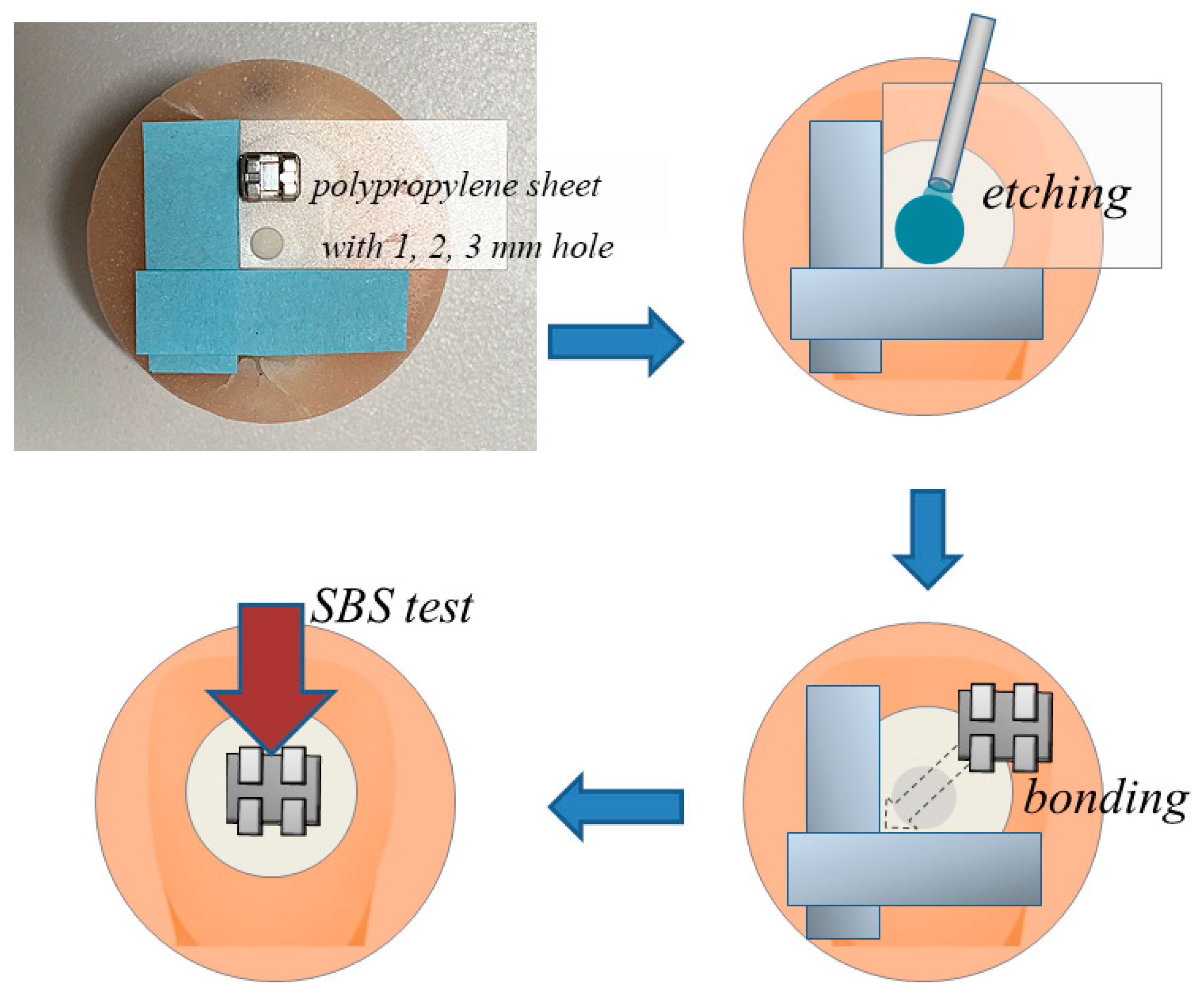
2. Materials and Methods

2.1. Shear Bond Strength

The samples, consisting of 72 bovine mandibular incisors, were divided into eight groups of nine specimens each. Bovine enamel was employed due to being an acceptable substrate for bonding testing [9]. The enamel was flattened and polished up to #2000-grit with silicon carbide paper. The pre-etching (Transbond XT Etching Gel; 3M Unitek, Monrovia, CA, USA https://www.3m.com/3M/en_US/p/c/dental-orthodontics/ accessed on 16 August 2021; and GC Ortho Etching Gel; GC Ortholy, Tokyo, Japan, https://www.gcortholy.com/ accessed on 16 August 2021, application time; 30 s) was performed in circles of different areas (diameter: 1.0, 2.0 or 3.0 mm). The area was standardised with a hole in a 0.2 mm-thick polypropylene sheet (Figure 1). The pre-etched surface was rinsed and dried for 20 s. A paste (Transbond XT [TB]; 3M Unitek (functional monomer-free), or Universal Bond [UB]; GC Ortholy (containing functional monomer)) was smeared on the bracket and pressed against the pre-etched enamel surface. After removing the excess paste, the mesio-distal portion of the bracket was irradiated with an LED curing unit (Valo; Ultradent Products, South Jordan, UT, USA, https://www.ultradent.com/ accessed on 16 August 2021) for 20 s. After being stored in distilled water (37 °C for 24 h), each specimen was tested in shear mode using a universal testing machine (5567; Instron, Norwood, MA, USA, https://www.instron.com/ accessed on 16 August 2021, experimental environment; 23 ± 1 °C and 50 ± 5% relative humidity) at a crosshead speed of 1 mm/min. Shear bond strength (SBS) was calculated as the peak load at failure divided by the bonded area (12.8 mm2) of the bracket base. For the baseline groups, the bracket was bonded to a pre-etched enamel surface that was the same size as the bracket base. The SBSs of the baseline groups were measured under the same conditions described above.

2.2. Adhesive Remnant Index

After the SBS test, the tooth and bracket sides of each debonded specimen were examined under an optical microscope (SZ-3003; As One, Osaka, Japan) at 15× magnification. The residual adhesive on each tooth was assessed based on the adhesive remnant index (ARI) [10].

2.3. Knoop Hardness Numbers of Orthodontic Adhesive Pastes

To define orthodontic adhesive paste thickness, an adhesive tape with a hole (internal diameter, 2 mm; thickness, 100 μm) was attached to transparent matrix tape (Matrix Tape and Dispenser; 3M Oral Care), which was then put on a flat enamel surface. The paste was condensed into the hole of the adhesive tape and a metal bracket was then pressed on the paste. Light irradiation was performed from two different sides for 20 s. The bonded specimens were stored under conditions of 100% humidity at 37 °C for 24 h, prior to measurement of the Knoop hardness number (KHN) at the centre of the adhesive paste bonded to the bracket (from the indentation), following application of a load (98.7 mN) with a microhardness tester (HMV-2; Shimadzu, Kyoto, Japan, https://www.shimadzu.com/ accessed on 16 August 2021, application time; 15 s). Ten specimens were prepared for each adhesive paste and the mean values were calculated.

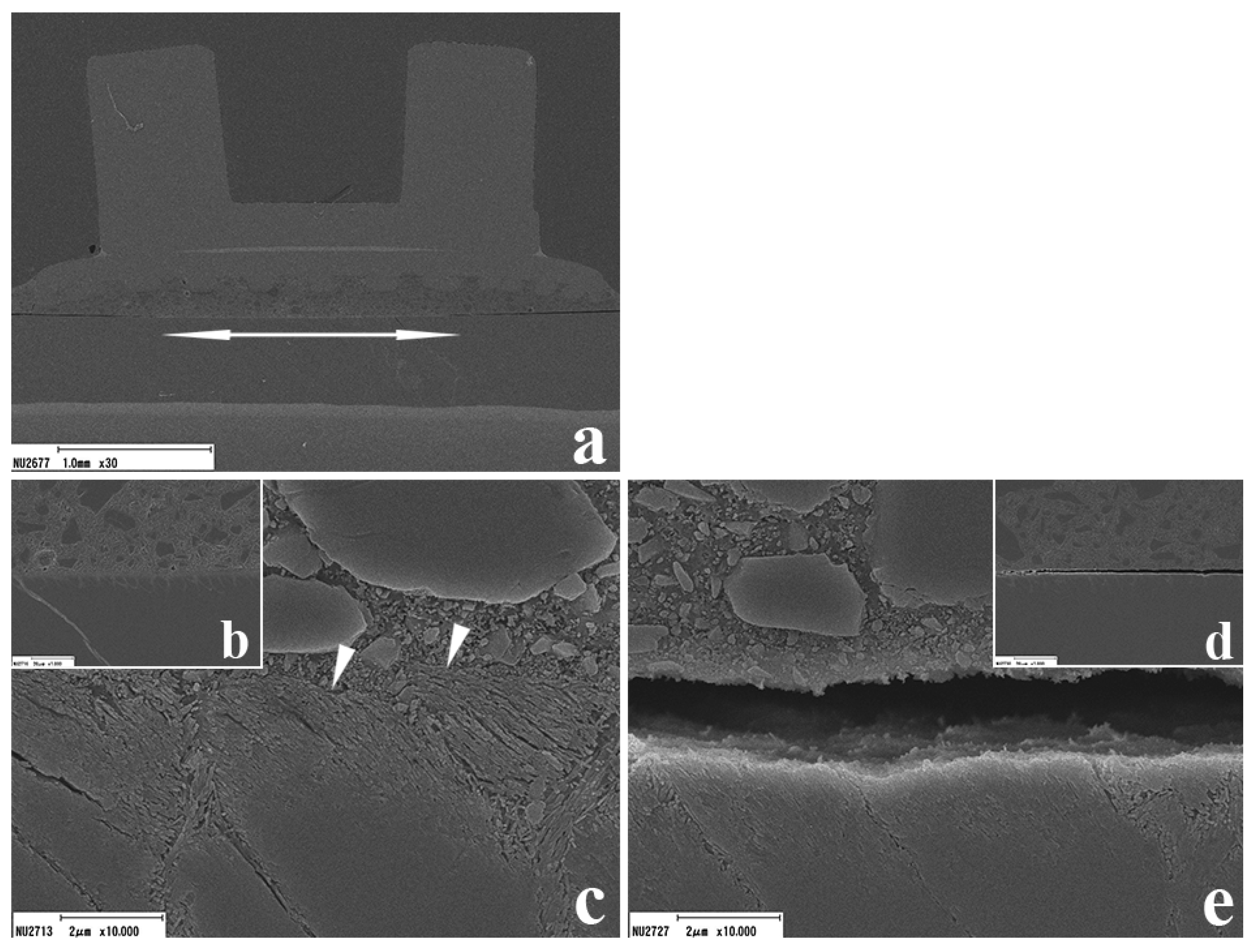
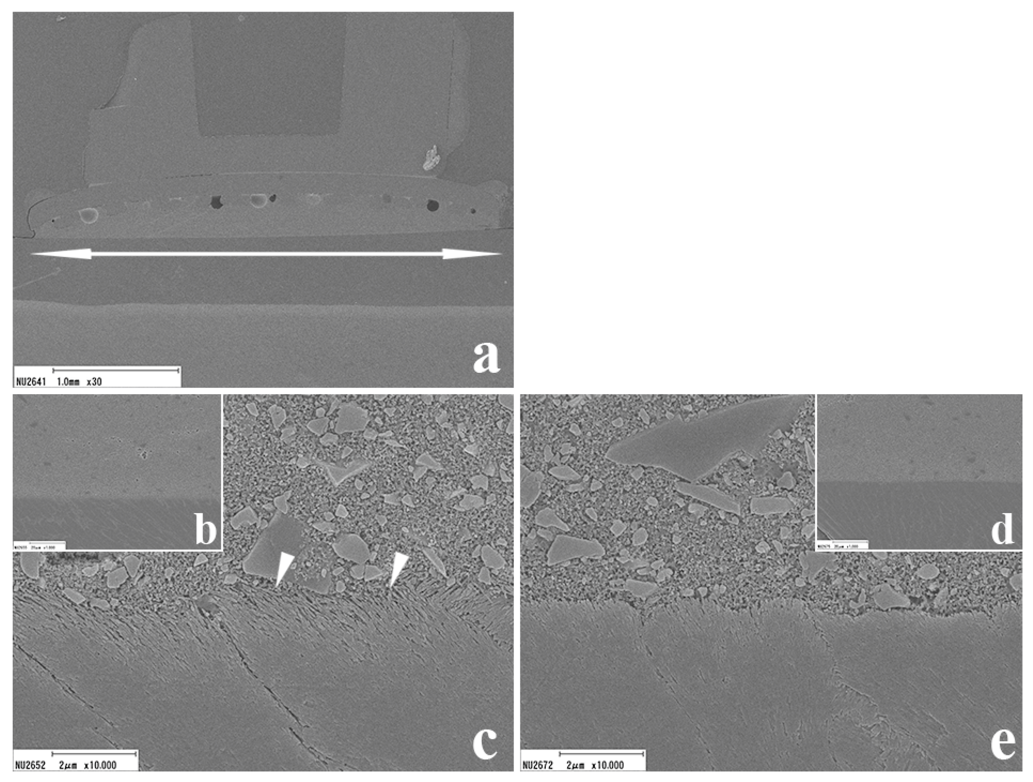
2.4. Scanning Electron Microscopy

The cross section of the bonded brackets was observed using scanning electron microscopy (SEM) (ERA-8800FE; Elionix Ltd., Tokyo, Japan, https://www.elionix.co.jp/ accessed on 16 August 2021) to determine the ultrastructure of the adhesive paste/enamel interfaces. Bonded specimens were fabricated as described for the SBS test and stored in distilled water at 37 °C for 24 h, before being embedded in epoxy resin (Epon 812; Nisshin EM, Tokyo, Japan) and sectioned longitudinally. All specimens were examined under the operating condition of a voltage of 10 kV.

2.5. Statistical Analysis

Statistical power analysis indicated the need for seven specimens for SBS tests. Descriptive statistics were calculated using a statistical analysis software (SPSS Inc., Chicago, IL, USA). Kolmogorov–Smirnov and Leven tests were performed to assess normality and homogeneity of variance. Analysis of variance followed by the Games–Howell post hoc test was used for statistical comparison of SBS values [11]. For comparison of ARI between the pre-etching areas and the adhesives, the Chi-squared test with Yates’ correction was performed [12]. Student’s t test was conducted for comparison of KHN.

3. Results

3.1. SBS

The SBSs of TB and UB are presented in Table 1. For the baseline groups, the mean SBS of TB was 12.3 MPa and that of UB was 13.3 MPa. For the 1 mm- and 2 mm-diameter pre-etched groups, the mean SBSs of TB were 2.7 and 3.5 MPa, and those of UB were 2.9 and 5.2 MPa, respectively. For the 3 mm-diameter pre-etched groups, the mean SBS of TB was 6.7 MPa, whereas that of UB was 10.0 MPa. The 1 and 2 mm groups with both adhesives, and the 3 mm group with TB, indicated significantly lower SBS values than the baseline groups. On the other hand, there was no significant difference in SBS between the 3 mm group and baseline group with UB. When comparing different materials with the same pre-etched area, UB showed higher SBS values than TB.

3.2. Adhesive Remnant Index

The ARI results are presented in Table 2. For TB, no significant differences in failure mode were observed among groups with different pre-etched areas. For UB, significant differences in failure mode were observed among the groups and the ARI tended to increase with increasing pre-etched area.

3.3. KHN of Orthodontic Adhesive Pastes

The KHNs of the tested orthodontic adhesive pastes are presented in Table 3. The mean KHN of UB was 6.2 ± 1.0 and that of TB was 16.5 ± 1.6.

3.4. SEM Observations

SEM images of adhesive paste/enamel interfaces in the 2 mm groups are shown in Figure 2 and Figure 3. For TB (Figure 2), although the pre-etched region (white arrow in Figure 2a) indicated excellent adaptation between demineralised enamel substrate and the adhesive paste, the non-etched region showed a gap between the enamel and adhesive paste. For UB (Figure 3), excellent adaptation between enamel substrate and the adhesive paste was observed in both the pre-etched and non-etched regions (Figure 3c,e). Adhesive paste interpenetration with enamel as resin tags was clearly observed in the pre-etched region with both adhesive pastes (Figure 2c and Figure 3c).

4. Discussion

In this study, bovine enamel was employed as a substitute for human enamel because of the difficulty of collecting the latter in the same condition [13]. To maximise the similarity to clinical orthodontic treatment with direct bonding of brackets, it would have been best to use intact enamel in the experiment. However, the undulations of bovine enamel are deeper and broader than those of human enamel. Therefore, the adherent enamel surfaces were ground flat to standardise the methodology and ensure both an appropriate adherent area for the bonded assembly and a uniform stress distribution [14,15].
Various types of functional monomer have been employed in different types of adhesive systems, such as phosphoric acid ester, carboxylic acid and alcohol functional monomers [16]. The functional monomers 10-methacryloyloxydecyl dihydrogen phosphate (MDP) and 4-methacryloxyethyl trimellitate anhydride (4-META) have often been employed in orthodontic adhesive systems [17,18]. In this study, UB contained MDP as a functional monomer, but there were no significant differences in SBS between UB and the functional monomer-free orthodontic adhesive paste TB, regardless of the pre-etched area. Therefore, the first null hypothesis, that the enamel SBS in different orthodontic adhesive pastes would not differ according to the presence or absence of functional monomers in the adhesive paste, was not rejected. An optimal orthodontic adhesive system would not fail during treatment, and would allow easy debonding of the brackets on completion of treatment [19]. Previous studies have reported that the ideal enamel bond strength of orthodontic brackets ranges from 6 to 13.7 MPa [5,6]. Excessive enamel bond strength may cause damage to the intact enamel surface when debonding brackets [20]. In this study, the baseline and 3 mm groups were within the range of ideal bond strength for both materials. Although UB showed higher SBS values than TB, the difference was not significant in any pre-etched group. This can be explained by the lower mechanical properties of UB due to the presence of functional monomers. The incompatibility between residual acidic monomer and the other resin monomers, and the low pH of functional monomers, may inhibit polymerization, resulting in lower mechanical properties [21,22]. This was supported by the results of the KHN and ARI evaluation. UB showed a significantly lower KHN and marked increases in the rates of ARI scores 2 and 3 than TB.
However, on SEM images, although UB in the 2 mm group showed excellent adaptation in both pre-etched and non-etched regions, TB in the 2 mm group showed gap formation between the enamel and adhesive paste in the non-etched region. This gap formation between the adhesive paste and enamel substrate may be a cause of caries, staining and bracket failure during orthodontic treatment. Therefore, orthodontic adhesive paste containing functional monomers may improve the integrity of the interface even in non-etched regions due to its chemical bonding ability.
On investigation of the influence of the pre-etched area on the enamel SBS, no significant differences were observed in SBS with UB between the 3 mm and baseline groups. On the other hand, the functional monomer-free adhesive paste TB showed significantly lower SBSs in the partially pre-etched groups than the baseline group, regardless of the pre-etched area. Therefore, the second null hypothesis, that the enamel bond strength would not differ by pre-etched area, was rejected. Although enamel HAp is considered less amenable to chemical reaction with functional monomers than dentin HAp [23,24], the functional monomer in the orthodontic adhesive may enhance enamel bond effectiveness. However, it may be difficult to reach the minimum enamel bond strength requirement without phosphoric acid pre-etching, even when orthodontic adhesive pastes contain functional monomers. Takamiya et al. investigated the bond effectiveness of five self-adhesive flowable resin composites without any pre-treatment, and recommended selective enamel etching for these materials [25]. Based on previous investigations of self-adhesive materials and the outcomes of this study, it may be necessary to pre-etch at least half of the bracket base area in the case of UB.
It is difficult to compare the outcomes of in vitro experiments and clinical situations directly. In addition, the bond strength value is influenced by many factors, such as the type of bond strength test, adherent substrate, material selection, adherent conditions, caries prophylaxis treatment, etc. [26,27,28]. In this study, the adherent enamel surface was ground flat to standardise conditions, but this introduces an important difference from the clinical situation. The outer surface of intact enamel, i.e., the prismless layer, has stronger resistance to acid than a prismatic one. In a previous investigation of self-etching adhesive systems for intact enamel using different etching strategies, unground enamel without pre-etching showed significantly lower bond performance than ground enamel, but no significant difference was observed between ground and unground enamel with pre-etching [29]. It was suggested that mechanical interlocking is more important for intact enamel adhesion than chemical bonding. Hence, phosphoric acid pre-etching is necessary for securing brackets during treatment, even when using orthodontic adhesive pastes marketed as self-adhesive.
Although there has been a study showing the enamel bonding effectiveness of UB, little information is available on this material [30]. On the other hand, TB has been used in many studies, often as a control material [31,32,33]. In this study, to standardise the bonding protocol between TB and UB, we omitted the priming step of TB. The primer composition of TB is similar to typical bonding agents and does not contain any functional monomers. In a previous study, SEM revealed no ultrastructure differences in the etched enamel region according to primer application [34]. In addition, previous laboratory studies demonstrated comparable tensile bond strength with and without primer application [35,36,37]. These findings suggest that primer application may not influence enamel bond effectiveness or morphological features in the vicinity of the interface of TB, and that the bonding procedures used in the baseline and partially pre-etched groups did not cause problems.
Regarding the ARI, a significant difference was detected in UB, but not TB, according to the pre-etched area. UB tended to show an increase in the rate of ARI scores of 2 and 3 with increasing pre-etched area. Remnant adhesive paste on the enamel surface should be removed with rotary rather than hand instruments [38]. Although grinding and polishing the adhesive remnant on the tooth surface may be time-consuming, this method is safe and gentle [39]. Therefore, adhesive paste containing functional monomers with partial pre-etching may be preferable to functional monomer-free adhesive paste because of adaptation and improved treatment of the debonded enamel surface.

5. Conclusions

Within the limitations of this study, we concluded that:
  • Enamel bond strengths of orthodontic adhesive pastes increased with increasing pre-etched area.
  • SEM revealed no gap formation in the vicinity of the interface with UB, in contrast to TB.
  • For UB, adhesive failure at the adhesive paste/bracket interface increased as a higher proportion of the bonding area was pre-etched.
These findings suggest that the pre-etching area markedly influences enamel bond effectiveness regardless of the presence or absence of functional monomers in orthodontic adhesive paste.

Author Contributions

Conceptualization, Y.N. and T.T.; methodology, Y.N., Y.T. and Y.U.; validation, Y.U. and M.I.; formal analysis, Y.N.; investigation, Y.T., A.U. and K.W.; resources, Y.T.; data curation, K.W.; writing—original draft preparation, Y.N.; writing—review and editing, T.T. and M.M.; visualization, Y.N.; supervision, M.M.; project administration, Y.N.; funding acquisition, Y.N. and T.T. All authors have read and agreed to the published version of the manuscript.

Funding

This work was supported in part by a Grant-in-Aid for Scientific Research (no. 19K10158) from the Japan Society for the Promotion of Science. This project was also supported by the Sato Fund in 2021.

Institutional Review Board Statement

Not applicable.

Data Availability Statement

The data are contained within the article and/or available from the corresponding author upon reasonable request.

Acknowledgments

Not applicable.

Conflicts of Interest

The authors declare no conflict of interest.

References

  1. de Assis, C.; Lemos, C.; Gomes, J.; Vasconcelos, B.; Moraes, S.; Braz, R.; Pellizzer, E.P. Clinical efficiency of self-etching one-step and two-step adhesives in NCCL: A systematic review and meta-analysis. Oper. Dent. 2020, 45, 598–607. [Google Scholar] [CrossRef] [PubMed]
  2. Patcas, R.; Eliades, T. Structure/property relationships in orthodontic ceramics. In Orthodontic Applications of Biomaterials; Eliades, T., Brantley, W.A., Eds.; Woodhead Publishing: Cambridge, UK, 2017; p. 225. [Google Scholar]
  3. Van Landuyt, K.L.; Yoshida, Y.; Hirata, I.; Snauwaert, J.; De Munck, J.; Okazaki, M.; Suzuki, K.; Lambrechts, P.; Van Meerbeek, B. Influence of the chemical structure of functional monomers on their adhesive performance. J. Dent. Res. 2008, 87, 757–761. [Google Scholar] [CrossRef] [PubMed]
  4. Takamizawa, T.; Barkmeier, W.W.; Latta, M.A.; Berry, T.P.; Tsujimoto, A.; Miyazaki, M. Simulated Wear of Self-Adhesive Resin Cements. Oper. Dent. 2016, 41, 327–338. [Google Scholar] [CrossRef] [PubMed]
  5. Imai, A.; Takamizawa, T.; Sugimura, R.; Tsujimoto, A.; Ishii, R.; Kawazu, M.; Saito, T.; Miyazaki, M. Interrelation among the handling, mechanical, and wear properties of the newly developed flowable resin composites. J. Mech. Behav. Biomed. Mater. 2019, 89, 72–80. [Google Scholar] [CrossRef]
  6. Hess, E.; Campbell, P.M.; Honeyman, A.L.; Buschang, P.H. Determinants of enamel decalcification during simulated orthodontic treatment. Angle Orthod. 2011, 81, 836–842. [Google Scholar] [CrossRef] [PubMed]
  7. Reynolds, I.R. A review of direct orthodontic bonding. Br. J. Orthod. 1975, 2, 171–179. [Google Scholar] [CrossRef]
  8. Retief, D.H. Failure at the dental adhesive-etched enamel interface. J. Oral Rehabil. 1974, 1, 265–294. [Google Scholar] [CrossRef]
  9. Soares, F.Z.; Follak, A.; da Rosa, L.S.; Montagner, A.F.; Lenzi, T.L.; Rocha, R.O. Bovine tooth is a substitute for human tooth on bond strength studies: A systematic review and meta-analysis of in vitro studies. Dent. Mater. 2016, 32, 1385–1393. [Google Scholar] [CrossRef]
  10. Årtun, K.; Bergland, S. Clinical trials with crystal growth conditioning as an alternative to acid-etch enamel pretreatment. Am. J. Orthod. 1984, 85, 333–340. [Google Scholar] [CrossRef]
  11. Namura, Y.; Takamizawa, T.; Uchida, Y.; Inaba, M.; Noma, D.; Takemoto, T.; Miyazaki, M.; Motoyoshi, M. Effects of composition on the hardness of orthodontic adhesives. J. Oral Sci. 2020, 62, 48–51. [Google Scholar] [CrossRef] [Green Version]
  12. Masucci, C.; Cipriani, L.; Defraia, E.; Franchi, L. Transverse relationship of permanent molars after crossbite correction in deciduous dentition. Eur. J. Orthod. 2017, 39, 560–566. [Google Scholar] [CrossRef] [PubMed]
  13. Yassen, G.H.; Platt, J.A.; Hara, A.T. Bovine teeth as substitute for human teeth in dental research: A review of literature. J. Oral Sci. 2011, 53, 273–282. [Google Scholar] [CrossRef] [PubMed] [Green Version]
  14. Braga, R.R.; Meira, J.B.; Boaro, L.C.; Xavier, T.A. Adhesion to tooth structure: A critical review of “macro” test methods. Dent. Mater. 2010, 26, e38–e49. [Google Scholar] [CrossRef] [PubMed]
  15. Sirisha, K.; Rambabu, T.; Shankar, Y.R.; Ravikumar, P. Validity of bond strength tests: A critical review: Part I. J. Conserv. Dent. 2014, 17, 305–311. [Google Scholar] [CrossRef] [PubMed] [Green Version]
  16. Yoshida, Y.; Nagakane, K.; Fukuda, R.; Nakayama, Y.; Okazaki, M.; Shintani, H.; Inoue, S.; Tagawa, Y.; Suzuki, K.; De Munck, J.; et al. Comparative study on adhesive performance of functional monomers. J. Dent. Res. 2004, 83, 454–458. [Google Scholar] [CrossRef]
  17. Costa, A.C.; Sabóia, V.; Marçal, F.; Sena, N.; De Paula, D.; Ribeiro, T.; Feitosa, V. In vitro evaluation of experimental self-adhesive orthodontic composites used to bond ceramic brackets. Materials 2019, 12, 419. [Google Scholar] [CrossRef] [Green Version]
  18. Saito, K.; Sirirungrojying, S.; Meguro, D.; Hayakawa, T.; Kasai, K. Bonding durability of using self-etching primer with 4-META/ MMA-TBB resin cement to bond orthodontic brackets. Angle Orthod. 2005, 75, 260–265. [Google Scholar] [CrossRef]
  19. Theodorakopoulou, L.P.; Sadowsky, P.L.; Jacobson, A.; Lacefield, W., Jr. Evaluation of the debonding characteristics of 2 ceramic brackets: An in vitro study. Am. J. Orthod. Dentofac. Orthop. 2004, 125, 329–336. [Google Scholar] [CrossRef]
  20. Azzeh, E.; Feldon, P.J. Laser debonding of ceramic brackets: A comprehensive review. Am. J. Orthod. Dentofac. Orthop. 2003, 123, 79–83. [Google Scholar] [CrossRef]
  21. Sanares, A.M.; Itthagarun, A.; King, N.M.; Tay, F.R.; Pashley, D.H. Adverse surface interactions between one-bottle light-cured adhesives and chemical-cured composites. Dent. Mater. 2001, 17, 542–556. [Google Scholar] [CrossRef]
  22. Zorzin, J.; Petschelt, A.; Ebert, J.; Lohbauer, U. pH neutralization and influence on mechanical strength in self-adhesive resin luting agents. Dent. Mater. 2012, 28, 672–679. [Google Scholar] [CrossRef] [PubMed]
  23. Yoshihara, K.; Yoshida, Y.; Hayakawa, S.; Nagaoka, N.; Irie, M.; Ogawa, T.; Van Landuyt, K.L.; Osaka, A.; Suzuki, K.; Minagi, S.; et al. Nanolayering of phosphoric acid ester monomer on enamel and dentin. Acta Biomater. 2011, 7, 3187–3195. [Google Scholar] [CrossRef]
  24. Yaguchi, T. Layering mechanism of MDP-Ca salt produced in demineralization of enamel and dentin apatite. Dent. Mater. 2017, 33, 23–32. [Google Scholar] [CrossRef] [PubMed]
  25. Takamiya, H.; Tsujimoto, A.; Teixeira, E.C.; Jurado, C.A.; Takamizawa, T.; Barkmeier, W.W.; Latta, M.A.; Miyazaki, M.; Garcia-Godoy, F. Bonding and wear properties of self-adhesive flowable restorative materials. Eur. J. Oral Sci. 2021, 31, e12799. [Google Scholar] [CrossRef]
  26. Finnema, K.J.; Ozcan, M.; Post, W.J.; Ren, Y.; Dijkstra, P.U. In-vitro orthodontic bond strength testing: A systematic review and meta-analysis. Am. J. Orthod. Dentofac. Orthop. 2010, 137, 615–622.e3. [Google Scholar] [CrossRef]
  27. Salz, U.; Bock, T. Testing adhesion of direct restoratives to dental hard tissue—A review. J. Adhes. Dent. 2010, 12, 343–371. [Google Scholar] [CrossRef] [PubMed]
  28. Lanteri, V.; Segù, M.; Doldi, J.D.H.; Butera, A.D.H. Pre-bonding prophylaxis and brackets detachment: An experimental comparison of different methods. Int. J. Clin. Dent. 2014, 7, 191–197. [Google Scholar]
  29. Takeda, M.; Takamizawa, T.; Imai, A.; Suzuki, T.; Tsujimoto, A.; Barkmeier, W.W.; Latta, M.A.; Miyazaki, M. Immediate enamel bond strength of universal adhesives to unground and ground surfaces in different etching modes. Eur. J. Oral Sci. 2019, 127, 351–360. [Google Scholar] [CrossRef] [PubMed]
  30. Ok, U.; Aksakalli, S.; Eren, E.; Kechagia, N. Single-component orthodontic adhesives: Comparison of the clinical and in vitro performance. Clin. Oral Investig. 2021, 25, 3987–3999. [Google Scholar] [CrossRef] [PubMed]
  31. Romano, F.L.; Tavares, S.W.; Nouer, D.F.; Consani, S.; Magnani, M.B.B.A. Shear bond strength of metallic orthodontic brackets bonded to enamel prepared with Self-Etching Primer. Angle Orthod. 2005, 75, 849–853. [Google Scholar] [CrossRef]
  32. Cunha, T.M.A.; Behrens, B.A.; Nascimento, D.; Retamoso, L.B.; Lon, L.F.S.; Tanaka, O.; Guariza Filho, O. Blood contamination effect on shear bond strength of an orthodontic hydrophilic resin. J. Appl. Oral Sci. 2012, 20, 89–93. [Google Scholar] [CrossRef] [PubMed] [Green Version]
  33. Wan, A.; Razak, W.S.; Sherriff, M.; Bister, D.; Seehra, J. Bond strength of stainless steel orthodontic brackets bonded to prefabricated acrylic teeth. J. Orthod. 2017, 44, 105–109. [Google Scholar] [CrossRef]
  34. Jörgensen, K.D.; Shimokobe, H. Adaptation of resinous restorative materials to acid etched enamel surfaces. Scand. J. Dent. Res. 1975, 83, 31–36. [Google Scholar] [CrossRef]
  35. O’Brien, K.D.; Watts, D.C.; Read, M.J. Light cured direct bonding—Is it necessary to use a primer? Eur. J. Orthod. 1991, 13, 22–26. [Google Scholar] [CrossRef]
  36. Tang, A.T.; Björkman, L.; Adamczak, E.; Andlin-Sobocki, A.; Ekstrand, J. In vitro shear bond strength of orthodontic bondings without liquid resin. Acta Odontol. Scand. 2000, 58, 44–48. [Google Scholar] [CrossRef] [PubMed]
  37. Bazargani, F.; Magnuson, A.; Löthgren, H.; Kowalczyk, A. Orthodontic bonding with and without primer: A randomized controlled trial. Eur. J. Orthod. 2016, 38, 503–507. [Google Scholar] [CrossRef] [PubMed]
  38. Janiszewska-Olszowska, J.; Szatkiewicz, T.; Tomkowski, R.; Tandecka, K.; Grocholewicz, K. Effect of orthodontic debonding and adhesive removal on the enamel - current knowledge and future perspectives—Asystematic review. Med. Sci. Monit. 2014, 20, 1991–2001. [Google Scholar] [CrossRef] [Green Version]
  39. Proffit, W.R. The third stage of comprehensive treatment: Finishing. In Contemporary Orthodontics, 5th ed.; Proffit, W.R., Fields, H.W., Sarver, D.M., Eds.; Mosby Elsevier: St Louis, MI, USA, 2013; pp. 582–605. [Google Scholar]
Figure 1. Bonding procedures.
Applsci 11 08251 g001
Figure 2. Representative SEM images of the orthodontic adhesive paste/enamel interface in TB. In the pre-etched area, excellent adaptation was observed (arrow), but gap formation was observed at both edges of the bracket base ((a): magnification 30×). In the pre-etched region ((b): magnification 1000×, (c): magnification 10,000×), compression of the enamel surface was observed (arrowheads). In the non-etched region, detachment between adhesive paste and enamel was observed ((d): magnification 1000×, (e): magnification 10,000×).
Applsci 11 08251 g002
Figure 3. Representative SEM images of the orthodontic adhesive paste/enamel interface in UB. The whole interface (arrow range) region showed excellent adaptation ((a): magnification 30×). In the pre-etched region ((b): magnification 1000 ×, (c): magnification 10,000×), a typical etching pattern and resin enamel tags were clearly observed (arrowheads). The non-etched region ((d): magnification 1000×, (e): magnification 10,000×) showed neither the typical etching pattern nor resin enamel tags.
Applsci 11 08251 g003
Table 1. Shear bond strength by pre-etched area.
Pre-Etching Area
(mm2)
Proportion of Bonding Area Pre-etchedTBUB
Mean ± SD (MPa)Group *Mean ± SD (MPa)Group *
1 mm0.80.062.7 ± 1.3a2.9 ± 1.1a
2 mm3.10.243.5 ± 1.3a5.2 ± 2.3a, b
3 mm7.10.556.7 ± 1.5b, c10.0 ± 2.6c, d
Baseline
(bracket base dimension: 3.2 mm × 4.0 mm)
12.81.0012.3 ± 3.0d13.3 ± 4.3d
* Different letters indicate a significant difference (p < 0.05). Abbreviation: SD, standard deviation.
Table 2. ARI scores by pre-etched area.
TBUB
Score01230123
1 mm 72009000
2 mm 54006030
3 mm 43200360
Baseline 62102025
Chi-square value
(p value)
1.6364
(p = 0.996)
27.4168
(p = 0.0012)
ARI scores: 0, no adhesive left on tooth surface; 1, less than 50% of adhesive left on tooth surface; 2, more than 50% of adhesive left on tooth surface; and 3, all adhesive left on the tooth surface [8].
Table 3. KHN for different orthodontic adhesive pastes.
TBUB
Mean ± SDGroup *Mean ± SDGroup *
16.5 ± 1.6a6.2 ± 1.0b
* Different letters indicate a significant difference (p < 0.05). Abbreviation: SD, standard deviation.
Publisher’s Note: MDPI stays neutral with regard to jurisdictional claims in published maps and institutional affiliations.

