
Investigation of Ancient Wall Painting Fragments Discovered in the Roman Baths from Alburnus Maior by Complementary Non-Destructive Techniques


Abstract
:1. Introduction
2. Materials and Methods
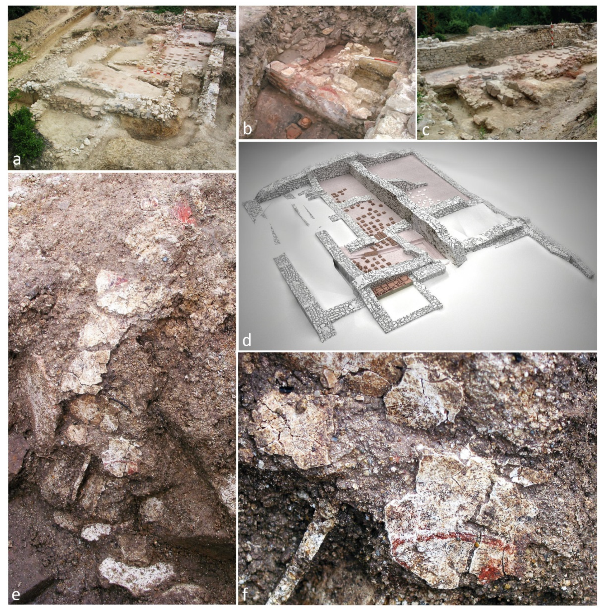
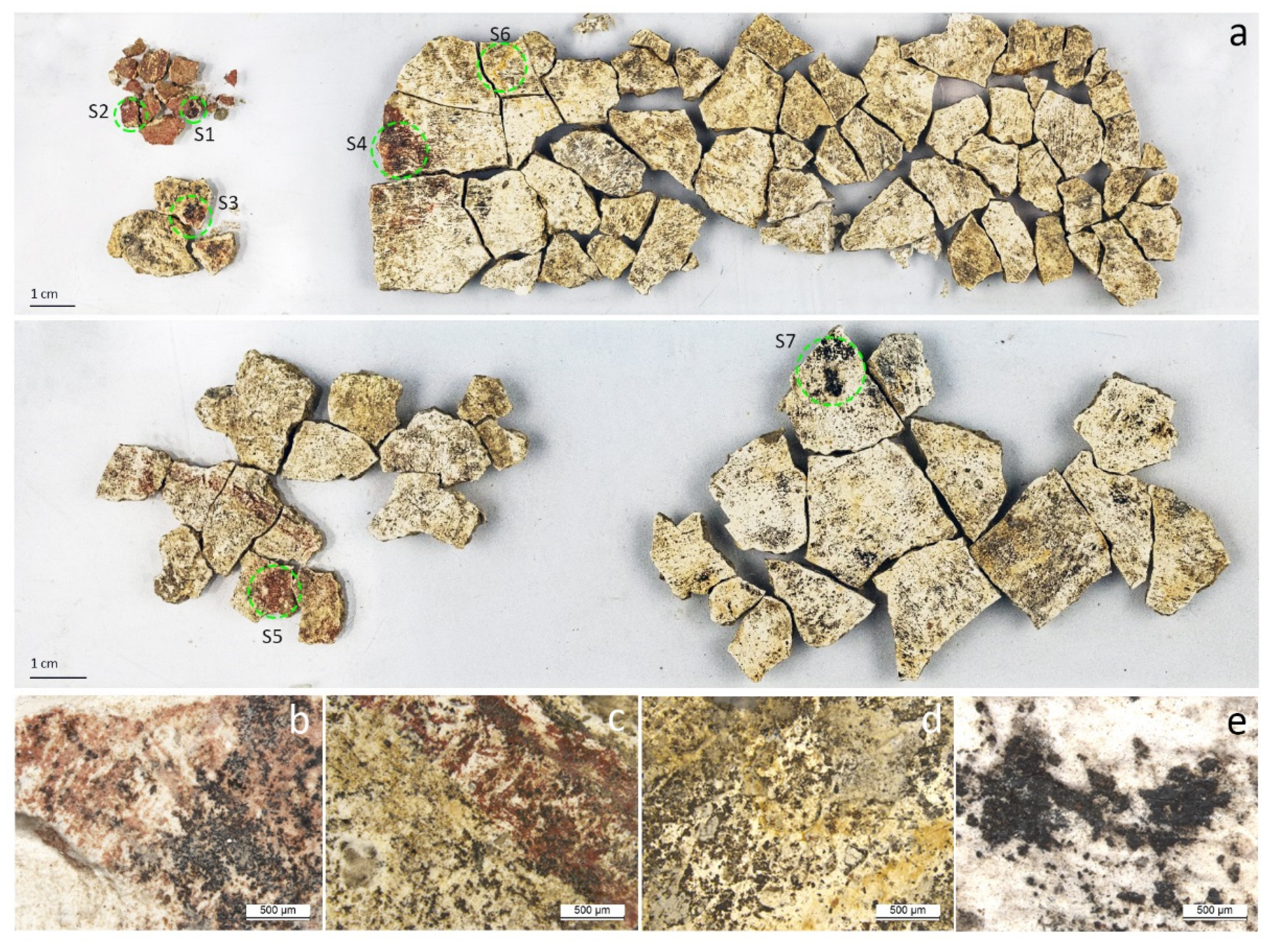
2.1. Archeological Background and Wall Painting Samples
2.2. Fourier Transform Infrared Spectroscopy (FTIR)
2.3. X-ray Fluorescence (XRF)
2.4. X-ray Diffraction (XRD)
2.5. Imaging Diagnostic Techniques
3. Results
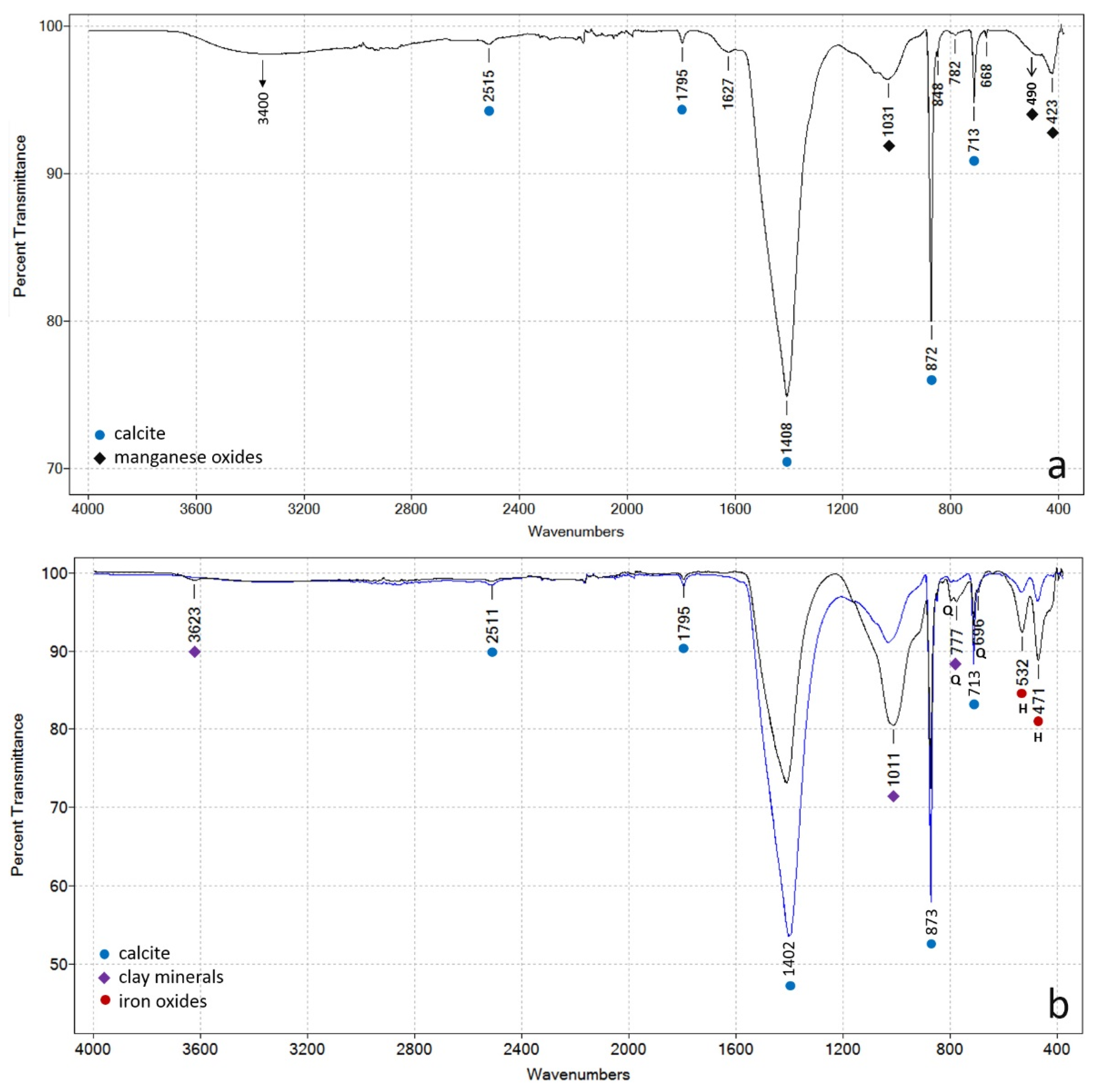
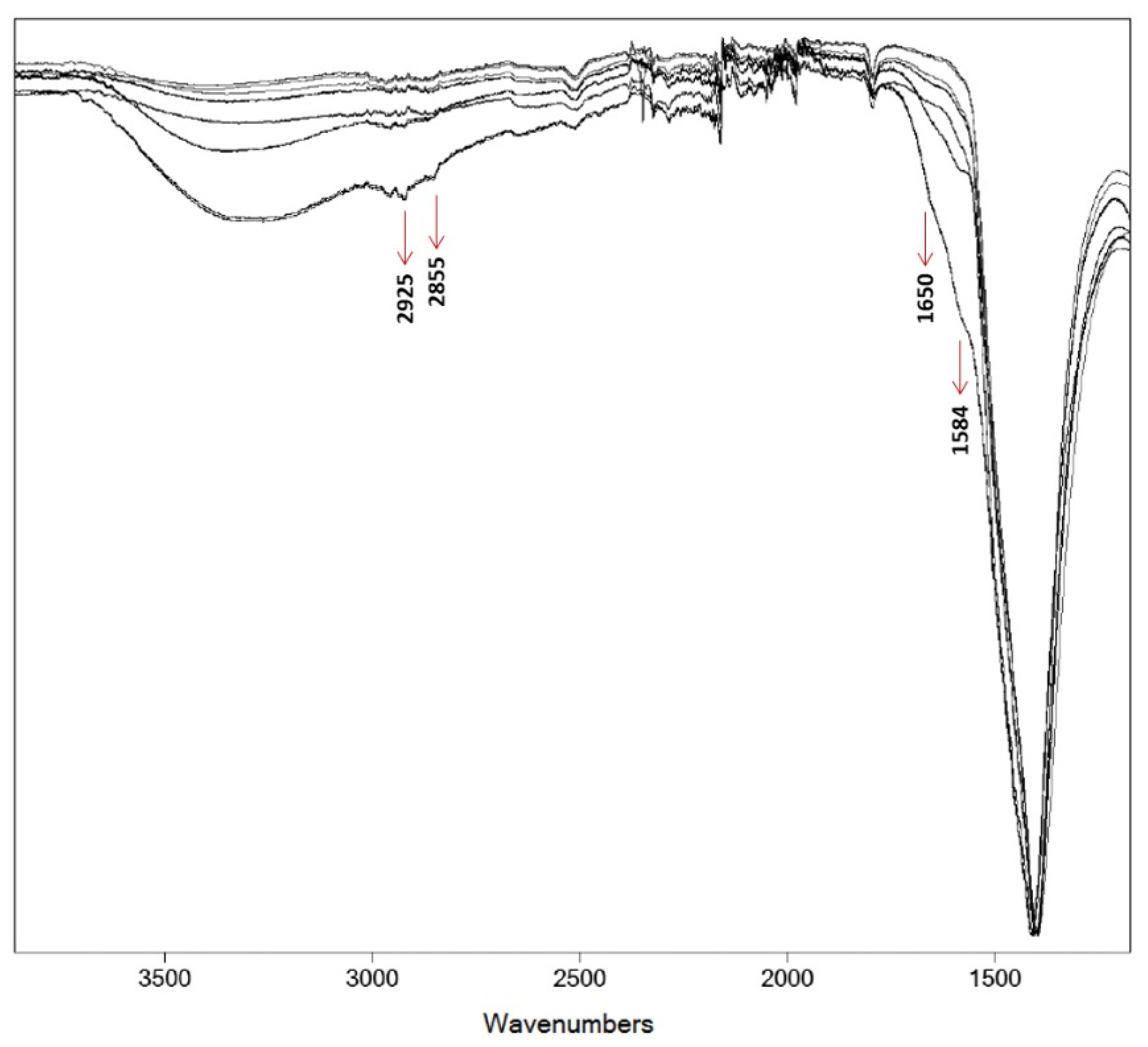
3.1. FTIR Analysis
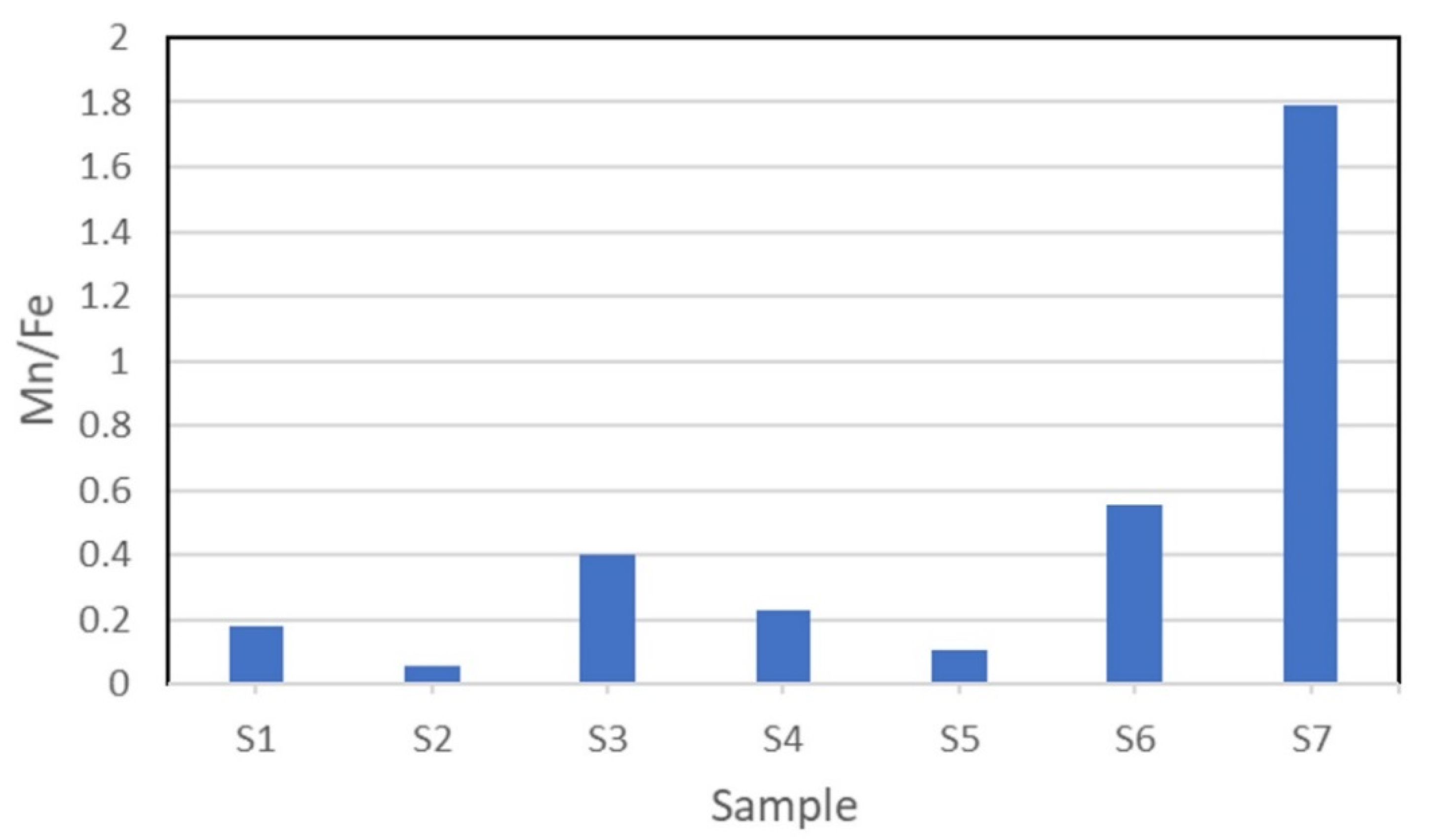
3.2. XRF Analysis
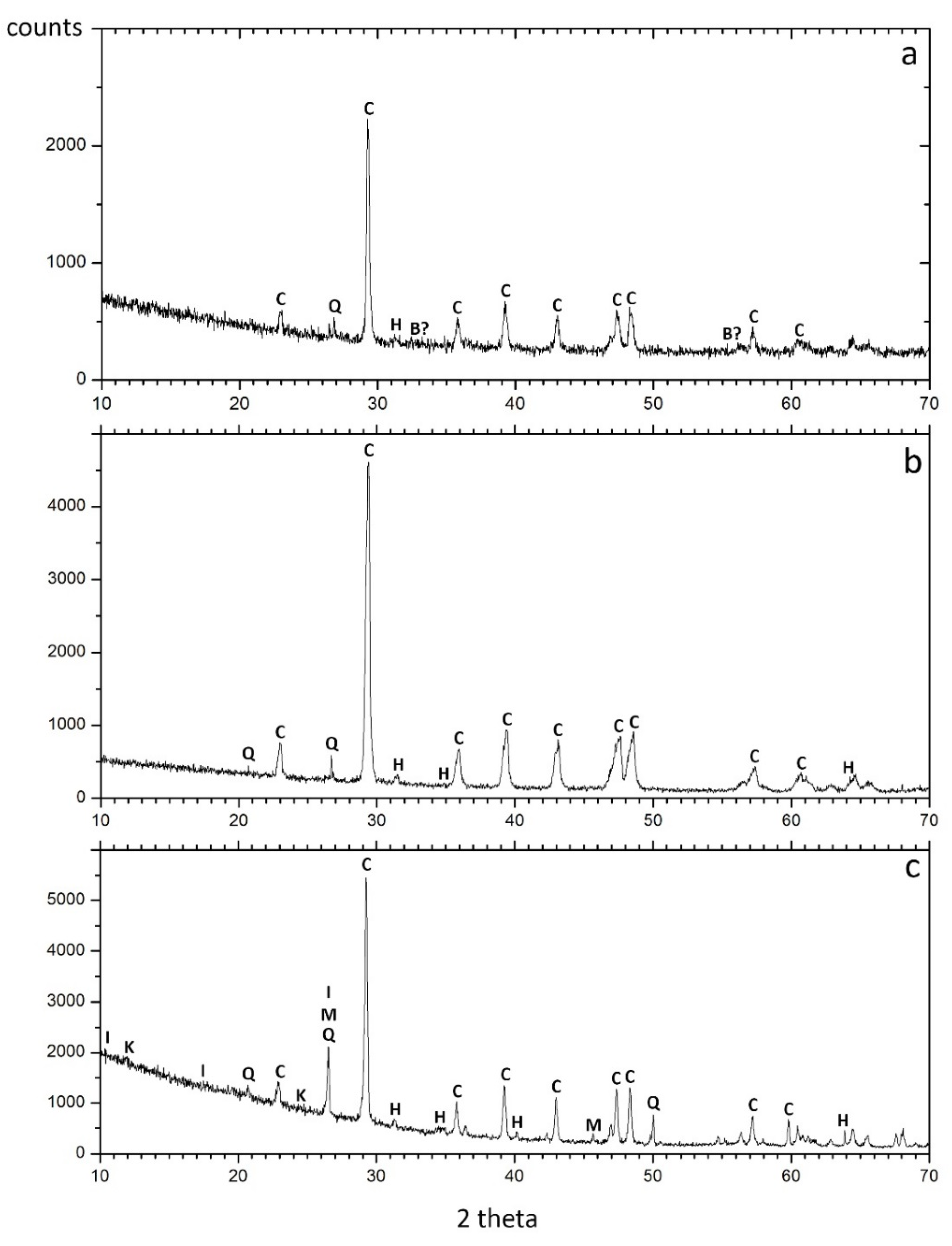
3.3. XRD Analysis
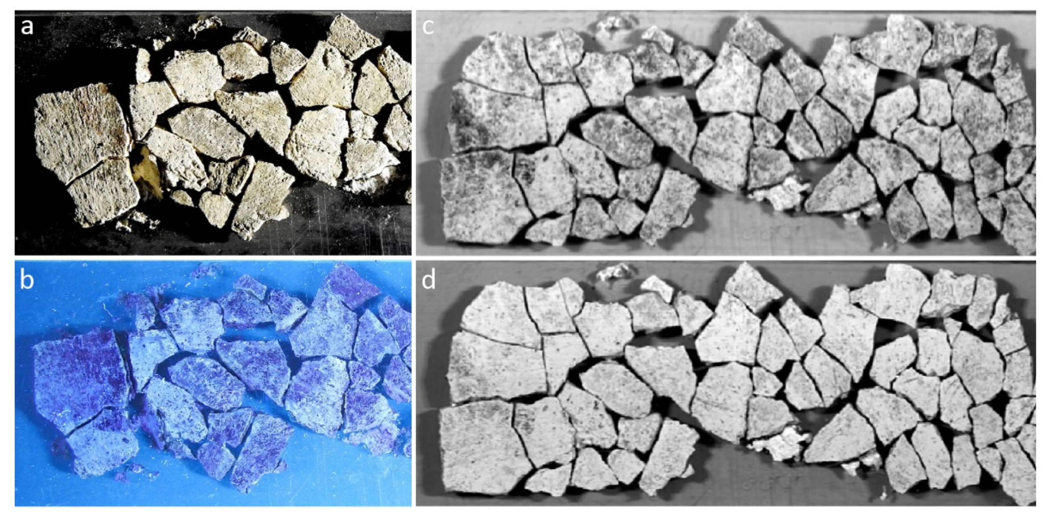
3.4. Imaging Documentation
4. Discussion
5. Conclusions
Author Contributions
Funding
Data Availability Statement
Conflicts of Interest
References
- Kosso, C.; Scott, A. The Nature and Function of Water, Baths, Bathing and Hygiene from Antiquity through the Renaissance; Brill: Leiden, The Netherlands, 2009. [Google Scholar]
- Nielsen, I. Thermae et Balnea: The Architecture and Cultural History of Roman Public Baths; Aarhus University Press: Aarhus, Denmark, 1993. [Google Scholar]
- DeLaine, J. Recent research on Roman baths. J. Rom. Archaeol. 1988, 1, 11–32. [Google Scholar] [CrossRef]
- Țentea, O.; Burkhardt, B. Bath and bathing in Dacia (1). Current state of research of the balnea. J. Anc. Hist. Archaeol. 2020, 7, 12–21. [Google Scholar] [CrossRef]
- Țentea, O. Bath and Bathing at Alburnus Maior; Mega Publishing House: Cluj-Napoca, Romania, 2015. [Google Scholar]
- Cortea, I.M.; Ghervase, L.; Țentea, O.; Pârău, A.C.; Rădvan, R. First Analytical Study on Second-Century Wall Paintings from Ulpia Traiana Sarmizegetusa: Insights on the Materials and Painting Technique. Int. J. Archit. Herit. 2020, 14, 751–761. [Google Scholar] [CrossRef]
- Piovesan, R.; Siddall, R.; Mazzoli, C.; Nodari, L. The Temple of Venus (Pompeii): A study of the pigments and painting techniques. J. Archaeol. Sci. 2011, 38, 2633–2644. [Google Scholar] [CrossRef]
- Coccato, A.; Mazzoleni, P.; Spinola, G.; Barone, G. Two centuries of painted plasters from the Lateran suburban villa (Rome): Investigating supply routes and manufacturing of pigments. J. Cult. Herit. 2021, 48, 171–185. [Google Scholar] [CrossRef]
- Ling, R. Roman Painting; Cambridge University Press: New York, NY, USA, 1991. [Google Scholar]
- Ciobanu, R. Pictura Murală Romană (La Peinture Murale Romaine); Alba Iulia: Editura Grinta, Romania, 2011. [Google Scholar]
- Ciobanu, R. The Paintings Hall with Hypocausts from Apulum II. Apulum 2005, 42, 123–136. [Google Scholar]
- Țentea, O.; Olteanu, B.C. Decorating Overlapping Buildings: A Domus and Palmyrene Temple at Colonia Dacica Sarmizegetusa. Theor. Rom. Archaeol. J. 2020, 3, 6. [Google Scholar] [CrossRef]
- Boroș, D.; Duca, V. Tencuiala pictată din amfiteatrul de la Porolissum (consideraţii tehnice). AMP 2008, 30, 113–122. [Google Scholar]
- Caggiani, M.C.; Coccato, A.; Mazzoleni, P.; D’Alessio, A.; Russo, A.; Barone, G. Integrated analytical approach to unveil the secrets of the recently discovered ‘Sphinx Room’: A new piece of Domus Aurea puzzle. Herit. Sci. 2020, 8, 124. [Google Scholar] [CrossRef]
- Edreira, M.C.; Feliu, M.J.; Fernández-Lorenzo, C.; Martín, C. Roman wall paintings characterization from Cripta del Museo and Alcazaba in Mérida (Spain): Chromatic, energy dispersive X-ray flurescence spectroscopic, X-ray diffraction and Fourier transform infrared spectroscopic analysis. Anal. Chim. Acta 2001, 434, 331–345. [Google Scholar] [CrossRef]
- Cerrato, E.J.; Cosano, D.; Esquivel, D.; Jiménez-Sanchidrián, C.; Ruiz, J.R. Spectroscopic analysis of pigments in a wall painting from a high Roman Empire building in Córdoba (Spain) and identification of the application technique. Microchem. J. 2021, 168, 106444. [Google Scholar] [CrossRef]
- Angelini, I.; Asscher, Y.; Secco, M.; Parisatto, M.; Artioli, G. The pigments of the frigidarium in the Sarno Baths, Pompeii: Identification, stratigraphy and weathering. J. Cult. Herit. 2019, 40, 309–316. [Google Scholar] [CrossRef]
- Asscher, Y.; Angelini, I.; Secco, M.; Parisatto, M.; Chaban, A.; Deiana, R.; Artioli, G. Combining multispectral images with X-ray fluorescence to quantify the distribution of pigments in the frigidarium of the Sarno Baths, Pompeii. J. Cult. Herit. 2019, 40, 317–323. [Google Scholar] [CrossRef]
- Fischer, C.; Kakoulli, I. Multispectral and hyperspectral imaging technologies in conservation: Current research and potential applications. Stud. Conserv. 2006, 51, 3–16. [Google Scholar] [CrossRef]
- Cucci, C.; Picollo, M.; Chiarantini, L.; Uda, G.; Fiori, L.; De Nigris, B.; Osanna, M. Remote-sensing hyperspectral imaging for applications in archaeological areas: Non-invasive investigations on wall paintings and on mural inscriptions in the Pompeii site. Microchem. J. 2020, 158, 105082. [Google Scholar] [CrossRef]
- Ţentea, O.; Burkhardt, B. Baths on the Frontiers of Roman Dacia; Mega Publishing House: Cluj-Napoca, Romania, 2017. [Google Scholar]
- Crupi, V.; Allodi, V.; Bottari, C.; D’Amico, F.; Galli, G.; Gessini, A.; La Russa, M.F.; Longo, F.; Majolino, D.; Mariotto, G.; et al. Spectroscopic investigation of Roman decorated plasters by combining FT-IR, micro-Raman and UV-Raman analyses. Vib. Spectrosc. 2016, 83, 78–84. [Google Scholar] [CrossRef]
- Miliani, C.; Rosi, F.; Daveri, A.; Brunetti, B.G. Reflection infrared spectroscopy for the non-invasive in situ study of artists’ pigments. Appl. Phys. A Mater. Sci. Process. 2012, 106, 295–307. [Google Scholar] [CrossRef]
- Pique, F.; Verri, G. Organic Materials in Wall Paintings (Project Report); The Getty Conservation Institute: Los Angeles, CA, USA, 2015. [Google Scholar]
- Bikiaris, D.; Daniilia, S.; Sotiropoulou, S.; Katsimbiri, O.; Pavlidou, E.; Moutsatsou, A.P.; Chryssoulakis, Y. Ochre-differentiation through micro-Raman and micro-FTIR spectroscopies: Application on wall paintings at Meteora and Mount Athos, Greece. Spectrochim. Acta-Part A Mol. Biomol. Spectrosc. 2000, 56, 3–18. [Google Scholar] [CrossRef]
- Salama, W.; El Aref, M.; Gaupp, R. Spectroscopic characterization of iron ores formed in different geological environments using FTIR, XPS, Mössbauer spectroscopy and thermoanalyses. Spectrochim. Acta-Part A Mol. Biomol. Spectrosc. 2015, 136, 1816–1826. [Google Scholar] [CrossRef] [PubMed]
- Helwig, K. The characterisation of iron earth pigments using infrared spectroscopy. In Proceedings of the Second Infrared and Raman User’s Group (IRUG 2) Conference, Victoria and Albert Museum, London, UK, 12–13 September 1995; pp. 83–92. [Google Scholar]
- Genestar, C.; Pons, C. Earth pigments in painting: Characterisation and differentiation by means of FTIR spectroscopy and SEM-EDS microanalysis. Anal. Bioanal. Chem. 2005, 382, 269–274. [Google Scholar] [CrossRef]
- Kendix, E.L.; Prati, S.; Joseph, E.; Sciutto, G.; Mazzeo, R. ATR and transmission analysis of pigments by means of far infrared spectroscopy. Anal. Bioanal. Chem. 2009, 394, 1023–1032. [Google Scholar] [CrossRef] [PubMed]
- Farmer, V.C. The Infrared Spectra of Minerals; Mineralogical Society of Great Britain and Ireland: London, UK, 1974. [Google Scholar]
- Vahur, S.; Teearu, A.; Leito, I. ATR-FT-IR spectroscopy in the region of 550–230 cm−1 for identification of inorganic pigments. Spectrochim. Acta A Mol. Biomol. 2010, 75, 1061–1072. [Google Scholar] [CrossRef]
- Zviagina, B.B.; Drits, V.A.; Dorzhieva, O.V. Distinguishing features and identification criteria for K-dioctahedral 1M micas (Illite-aluminoceladonite and illite-glauconite-celadonite series) from middle-infrared spectroscopy data. Minerals 2020, 10, 153. [Google Scholar] [CrossRef] [Green Version]
- Ekosse, G.-I.E. Fourier transform infrared spectrophotometry and X-ray powder diffractometry as complementary techniques in characterizing clay size fraction of kaolin. J. Appl. Sci. Environ. Manag. 2005, 9, 43–48. [Google Scholar] [CrossRef] [Green Version]
- Hahn, A.; Vogel, H.; Andó, S.; Garzanti, E.; Kuhn, G.; Lantzsch, H.; Schüürman, J.; Vogt, C.; Zabel, M. Using Fourier transform infrared spectroscopy to determine mineral phases in sediments. Sediment. Geol. 2018, 375, 27–35. [Google Scholar] [CrossRef]
- Khang, V.C.; Korovkin, M.V.; Ananyeva, L.G. Identification of clay minerals in reservoir rocks by FTIR spectroscopy. IOP Conf. Ser. Earth Environ. Sci. 2016, 43, 012004. [Google Scholar] [CrossRef] [Green Version]
- Kang, L.; Zhang, M.; Liu, Z.H.; Ooi, K. IR spectra of manganese oxides with either layered or tunnel structures. Spectrochim. Acta A Mol. Biomol. 2007, 67, 864–869. [Google Scholar] [CrossRef]
- Siddall, R. Mineral pigments in archaeology: Their analysis and the range of available materials. Minerals 2018, 8, 201. [Google Scholar] [CrossRef] [Green Version]
- Eastaugh, N.; Walsh, V.; Chaplin, T.; Siddall, E. Pigment Compendium: A Dictionary and Optical Microscopy of Historical Pigments, 1st ed.; Butterworth-Heinemann: London, UK, 2008. [Google Scholar]
- Westlake, P.; Siozos, P.; Philippidis, A.; Apostolaki, C.; Derham, B.; Terlixi, A.; Perdikatsis, V.; Jones, R.; Anglos, D. Studying pigments on painted plaster in Minoan, Roman and Early Byzantine Crete. A multi-analytical technique approach. Anal. Bioanal. Chem. 2012, 402, 1413–1432. [Google Scholar] [CrossRef]
- Popelka-Filcoff, R.S.; Robertson, J.D.; Glascock, M.D.; Descantes, C. Trace element characterization of ochre from geological sources. J. Radioanal. Nucl. Chem. 2007, 272, 17–27. [Google Scholar] [CrossRef]
- Marcaida, I.; Maguregui, M.; Morillas, H.; Prieto-Taboada, N.; Fdez Ortiz de Vallejuelo, S.; Veneranda, M.; Madariaga, J.M.; Martellone, A.; De Nigris, B.; Osanna, M. In situ non-invasive characterization of the composition of Pompeian pigments preserved in their original bowls. Microchem. J. 2018, 139, 458–466. [Google Scholar] [CrossRef]
- Guineau, B.; Lorblanchet, M.; Gratuze, B.; Dulin, L.; Roger, P.; Akrich, R.; Muller, F. Manganese black pigments in prehistoric paintings: The case of the Black Frieze of Pech Merle (France). Archaeometry 2001, 43, 211–225. [Google Scholar] [CrossRef]
- Weihe, H.; Piligkos, S.; Barra, A.L.; Laursen, I.; Johnsen, O. EPR of Mn2+ impurities in calcite: A detailed study pertinent to marble provenance determination. Archaeometry 2009, 51, 43–48. [Google Scholar] [CrossRef]
- Butterman, W.C.; Reese, R.G.J. Mineral Commodity Profiles-Rubidium; Report/03-045; U.S. Geological Survey: Reston, VA, USA, 2003. [Google Scholar] [CrossRef]
- Beck, L.; Rousselière, H.; Castaing, J.; Duran, A.; Lebon, M.; Moignard, B.; Plassard, F. First use of portable system coupling X-ray diffraction and X-ray fluorescence for in-situ analysis of prehistoric rock art. Talanta 2014, 129, 459–464. [Google Scholar] [CrossRef]
- Bhattacharyya, P.K.; Dasgupta, S.; Fukuoka, M.; Roy, S. Geochemistry of braunite and associated phases in metamorphosed non-calcareous manganese ores of India. Contrib. Mineral. Petrol. 1984, 87, 65–71. [Google Scholar] [CrossRef]
- Cuní, J. What do we know of Roman wall painting technique? Potential confounding factors in ancient paint media analysis. Herit. Sci. 2016, 4, 44. [Google Scholar] [CrossRef] [Green Version]
- Tuñón, J.; Sánchez, A.; Parras, D.J.; Amate, P.; Montejo, M.; Ceprián, B. The colours of Rome in the walls of Cástulo (Linares, Spain). Sci. Rep. 2020, 10, 12739. [Google Scholar] [CrossRef] [PubMed]
- Siddall, R. Not a day without a line drawn: Pigments and painting techniques of Roman Artists. Infocus Mag. 2006, 2, 1–14. [Google Scholar] [CrossRef] [Green Version]
- Bioucas-Dias, J.M.; Plaza, A.; Dobigeon, N.; Parente, M.; Du, Q.; Gader, P.; Chanussot, J. Hyperspectral unmixing overview: Geometrical, statistical, and sparse regression-based approaches. IEEE J. Sel. Top. Appl. Earth Obs. Remote. Sens. 2012, 5, 354–379. [Google Scholar] [CrossRef] [Green Version]
- Rohani, N.; Pouyet, E.; Walton, M.; Cossairt, O.; Katsaggelos, A.K. Nonlinear Unmixing of Hyperspectral Datasets for the Study of Painted Works of Art. Angew. Chemie-Int. Ed. 2018, 57, 10910–10914. [Google Scholar] [CrossRef]
- Capobianco, G.; Prestileo, F.; Serranti, S.; Bonifazi, G. Hyperspectral imaging-based approach for the in-situ characterization of ancient Roman wall paintings. Period. Mineral. 2015, 84, 407–418. [Google Scholar] [CrossRef]
- de La Rie, E.R. Fluorescence of paint and varnish layers (Part I). Stud. Conserv. 1982, 27, 1–7. [Google Scholar] [CrossRef]
- Amadori, M.L.; Barcelli, S.; Poldi, G.; Ferrucci, F.; Andreotti, A.; Baraldi, P.; Colombini, M.P. Invasive and non-invasive analyses for knowledge and conservation of Roman wall paintings of the Villa of the Papyri in Herculaneum. Microchem. J. 2014, 118, 183–192. [Google Scholar] [CrossRef]
- Gelzo, M.; Corso, G.; Pecce, R.; Arcari, O.; Piccioli, C.; Dello Russo, A.; Arcari, P. An enhanced procedure for the analysis of organic binders in Pompeian’s wall paintings from Insula Occidentalis. Herit. Sci. 2019, 7, 12. [Google Scholar] [CrossRef]
- Fantoni, R.; Caneve, L.; Colao, F.; Fiorani, L.; Palucci, A.; Dell’Erba, R.; Fassina, V. Laser-induced fluorescence study of medieval frescoes by Giusto de’ Menabuoi. J. Cult. Herit. 2013, 14, S59–S65. [Google Scholar] [CrossRef] [Green Version]
- Angheluta, L.; Moldovan, A.; Rădvan, R. The teleoperation of a lif scanning device. UPB Sci. Bull. Ser. A Appl. Math. Phys. 2011, 73, 193–200. [Google Scholar]
- Allag, C.; Barbet, A. Techniques de Préparation des Parois Dans la Peinture Murale Romaine; Ecole Française de Rome: Rome, Italy, 1972. [Google Scholar]
- Barbet, A. Peinture Murale Romaine Dans Les Provinces De l’Empire, 1st ed.; British Archaeological Reports: Oxford, UK, 1983. [Google Scholar]
- Miriello, D.; Barca, D.; Bloise, A.; Ciarallo, A.; Crisci, G.M.; De Rose, T.; Gattuso, C.; Gazineo, F.; La Russa, M.F. Characterisation of archaeological mortars from Pompeii (Campania, Italy) and identification of construction phases by compositional data analysis. J. Archaeol. Sci. 2010, 37, 2207–2223. [Google Scholar] [CrossRef]
- Alonso-Olazabal, A.; Ortega, L.A.; Zuluaga, M.C.; Ponce-Antón, G.; Echevarría, J.J.; Fernández, C.A. Compositional characterization and chronology of roman mortars from the archaeological site of arroyo de la dehesa de velasco (Burgo de osma-ciudad de osma, Soria, Spain). Minerals 2020, 10, 393. [Google Scholar] [CrossRef]
- Ionescu, C.; Ghergari, L.; Țentea, O. Interdisciplinary (mineralogical-geological-archaeological) study on the tegular material belonging to the legion XIII Gemina from Alburnus Maior (Roșia Montanã) and Apulum (Alba Iulia): Possible raw materials sources. Cercet. Arheol. 2006, 13, 413–436. [Google Scholar] [CrossRef]


| Sample | Typology | Color/Typology | Detected Elements |
|---|---|---|---|
| S1 | paint layer | red with black | Fe, Ca, Mn, Cu, Ti, Pb, K, Hg, Zn, S, Sr, Si, P |
| S2 | paint layer | red | Ca, Fe, Mn, Cu, Ti, K, Pb, S, Si, Zn, Hg, Sr |
| S3 | paint layer | yellow, red, and black | Ca, Fe, Mn, Sr, K, Cu, Pb, Ti, Zn, Si, S, Rb, Cr, Al |
| S4 | paint layer | red with black | Ca, Fe, Mn, Sr, K, Ti, As, Zn, Pb, Cu, Si, Rb, S, P |
| S5 | paint layer | red with yellow | Ca, Fe, Mn, K, Sr, Ti, Pb, Cu, Zn, Si, Cr, Rb, S, P |
| S6 | paint layer | white with yellow | Ca, Fe, Mn, Sr, Pb, Cu, Ti, Ba, Zn, S, Si, Rb, P, Al |
| S7 | paint layer | black | Ca, Mn, Fe, Cu, Ti, Pb, Si, S, Sr, Zn |
| S8 | fine plaster layer | white | Ca, Fe, Sr, Mn, Cu, Ti |
| S9 | coarse plaster | greyish-white | Ca, Fe, K, Mn, Rb, Ti, As, Sr, Cu, Si |
Publisher’s Note: MDPI stays neutral with regard to jurisdictional claims in published maps and institutional affiliations. |
© 2021 by the authors. Licensee MDPI, Basel, Switzerland. This article is an open access article distributed under the terms and conditions of the Creative Commons Attribution (CC BY) license (https://creativecommons.org/licenses/by/4.0/).
Share and Cite
Cortea, I.M.; Ratoiu, L.; Ghervase, L.; Țentea, O.; Dinu, M. Investigation of Ancient Wall Painting Fragments Discovered in the Roman Baths from Alburnus Maior by Complementary Non-Destructive Techniques. Appl. Sci. 2021, 11, 10049. https://0-doi-org.brum.beds.ac.uk/10.3390/app112110049
Cortea IM, Ratoiu L, Ghervase L, Țentea O, Dinu M. Investigation of Ancient Wall Painting Fragments Discovered in the Roman Baths from Alburnus Maior by Complementary Non-Destructive Techniques. Applied Sciences. 2021; 11(21):10049. https://0-doi-org.brum.beds.ac.uk/10.3390/app112110049
Chicago/Turabian StyleCortea, Ioana Maria, Lucian Ratoiu, Luminița Ghervase, Ovidiu Țentea, and Mihaela Dinu. 2021. "Investigation of Ancient Wall Painting Fragments Discovered in the Roman Baths from Alburnus Maior by Complementary Non-Destructive Techniques" Applied Sciences 11, no. 21: 10049. https://0-doi-org.brum.beds.ac.uk/10.3390/app112110049