Article

Evaluation of Two Different Types of Mineral Trioxide Aggregate Cements as Direct Pulp Capping Agents in Human Teeth

1
Department of Conservative Dentistry & Endodontics, Manipal College of Dental Sciences, Manipal Academy of Higher Education, Manipal 576104, India
2
Department of Oral Pathology & Microbiology, Manipal College of Dental Sciences, Manipal Academy of Higher Education, Manipal 576104, India
3
Department of Orthodontics and Dentofacial Orthopaedics, Manipal College of Dental Sciences, Manipal Academy of Higher Education, Manipal 576104, India
4
Department of Oral and Maxillofacial Surgery, Seoul National University School of Dentistry, Seoul 03080, Korea
5
Department of Reconstructive Dentistry and Gerodontology, School of Dental Medicine, University of Bern, 3012 Bern, Switzerland
6
Division of Dental Biomaterials, Clinic for Reconstructive Dentistry, Center of Dental Medicine, University of Zurich, 8001 Zurich, Switzerland
*
Author to whom correspondence should be addressed.
Submission received: 23 September 2021 / Revised: 1 November 2021 / Accepted: 3 November 2021 / Published: 7 November 2021
(This article belongs to the Special Issue Clinical Applications for Dentistry and Oral Health)

Abstract

:
Traumatic human dental injuries involving the pulp might necessitate direct capping procedures. This clinical study aimed to analyse the histological outcomes using two different direct capping materials. Twenty patients with bilateral premolars, scheduled for orthodontic extraction, were selected. The teeth were treated either using ProRoot MTA or RetroMTA. All patients were recalled after 30 and 60 days for teeth extraction. The histopathologically stained specimens were blindly evaluated using hard tissue bridge formation, inflammatory reaction and pulpal findings criteria. Data were evaluated statistically. Results: After 60 days, only the parameter for hard tissue bridge formation showed significant difference in the ProRoot MTA group (p = 0.010), while both direct capping materials performed similarly regarding inflammatory pulp reaction and pulpal findings. Although, during the first 30 days, RetroMTA presented better results in terms of continuity, morphology, hard tissue bridge localisation, and extension/general state of the inflammatory reaction, the continuity was better at 60 days when ProRoot MTA was applied. Treatment with RetroMTA healed the pulpal tissue faster compared with ProRoot MTA but it seemed to be rather a reparative process.

1. Introduction

Dental trauma is a significant problem in young people. It has been reported that most dental traumatic injuries involve anterior teeth [1]. These traumatic injuries can cause irreparable damage to the young permanent teeth, not only at the time of accident, but also during the post-treatment period. Maintaining the vitality of the dentin–pulp complex is a high priority in cases of traumatic pulp exposures, since vital functioning pulp is capable of initiating several defence mechanisms to protect the body from bacterial invasion. Once pulpal tissue is exposed to the oral environment, among many other processes, repair or regeneration might occur, which is achieved by encouraging the formation of a “dentin bridge”, using direct pulp capping (DPC) procedures. These involve the medical or addressing material application upon exposed tissue. Several prior studies indicated calcium hydroxide as gold standard for pulp capping [2,3,4]. However, performed studies have also reported disadvantages using calcium hydroxide such as dentin barrier tunnels, obliteration of the pulp chamber caused by extensive dentin formation, high oral fluid solubility, and a lack of degradation and adhesion after acid etching [5,6,7,8]. Mineral trioxide aggregate is one commonly used pulp capping agent, which was first evaluated by Pitt Ford et al. in monkey’s teeth, showing a superior performance of MTA compared with calcium hydroxide [9].
MTA has several advantages over calcium hydroxide, such as lower solubility, improved mechanical strength, better marginal adaptation, and sealing ability [10]. However, ProRoot MTA has disadvantages such as long setting time and causes discoloration of teeth [11,12]. Hence, several MTA-like materials have been developed with improved physical properties. Recently, RetroMTA (BioMTA, Seoul, Korea), consisting of a hydraulic calcium zirconia complex, was introduced for pulp capping, pulpotomy, perforation repair, and as a base material [13,14]. The complex is a mixture of calcium carbonate, aluminium oxide, silicone dioxide, and zirconium oxide.
In RetroMTA, similar components are present as in ProRoot MTA but less heavy metal contents than ProRoot MTA (Table 1).
Also, zirconium oxide has been added as an alternative radiopacifier to bismuth oxide to prevent discoloration [15]. It has been stated that RetroMTA has good biocompatibility, strong antibacterial effect, good sealing ability, fast setting (2.5 min) and no wash out. Chung C.J. et al. reported RetroMTA to have similar effects as ProRoot MTA on human pulp cells and can be used as pulp capping material. Only few clinial studies compared the efficacy of RetroMTA and ProRoot MTA as direct pulp capping material. Therefore, the aim of this clinical trial was to compare the efficacy of RetroMTA and ProRoot MTA in human dental pulp tissue healing when used as direct capping agent. The null hypothesis was there is no significant difference in hard tissue formation and inflammatory pulp response with RetroMTA and ProRoot MTA when evaluated after 30 or 60 days of application.

2. Materials and Methods

2.1. Inclusion and Exclusion Criteria

Twenty patients (ages 18–30) having contra lateral premolar teeth, scheduled for extraction because of orthodontic reasons were selected. A total of 40 teeth, 2 in each patient were selected. A split mouth design was used, where both MTA materials were applied in the same patient. Sample size was calculated with 95% confidence level and 80% power, according to existing literature [16]. Patients suffering from any systemic disease were excluded. The exclusion criteria were teeth with decay, restorations, trauma, periapical lesions, and periodontal pathologies. If teeth could not be isolated using a rubber dam and if patients were allergic to local anaesthesia, with systemic diseases, or when extraction is contraindicated, were also excluded. Informed consent of each patient was a prerequisite for participation in the study.

2.2. Clinical Procedure

Ethical clearance was approved from the institutional review board (IEC 859/2016). This study was registered in Clinical Trial Registry of India (CTRI/2017/10/010303) and carried out at the Department of Conservative Dentistry & Endodontics, Manipal College of Dental Sciences, Manipal, Karnataka, India. Following a detailed explanation of the experimental study purpose, clinical procedures, and possible risks, informed consent was obtained from each patient. Absence of decay, restorations, trauma, periapical lesions, and periodontal pathologies was assured throughout clinical and radiographical examination of all teeth. Teeth which could not be isolated under a rubber dam were excluded. The pulp sensibility of all the experimental teeth was tested using thermal (cold test, Endo-Frost, Coltene, Cuyahoga Falls, OH, USA) and electric stimuli (Parkell Electronics Division, Farmingdale, NY, USA). In each patient, local anaesthesia (Septodont, Saint-Maur-des-Fosses, France) was performed, followed by rubber dam isolation, and cleansing of the surrounding regions (clamp, dam, and tooth) using a 0.2% chlorhexidine gluconate solution. High speed diamond burs (Horico Dental, Berlin, Germany) under water coolant were used to prepare class I occlusal standardized cavities with an occlusal depth of 3 ± 0.2 mm, mesiodistal width of 4 ± 0.5 mm, and buccolingual width of 3 ± 0.2 mm. After exposure of the pulp in the cavity centre on the pulpal floor using a sterilized round bur under water cooling, haemostasis was obtained by application of gentle pressure using a saline solution moistened cotton pellet. The tooth was then randomly treated (coin toss method) with either ProRoot MTA (Dentsply Sirona) or RetroMTA (BioMTA). The same procedure was applied on the contra lateral premolar. Both MTA cements were prepared and applied according to manufacturer’s instructions. After application of the pulp capping material, a thin layer or resin-modified glass ionomer cement (Ionolux, VOCO GmbH, Cuxhaven, Germany) was placed over the tested capping materials as a liner and the access hole was restored using a universal resin composite (Filtek Z350 XT, 3M ESPE, St. Paul, MN, USA). All the procedures were carried out by a single operator, experienced in performing vital pulp therapy. All patients were recalled after 30 and 60 days for teeth extraction and histopathological analysis. In this period, if the patient experienced any pain in the restored test with the test agents, it was extracted immediately.

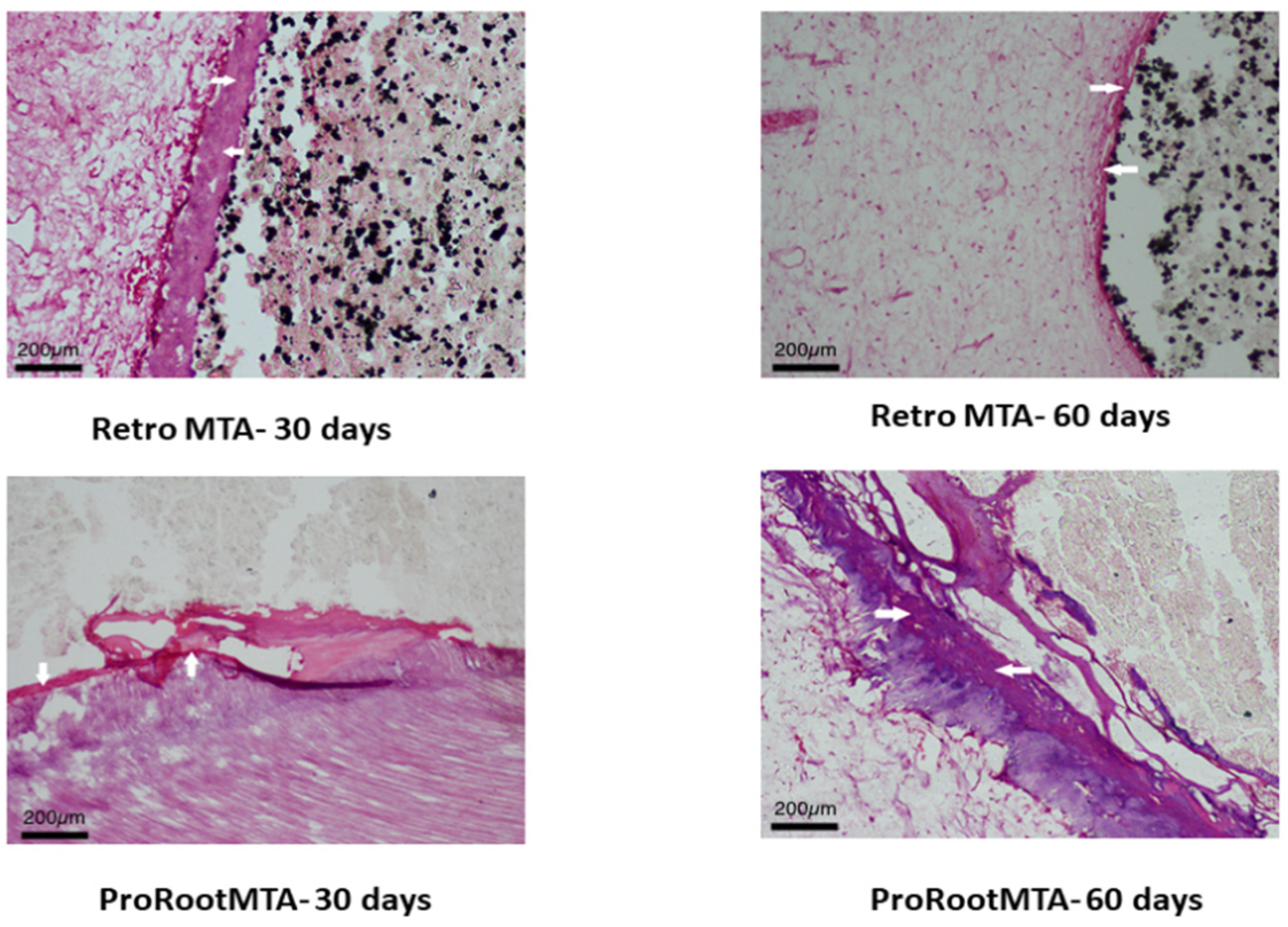
2.3. Tissue Processing for Microscopic Examination

A total of 10 teeth from each group were extracted after 30 days, and 10 further teeth were extracted after 60 days, for histopathological analysis. Presence and/or absence of post-operative tenderness to percussion, sensitivity, and pain were evaluation by questionnaires of the patients. Furthermore, Pulp sensibility tests were repeated according to the preoperatively performed ones at the time points 30 and 60 days. Oral surgeons extracted the teeth atraumatically, and orthodontists continued the orthodontic treatment. Following extraction, the apical 3 mm of the roots were sectioned to enable penetration of the 10% buffered formalin solution for 72 h. After blind coding for histomorphological processing and examination, the specimens were decalcified using 50% formic acid-sodium citrate for a 6 to 8 weeks period and prepared and embedded in paraffin. Between 10 and 12 sections, 4 or 5 6 micrometre sections were obtained by cutting using a microtome in a parallel direction to the vertical axis of the tooth. Sections were then mounted on glass slides, stained with haematoxylin-eosin, blindly evaluated by an experienced and calibrated oral and maxillofacial pathologist, according to the criteria in Table 2, Table 3 and Table 4. Each histomorphological event was evaluated with a 1–4 score system, with 1 being the best and 4 the worst result [17].

2.4. Statistical Analysis

Statistical analysis was performed with R Project for Statistical Computing 3.3.0 and Microsoft Excel 2013. The normality of the data distribution and the homogeneity of group variances were verified by the Kolmogorov–Smirnov test. Chi square test was performed for the comparison between the Proroot MTA and RetroMTA. p < 0.05 was considered as significant.

3. Results

All selected patients and treated teeth could be evaluated. The score percentages for each group in each criterion are shown after 30 days in Table 5, and after 60 days in Table 6.
Considering hard tissue bridge formation after 30 days, significant differences could be found when ProRoot and RetroMTA were compared. As for the parameters—continuity (p = 0.029), morphology (p = 0.023), and localisation (p < 0.001)—the RetroMTA performed significantly better compared with ProRoot MTA.
After 60 days, only continuity, the hard tissue bridge formation parameter, showed significant difference, in favour of the direct capping material ProRoot MTA (p = 0.010) (Figure 1).
The pulpal inflammatory response with the criteria (intensity, extension, and general state) no significance could be observed 30 days after the treatment for the parameter intensity (p = 1.00), while RetroMTA performed better in terms of extension (p = 0.007) and general state (p = 0.033).
Pulpal findings including the parameters (giant cells and particles) performed only significantly different for the parameter particles when both direct capping materials were compared after 30 days, in favour of RetroMTA (p = 0.048).
After 60 days, both direct capping materials Pro Root and RetroMTA performed similar regarding inflammatory response of the pulp and pulpal findings.

4. Discussion

This clinical study was conducted in teeth with a healthy uninflamed pulp using two different capping materials ProRoot MTA and RetroMTA, which are hydraulic biocompatible cements [18] with similar biological effects [19]. The null hypothesis was rejected for the parameters of hard tissue formation, inflammatory pulp response, and pulpal findings, except for intensity and giant cells number after 30 days, and for continuity after 60.
As for the hard tissue bridge, the RetroMTA results of this study showed better results regarding the parameters continuity, morphology, and localisation, while ProRoot MTA performed better regarding the parameter thickness. However, after 60 days, the only significant parameter was continuity, in favour of ProRoot MTA. The complete bridge formation is expected to take place 60–90 days after pulp capping [20,21].
The findings of this study are in accordance with one histological study, which evaluated direct pulp capping performance in dog’s teeth, where no significance could be found between the two test pulp capping materials RetroMTA and ProRoot MTA, where a hard tissue bridge was formed in direct contact to the vital pulp [18]. Kang et al. also sustained high success rate up to 1 year, with no significant differences between outcomes treated with ProRoot MTA (96.0%) and RetroMTA(96.0%) [22].
The dentine bridge, in the form of a calcified barrier, is usually considered a favourable response to capping therapy. In conjunction to vital signs, it supports that the pulp condition is healthy, leading to physiologic dentin deposition. Mass, E. et al. reported that a calcified barrier was found in almost 70% of teeth [23], compared with 55%–64% in previous studies [24,25].
The thickness of the dentine bridge in the 60 days sample of RetroMTA is less than 30 days. It is proof that the thickness of the dentinal bridge may vary depending on the location and angle of sectioning; therefore, it is more critical to evaluate the condition of pulp beside the bridge thickness, continuity, and quality, while assessing the success of the direct pulp capping material. The initiation is induced throughout initiation of reparative dentinogenesis in the case of MTA [26,27]. Pulp cells are aligned in proximity to crystals, built in a homogenous zone along the pulp–MTA interface. After MTA hardening, calcium oxide is formed, which can react with tissue fluids to produce calcium hydroxide [28]. After contact with pulp tissue, MTA presents some structures that are similar to calcite crystals, which attract fibronectin, which is generally responsible for cellular adhesion and differentiation [29]. Some studies found necrotic tissue next to the hard tissue bridge, which is assumed to be caused by MTA throughout coagulation in contact with pulp connective tissue [26]. The randomized controlled trial, conducted by Kang et al. [23], using three different MTA materials (ProRoot, Ortho, and RetroMTA), stated that the partial pulpotomy performed in permanent teeth showed favourable results, while clinical and radiographic results showed no difference after one year. In contradiction, one study found significantly worse results after 8 weeks using RetroMTA concerning pulp morphology and hard tissue bridge thickness compared to ProRoot. The hard tissue was described as a tubular reparative dentine. However, one study reported a lack of mineralized tissue and rather regular dentin with parallel tubules and rather amorphous and irregularly calcified tissue with lacunae of necrotic debris [23]. This finding may explain the changes after 60 days, and the less favourable performance of RetroMTA after 60 days. The quality of the hard tissue bridge relates to the pulpal health state after application of MTA and the pulp capping procedure [30,31].
For tooth survival, the pulp status and inflammatory response after pulp capping material application is more important compared to the thickness of the dentine bridge. The inflammatory response of the pulp with the criteria (intensity, extension, and general state) showed no significance between both tested cements after 60 days. RetroMTA is a hard-setting hydrophilic cement consisting of fine, hydrophilic particles that set in the presence of moisture and form a hard barrier. In contrast to Ca(OH)2 suspension, calcium silicate cements are not resorbable; therefore, they provide long-term protection against the invasion of microorganisms [32,33]. The quality of the hard tissue influences the state of pulpal health after direct pulp capping [30,31]. Furthermore, the formation of a hard tissue bridge does not mean that the pulp tissue will be sealed completely, because the hard tissue is permeable initially [34]. The formation of a mineralized barrier after capping with RetroMTA was reported as unpredictable, as it was incomplete and showed channels, which can be supported by the findings of this study as initial better results after 30 days compared to ProRoot MTA disappear, while the values for ProRoot MTA remain stable. The inflammation intensity and extension of RetroMTA teeth increased, while the general state of the pulp decreased.
No significant difference regarding the presence of microorganisms was found after 60 days. This means that, although the MTA particles dissolved faster in the RetroMTA group, the bacteriostatic action of both MTAs, per se, was similar, and was enough to reduce the number of viable bacteria near the pulp exposure.
Study limitations were the healthy uninflamed teeth, which don’t present the response that occurs in a clinical setting of an inflamed pulp, as the pathological changes of inflammation, odontoblasts absence, and tertiary dentine formation are missing. As for the bacteria detection, low sensibility of the histochemical staining technique for the detection of bacteria makes their identification difficult, mainly when there is a small number of such microorganisms [35]. Moreover, bacteria are easily removed from dental tissue during histologic preparation [36,37]. Although the use of healthy teeth allows standardisation, the effects of ProMTA and RetroMTA should also be evaluated in a clinical setting using infected and inflamed teeth.

5. Conclusions

The results of the present study indicated that RetroMTA might be a valuable ProRoot MTA alternative, regarding the tested parameters pulpal findings, including inflammatory response and hard tissue bridge formation. Although, during the first 30 days period, RetroMTA presented better results in terms of continuity, localisation, and morphology of the formed hard tissue bridge and extension and inflammatory pulp response, the continuity improved after 60 days when ProRoot MTA was applied.
MTA seemed to heal the pulp tissue at a faster rate for RetroMTA compared to ProRoot MTA.

Author Contributions

Conceptualization, N.V.B., S.R., N.R., A.U., J.-S.Y. and M.Ö.; methodology, N.V.B., S.R., N.R., A.U., J.-S.Y. and M.Ö.; software, N.V.B., S.R., N.R., A.U. and J.-S.Y.; validation, N.V.B., S.R., N.R., A.U., J.-S.Y. and M.Ö.; formal analysis, N.V.B., S.R., N.R., A.U., J.-S.Y. and M.Ö.; investigation, N.V.B., S.R., N.R., A.U., J.-S.Y. and M.Ö.; resources, N.V.B., S.R., N.R., A.U., J.-S.Y. and M.Ö.; data curation, N.V.B., S.R., N.R., A.U. and J.-S.Y.; writing—original draft preparation, N.V.B., S.R., N.R., A.U., J.-S.Y., N.A.-H.H. and M.Ö.; writing—review and editing, N.V.B., S.R., N.R., A.U., J.-S.Y., N.A.-H.H. and M.Ö.; visualization, N.V.B., S.R., N.R., A.U., J.-S.Y., N.A.-H.H. and M.Ö.; supervision, N.V.B., S.R., N.R., A.U., J.-S.Y., N.A.-H.H. and M.Ö.; project administration, N.V.B., S.R., N.R., A.U., J.-S.Y., N.A.-H.H. and M.Ö.; funding acquisition, N.V.B., S.R., N.R., A.U., J.-S.Y. and M.Ö. All authors have read and agreed to the published version of the manuscript.

Funding

This research received no external funding.

Institutional Review Board Statement

The study was conducted according to the guidelines of the Declaration of Helsinki, and was approved by the Institutional Review Board (IEC 859/2016) of Manipal University (CTRI/2017/10/010303).

Informed Consent Statement

Informed consent was obtained from all subjects involved in the study.

Conflicts of Interest

The authors declare no conflict of interest.

References

  1. Caladas, A.F., Jr.; Burgos, M.E. A retrospective study of traumatic dental injuries in a Brazilian dental trauma clinic. Dent. Traumatol. 2001, 17, 250–253. [Google Scholar] [CrossRef]
  2. Olsson, H.; Petersson, K.; Rohlin, M. Formation of a hard tissue barrier after pulp cappings in humans. A systematic review. Int. Endod. J. 2006, 39, 429–442. [Google Scholar] [CrossRef] [PubMed]
  3. Pereira, J.C.; Segala, A.D.; Costa, C.A.S. Human pulpal response to direct pulp capping with an adhesive system. Am. J. Dent. 2000, 13, 139–147. [Google Scholar]
  4. Costa, C.A.S.; Nascimento, A.B.L.; Teixeira, H.M.; Fontana, U.F. Response of human pulps capped with a self-etching adhesive system. Dent. Mater. 2001, 17, 230–240. [Google Scholar] [CrossRef]
  5. Hilton, T.J. Keys to clinical success with pulp capping: A review of the literature. Oper. Dent. 2009, 34, 615–625. [Google Scholar] [CrossRef] [PubMed] [Green Version]
  6. Cox, C.F.; Subay, R.K.; Ostro, E.; Suzuki, S.; Suzuki, S.H. Tunnel defects in dentin bridges: Their formation following direct pulp capping. Oper. Dent. 1996, 21, 4–11. [Google Scholar] [PubMed]
  7. Cox, C.F.; Hafez, A.A.; Akimoto, N.; Otsuki, M.; Suzuki, S.; Tarim, B. Biocompatibility of primer; adhesive and resin composite systems on non-exposed and exposed pulps of non-human primate teeth. Am. J. Dent. 1998, 11, S55–S63. [Google Scholar]
  8. Cox, C.F.; Tarim, B.; Kopel, H.; Gurel, G.; Hafez, A. Technique sensitivity: Biological factors contributing to clinical success with various restorative materials. Adv. Dent. Res. 1998, 5, 85–90. [Google Scholar] [CrossRef]
  9. Pitt Ford, T.R.; Torabinejad, M.; Abedi, H.R.; Bakland, L.K.; Kariyawasam, S.P. Using mineral trioxide aggregate as a pulp-capping materials. J. Am. Dent. Assoc. 1996, 127, 1491–1494. [Google Scholar] [CrossRef]
  10. Parirokh, M.; Torabinejad, M. Mineral Trioxide aggregate: A comprehensive literature review—Part III: Clinical applications; drawbacks; and mechanism of action. J. Endod. 2010, 36, 400–412. [Google Scholar] [CrossRef]
  11. Boutsioukis, C.; Noula, G.; Lambrianidis, T. Ex vivo study of the efficiency of two techniques for the removal of mineral trioxide aggregate used as a root canal filling material. J. Endod. 2008, 34, 1239–1242. [Google Scholar] [CrossRef] [PubMed]
  12. Felman, D.; Parashos, P. Coronal tooth discoloration and white mineral trioxide aggregate. J. Endod. 2013, 39, 484–487. [Google Scholar] [CrossRef] [PubMed]
  13. Lee, H.; Shin, Y.; Kim, S.O.; Lee, H.S.; Choi, H.J.; Song, J.S. Comparative study of pulpal responses to pulpotomy with ProRoot MTA, RetroMTA, and TheraCal in dogs’ teeth. J. Endod. 2015, 41, 1317–1324. [Google Scholar] [CrossRef]
  14. Üstün, Y.; Topçuoğlu, H.S.; Akpek, F.; Aslan, T. The effect of blood contamination on dislocation resistance of different endodontic reparative materials. J. Oral Sci. 2015, 57, 185–190. [Google Scholar] [CrossRef] [Green Version]
  15. Kang, S.H.; Shin, Y.S.; Lee, H.S.; Kim, O.S.; Shin, Y.; Jung, I.Y.; Song, J.S. Color changes of teeth after treatment with various mineral trioxide aggregate–based materials: An ex vivo study. J. Endod. 2015, 41, 737–741. [Google Scholar] [CrossRef]
  16. Abdul, M.S.M.; Murali, N.; Rai, P.; Mirza, M.B.; Salim, S.; Aparna, M.; Singh, S. Clinico-Histological Evaluation of Dentino-Pulpal Complex of Direct Pulp Capping Agents: A Clinical Study. J. Pharm. Bioallied Sci. 2021, 13, S194–S198. [Google Scholar] [CrossRef]
  17. Chung, C.J.; Kim, E.; Song, M.; Park, J.W.; Shin, S.J. Effects of two fast-setting calcium-silicate cements on cell viability and angiogenic factor release in human pulp-derived cells. Odontology 2016, 104, 143–151. [Google Scholar] [CrossRef]
  18. Mestrener, S.R.; Holland, R.; Dezan, E., Jr. Influence of age on the behavior of dental pulp of dog teeth after capping of an adhesive system or calcium hydroxide. Dent. Traumatol. 2003, 19, 255–261. [Google Scholar] [CrossRef] [PubMed]
  19. Sawicki, L.; Pameijer, C.H.; Emerich, K.; Adamowicz-Klepalskak, B. Histological evaluation of mineral trioxide aggregate and calcium hydroxide in direct pulp capping of human immature permanent teeth. Am. J. Dent. 2008, 21, 262–266. [Google Scholar] [PubMed]
  20. Dammaschke, T.; Nowicka, A.; Lipski, M.; Ricucci, D. Histological evaluation of hard tissue formation after capping with a fast-setting mineral trioxide aggregate in humans. Clin. Oral Investig. 2019, 23, 4289–4299. [Google Scholar] [CrossRef]
  21. Accorinte, M.L.R.; Loguercio, A.D.; Reis, A.; Bauer, J.R.; Grande, R.H.; Murata, S.S.; Souza, V.; Holland, R. Evaluation of two mineral trioxide aggregate compounds as pulp-capping agents in human teeth. Int. Endod. J. 2009, 42, 122–128. [Google Scholar] [CrossRef]
  22. Kang, C.M.; Sun, Y.; Song, J.S.; Pang, N.S.; Roh, B.D.; Lee, C.Y.; Shin, Y. A randomized controlled trial of various MTA materials for partial pulpotomy in permanent teeth. J. Dent. 2017, 60, 8–13. [Google Scholar] [CrossRef]
  23. Bakhtiar, H.; Aminishakib, P.; Ellini, M.R.; Mosavi, F.; Abedi, F.; Esmailian, S.; Esnaashari, E.; Nekoofar, M.H.; Sezavar, M.; Mesgarzadeh, V.; et al. Dental pulp response to RetroMTA after partial pulpotomy in permanent human teeth. J. Endod. 2018, 44, 1692–1696. [Google Scholar] [CrossRef]
  24. Mass, E.; Zilberman, U. Long-term radiologic pulp evaluation after partial pulpotomy in young permanent molars. Quintessence Int. 2011, 42, 547–554. [Google Scholar] [PubMed]
  25. Barrieshi-Nusair, K.M.; Qudeimat, M.A. A prospective clinical study of mineral trioxide aggregate for partial pulpotomy in cariously exposed permanent teeth. J. Endod. 2006, 32, 731–735. [Google Scholar] [CrossRef] [PubMed]
  26. Qudeimat, M.A.; Barrieshi-Nusair, K.M.; Owais, A.I. Calcium hydroxide vs mineral trioxide aggregates for partial pulpotomy of permanent molars with deep caries. Eur. Arch. Paediatr. Dent. 2007, 8, 99–104. [Google Scholar] [CrossRef] [PubMed]
  27. Nair, P.N.R.; Duncan, H.F.; Pitt Ford, T.R.; Luder, H.U. Histological; ultrastructural and quantitative investigations on the response of healthy human pulps to experimental capping with mineral trioxide aggregate: A randomized controlled trial. Int. Endod. J. 2008, 41, 128–150. [Google Scholar]
  28. Faraco Junior, I.M.; Holland, R. Response of the pulp of dogs to capping with mineral trioxide aggregate or calcium hydroxide cement. Dent. Traumatol. 2001, 17, 163–166. [Google Scholar] [CrossRef] [PubMed]
  29. Tziafas, D.; Pantelidou, O.; Alvanou, A.; Belibasakis, G.; Papadimitriou, S. The dentinogenic effect of mineral trioxide aggregate (MTA) in short-term capping experiments. Int. Endod. J. 2002, 35, 245–254. [Google Scholar] [CrossRef] [PubMed]
  30. Koh, E.T.; McDonald, F.; Pitt Ford, T.R.; Torabinejad, M. Cellular response to mineral trioxide aggregate. J. Endod. 1998, 24, 543–547. [Google Scholar] [CrossRef]
  31. Seux, D.; Coulbe, M.L.; Hartmann, D.J.; Gauthier, J.P.; Magloire, H. Odontoblast-like cytodifferentiation of human dental pulp cells in vitro in the presence of a calcium hydroxide-contain cement. Arch. Oral Biol. 1991, 36, 117. [Google Scholar] [CrossRef]
  32. Schröder, U. Evaluation of healing following experimental pulpotomy of intact human teeth and capping with calcium hydroxide. Odontol. Revy 1972, 23, 329–340. [Google Scholar] [PubMed]
  33. About, I. Recent trends in tricalcium silicates for vital pulp therapy. Curr. Oral Health Rep. 2018, 5, 178–185. [Google Scholar] [CrossRef]
  34. Dammaschke, T.; Camp, J.H.; Bogen, G. MTA in vital pulp therapy. In Mineral Trioxide Aggregate—Properties and Clinical Applications; Torabinejad, M., Ed.; Wiley Blackwell: Ames, IA, USA, 2014; pp. 71–110. [Google Scholar]
  35. Dammaschke, T. Dentine and hard tissue formation after indirect and direct pulp capping. Int. Dent. 2012, 7, 52–58. [Google Scholar]
  36. Accorinte, M.L.R.; Loguercio, A.D.; Reis, A.; Carneiro, E.; Grande, R.H.; Murata, S.S.; Holland, R. Response of human dental pulp capped with MTA and calcium hydroxide powder. Oper. Dent. 2008, 33, 488–495. [Google Scholar] [CrossRef]
  37. Min, K.S.; Park, H.J.; Lee, S.K.; Park, S.H.; Hong, C.U.; Kim, H.W.; Lee, H.H.; Kim, E.C. Effect of mineral trioxide aggregate on dentin bridge formation and expression of dentin sialoprotein and heme oxygenase-1 in human dental pulp. J. Endod. 2008, 34, 666–670. [Google Scholar] [CrossRef] [PubMed]
Figure 1. Hard tissue bridge (arrow) and inflammatory cells (star) demonstrated in relation to ProRoot MTA and RetroMTA at 30 days and 60 days (haematoxylin and eosin, original magnification ×10).
Applsci 11 10455 g001
Table 1. Composition of the experimental MTA cements used.
ProRoot MTARetroMTA
Tricalcium silicate Calcium carbonate
Dicalcium silicateSilicon dioxide
Aluminium oxide
Calcium Zirconium complex
Tricalcium aluminate
Tetracalcium aluminoferrite
Calcium oxide
Bismuth Oxide
Table 2. Evaluation parameters and scores used for hard tissue bridge formation (continuity, morphology, thickness of dental bridge, and localization).
ScoreContinuityMorphologyThickness of Dental BridgeLocalization
1CompleteDentin or dentin associated with an irregular hard tissueUp to 250 µmClosure to the exposition area without invading the pulp space
2Little communication of the capping material with the dental pulpOnly irregular hard tissue deposition150–249 µmBridge invading pulp space next to the opposite dentin wall
3Only lateral deposition of hard tissue on the walls of the cavity of pulp exposureOnly a slight layer of hard tissue deposition1–149 µmBridge reached the opposite dentin wall
4Absence of hard tissue bridge and absence of lateral deposition of hard tissueNo hard tissue depositionPartial or absent bridgeNo bridge or only hard tissue deposition on the walls of the exposition cavity site
Table 3. Scores used for the evaluation of the inflammatory response of pulp.
ScoreIntensity of Inflammatory Reaction (Acute and Chronic)
1Absent or very few inflammatory cells
2Mild: average number less than 10 inflammatory cells
3Moderate: average number 10–25 inflammatory cells
4Severe: average number greater than 25 inflammatory cells
ScoreExtension of The Inflammatory Reaction (Acute and Chronic)
1Absent
2Mild: inflammatory cells only next to dentin bridge or area of pulp exposition
3Moderate: inflammatory cells are observed in part of coronal pulp
4Severe: all coronal pulp is infiltrated or necrotic
ScoreGeneral State of The Pulp
1No inflammatory reaction
2With inflammatory reaction
3Abscess
4Necrosis
Table 4. Scores used for other pulpal findings.
ScoreGiant CellsParticles of Capping Materials
1Absent Absent
2MildMild
3ModerateModerate
4Pulp necrosisLarge number
Table 5. Percentage of scores (%) attributed to each group in each criterion of hard tissue bridge after 30 days for both cement types: ProRoot MTA and RetroMTA.
30 DaysProRoot MTARetroMTAX2p
12341234
Continuity10-6030106020109.00.029
Morphology20-602080-10107.5030.023
Thickness--9010--1090 <0.001
Localisation50--50305010109.1670.027
Intensity9010--100----1.0
Extension2080--8020--7.20.007
General state5050--100----0.033
Giant cells100---100----1
Particles10-7020403020107.9110.048
Table 6. Percentage of scores (%) attributed to each group in each criterion of hard tissue bridge after 60 days for both cement types: ProRoot MTA and RetroMTA.
60 DaysProRoot MTARetroMTAX2p
12341234
Continuity10-603020-206011.330.01
Morphology20-60206010-302.0770.557
Thickness--9010--10901.250.264
Localisation50--505040-102.0190.364
Intensity9010--502030-3.5330.171
Extension2080--603010-1.1430.565
General state5050--7030--0.2670.606
Giant cells100---100----1
Particles10-7020--30702.2770.32
Publisher’s Note: MDPI stays neutral with regard to jurisdictional claims in published maps and institutional affiliations.

