Article

A Novel Method for Estimating the Dosage of Cold Atmospheric Plasmas in Plasma Medical Applications

1
Department of Engineering Physics, Tsinghua University, Beijing 100084, China
2
School of Mechanical Engineering, Tsinghua University, Beijing 100084, China
3
School of Medicine, Tsinghua University, Beijing 100084, China
4
College of Mechanical Engineering, North China University of Science and Technology, Tangshan 063210, China
5
School of Pharmaceutical Science, Tsinghua University, Beijing 100084, China
*
Authors to whom correspondence should be addressed.
These two authors contributed equally to this work.
Submission received: 9 October 2021 / Revised: 8 November 2021 / Accepted: 12 November 2021 / Published: 24 November 2021

Abstract

:
Cold atmospheric plasmas (CAPs) used in plasma medicine have shown great potential in various aspects including wound healing, dermatology, cancer therapy, etc. It is one of the important issues to determine the plasma dosage in plasma medicine because it dominates the specific plasma treatment results. However, the multi-process interactions between CAPs and biological materials make it rather challenging to give an accurate and versatile definition for plasma dosage. In this study, the ratio of the discharge energy to the number of the treated in vitro kidney cells (mJ/cell) was employed as the unit of the plasma dosage. Additionally, inspired by basic knowledge of pharmacy, the median lethal dose (LD50) was employed to help estimate the plasma dosage. The experimental results show that the value of LD50 using the newly designed CAP Bio-Med Platform for the kidney cells is 34.67 mJ/cell. This biology-based method has the advantages of easy operation, independence of specific CAP sources, and also independence of complex interactions between CAPs and the treated biological targets, and consequently, may provide a new direction to quantitatively define the plasma dosage in various plasma medical applications.

1. Introduction

Plasma medicine [1] is one of the most widely concerned interdisciplinary fields of cold atmospheric plasmas (CAPs) owing to its great potential in clinical treatment. Up to now, it has been proved that CAPs are efficient in aspects ranging from sterilization [2] and disinfection [3] to wound healing [4,5], skin disease treatment [6], and even cancer therapy [7]. These results show that the CAP treatment can be more effective than the traditional method for certain diseases with few side effects. Based on the state-of-the-art knowledge, the CAPs contain abundant reactive oxygen and nitrogen species (RONS), which can regulate cell metabolisms and play a dominant role in the plasma-induced biological effects [8,9,10]. Some of the most frequently discussed RONS are hydroxyl (OH), atomic oxygen (O), nitric oxide (NO), and hydrogen peroxide (H2O2). The generation and transport characteristics of the RONS largely depend on the CAP sources and the ambient conditions. Generally, the CAP sources used in plasma medicine are operated at atmospheric pressure and have gas temperatures close to room temperature, making it feasible and convenient to be applied for the treatment of biological materials such as living tissues, cells, etc. Detailed descriptions and features of various kinds of CAP sources used in plasma medicine can be found in some topical reviews [7,9,10].
From the very beginning of the research on plasma medicine, the treatment safety issues have been widely investigated. The inactivation of microorganisms by plasma treatment has been proved and applied before 2000 [7], whereas Stoffels et al. first reported the non-destructive CAP treatment on mammalian cells under moderate conditions in the early 2000s [11]. Later, they found that the CAP treatment for a too-long time period would cause mammalian cell necrosis [12]. Using different CAP generators, Fridman et al. came to a similar conclusion that the cells were hardly damaged with a relatively low CAP treatment energy density, whereas with a medium- to high-level treatment, the cells began to be harmed and even died [13]. That is to say, different CAP treatment intensities will lead to completely different results in plasma medicine. To quantitatively describe how much CAPs are interacting with the targets (e.g., living tissues, cells, etc.), the concept of “plasma dosage” should be introduced. Additionally, only with a clear definition of the “plasma dosage” the users can accurately determine, and thus, carefully adjust, the action intensity of the CAPs to obtain the desired results in plasma medicine.
Many studies have involved plasma dosage when investigating possible factors which intrinsically influence the CAP treatment effects. On the one hand, from the aspect of physical plasmas, various factors including power input into the CAP system, chemical compositions of the working gas, amplitudes and waveforms of discharge voltage and current, gas temperatures, treatment times and distances are all believed to be significant factors in plasma dosage [8,9,12,13,14,15,16]. In addition, some biological experimental parameters such as the thicknesses of the cell culture medium and the numbers of cells can also have obvious influences on the CAP treatment results [17]. A recent study by Cheng et al. provided an important attempt to define plasma dosage by the simplified equivalent total oxidation potential of several important RONS [8]. However, as shown in Figure 1, the interactions between the CAPs and the treated biological targets involve multiple physical, chemical and biological processes, leading to the significantly complicated, nonlinear, and multi-variable natures. These have made it extremely difficult to propose an accurate and versatile definition for plasma dosage, considering all the possible mechanisms. More recently, in order to adjust and/or control the effective CAP dosages, some researchers begin to focus on machine learning and neural network [18,19], which further proves the complexity and difficulty of the plasma dosage definition.
In our opinion, since the concept of “dosage” is frequently used in pharmaceuticals, the definition of plasma dosage can refer to the basic knowledge in pharmacy. The median lethal dose (LD50) is one of the most typical expressions to describe the drug toxicity which represents the exact dose that can kill half of the experimental subjects (e.g., cells, animals, etc.). The measurement of the LD50 values usually serves as an important and basic step for the systematic assessment and evaluation of the drug toxic characteristics [20,21,22]. Inspired by this methodology in pharmacy, we intend to introduce a novel method to estimate the plasma dosage: treating the in vitro cells with different CAP intensities to find the value of LD50 which corresponds to the situation under which the CAP treatment can kill a half number of the cells. The value of LD50 can serve as an indicator to help the users determine and control the plasma dosage used in their practical studies. Distinguished from the physics- and/or chemistry-based method to define or control the plasma dosage [8,18,19,23,24,25], this biology-based approach has the advantages of being versatile to various CAP sources and easy to be conducted without considering the specific and complex interaction processes between the CAPs and the treated biological targets.

2. Materials and Methods

2.1. Cells and Cell Culture

The flexibility of CAPs has enabled them to be applied in the internal clinical treatment [26]; therefore, the human embryonic kidney cell line was employed to conduct the primary experiments, considering the possible CAP applications in viscus therapy. Before CAP treatment, the cells were grown in Dulbecco’s Modified Eagle Medium (DMEM) supplemented with 10% fetal bovine serum and 1% Penicillin–Streptomycin. The cells were not treated by CAPs until the logarithmic growth phase was reached. Before the treatment, the cells were observed by the XD biological microscope (Sunny Optical Technology Co., Ltd., Ningbo, China) to make sure that they were in the logarithmic growth phase. Then, the cells were seeded near confluence in 96-well plates (5000 cells per well). Additionally, the cells were then kept in the cell incubator (Thermo Fisher Scientific Co., Ltd., Waltham, MA, USA) for 24 h with 37 °C and 5% CO2 prior to the CAP treatment. The cell line, the reagents, and their sources of procurement used in this study are listed in Table 1.

2.2. CAP Bio-Med Platform and Treatment of Cells

As shown in Figure 2a, the CAP Bio-Med Platform employed in this study is a newly designed all-in-one device developed by the Plasma Health Scientech Group (PHSG) of the Tsinghua University of China. The upper part of the device has double doors to form a confined space. The UV lamp attached to the ceiling can be applied for atmosphere sterilization and disinfection. The CAP Bio-Med Platform is a multi-function device and mainly consists of three units:
(i)
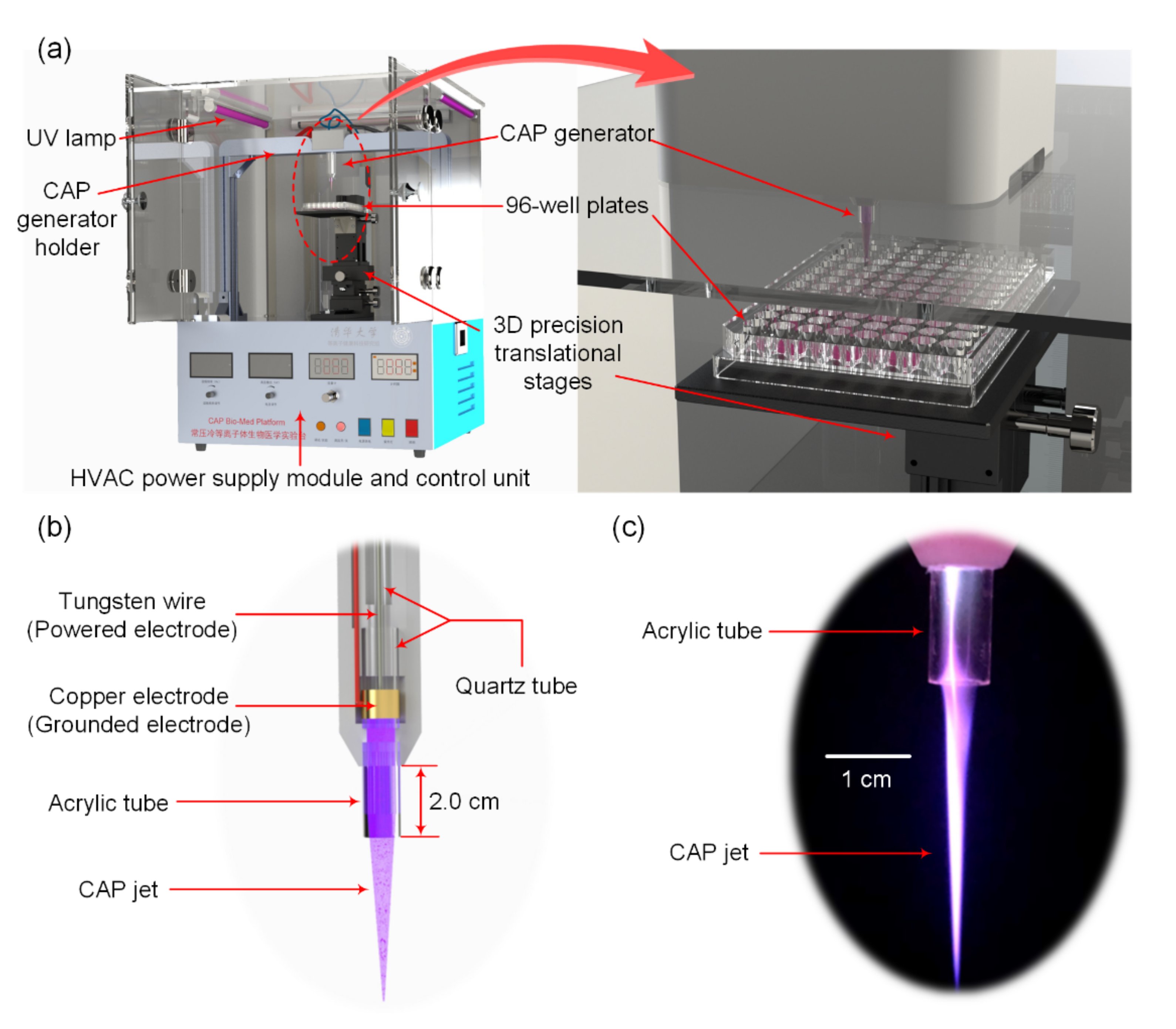
The CAP unit: It includes a co-axial type of atmospheric pressure dielectric barrier discharge (AP-DBD) plasma generator and the high-voltage alternating-current (HVAC) power supply module. As shown in Figure 2b, a tungsten wire confined inside a quartz tube works as the powered electrode, whereas a copper slice works as the grounded electrode. An acrylic tube is connected to the exit of the CAP generator to keep the treatment distance fixed at 2.0 cm and simultaneously protect the CAP jet from the entrainment of the surrounding air. The plasma jet shown in Figure 2c is uniform and stable with a gas temperature close to the room temperature, which meets the basic requirements of plasma medical applications.
(ii)
The control unit: It includes the helium gas flow rate control module (0–20 slpm), the driving frequency control module (0–25 kHz), the discharge voltage control module (0–15 kV), and the discharge time control module (0–999 s). These control modules can ensure that the experimental parameters can be conveniently adjusted in a wide range. Moreover, the discharge will automatically stop when the pre-set discharge time is up. The HVAC power supply and the preceding control modules are integrated inside the lower part of the device as illustrated in Figure 2a.
(iii)
The positioning unit: It includes a customized holder that supports the AP-DBD generator and the three-dimensional (3D) precision translational stages (LWX60-L200 and LWE4090, Misi Automation Equipment Co., Ltd., Dongguan, China). The CAP generator holder is used to fix the generator and to keep it vertical to the treated materials. When treating the cells, a 96-well plate can be put on the horizontal plane attached to the precision translational stage as shown in Figure 2a, so that the distance between the plasma jet exit and the upper surface of the 96-well plate can be adjusted precisely.
For the cell treatment, the plasma dosages were varied by changing the treatment time and simultaneously keeping all the other parameters unchanged. The discharge frequency was fixed at f = 23 kHz and the pure helium (99.999%) gas flow rate was Q = 8.0 slpm. The amplitude of the discharge voltage was fixed to be Vd = 3.5 kV measured by a digital oscilloscope (DPO4034, Tektronix, Beaverton, OR, USA) and a voltage probe (P6015A, Tektronix, Beaverton, OR, USA). Under such conditions, the free CAP jet was approximately 5.0 cm long, determined by a visible image processing technique [27,28]. With an unchanged discharge voltage, the power input was also constant and should be measured, because it was an important factor for plasma dosage as discussed in Section 1 and would influence various interaction processes as shown in Figure 1. The CAP treatment times (t) were 0 (control), 2, 3, 3.5, 4, 4.5, 5, 7, and 10 min. All the experiments were repeated three times. Starting from the moment that the discharge was triggered, the gas temperatures of the CAP jet were measured and recorded every 30 s to estimate the thermal effects.
To qualitatively analyze and quantitatively determine the value of LD50, both the optical and electrical features of the CAP generator were measured. First, the reactive species in the CAP jet were detected by the spectrometer (Avaspec Multi-Channel Spectrometer, Avantes, Apeldoom, the Netherlands) with a fiber probe to show whether RONS exist in the CAP jet which are important for the plasma medical applications. The discharge power (Pin) was obtained by the voltage/charge Lissajous figure method [29] with the aid of a digital oscilloscope and two voltage probes. To quantitatively obtain the LD50 value of the CAP jet to the HEK293 cells, the ratio of the discharge energy (W) to the number of cells (N) in each well, i.e., (W/N), was used when half of the cells in a well were killed. For a certain CAP treatment time t, the value of (W/N) could be calculated by:
W/N = Pin × t/N.

2.3. Cell Viability Measurement by the MTT Assay

In this paper, only the MTT (3-(4,5-Dimethylthiazol-2-yl)-2,5-diphenyltetrazolium bromide) assay method was used to evaluate the cell viability after the CAP treatment as a preliminary study. On the one hand, the reliability of the MTT assay for evaluating the cell viability has been discussed by Yang and Acosta with a conclusion that the results obtained by MTT assay were comparable with those of the Lactate dehydrogenase (LDH) release assay [30]. On the other hand, the advantages of ease of operation, sensitivity, rapidity and low cost have made the MTT assay as one of the most widely used methods for measuring the cytotoxic effects and for evaluating the cell viability [31,32,33,34,35].
After CAP treatment, the 96-well plates were put into the cell incubator to cultivate for a certain time (e.g., 0, 6, 12, 18 or 24 h), then, 10 μL MTT solution was added to each well. After another 4 h incubation, the formed formazan crystals were dissolved in 110 μL dimethyl sulfoxide (DMSO) in each well. Finally, the absorbance was detected at 570 nm by a microplate reader (Tecan Trading AG, Männedorf, Switzerland). Since the cell numbers had a linear relationship with the absorbance, the cell viability was calculated by:
A i A control × 100 %
where A was the value of the absorbance, the subscript i represented the treatment time, i.e., i = 2, 3, 3.5, 4, 4.5, 5, 7, 10.

2.4. Statistical Analysis

The statistical analyses were performed with a one-way analysis of variance (ANOVA) using the Prism Software (GraphPad, San Diego, CA, USA). The data were reported as the mean ± standard error. Additionally, compared with the control group, the p-value **** p < 0.0001 was considered to be statistically significant.

3. Experimental Results

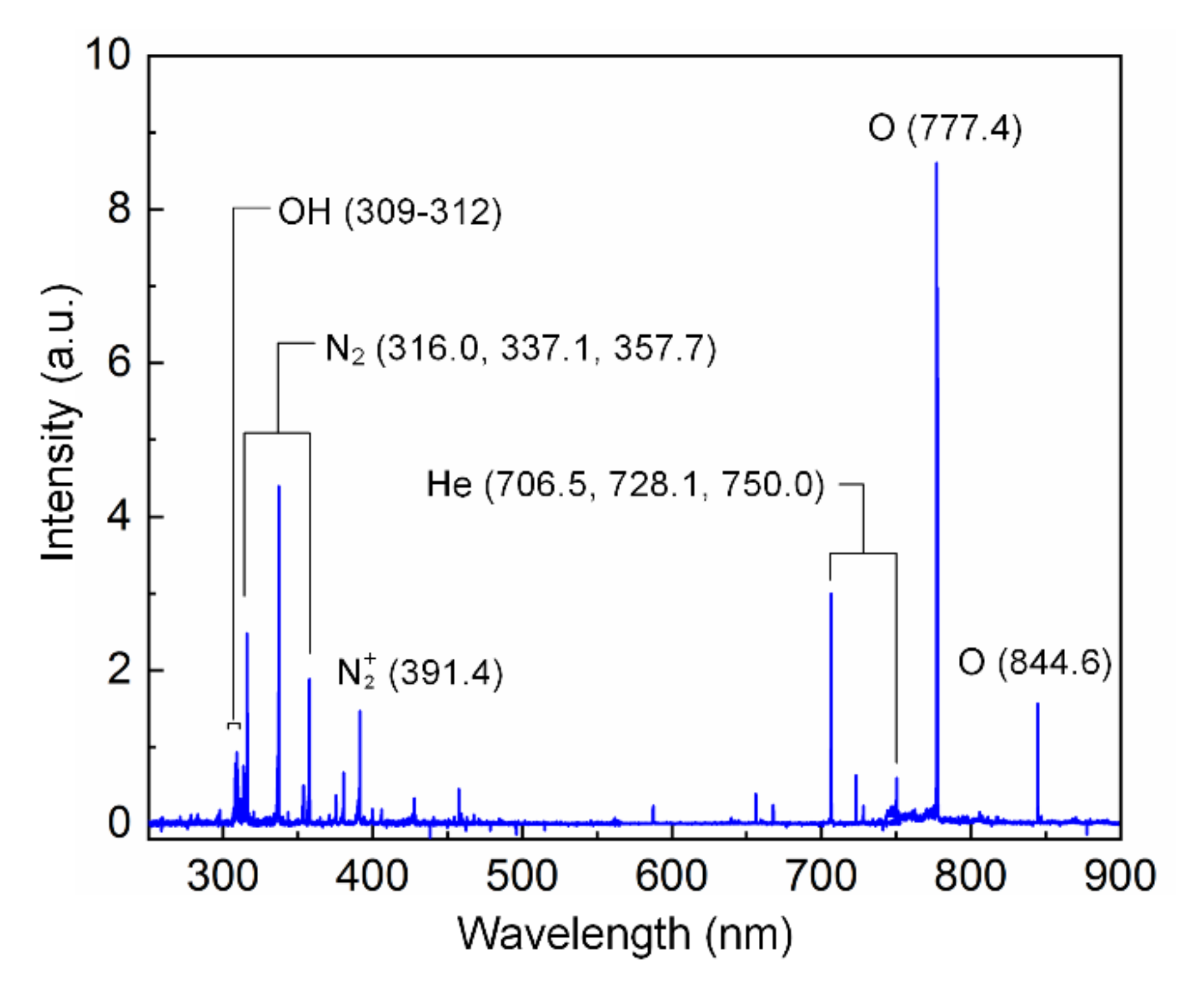
3.1. CAP Jet Features

Under the discharge conditions listed in Section 2.2, the discharge power obtained from the Lissajous figure method was Pin = 0.8 W. As shown in Figure 3, the CAP jet issued from the generator contained various kinds of reactive species. Besides the abundant helium species, several kinds of RONS, i.e., nitrogen molecules, hydroxyl (OH), and oxygen atoms (O) [36], also had relatively high intensities, indicating that the CAP device was a suitable source for plasma medicine and was promising for investigating the plasma dosage. In addition, with an initial room temperature of 22 °C, the gas temperature of the CAP jet did not exceed 24 °C after the discharge was on for 10 min, proving that the CAP jet had good thermal stability and safety during the whole CAP treatment process.
Here, there is one issue we hope to emphasize. As illustrated in Figure 2a, during the CAP treatment, the plasma jet directly interacted with the cell culture in the 96-well plate which worked as a liquid substrate. Although previous studies have shown that different substrates could influence the features of the impinged plasma jets, for example, the number densities of some important RONS such as hydroxyl (OH) and atomic oxygen (O) increased obviously due to the existence of water [37] compared with the plasma free jet, in our opinion, determination of the cell viabilities would, to some extent, not be affected since the cell culture used in all the experiments was completely the same in this study. Our goal in this paper is to determine the plasma dosage by avoiding the considerations of the extremely complicated interactions between the CAPs and the treated biological targets (Figure 1).

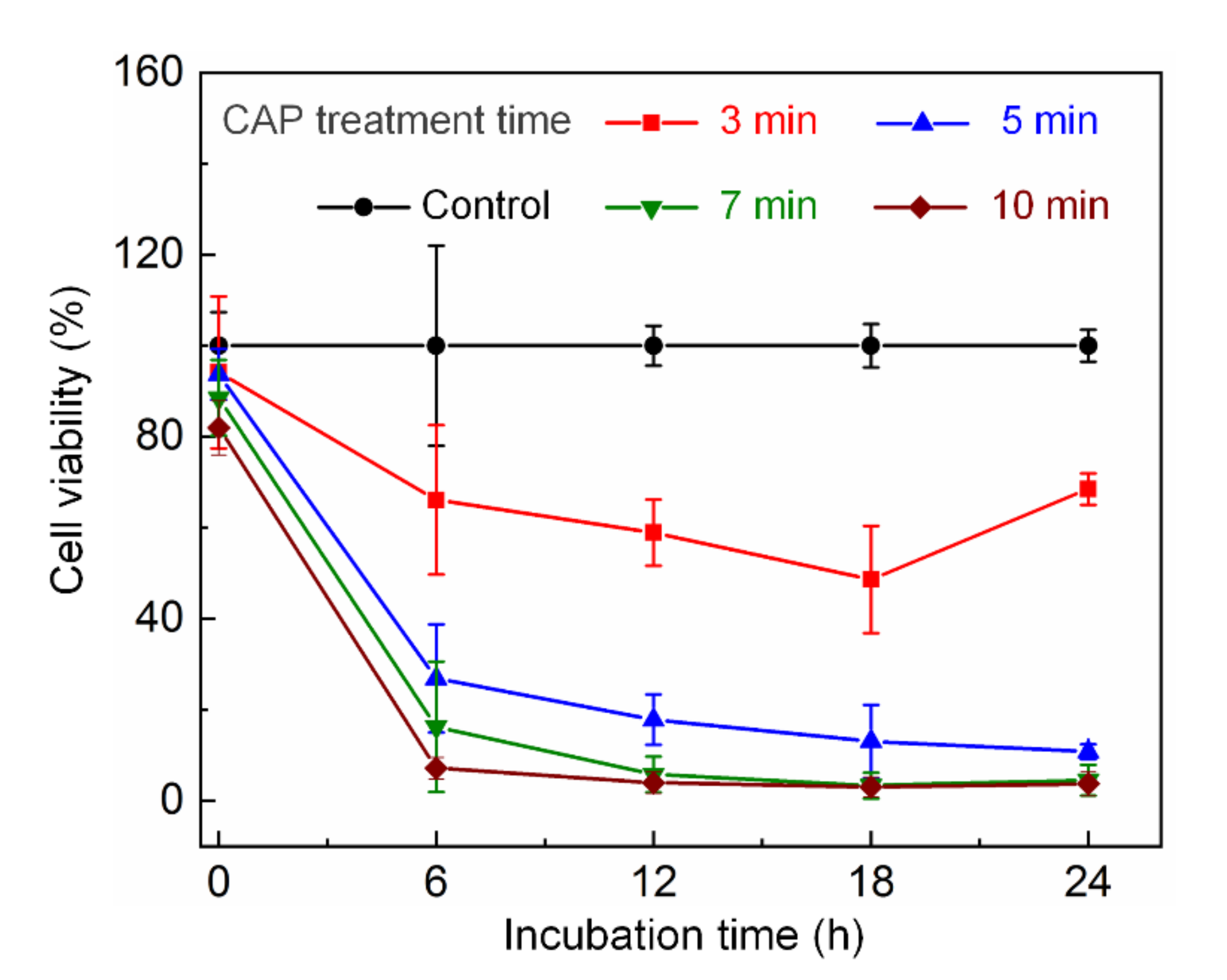
3.2. Selection of the Incubation Time

In the previous studies concerning the cell viability after CAP treatment, the cells were incubated for a certain period of time before the viability assays were conducted. However, different researchers used different incubation durations and seldom discussed the reasons to select such a duration [11,12,13]. Therefore, as the first step of this study, we incubated the cells after CAP treatment for different durations, i.e., 0, 6, 12, 18, and 24 h, and compared the measured cell viabilities. It could be seen clearly from Figure 4 that, under a fixed CAP treatment time, the cell viabilities seemed to remain unchanged with the longer incubation time after 12 h. The only exclusion was that, when the CAP treatment time was 3 min, the cell viability after 24 h incubation was higher than that after 18 h incubation. The possible reason might be explained as follows: the low dosage of plasma treatment, e.g., 3 min in this study, could not lead to an unrecoverable lethal effect to the treated cells, and consequently, the cell activity could still increase gradually after a long term incubation. This is, to some extent, consistent with the experimental results presented in Section 4 in which the plasma dosage corresponding to the 3 min CAP treatment time is lower than the value of LD50. The sub-lethal effects of CAP-treated living biomaterials form a very important and complex research aspect, which is beyond the scope of this paper. Therefore, we focus on the lethal effects of the CAP-treated HEK293 cells in this paper, and use the cell viabilities corresponding to the 24 h incubation after CAP treatment to calculate the value of LD50 in the following sub-sections.

3.3. Cell Viability in Response to the CAP Treatment

Since the discharge power and the initial number of cells in each well were both fixed, the different values of (W/N) in Figure 5, i.e., 19.44, 29.16, 34.02, 38.88, 43.74, 48.60, 68.04, and 97.20 mJ/cell, actually corresponded to different plasma treatment times of 2, 3, 3.5, 4, 4.5, 5, 7, and 10 min. The experiments were repeated three times on different days in this study. It could be seen from Figure 5 that: (i) with increasing in the treatment time, the cell viability decreased obviously; (ii) the value of (W/N) corresponding to LD50 should lie between 30 and 45 mJ/cell.

4. Discussions

4.1. Elimination of Thermal Effects

Most of the studies on plasma dosage highlight the dominant role of the multiple RONS, whereas the role of the thermal effect, in our opinion, should be considered in the first place. Normally, the biological materials are especially sensitive to the ambient temperature. For example, it was reported that treatment to the human skins for only 6 s under 59 °C could lead to a blister, whereas at the single cell level of in vitro experiments, cell necrosis was always observed when the gas temperature was higher than 42 °C [38]. Though the specific cell necrosis rate also relates to other parameters such as the duration and the type of the cells, it is anticipated that, when the gas temperature of the CAP jet is too high, the cell death resulting from thermal damages will appear, which means that the discussions on plasma dosage will be meaningless. Therefore, the gas temperature used for plasma medicine should be carefully controlled within an appropriate range.
In this study, the rise in gas temperature within the 10 min discharge was less than 2 °C, and its absolute value was always lower than 24 °C, which was still close to room temperature and much lower than the environment temperature in the cell incubator. Therefore, it is reasonable to conclude that no damages to the cells were caused by the thermal effect in our study.

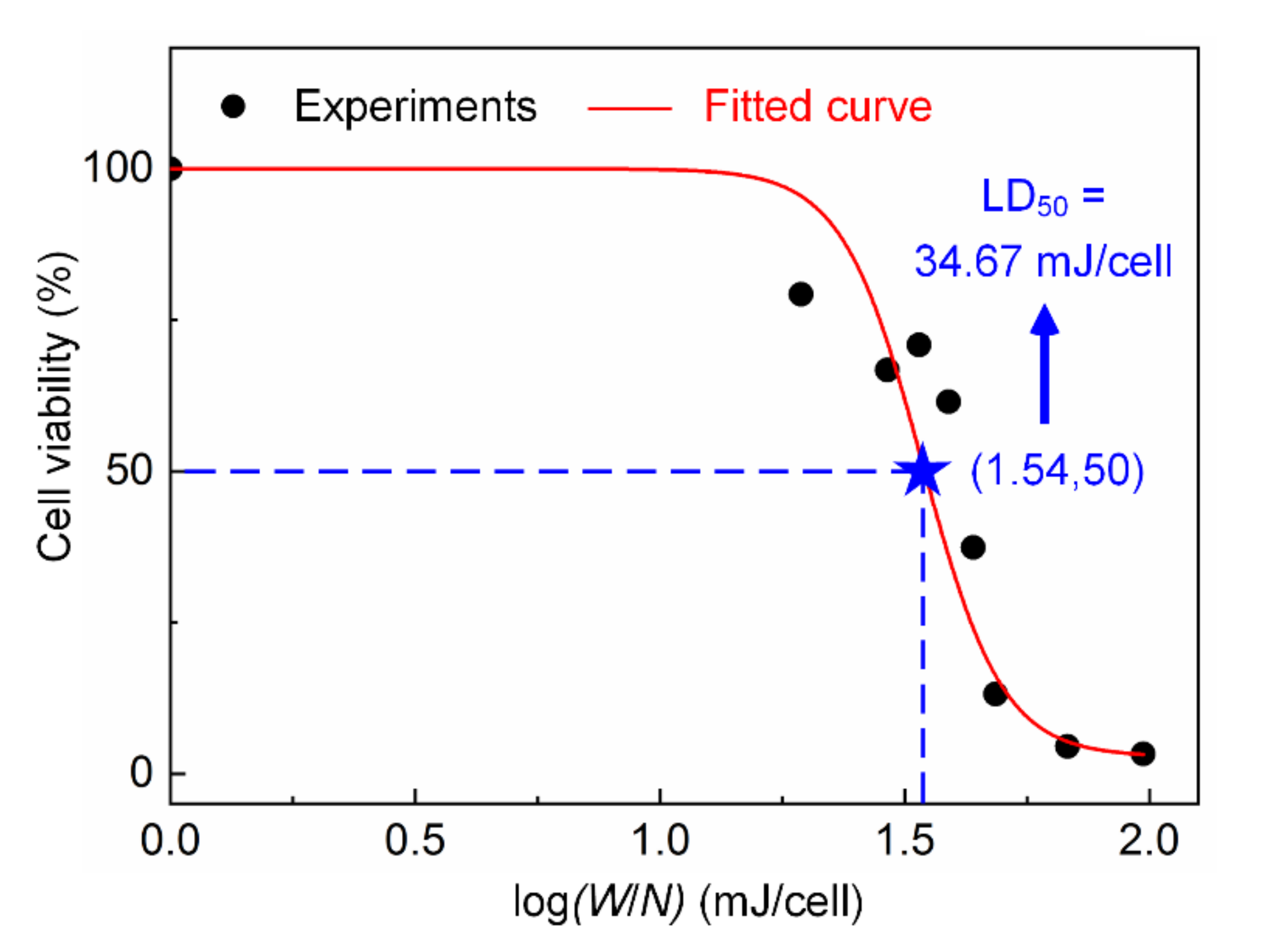
4.2. Determination of LD50

In order to obtain the value of LD50 of the CAP jet to the HEK293 cells, the cell viabilities in Figure 5 are plotted versus the logarithmic values of (W/N), forming an S-shaped curve, as shown in Figure 6. The fitted value of log(W/N) corresponding to the cell viability of 50% is 1.54. Thus, the value of LD50 is 34.67 mJ/cell. Although the obtained LD50 value in this section is not versatile for other CAP sources and biological materials, this method combining plasma physics and pharmacy provides a novel idea that is easy to conduct and realize for estimating the plasma dosage in plasma medicine. The value of LD50 plays a critical role as an indicator for studying the plasma biomedical effects provided that various types of CAP sources and cell lines are used in future studies. On the one hand, this dosage equals a 217-s CAP treatment to the HEK293 cells under the operating parameters as listed in Section 2.2. Thus, when studying the sub-lethal effects of the HEK293 cells using the CAP Bio-Med Platform, it would be better to limit the plasma treatment time to be shorter than 217 s under the discharge power of 0.8 W, or control the discharge power to be lower than 0.8 W when the plasma treatment time lasts 217 s or even longer. On the other hand, the LD50 value of 34.67 mJ/cell is also an important reference datapoint when studying the lethal and/or sub-lethal effects of other cell lines using different types of CAPs.
The published results concerning plasma dosage can be roughly divided into two types: one is the quantitative definition of plasma dosage [8], and the other skips the definition of plasma dosage but controls specific effects with the help of advanced algorithms such as machine learning [19,23,24,25]. For the first type, the number densities of all the considered RONS should be precisely measured or calculated with great efforts; in particular, whenever the discharge parameters or even the environmental conditions are changed, all the measurements or calculations need to be re-conducted. While for the second type, the design, validation, and optimization of the algorithm with the considerations of complex processes as illustrated in Figure 1 are also a very challenging job. In this study, the measurement of LD50 value is determined based on the biological effects of the CAP treatment with no considerations on the complicated physical and biochemical processes, as well as specific geometrical configurations and operating conditions of the CAP generators; consequently, it is a time-saving and feasible method for estimating the plasma dosage in practice.

5. Conclusions

Inspired by the methodology in pharmacy to quantitatively describe the drug dose using the concept of LD50, this study provides a proof of concept to estimate the plasma dosage in the field of plasma medicine by measuring the value of LD50 with the CAP treatment of the in vitro kidney cells. This novel method opens a new and promising direction for a quantitative definition of the plasma dosage with the advantages of versatility and easy manipulation. In future research, more systematic research needs to be conducted in order to deepen our understandings to the CAP cell treatment effects quantitatively. For example, the cell morphological changes after CAP treatment with the aid of cell staining and a high resolution image recording facility can be investigated; complementary assays on the cell viability can be conducted to further improve the accuracy of the LD50 values; the sub-lethal effects on living cells when the CAP treatment dosage is lower than the value of LD50 should also be studied in detail. Consequently, a database including the macroscopic morphologies, CAP sub-lethal effects, and values of LD50 for various cell lines using different types of CAP sources can be established. This may form the basis for investigating the quite complex biomedical effects of CAPs in plasma medicine.

Author Contributions

Conceptualization, Y.Z. and H.-P.L.; methodology, Y.Z., H.-P.L., J.L., L.-X.Z. and W.-W.D.; software, T.H., X.Z. and Z.-F.Z.; validation, J.L., L.-X.Z. and T.H.; formal analysis, X.-Y.C. and L.-X.Z.; investigation, J.L., L.-X.Z., T.H. and Y.Y.; resources, T.H., N.Z. and Y.Y.; data curation, J.L., L.-X.Z. and T.H.; writing—original draft preparation, J.L., L.-X.Z. and T.H.; writing—review and editing, Y.Z. and H.-P.L.; visualization, J.L., L.-X.Z. and T.H.; supervision, Y.Z. and H.-P.L.; funding acquisition, Y.Z. and H.-P.L. All authors have read and agreed to the published version of the manuscript.

Funding

This work has been supported by the Tsinghua University Initiative Scientific Research Program (No. 20182000306), the projects from Tsinghua Precision Medicine Foundation, Tsinghua-Foshan Innovation Special Fund (TFISF, No. 2020THFS0118), and partly supported by the National Natural Science Foundation of China (Nos. 11475103, 21627812).

Conflicts of Interest

The authors declare no conflict of interest.

References

  1. Laroussi, M. Plasma medicine: A brief introduction. Plasma 2018, 1, 47–60. [Google Scholar] [CrossRef] [Green Version]
  2. Vassallo, E.; Pedroni, M.; Silvetti, T.; Morandi, S.; Brasca, M. Inactivation of Staphylococcus aureus by the synergistic action of charged and reactive plasma particles. Plasma Sci. Technol. 2020, 22, 085504. [Google Scholar] [CrossRef]
  3. Scholtz, V.; Pazlarova, J.; Souskova, H.; Khun, J.; Julak, J. Nonthermal plasma—A tool for decontamination and disinfection. Biotechnol. Adv. 2015, 33, 1108–1119. [Google Scholar] [CrossRef] [PubMed]
  4. Li, D.; Li, G.; Li, J.; Liu, Z.-Q.; Zhang, X.; Zhang, Y.; Li, H.-P. Promotion of wound healing of genetic diabetic mice treated by a cold atmospheric plasma jet. IEEE Trans. Plasma Sci. 2019, 47, 4848–4860. [Google Scholar] [CrossRef]
  5. Li, G.; Li, D.; Li, J.; Jia, Y.-N.; Zhu, C.; Zhang, Y.; Li, H.-P. Promotion of the wound healing of in vivo rabbit wound infected with methicillin-resistant Staphylococcus aureus treated by a cold atmospheric plasma jet. IEEE Trans. Plasma Sci. 2021, 49, 2329–2339. [Google Scholar] [CrossRef]
  6. Zhai, S.; Kong, G.; Xia, Y. Application of atmospheric pressure cold plasma in dermatology. J. Diagn. Ther. Dermato.-Venereol. 2020, 20, 134–140. (In Chinese) [Google Scholar]
  7. Xu, Z.; Lan, Y.; Ma, J.; Shen, J.; Han, W.; Hu, S.H.; Ye, C.; Xi, W.; Zhang, Y.; Yang, C.; et al. Applications of atmospheric pressure plasma in microbial inactivation and cancer therapy: A brief review. Plasma Sci. Technol. 2020, 22, 103001. [Google Scholar] [CrossRef]
  8. Cheng, H.; Xu, J.-X.; Li, X.; Liu, D.-W.; Lu, X.-P. On the dose of plasma medicine: Equivalent total oxidation potential (ETOP). Phys. Plasmas 2020, 27, 063514. [Google Scholar] [CrossRef]
  9. Von Woedtke, T.; Reuter, S.; Masur, K.; Weltmann, K.-D. Plasmas for medicine. Phys. Rep. 2013, 530, 291–320. [Google Scholar] [CrossRef]
  10. Graves, D.B. The emerging role of reactive oxygen and nitrogen species in redox biology and some implications for plasma applications to medicine and biology. J. Phys. D Appl. Phys. 2012, 45, 263001. [Google Scholar] [CrossRef]
  11. Stoffels, E.; Kieft, I.E.; Sladek, R.E. Superficial treatment of mammalian cells using plasma needle. J. Phys. D Appl. Phys. 2003, 36, 2908–2913. [Google Scholar] [CrossRef]
  12. Kieft, I.E.; Darios, D.; Roks, A.J.M.; Stoffels, E. Plasma treatment of mammalian vascular cells: A quantitative description. IEEE Trans. Plasma Sci. 2005, 33, 771–775. [Google Scholar] [CrossRef]
  13. Dobrynin, D.; Fridman, G.; Friedman, G.; Fridman, A. Physical and biological mechanisms of plasma interaction with living tissue. New J. Phys. 2009, 11, 115020. [Google Scholar] [CrossRef]
  14. Baik, K.Y.; Kim, Y.H.; Ryu, Y.H.; Kwon, H.S.; Park, G.; Uhm, H.S.; Choi, E.H. Feeding-gas effects of plasma jets on Escherichia coli in physiological solutions. Plasma Process. Polym. 2013, 10, 235–242. [Google Scholar] [CrossRef]
  15. Metelmann, H.R.; Woedtke, T.V.; Weltmann, K.D. Comprehensive Clinical Plasma Medicine; Springer: Cham, Switzerland, 2018; pp. 84–85. [Google Scholar]
  16. Gidon, D.; Graves, D.B.; Mesbah, A. Effective dose delivery in atmospheric pressure plasma jets for plasma medicine: A model predictive control approach. Plasma Sources Sci. Technol. 2017, 26, 085005. [Google Scholar] [CrossRef]
  17. Xu, X.; Dai, X.; Xiang, L.; Cai, D.; Xiao, S.; Ostrikov, K. Quantitative assessment of cold atmospheric plasma anti-cancer efficacy in triple-negative breast cancers. Plasma Process. Polym. 2018, 15, e1800052. [Google Scholar] [CrossRef]
  18. Mesbah, A.; Graves, D.B. Machine learning for modeling, diagnostics, and control of non-equilibrium plasmas. J. Phys. D Appl. Phys. 2019, 52, 30LT02. [Google Scholar] [CrossRef] [Green Version]
  19. Bonzanini, A.D.; Paulson, J.A.; Graves, D.B.; Mesbah, A. Toward safe dose delivery in plasma medicine using projected neural network-based fast approximate NMPC. IFAC-PapersOnLine 2020, 53, 5279–5285. [Google Scholar] [CrossRef]
  20. Moini, J. Fundamental Pharmacology for Pharmacy Technicians; Cengage Learning: New York, NY, USA, 2009; p. 39. [Google Scholar]
  21. Haveles, E.B. Applied Pharmacology for the Dental Hygienist; Elsevier: Amsterdam, The Netherlands, 2020; p. 48. [Google Scholar]
  22. Akhila, J.S.; Shyamjith, D.; Alwar, M.C. Acute toxicity studies and determination of median lethal dose. Curr. Sci. 2007, 93, 917–920. [Google Scholar] [CrossRef]
  23. Gidon, D.; Abbas, H.S.; Bonzanini, A.D.; Graves, D.B.; Mesbah, A. Data-driven LPV model predictive control of a cold atmospheric plasma jet for biomaterials processing. Control. Eng. Pract. 2021, 109, 104725. [Google Scholar] [CrossRef]
  24. Gidon, D.; Graves, D.B.; Mesbah, A. Spatial thermal dose delivery in atmospheric pressure plasma jets. Plasma Sources Sci. Technol. 2019, 28, 025006. [Google Scholar] [CrossRef] [Green Version]
  25. Gidon, D.; Graves, D.B.; Mesbah, A. Predictive control of 2D spatial thermal dose delivery in atmospheric pressure plasma jets. Plasma Sources Sci. Technol. 2019, 28, 085001. [Google Scholar] [CrossRef]
  26. Lu, X.; Cao, Y.; Yang, P.; Xiong, Q.; Xiong, Z.; Xian, Y.; Yuan, P. An RC plasma device for sterilization of root canal of teeth. IEEE Trans. Plasma Sci. 2009, 37, 668–673. [Google Scholar] [CrossRef]
  27. Ge, N.; Wu, G.-Q.; Li, H.-P.; Wang, Z.; Bao, C.-Y. Evaluation of the two-dimensional temperature field and instability of a dual-jet DC arc plasma based on the image chain coding technique. IEEE Trans. Plasma Sci. 2011, 39, 2884–2885. [Google Scholar] [CrossRef]
  28. Guo, H.; Li, P.; Li, H.-P.; Ge, N.; Bao, C.-Y. In situ measurement of the two-dimensional temperature field of a dual-jet direct-current arc plasma. Rev. Sci. Instrum. 2016, 87, 033502. [Google Scholar] [CrossRef]
  29. Kogelschatz, U. Dielectric-barrier discharges: Their history, discharge physics, and industrial applications. Plasma Chem. Plasma Process. 2003, 23, 1–46. [Google Scholar] [CrossRef]
  30. Yang, W.; Acosta, D. Cytotoxicity potential of surfactant mixtures evaluated by primary cultures of rabbit cornea1 epithelial cells. Toxicol. Lett. 1994, 70, 309–318. [Google Scholar] [CrossRef]
  31. Cree, I.A. (Ed.) Cancer Cell Culture: Methods and Protocols, 2nd ed.; Springer Science & Business Media: London, UK, 2011; pp. 223–224. [Google Scholar] [CrossRef]
  32. Kamiloglu, S.; Sari, G.; Ozdal, T.; Capanoglu, E. Guidelines for cell viability assays. Food Front. 2020, 1, 332–349. [Google Scholar] [CrossRef]
  33. Kaspers, G.J.; Wijnands, J.J.; Hartmann, R.; Huismans, L.; Loonen, A.H.; Stackelberg, A.; Henze, G.; Pieters, R.; Hählen, K.; Van Wering, E.R.; et al. Immunophenotypic cell lineage and in vitro cellular drug resistance in childhood relapsed acute lymphoblastic leukaemia. Eur. J. Cancer 2005, 41, 1300–1303. [Google Scholar] [CrossRef]
  34. Hubeek, I.; Peters, G.J.; Broekhuizen, R.; Zwaan, C.M.; Kaaijk, P.; Van Wering, E.S.; Gibson, B.E.S.; Creutzig, U.; Janka-Schaub, G.E.; Boer, M.D.; et al. In vitro sensitivity and cross-resistance to deoxynucleoside analogs in childhood acute leukemia. Haematologica 2006, 91, 17–23. [Google Scholar] [CrossRef]
  35. Bahuguna, A.; Khan, I.; Bajpai, V.K.; Kang, S.C. MTT assay to evaluate the cytotoxic potential of a drug. Bangladesh J. Pharmacol. 2017, 12, 115–118. [Google Scholar] [CrossRef]
  36. Lu, X.; Naidis, G.V.; Laroussi, M.; Reuter, S.; Graves, D.B.; Ostrikov, K. Reactive species in non-equilibrium atmospheric-pressure plasmas: Generation, transport, and biological effects. Phys. Rep. 2016, 630, 1–84. [Google Scholar] [CrossRef] [Green Version]
  37. Yue, Y.; Pei, X.; Gidon, D.; Wu, F.; Wu, S.; Lu, X. Investigation of plasma dynamics and spatially varying O and OH concentrations in atmospheric pressure plasma jets impinging on glass, water and metal substrates. Plasma Sources Sci. Technol. 2018, 27, 064001. [Google Scholar] [CrossRef]
  38. Dewhirst, M.W.; Viglianti, B.L.; Lora-Michiels, M.; Hanson, M.; Hoopes, P.J. Basic principles of thermal dosimetry and thermal thresholds for tissue damage from hyperthermia. Int. J. Hyperth. 2003, 19, 267–294. [Google Scholar] [CrossRef] [PubMed]
Figure 1. Schematic of the complex interactions between CAPs and treated biological targets (cells).
Applsci 11 11135 g001
Figure 2. (a) Picture of the CAP Bio-Med Platform; (b) schematic of the geometrical configuration of the CAP generator; and (c) typical image of the CAP jet.
Applsci 11 11135 g002
Figure 3. Emission spectrum of the helium CAP jet. The discharge parameters are: f = 23 kHz, Vd = 3.5 kV, Q = 8.0 slpm, Pin = 0.8 W.
Applsci 11 11135 g003
Figure 4. Variations of the HEK293 cell viability under different incubation durations and CAP treatment times.
Applsci 11 11135 g004
Figure 5. Variation of the cell viability with the value of (W/N). **** p < 0.0001.
Applsci 11 11135 g005
Figure 6. S-shaped curve of the cell viability versus the logarithmic value of (W/N).
Applsci 11 11135 g006
Table 1. Materials and their sources of procurement used in this study.
MaterialsSources of Procurement
Cell lineHuman Embryonic Kidney 293 (HEK293) cellsCell Center of Peking Union Medical College, Beijing, China
ReagentsDulbecco’s Modified Eagle Medium (DMEM)Thermo Fisher Scientific Co., Ltd., Waltham, MA, USA
Fetal bovine serumThermo Fisher Scientific Co., Ltd., Waltham, MA, USA
Penicillin–StreptomycinThermo Fisher Scientific Co., Ltd., Waltham, MA, USA
CO2Beijing Praxair Practical Gas Co., Ltd.,
Beijing, China
MTT (3-(4,5-Dimethylthiazol-2-yl)-2,5-diphenyltetrazolium bromide) solutionBeijing Leagene Biotech. Co., Ltd.,
Beijing, China
Dimethyl sulfoxide (DMSO)Beijing Leagene Biotech. Co., Ltd.,
Beijing, China
Publisher’s Note: MDPI stays neutral with regard to jurisdictional claims in published maps and institutional affiliations.

