Article

Effect of Shrinking and No Shrinking Dentine and Enamel Replacing Materials in Posterior Restoration: A 3D-FEA Study

by
Pietro Ausiello
1,
Amanda Maria de Oliveira Dal Piva
2,
Alexandre Luiz Souto Borges
2,
Antonio Lanzotti
3,
Fausto Zamparini
4,*,
Ettore Epifania
1 and
João Paulo Mendes Tribst
5
1
School of Dentistry, University of Naples Federico II, via S. Pansini 5, 80131 Naples, Italy
2
Department of Dental Materials and Prosthodontics. Institute of Science and Technology, São Paulo State University-UNESP, São José Dos Campos 12220000, Brazil
3
Fraunhofer JL IDEAS, Department of Industrial Engineering, University of Naples Federico II, 80125 Naples, Italy
4
Endodontic Clinical Section, School of Dentistry, Department of Biomedical and Neuromotor Sciences, University of Bologna-Alma Mater Studiorum, 40125 Bologna, Italy
5
School of Dentistry, University of Taubaté–UNITAU, Taubaté, São Paulo 12020-340, Brazil
*
Author to whom correspondence should be addressed.
Submission received: 11 February 2021 / Revised: 23 February 2021 / Accepted: 26 February 2021 / Published: 3 March 2021

Abstract

:
The aim of the present study was to investigate the effect of shrinking and no shrinking dental filling materials combination in posterior restorations under the combined effects of polymerization shrinkage and occlusal load by means of 3D Finite Elements Analysis. Six computer-generated and restored class I or class II cavities models of a lower molar were designed in the CAD software and evaluated according to the cavity and restorative procedure. Different shrinking and no shrinking adhesive materials combination with diverse Young’s modulus were considered. A food bolus was modeled on the occlusal surface replicating the chewing load using static linear analyses Polymerization shrinkage was simulated for the shrinking different restorative materials. The maximum principal stress was selected as analysis criteria. All models exhibited higher stresses along the dentine restoration interfaces with different magnitude and a similar stress trend along enamel restoration interface. Stress values up to 22 MPa and 19 MPa were recorded in the enamel and restoration, respectively. The use of elastic not shrinking material layer in combination with bulk fill composite reduced the stress magnitude in dentine and enamel to replace dental tissues. Class I and class II posterior cavities adhesively restored with shrinking filling material’s combination showed the most unfavorable stress concentrations and the multilayer technique is a promising restorative alternative in posterior adhesive restorations when deep dentin and enamel volumes are missing.

1. Introduction

Dental nano-filled resin composites are considered advanced materials in restorative dentistry thanks to their improved aesthetical and functional properties [1]. They are indicated as direct fillings by adhesive techniques when efficient mechanical recovering of weakened or fractured teeth is necessary [2,3].
Cytotoxic risks of the filling composite polymeric matrix components have been also introduced in the past to limit their extensive clinical use and related to the resin monomers molecules [4]. However, no direct clinical signs have been described about this effect even when decay has been removed to limit the effects on the tooth biomechanical response [5] and filling composite adhesively placed in deep dentin.
Dental filling direct posterior resin composites, in fact, are used to substitute lost enamel and dentine tissues. Leakage and secondary caries often represent one of the main adhesive restoration failure reason because of they normally show a volumetric polymerization shrinkage, considered as post gel shrinkage, from 1% up to 4.5% [6]. The composition of organic matrix monomers and the volume percent of inorganic filler affect the polymerization shrinkage, contributing to the prevalence of residual stresses during the composite cure [7].
But stress kinetics in posterior dental restorations is intensely linked to the polymer characteristics and to the cavity design [8]. In adhesive class I restorations, in fact, it has been showed that shrinkage depends more on high c-factor configuration and it may results in cervical dentine leakage and gap generation if the stress exceeds the resin bond strength to dentine and enamel [9].
Stresses arising from shrinkage in class II cavities adhesively restored critically react at the internal and marginal surfaces in deep cavities. They are potentially able to determine in the time clinical failure by gap creation [10]. This relationship is more complex than a simply ratio of bonded/free surfaces [11,12]. The elastic modulus of the involved materials plays a significant role when they are associated with cavity geometry (boundary conditions) in the posterior adhesive restoration survival [13,14].
Flowable resin composite have been introduced with this aim to replace dentine under composite restorations being more adaptable to traditional composite to break polymerization stresses [15]. In this sense, in vivo, in vitro and in silico investigations have validated this aspect, reporting that flowable composites are able to reduce polymerization shrinkage and occlusal loading stresses in combination with multi-layer composite technique [16,17,18,19,20,21,22].
Alternatively, various studies suggested to use no shrinking adhesive dental materials such as glass ionomers in combination with composite to reduce the critical resin based materials stress distribution in enamel and dentine replacement [19].
In the last years, bulk-fill resin composites were developed as replacing materials to overcome the multilayering clinical limitations, due to the newer chemical low shrinking monomers structure, better translucency and reduced shrinkage stress development [21,22]. Different authors [23,24] have already declared that bulk-fill composites can safety be used in 4 mm or deeper cavities in posterior teeth. A literature review [25] indicated that more scientific and clinical trainings are still obligatory to completely confirm the clinical benefits of bulk-fill composites, still existing divergent results on shrinkage problematics evaluation in posterior restorations when enamel and dentine need to be replaced [26].
The aim of the present research is to clarify and to explore the effect of shrinking and no shrinking dental filling materials combination in posterior restorations, under the combined effects of polymerization shrinkage and occlusal load [27,28] by means of 3D Finite Elements Analysis. The null hypothesis was that there would be no differences in the adhesive posterior cavities restored with the application of shrinking and no shrinking dental materials under resin composite versus single “bulk” composite ones in replacing enamel and dentine.

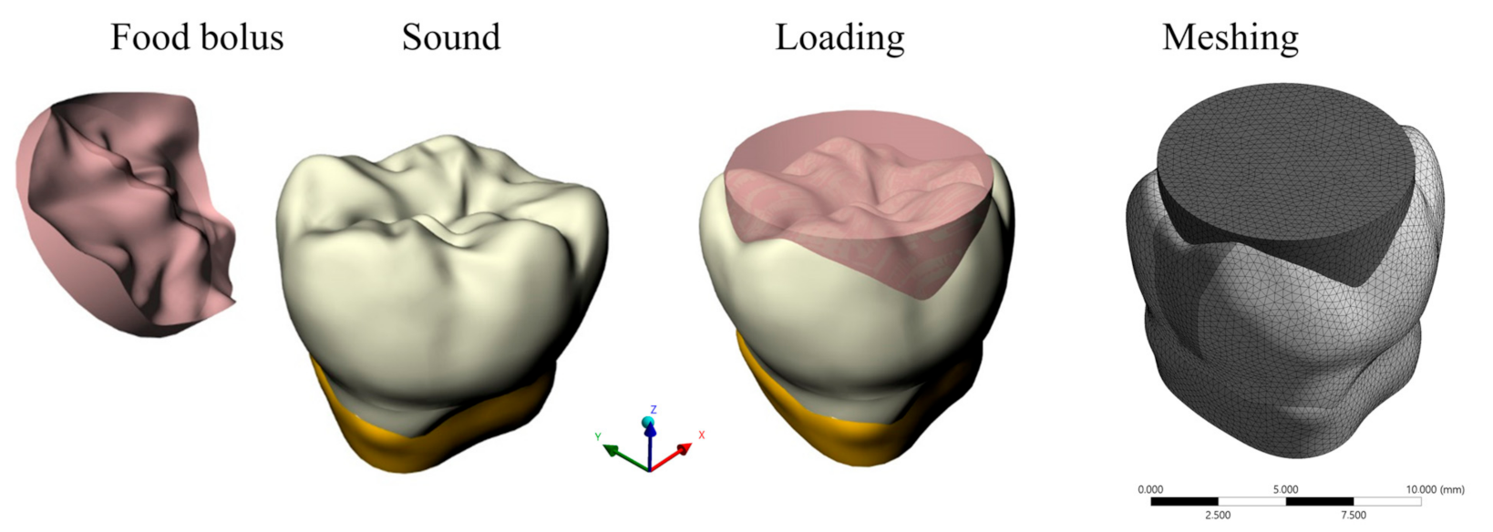
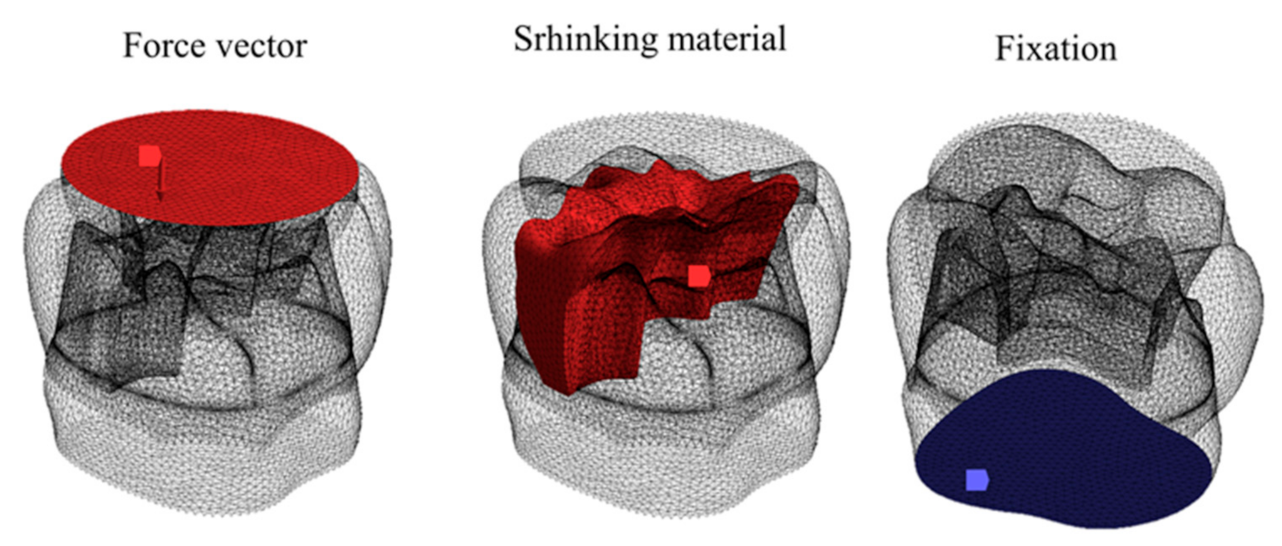
2. Materials and Methods

The mechanical behavior of materials and applications in Dentistry have been extensively investigated by means of CAD–FEM (Computer Aided Design and Finite Element Method) methods [10,27,28]. Therefore, the present study has been carried out using previously reported models with properly known elastic response.
Starting from these 3D CAD models, six models simulating six posterior restorations consisting in class I (occlusal) and class II MO (mesio-occlusal) lower molar cavities (Figure 1) were exported to the analysis software to investigate the influence on the stress distribution of resin composite (shrinking) and glass ionomer cement (no shrinking) material combinations in a bi-layer or bulk technique to substitute enamel and dentine [10,27,28]. These six models were nominated as A, B, C, D, E and F and were indicated in the Table 1. Their geometric features are exposed in Figure 1.
According to the modelling process from previous studies [27,28], the sound tooth lower molar model was got by means of reverse engineering techniques. The shapes and volumes of dentin and enamel were digitized using a micro-CT scanner (Bruker microCT, Kontich, Belgium). Images and data were processed via InVesalius 3.1.1 software and 3-D tessellated surfaces were elaborated. Cross-section curves were generated, and the parametric 3D CAD model was created using loft surfaces in (Rhinoceros® 4.0 Robert McNeel & Associates, Seattle, WA, USA) starting from 3-D tessellated surfaces. Boolean seperations guaranteed the interfacial dentin and enamel boundaries congruence. The tooth model was cut 2.5 mm below the cervical area to obtain the final model. The bucco-palatal and mesio-distal sizes were 10.60 mm and 12.36 mm respectively and the enamel thickness was of about 1.5 mm [10,26,27].
A class I cavity 4.0 mm deep was modelled for the restored models A, B and C obtained via Boolean operations between the cavity, enamel and dentin surfaces [27]. Class I pulp and lateral walls had rounded modelled angles. A class II MO cavity 4.0 mm deep in the central and 5.5 mm in the box. A bevel of 0.5 mm was shaped all along the enamel [8]. The margins of class II MO were modelled, and the restored models D, E and F were obtained via Boolean actions between the cavity, dentine and enamel surfaces. The class II MO lateral and internal surfaces had rounded modelled angles.
Masticatory function has been reported as depending on the contact between tooth surface and food bolus. In this study it was simulated by modelling food bolus geometry on the occlusal surface [28]. The dental model was positioned in a coordinate system, where X- and Y-axes indicated the bucco-lingual and mesio-distal directions, respectively. The Z-axis was vertically oriented (Figure 2).

Numerical Simulation
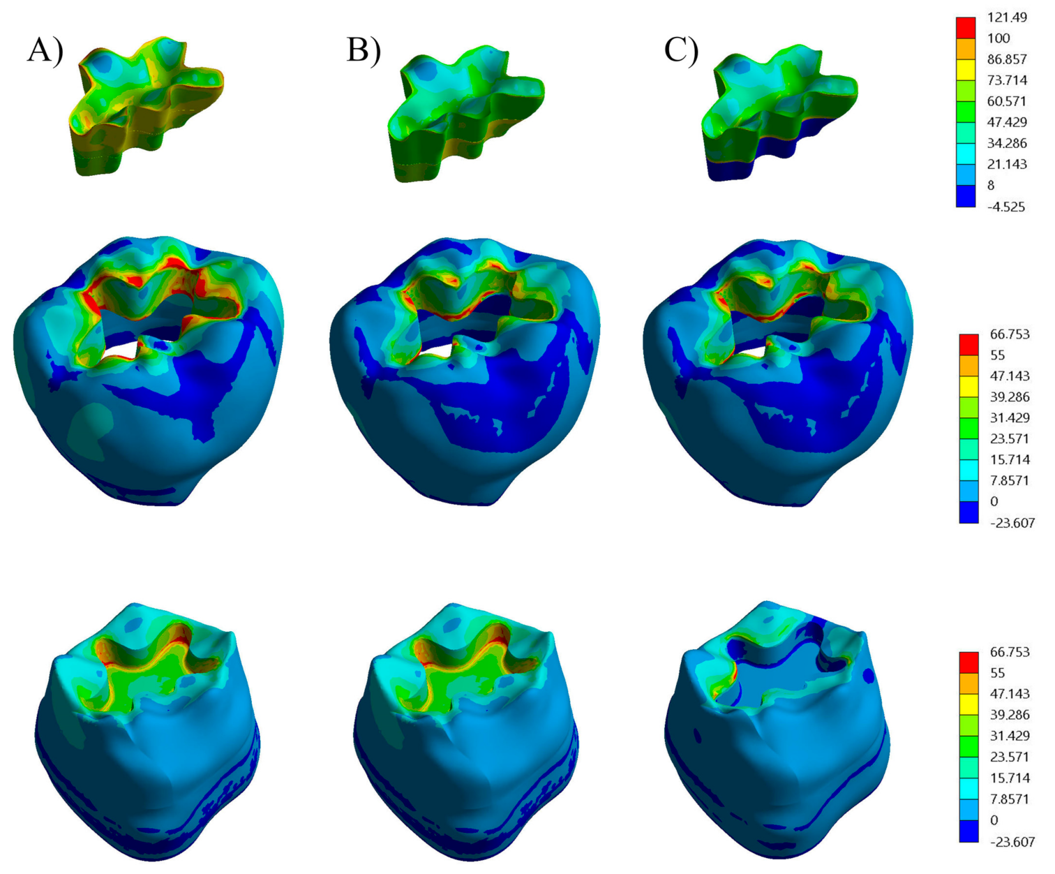
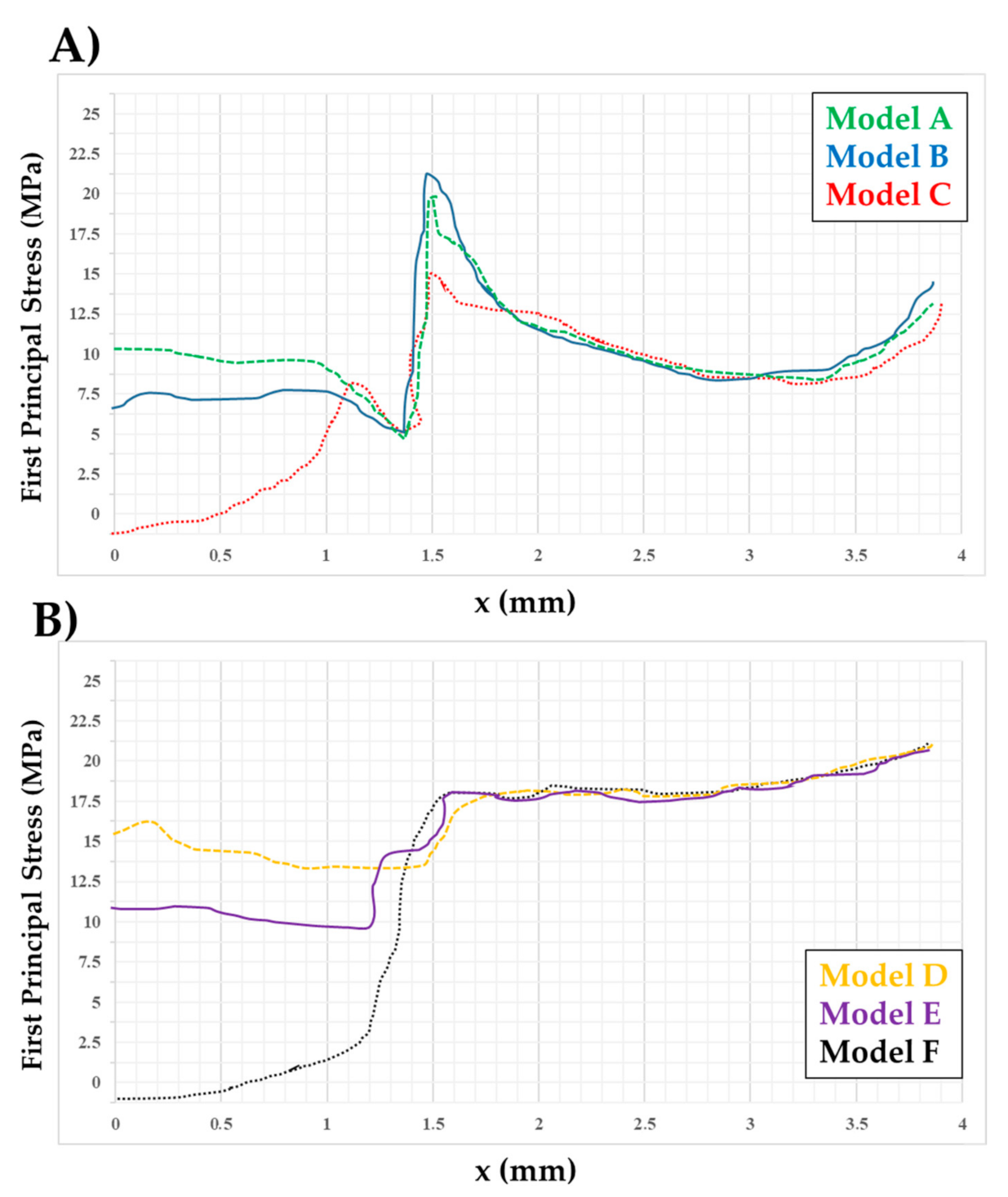
The response of six restored models was mechanically analysed with 3D- FEA (ANSYS 19.2, ANSYS Inc., Houston, TX, USA). Three models of a restored tooth for each cavity design and with a multilayer construction were created and investigated using different material combinations, as summarized in Table 1.
All the volumes were discretized by 4-node tetrahedral elements with a total size extending from 0.06 mm to 0.2 mm. To minimalize the results mesh-dependency due to the small curvature radius and notch effects, mesh improvement techniques were used according to the mesh convergence test. 2. All the FEA analyses were focused on load during the closing phase of the chewing cycle [10]. The variability of chewing function was considered dependent on the contact between food and tooth surface [10]. Solid food apple pulp was displayed on the occlusal surface (Figure 2) and slide-type contact elements were used between the food and the tooth surface. For class I the total number of elements were 445,242 with 97,364 nodes in the model A, and 438,092 elements with 97,412 in models B and C. For class II the total number of elements were 404,766 with 88,513 nodes in the model C, and 395,712 elements with 86,333 in models E and F.
It was stated that the shrinkage stresses in composite material restored teeth may be lower than those calculated on elastic model basis (Figure 3). Throughout the polymerization, kinetic stress relaxation goes together with the viscous flow of composites and the E modulus and viscosity rapidly upsurge. Seeing this behaviour, a basic approach has been used [29,30]. Effective linear shrinkage sr = 0.001 was accepted in the analyses. The mechanical properties allocated to each material and the scales of linear shrinkage (%) of materials are potted in Table 2. Adhesive layers and shrinking materials polymerization shrinkage were simulated with the thermal expansion approach by transmission a one-degree drop in temperature [13,29]. Physiological masticatory loads were simulated as an occlusal static load of 600 N and a transversal load of 20 N [10,27,28,30]. These loads vertically and buccolingually applied on the food and simultaneously in combination with shrinkage effects [28].
The model’s lower surfaces were constrained in all the directions. Statically and linearly analyses were carried out. The analyses were performed considering a non-failure condition and all materials were assumed to present an elastic behaviour.

3. Results

The resultant stress distributions for the models were compared and examined. By way of these materials exhibit a brittle mechanical behavior [10], First Principal Stress was designated as analysis criterion. Figure 4 displays the first principal stress distributions for enamel, dentin and restorative shrinking and no shrinking material for class I, and, Figure 5 illustrates first principal stress distributions for enamel, dentin and restorative material for class II models following the occlusal and transversal loading in combination with shrinkage result. Additional quantitative results were detected by inspection line, defined laterally to the cavity wall.
Looking at the cross section, first principal stresses were planned along the inspection patch and compared for the diverse models as previously reported [28,29].
All the class II MO (D, E and F models in Figure 5) restorations displayed high stress concentration close to the occlusal surfaces, in comparison with class II MO (A, B and C models). However, regardless the cavity design, the models exhibited a similar stress trend along enamel–restoration interfaces. The highest stress values in the restoration, were located nearby the top corner of the dentin-enamel interface (Figure 4) and on the occlusal surfaces.
Irrespective the cavity design, models A and B (Figure 4 and Figure 5) exhibited a similar stress tendency along dental tissues and restoration interface as well models D and E. A different stress trend was observed in models C and F along dental enamel and restoration interface while the stress declines with depth along the dentine and restoration interface and have a tendency to zero at the cavity floor (Figure 4 and Figure 5). For class I cavity, in Figure 4 it is evident the modification of stress distribution internally between models A and C. In model C, where no shrinking material has been replicated in the deepest 1.5 mm layer, while in model A, the highest stress was concentrated in the restoration. The same difference we found in class II (Figure 5) for internal stress distribution among model D and F. Model F, where no shrinking material has been imitated in the deepest 1.5 mm layers, while in model D, the highest stress was concentrated in the enamel.
The stress trend was plotted in Figure 5 and the stress peaks in enamel tissue, dentin tissue and restoration are summarized in Table 3.
The lowest stress magnitude in enamel was calculated in 9.0 MPa when class I design has been restored with glass-ionomer cement (no shrinking) associated with bulk-fill resin composite (Model C). However, this value increased 209% when the cavity has been prolonged to a class II and the material was the Bulk-fill resin composite (23.3 MPa). The same behavior can be observed in dentin tissue, comparing the lowest stress peak of 2.3 MPa for class I restored with glass-ionomer cement (no shrinking) associated with Bulk-fill composite with class II and the Bulk-fill composite (18.9 MPa), almost 87.93% lower stress for the first situation (Figure 6 and Figure 7).

4. Discussion

The null hypothesis was that there would be no difference in the posterior cavities restored with the application of shrinking and no shrinking material’s combination, in a bulk or in a multilayer technique. The results showed that different models exhibited higher stresses along the dentine–restoration interfaces with different magnitude according to the restorative material combination. Thus, the hypothesis was rejected.
It has been exposed that teeth have complex behavior during mastication in reason of anatomical arrangements based on the different enamel and dentine combination. Chewing is similarly influenced by jaw muscles response for differently rigid foods [30,31]. In addition, the present study showed that when different restorative materials were applied, the chewing load dissipation can also be affected regardless class I or class II designs. Stress distributions within posterior teeth are a function of 3D shape, stiffness and loading, even though the E modulus of most of the components are known [32]. Dental human enamel has a higher elastic modulus than dentine which is a more compliant tissue. This mechanical combination in posterior teeth permits a higher fracture resistance under chewing and a more uniform stress distribution [33,34]. Therefore, the restorative procedure should be performed in order to recover the natural tooth mechanical behavior, by means of resistant and compliance biomimetic materials in different layers placed [35].
Premolars and molars adhesively restored using different shrinking resin-based composites has been mechanically investigated under occlusal loading under laboratory conditions [35,36,37] or by means of FEA of restored teeth [13,26,27,28,31,34]. However, the combination of chewing load application and polymerization shrinkage effect should be applied as resultant of mechanical behavior in restored tooth structures [10]. The major concept is that adhesive materials are aimed to rebuild a durable adhesive bridge between the contrasting walls of the restored cavity [29] targeting to replicate properties and function of the dentine and enamel lost as closely as possible and reducing debonding, leakage at the margins and fracture risk [10,22,38,39]. Morphology, function and aesthetics must also be replicated. For that reason, dentin and enamel should be considered as individual tissue with unique characteristics that the biomimetic restoration should aim to replace.
Numerous analyses on adhesive posterior resin composite restorations were carried out using in vitro methods [40,41,42] or using in silico simulations [10,32,43] and have verified the influence of polymerization shrinkage, cusps deflection occlusal loading, enamel crack propagation and cavities design in the stress distribution. The information on how stresses redistribute in posterior restored teeth assists to prevent post restorative problems [9]. FEA presents several advantages than in vitro tests because of it permits the management of complex settings and because it delivers a superior understanding of detailed results about the internal stress of tooth, restorations and materials [16,29]. Finite Elements Analysis needs a detailed modelling and a multifaceted scheming with correct boundary conditions [10,43]. To minimize the stresses arising from composite polymerization during restoration of teeth there is a proposed approach in composite layering of class I composite restoration from bulk placement [44].
The objective was obtained by covering the cavity surfaces with a thin resin composite layer, vertically and horizontally placed. This layer was named the “prelayer” and it was followed, in a second step, by further composite horizontal layering. The FEA of this modified class I technique showed a 75% reduction in the shrinkage stresses. The “prelayer” assured a strong linking of the remaining layers with the tooth tissue due to appropriate shrinkage vectors. After the “prelayer” setting, the cavity becomes smaller. Resin composite thickness on one hand and its E modulus on the other one influence stress distribution at all the adhesive interfaces determining a variable dental cusps deformation [44].
Potential adverse effects were reported as a consequence of bulk fill composite 1% linear polymerization shrinkage which affect the marginal adaptation of this material with the cavity walls [28]. Additionally, the new generation of conventional and bulk-fill composites did not reduce shrinkage stress in endodontically-treated molars [45]. In this context, where composites substitute lost enamel and dentin, shrinking stress of the materials has deeper consequences than occlusal loading stress. Therefore, to reproduce dentin and enamel with only bulk-fill material seems not to be the most promising approach regardless the cavity design [38,39,43].
The present stress results were perhaps not dependent on the cavity configuration only but also upon the linearly polymerization shrinkage and E modulus restorative materials [11].
The results (Figure 5 and Figure 6) confirm the role that shrinkage distinctly get in determining more failure risk consequences than occlusal loading [40].
These data partially limit the role of the c-factor (cavity configuration) at the class II restorations interfaces. However, it correlates the stresses to the cavity compliance. A possible clarification of this behavior can be accredited to the proportion thickness/E modulus of the restorative materials. In spite of the bulk-fill composites time convenient application, clinicians should not fully substitute the traditional dentine and enamel replacing materials by a single shrinking material, but they could combine their use when large and deep cavities are considered [10,41,46].
C-factor and of bulk-fill composite have a great impact on bonding resistance of the cavity restorations [42]. These findings confirm that a mathematical analysis can be helpful to study the shrinking materials problems and to translate them to clinical recommendations [11]. results of the present study confirm the influence of the shrinking or no shrinking restorative material, E modulus and layering technique to restore different volumes of the lost dentine and enamel [46].

5. Conclusions

This 3D FEA study, with the limits arising from isotropic elastic mechanical material’s behavior, suggests:
  • Class I and class II posterior cavities adhesively restored with shrinking filling material’s combination showed the most unfavorable stress concentrations in replacing dentine and enamel tissues;
  • In these same posterior adhesive restorations, residual lower shrinkage stress and occlusal loading stress magnitude were detected when combining non-shrinking filling material with bulk filling composite;
  • Multilayer technique confirmed to be an adequate restorative option in posterior adhesive restorations when deep dentin and enamel volumes were missing.

Author Contributions

Conceptualization, P.A.; A.L. and J.P.M.T.; methodology, P.A.; A.L.S.B.; A.L. and J.P.M.T.; software, P.A.; A.L.S.B.; J.P.M.T.; validation, P.A.; A.M.d.O.D.P.; A.L.S.B.; A.L. and J.P.M.T.; formal analysis, P.A.; A.M.d.O.D.P. and J.P.M.T.; investigation, P.A.; A.M.d.O.D.P.; A.L.S.B.; A.L. and J.P.M.T.; resources, P.A.; A.M.d.O.D.P.; A.L.S.B.; A.L. and J.P.M.T.; data curation, P.A.; A.M.d.O.D.P. and J.P.M.T.; writing—original draft preparation, P.A.; A.M.d.O.D.P.; A.L. and J.P.M.T.; writing—review and editing, P.A.; A.M.d.O.D.P.; A.L.S.B.; A.L.; F.Z.; E.E. and J.P.M.T.; visualization, P.A.; A.L. and J.P.M.T.; supervision, P.A.; A.L. and J.P.M.T.; project administration, P.A.; and J.P.M.T.; funding acquisition, P.A. and A.L. All authors have read and agreed to the published version of the manuscript.

Funding

This research received no external funding.

Institutional Review Board Statement

Not applicable.

Informed Consent Statement

Not applicable.

Data Availability Statement

The data presented in this study are available on request from the corresponding author.

Acknowledgments

None.

Conflicts of Interest

The authors declare no conflict of interest.

References

  1. Yadav, R.; Kumar, M. Dental restorative composite materials: A review. J. Oral Biosci. 2019, 61, 78–83. [Google Scholar] [CrossRef] [PubMed]
  2. Khosravani, M.R. Mechanical behavior of restorative dental composites under various loading conditions. J. Mech. Behav. Biomed. Mater. 2019, 93, 151–157. [Google Scholar] [CrossRef]
  3. Borgia, E.; Baron, R.; Borgia, J.L. Quality and Survival of Direct Light-Activated Composite Resin Restorations in Posterior Teeth: A 5- to 20-Year Retrospective Longitudinal Study. J. Prosthodont. 2019, 28, e195–e203. [Google Scholar] [CrossRef] [Green Version]
  4. Treglia, A.S.; Turco, S.; Ulianich, L.; Ausiello, P.; Lofrumento, D.D.; Nicolardi, G.; Miele, C.; Garbi, C.; Beguinot, F.; di Jeso, B. Cell fate following ER stress: Just a matter of quo ante recovery or death? Histol. Histopathol. 2012, 27, 1–12. [Google Scholar]
  5. Weimann, D.; Morgenthal, A.; Schwendicke, F.; Fleck, C.; Razi, H. Substantial regional differences in the biomechanical behavior of molar treated with selective caries tissue removal technique: A finite element study. Dent. Mater. 2020, S0109-5641, 30328–30336. [Google Scholar] [CrossRef]
  6. Davidson, C.L.; Feilzer, A.J. Polymerization shrinkage and polymerization shrinkage stress in polymer-based restoratives. J. Dent. 1997, 25, 435–440. [Google Scholar] [CrossRef]
  7. Schneider, L.F.J.; Cavalcante, L.M.; Silikas, N. Shrinkage stresses generated during resin-composite applications: A review. J. Dent. Biomech. 2010, 2010, 131630. [Google Scholar] [CrossRef] [PubMed]
  8. Apicella, A.; Di Palma, L.; Aversa, R.; Ausiello, P. DSC kinetic characterization of dental composites using different light sources. J. Adv. Mater. 2002, 34, 22–25. [Google Scholar]
  9. Watts, D.C.; Satterthwaite, J.D. Axial shrinkage-stress depends upon both C-factor and composite mass. Dent. Mater. 2008, 24, 1–8. [Google Scholar] [CrossRef]
  10. Ausiello, P.; Ciaramella, S.; De Benedictis, A.; Lanzotti, A.; Tribst, J.P.M.; Watts, D.C. The use of different adhesive filling material and mass combinations to restore class II cavities under loading and shrinkage effects: A 3D-FEA. Comput. Methods Biomech. Biomed. Eng. 2020, 22, 1–11. [Google Scholar] [CrossRef]
  11. Jantarat, J.; Palamara, J.E.A.; Messer, H.H. An investigation of cuspal deformation and delayed recovery after occlusal loading. J. Dent. 2001, 29, 363–370. [Google Scholar] [CrossRef]
  12. Fu, J.; Aregawi, W.A.; Fok, A.S.L. Mechanical manifestation of the C-factor in relation to photopolymerization of dental resin composites. Dent. Mater. 2020, 36, 1108–1114. [Google Scholar] [CrossRef]
  13. Ausiello, P.; Apicella, A.; Davidson, C.L. Effect of adhesive layer properties on stress distribution in composite restorations—A 3D finite element analysis. Dent. Mater. 2002, 18, 295–303. [Google Scholar] [CrossRef]
  14. Al Sunbul, H.; Silikas, N.; Watts, D.C. Polymerization shrinkage kinetics and shrinkage-stress in dental resin-composites. Dent. Mater. 2016, 32, 998–1006. [Google Scholar] [CrossRef] [Green Version]
  15. Frankenberger, R.; Reinelt, C.; Glatthöfer, C.; Krämer, N. Clinical performance and SEM marginal quality of extended posterior resin composite restorations after 12 years. Dent. Mater. 2020, 36, e217–e228. [Google Scholar] [CrossRef]
  16. Baroudi, K.; Rodrigues, J.C. Flowable resin composites: A systematic review and clinical considerations. J. Clin. Diagn. Res. 2015, 9, ZE18–ZE24. [Google Scholar] [CrossRef]
  17. Boruziniat, A.; Gharaee, S.; Sarraf Shirazi, A.; Majidinia, S.; Vatanpour, M. Evaluation of the efficacy of flowable composite as lining material on microleakage of composite resin restorations: A systematic review and meta-analysis. Quintessence Int. 2016, 47, 93–101. [Google Scholar] [CrossRef] [PubMed]
  18. Martorelli, M.; Ausiello, P. A novel approach for a complete 3D tooth reconstruction using only 3D crown data. Int. J. Interact. Design Manuf. 2013, 7, 125–135. [Google Scholar] [CrossRef]
  19. Ausiello, P.; Ciaramella, S.; Garcia-Godoy, F.; Martorelli, M.; Sorrentino, R.; Gloria, A. Stress distribution of bulk-fill resin composite in class II restorations. Am. J. Dent. 2017, 30, 227–232. [Google Scholar] [PubMed]
  20. Anatavara, S.; Sitthiseripratip, K.; Senawongse, P. Stress relieving behaviour of flowable composite liners: A finite element analysis. Dent. Mater. J. 2016, 35, 369–378. [Google Scholar] [CrossRef] [PubMed] [Green Version]
  21. Han, S.H.; Park, S.H. Incremental and bulk-fill techniques with bulk-fill resin composite in different cavity configurations. Oper. Dent. 2018, 43, 631–641. [Google Scholar] [CrossRef] [PubMed]
  22. Correia, A.; Andrade, M.R.; Tribst, J.; Borges, A.; Caneppele, T. Influence of bulk-fill restoration on polymerization shrinkage stress and marginal gap formation in class V restorations. Oper. Dent. 2020, 45, E207–E216. [Google Scholar] [CrossRef]
  23. Rosatto, C.M.; Bicalho, A.A.; Veríssimo, C.; Bragança, G.F.; Rodrigues, M.P.; Tantbirojn, D.; Versluis, A.; Soares, C.J. Mechanical properties, shrinkage stress, cuspal strain and fracture resistance of molars restored with bulk-fill composites and incremental filling technique. J. Dent. 2015, 43, 1519–1528. [Google Scholar] [CrossRef]
  24. Ferracane, J.L.; Lawson, N.C. Probing the hierarchy of evidence to identify the best strategy for placing class II dental composite restorations using current materials. J. Esthet. Restor. Dent. 2020, 18. [Google Scholar] [CrossRef] [PubMed]
  25. Van Ende, A.; De Munck, J.; Lise, D.P.; Van Meerbeek, B. Bulk-Fill composites: A review of the current literature. J. Adhes. Dent. 2017, 19, 95–109. [Google Scholar] [CrossRef] [Green Version]
  26. Kaisarly, D.; Meierhofer, D.; Gezawi, M.; El Rösch, P.; Kunzelmann, K.H. Effects of flowable liners on the shrinkage vectors of bulk-fill composites. Clin. Oral Investig. 2021. [Google Scholar] [CrossRef]
  27. Ausiello, P.P.; Ciaramella, S.; Lanzotti, A.; Ventre, M.; Borges, A.L.; Tribst, J.P.; Dal Piva, A.; Garcia-Godoy, F. Mechanical behavior of Class I cavities restored by different material combinations under loading and polymerization shrinkage stress. A 3D-FEA study. Am. J. Dent. 2019, 32, 55–60. [Google Scholar] [PubMed]
  28. Ausiello, P.; Ciaramella, S.; Di Rienzo, A.; Lanzotti, A.; Ventre, M.; Watts, D.C. Adhesive class I restorations in sound molar teeth incorporating combined resin-composite and glass ionomer materials: CAD-FE modeling and analysis. Dent. Mater. 2019, 35, 1514–1522. [Google Scholar] [CrossRef]
  29. Ausiello, P.; Ciaramella, S.; Martorelli, M.; Lanzotti, A.; Gloria, A.; Watts, D.C. CAD-FE modeling and analysis of class II restorations incorporating resin-composite, glass ionomer and glass ceramic materials. Dent. Mater. 2017, 33, 1456–1465. [Google Scholar] [CrossRef] [Green Version]
  30. Williams, S.H.; Wright, B.W.; Truong, V.D.; Daubert, C.R.; Vinyard, C.J. Mechanical properties of foods used in experimental studies of primate masticatory function. Am. J. Primatol. 2005, 67, 329–346. [Google Scholar] [CrossRef]
  31. Williams, K.R.; Edmundson, J.T.; Rees, J.S. Finite element stress analysis of restored teeth. Dent. Mater. 1987, 3, 200–206. [Google Scholar] [CrossRef]
  32. Hada, Y.S.; Panwar, S. Comparison of the fracture resistance of three different recent composite systems in large Class II mesio-occlusal distal cavities: An in vitro study. J. Conserv. Dent. 2019, 22, 287–291. [Google Scholar] [CrossRef]
  33. Tribst, J.P.; Dal Piva, A.M.; Borges, A.L. Biomechanical behavior of indirect composite materials: A 3D-FEA study. Braz. Dent. Sci. 2017, 20, 52–57. [Google Scholar] [CrossRef] [Green Version]
  34. da Rocha, D.M.; Tribst, J.P.M.; Ausiello, P.; Dal Piva, A.M.O.; da Rocha, M.C.; Di Nicoló, R.; Borges, A.L.S. Effect of the restorative technique on load-bearing capacity, cusp deflection, and stress distribution of endodontically-treated premolars with MOD restoration. Restor. Dent. Endod. 2019, 44, e33. [Google Scholar] [CrossRef] [PubMed]
  35. Bonilla, E.D.; Hayashi, M.; Pameijer, C.H.; Le, N.V.; Morrow, B.R.; Garcia-Godoy, F. The effect of two composite placement techniques on fracture resistance of MOD restorations with various resin composites. J. Dent. 2020, 101, 103348. [Google Scholar] [CrossRef]
  36. Frankenberger, R.; Nassiri, S.; Lücker, S.; Lygidakis, N.N.; Krämer, N. The effect of different liners on the bond strength of a compomer to primary teeth dentine: In vitro study. Eur. Arch. Paediatr. Dent. 2021. [Google Scholar] [CrossRef] [PubMed]
  37. Peumans, M.; Politano, G.; Bazos, P.; Severino, D.; Van Meerbeek, B. Effective Protocol for Daily High-quality Direct Posterior Composite Restorations: Layering and Finishing. J. Adhes. Dent. 2020, 22, 597–613. [Google Scholar] [CrossRef] [PubMed]
  38. Aggarwal, V.; Singla, M.; Yadav, S.; Yadav, H. Effect of flowable composite liner and glass ionomer liner on class II gingival marginal adaptation of direct composite restorations with different bonding strategies. J. Dent. 2014, 42, 619–625. [Google Scholar] [CrossRef]
  39. Van Ende, A.; De Munck, J.; Van Landuyt, K.; Van Meerbeek, B. Effect of bulk-filling on the bonding efficacy in occlusal Class I cavities. J. Adhes. Dent. 2016, 18, 119–124. [Google Scholar] [CrossRef] [PubMed] [Green Version]
  40. Politi, I.; McHugh, L.E.J.; Al-Fodeh, R.S.; Fleming, G.J.P. Modification of the restoration protocol for resin-based composite (RBC) restoratives (conventional and bulk fill) on cuspal movement and microleakage score in molar teeth. Dent. Mater. 2018, 34, 1271–1277. [Google Scholar] [CrossRef]
  41. Schneider, L.F.; Cavalcante, L.M.; Prahl, S.A.; Pfeifer, C.S.; Ferracane, J.L. Curing efficiency of dental resin composites formulated with camphorquinone or trimethylbenzoyldiphenylphosphine oxide. Dent. Mater. 2012, 28, 392–397. [Google Scholar] [CrossRef]
  42. McHugh, L.E.J.; Politi, I.; AlFodeh, R.S.; Fleming, G.J.P. Implications of resin-based composite (RBC) restoration on cuspal deflection and microleakage score in molar teeth: Placement protocol and restorative material. Dent. Mater. 2017, 33, e329–e335. [Google Scholar] [CrossRef]
  43. Kim, Y.J.; Kim, R.; Ferracane, J.L.; Lee, I.B. Influence of the compliance and layering method on the wall deflection of simulated cavities in bulk-fill composite restoration. Oper. Dent. 2016, 41, e183–e194. [Google Scholar] [CrossRef] [PubMed]
  44. Kowalczyk, P. Influence of the shape of the layers in photo-cured dental restorations on the shrinkage stress peaks-FEM study. Dent. Mater. 2009, 25, e83–e91. [Google Scholar] [CrossRef] [PubMed]
  45. Oliveira Schliebe, L.R.S.; Lourenço Braga, S.S.; da Silva Pereira, R.A.; Bicalho, A.A.; Veríssimo, C.; Novais, V.R.; Versluis, A.; Soares, C.J. The new generation of conventional and bulk-fill composites do not reduce the shrinkage stress in endodontically-treated molars. Am. J. Dent. 2016, 29, 333–338. [Google Scholar] [PubMed]
  46. Sun, T.; Shao, B.; Liu, Z. Effects of the lining material, thickness and coverage on residual stress of class II molar restorations by multilayer technique. Comput. Methods Programs Biomed. 2021. [Google Scholar] [CrossRef]
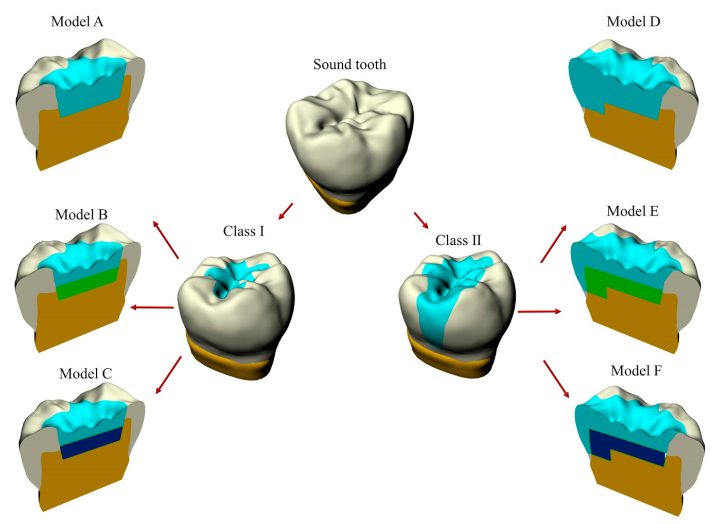
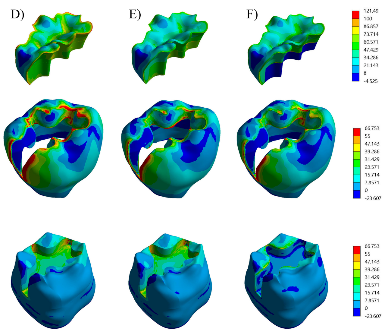
Figure 1. The geometric features of the analyzed model-cavity according to the filling.
Applsci 11 02215 g001
Figure 2. Loading condition with food bolus and meshing finite element model.
Applsci 11 02215 g002
Figure 3. Boundary conditions applied in the present simulation.
Applsci 11 02215 g003
Figure 4. Global contour plots of each restorative procedure First principal stress (MPa) in a class I cavity: (A) Bulk-fill composite, (B) Flowable composite + Bulk-fill composite and (C) Glass-Ionomer + Bulk-fill composite.
Applsci 11 02215 g004
Figure 5. Global contour plots of each restorative procedure First principal stress in a class I cavity: (D) Bulk-fill composite, (E) Flowable composite + Bulk-fill composite and (F) Glass-Ionomer + Bulk-fill resin composite.
Applsci 11 02215 g005
Figure 6. First principal stresses (MPa) plotted along the inspection path according to the cavity shape and restorative material: (A) Trend in tooth tissue for the Class I cavity, (B) Trend in tooth tissue for the Class II cavity.
Applsci 11 02215 g006
Figure 7. First principal stresses (MPa) plotted along the inspection path according to the cavity shape and restorative material: (A) Trend in trend in material restoration for the Class I cavity. (B) Trend in trend in material restoration for the Class I cavity.
Applsci 11 02215 g007
Table 1. The groupings restorative material.
ModelsBonding
Layer
Material
Lower Layer
Material
Upper Layer
Single Material
Layer
A and D10 µm thick--Bulk fill composite
(4 mm in-between cavity and 5.5 mm at the box)
B and E10 µm thickFlowable composite
(1.5 and 3 mm thick in-between cavity and box)
Bulk fill composite
(2.5 mm thick)
-
C and F10 µm thickGlass ionomer (GIC)
(1.5 and 3 mm thick in-between cavity and box)
Bulk fill composite
(2.5 mm thick)
-
Table 2. Materials Mechanical properties: modulus of Young, Poisson ratio and linear shrinkage.
MaterialModulus
of Young
(GPa)
Poisson
Ratio
Linear
Shrinkage (%)
Dentin18.00.30--
Enamel80.00.30--
Food apple pulp3.40.1--
Adhesive layer4.00.301.0
Flowable composite8.00.251.0
Glass inomer8.00.25--
Bulk fill composite12.00.251.0
Table 3. Material mechanical properties: Modulus of Young, Poisson ratio and linear shrinkage.
Cavity ShapeModelEnamelDentinRestoration
Class IA10.4 MPa6.3 MPa19.5 MPa
B10.2 MPa6.0 MPa18.2 MPa
C9.0 MPa2.3 MPa13.0 MPa
Class IID23.3 MPa12.9 MPa18.9 MPa
E22.1 MPa12.2 MPa18.3 MPa
F21.7 MPa5.3 MPa17.9 MPa
Publisher’s Note: MDPI stays neutral with regard to jurisdictional claims in published maps and institutional affiliations.

