Article

Effect of Citric Acid on Color Changes of Calcium Silicate-Based Cements an In Vitro Study

1
Department of Conservative Dentistry and Endodontics, Pomeranian Medical University in Szczecin, 71-454 Szczecin, Poland
2
Department of Periodontology and Operative Dentistry, Westphalian Wilhelms-University, 48149 Münster, Germany
3
Department of Preclinical Conservative Dentistry and Preclinical Endodontics, Pomeranian Medical University in Szczecin, 71-454 Szczecin, Poland
4
Doctoral Studies of the Faculty of Dentistry, Pomeranian Medical University in Szczecin, 71-454 Szczecin, Poland
5
Department of Experimental and Clinical Pharmacology, Pomeranian Medical University in Szczecin, 71-454 Szczecin, Poland
*
Author to whom correspondence should be addressed.
Submission received: 17 January 2021 / Revised: 3 February 2021 / Accepted: 3 March 2021 / Published: 6 March 2021

Abstract

:
The aim of the present in vitro study was to investigate the effects of 10% and 40% citric acid (CA) on the color of calcium silicate–based cements (CSCs) in comparison to the effects of common root canal irrigants. Samples of six CSCs (n = 6)—ProRoot MTA (Dentsply, Tulsa, OK, USA), Biodentine (Septodont, Saint-Maur-des-Fossés, France), MTA Plus (Avalon Biomed Inc, by Prevest Denpro Limited, Jammu, India), MTA Repair HP (Angelus, Londrina, PR, Brazil), Ortho MTA (BioMTA, Seoul, Korea), and Retro MTA (BioMTA, Seoul, Korea)—were immersed in 10% and 40% CA as well as 15% EDTA, 2% NaOCl, 2% CHX, and 0.9% NaCl for 15 min, 1 h, and 24 h. ΔE values, representing the difference between the final and baseline values of the color components, were then determined using a VITA Easyshade Compact 5.0 spectrophotometer. Naked-eye evaluation of the changes in color and structures of the materials was performed using our own scale. Upon immersion of the materials in both 10% and 40% CA, there were statistically significant differences between spectrophotometric color measurement results for all CSCs (P < 0.05). However, CA does not cause dark discoloration, observable with the naked eye, of any of the materials, such as NaOCl and CHX. Significant statistical differences were also found between all CSCs in terms of submersion duration (P < 0.05). CA, which could be an alternative to EDTA use, caused greater CSCs discoloration and changed some of their structures. Unless required by the therapeutic procedure, clinicians should pay attention to the fact that the irrigant may affect the CSCs discoloration and minimize the contact time of irrigant with CSCs.

1. Introduction

In dentistry, calcium silicate–based cements (CSCs) are increasingly used instead of calcium hydroxide products [1]. They are biocompatible materials that are used in an indirect or direct pulp capping, apexification, and pulp regeneration. CSCs are also used for perforation filling, resection procedures, and, more recently, as root canal sealers [2,3,4]. They form a tight barrier against the migration of microorganisms, stimulate the healing of tissues without causing inflammation, exhibit biocompatibility and negligible neuro- and cytotoxicity, and often have bactericidal as well as fungicidal properties [2,3,5]. However, their use in the esthetic zone of the teeth is problematic owing to their susceptibility to staining [6,7]. Tooth discoloration may be the result of intra- or postendodontic procedural errors and can be related to the sealant used to fill the canal or the prosthetic restoration used. Many studies have reported that various medicaments and materials used during root canal treatment can cause coronal tooth discoloration [8,9,10]. Blood and light also significantly affect tooth staining; the size of the lesion depends on the type of material contacted and the time elapsed [7,11].
The current gold standard among CSCs is mineral trioxide aggregate (ProRootMTA (Dentsply, Tulsa, OK, USA) [7]. However, this CSC has several disadvantages, such as its long setting time, and low pressure stability. Furthermore, it contains the X-ray contrast agent bismuth oxide. Such contrast agents are added to CSCs to increase their visibility on radiographs, but they can cause dental hard tissue staining [12,13,14,15,16].
Accordingly, many CSCs have been developed and are commercially available. These include MTA Plus (compounded by PrevestDenpro, Jammu, India for Avalon Biomed Inc. Bradenton, FL, USA), Ortho MTA (BioMTA, Seoul, Korea), Retro MTA (BioMTA, Seoul, Korea), and MTA Repair HP (Angelus, Londrina, PR, Brazil). In some new CSCs, bismuth oxide has been replaced by zirconium oxide, e.g., Biodentine (Septodont, Saint-Maur-des-Fossés, France) and Retro MTA, or by calcium tungstate, e.g., MTA Repair HP. The compositions of these CSCs are given in Table 1.
The success of conventional and regenerative endodontic procedures is influenced by many factors, including the quality of diagnosis, tissue development, and the material used [17]. A particularly important stage of endodontic treatment is canal disinfection, which involves the use of a suitable irrigant [18]. A good irrigant should be an effective antimicrobial agent and organic tissue solvent, non-irritating, stable, and easily stored. Furthermore, it should remove the smear layer.
In conventional endodontics, a commonly used irrigant regime is sodium hypochlorite (NaOCl) at concentrations of 1% to 8%, 2% chlorhexidine (CHX) as an antimicrobial agent, and ethylenediaminetetraacetic acid (EDTA) as a demineralizing agent [18]. According to the guidelines of The American Association of Endodontists Clinical Considerations for Regenerative Procedures, for pulp regeneration treatment, the canal should be prepared with 1.5% NaOCl solution for disinfection and then EDTA solution [19]. EDTA increases surface roughness and thus improves adherence and growth of cells. The above guidelines are currently considered to represent the gold standard in regenerative endodontic procedures [20].
However, EDTA can be replaced by citric acid (CA) at concentrations of 10–40% [18,21,22]. Anhydrous CA is a tricarboxylic acid found in citrus fruits that is frequently used as an excipient in pharmaceutical preparations owing to its antioxidant properties. It maintains the stability of active ingredients and is used as a preservative. It is also used as an acidulant to control pH and acts as an anticoagulant by chelating the calcium in blood [23]. CA exhibits good chemical stability and shows slight anti-microbial effects. Upon irrigation with 10% CA, almost twice as much TGF-β1 is released when compared to irrigation with 17% EDTA. The viabilities of stem cells from the apical papilla (SCAP) after root canal irrigation with 17% EDTA and 10% CA are similar [21,22]. Furthermore, conditioning root canals with CA significantly increases dentin roughness compared to EDTA treatment [24,25].
However, until now, no work on the influence of citric acid on CSCs has been published, nor have the effects of the aforementioned root canal irrigants on MTA Repair HP been studied. Thus, the aim of the present study was to investigate the effects of 10% and 40% CA in comparison to those of irrigation solutions such as 15% EDTA, 2% NaOCl, 2% CHX, and 0.9% sodium chloride (NaCl) as a control group on the color change of the CSCs Biodentine, ProRoot MTA, Retro MTA, Ortho MTA, MTA Plus, and MTA Repair HP.
The null hypothesis is that CA does not discolor these bioceramic materials.

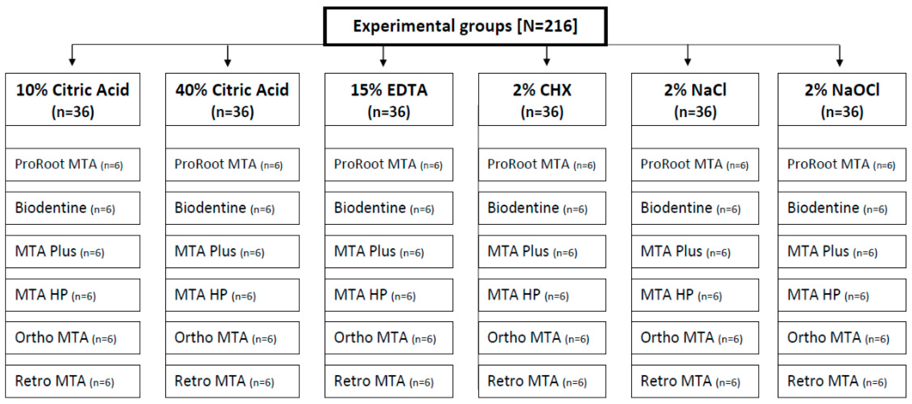
2. Materials and Methods

2.1. Sample Preparation

Six sample groups were used, each containing six materials: Biodentine, ProRoot MTA, Retro MTA, Ortho MTA, MTA Plus, MTA Repair HP (Figure 1). The number of samples was based on a sample size calculation, performed in G*Power 3.1.9.7. software. All materials were prepared in accordance with the manufacturer’s instructions (Table 1). The samples were formed into cylindrical specimens of 4.62 mm diameter and 2 mm height using a sterile catheter and stored at 37 °C and 100% humidity for the time required to achieve setting of the materials. After 24 h, the samples were randomly immersed in 10% CA (Cerkamed, StalowaWola, Poland), 40% CA (Cerkamed, StalowaWola, Poland), 15% EDTA (Cerkamed, StalowaWola, Poland), 2% NaOCl (Cerkamed, StalowaWola, Poland), 2% CHX (Cerkamed, StalowaWola, Poland), or 0.9% NaCl (Fresenius Kabi, Warsaw, Poland) at 37 °C.

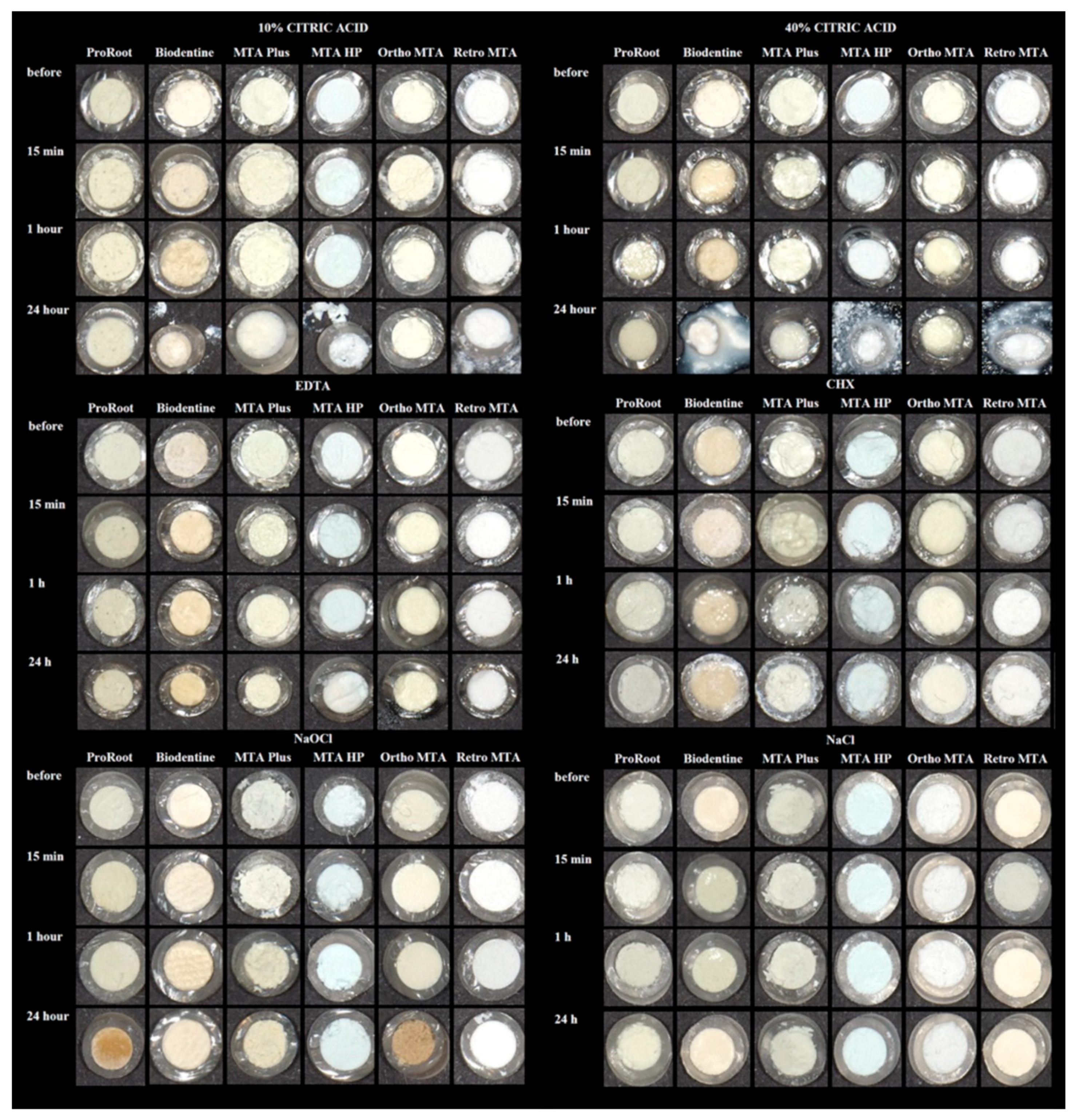
2.2. Material Discoloration Measurement

The measurements were made using a VITA Easyshade Compact 5.0 (VITA Zahnfabrik, Bad Säckingen, Germany) digital spectrophotometer before the procedure and after 15 min, 1 h, or 24 h of immersion. The measurements were made in a dark room under constant measurement conditions and by two blinded operators. The device was calibrated before each measurement, and the samples were dried. The readings were repeated three times. Differences in the colors of the materials were evaluated in terms of values in the CIELAB color space. ΔE values representing the difference between the final and baseline values were calculated using the following equation:
Δ E = [ L 2 L 1 ] 2 + [ a 2 a 1 ] 2 + [ b 2 b 1 ] 2 .
After the appropriate soaking time, the samples were dried and evaluated with the naked eye by two blinded operators. Images of the samples were taken before and after immersion using an Eye Special C-II digital camera (Shofu, Inc., Kyoto, Japan—exposure: 1000 s; aperture f/8.2; focal distance 32 mm; ISO-100). It is an automatic device dedicated solely to analyzing the teeth and mouth of a patient. Clinical evaluation of the changes in color and structures of the materials was performed using our own scale. The color changes were defined as: 0 = no change, 1 = darkening, 2 = brightening. The structural changes of the materials were defined as: a = stable structure, b = disintegration of the structure.

2.3. Statistical Analysis
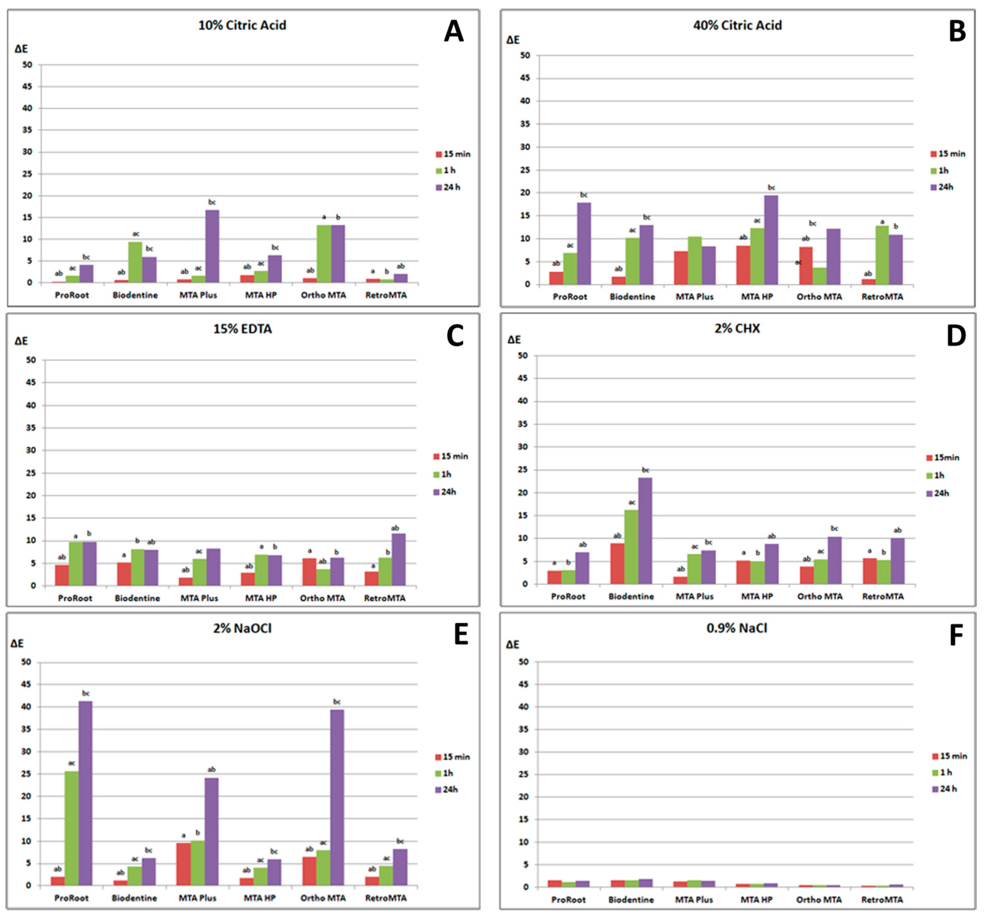
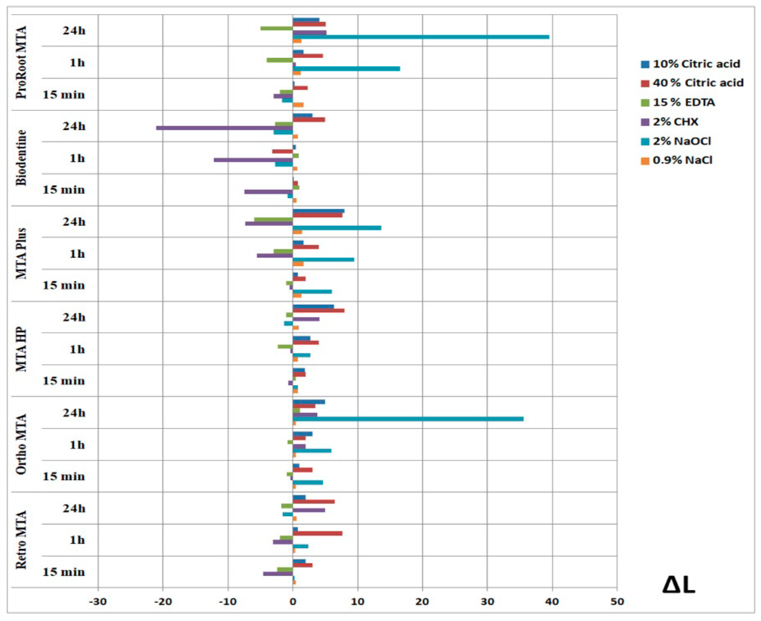
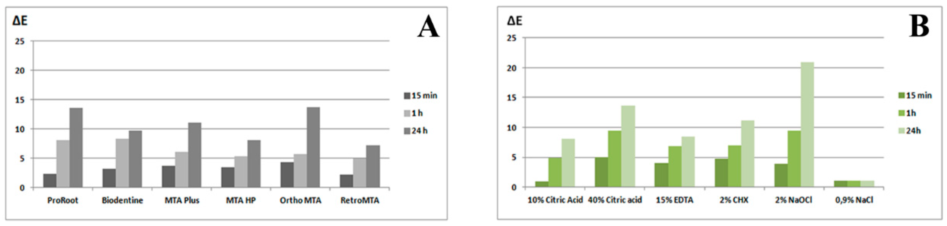
Statistical analysis was performed using IBM SPSS 24.0 (IBM, Warsaw, Poland). In order to assess the compatibility of distributions with the normal distribution, Kolmogorov–Smirnov tests were performed. The Wilcoxon test for dependent samples was used for each material to assess the significance of the differences between the individual measurements over time. In order to assess the significance of differences in the magnitude of changes over time between individual materials, absolute changes were calculated, then the groups were compared by the Kruskal–Wallis test. The level of statistical significance was set at p < 0.05.

3. Results

Our results are presented in Figure 2 as photographs and in Figure 3 as charts. In addition, the ΔL values are shown in Figure 4. All tested materials submerged in CA, EDTA, CHX, and NaOCl showed a significant change in color compared to those not subjected to immersion (p < 0.05). The materials submerged in 0.9% NaCl did not show color changes. Significant statistical differences were also found between all the studied bioceramic materials in terms of submersion time (p < 0.05). Figure 5 shows the analysis charts of average color variation (∆E) over time considering only the materials, independent of the irrigant used (A) and average ∆E over time considering only treatment (irrigant) variation independent of the material used (B).

3.1. Color Changes Caused by 10% and 40% CA

There were statistically significant differences between spectrophotometric color measurement results before immersion and after immersion for 15 min, 1 h, and 24 h for all CSCs. Statistical differences between different wetting times are shown in Figure 3 (p < 0.05).
Upon immersion of the materials in both 10% and 40% CA, the type of staining was the least predictable (Table 2). Naked-eye observation revealed that the CSCs either did not change color (scale 0) or became whiter, such as with MTA Repair HP (scale 2). In no case did the CSCs become darker. After storage in CA (both 10% and 40%) for 24 h, Biodentine, Retro MTA, and MTA Repair HP appeared corroded on the surface (scale b) for both 10% and 40% solutions (Figure 2).

3.2. Color Changes Caused by 15% EDTA

According to spectrophotometric measurements, there was less color change after immersion in EDTA than after immersion in CA (p < 0.05). There were statistically significant differences between color measurements before immersion and after immersion for 15 min, 1 h, and 24 h for all materials (P < 0.05) (Figure 3). No changes in the colors or structures of the materials were observed with the naked eye (scale 0, a) (Figure 2 and Table 2).

3.3. Color Changes Caused by 2% CHX

Upon immersion in CHX, the largest spectrophotometric change was observed for Biodentine. There were statistically significant differences between the color measurements before immersion and after immersion for 15 min, 1 h, and 24 h for all materials (Figure 3) (p < 0.05). Visibly to the naked eye, Biodentine darkened upon 24 h immersion in CHX (scale 1, b). No changes in the colors or structures of the other materials were observed (scale 0, a) (Figure 2 and Table 2).

3.4. Color Changes Caused by 2% NaOCl

The largest spectrophotometric changes in color upon immersion in NaOCl were observed for Ortho MTA, MTA Plus, and ProRoot MTA. There were statistically significant differences between color measurements before immersion and after 15 min, 1 h, and 24 h for all materials (p < 0.05) (Figure 3).
When observed with the naked eye, ProRootMTA and Ortho MTA turned dark brown upon 24 h immersion in NaOCl (scale 1). The color changes of the other materials were not observable by the naked eye (scale 0). The structures of the materials did not change (scale a) (Figure 2 and Table 2).

3.5. Color Changes Caused by 0.9% NaCl

No changes in the color or structure of the materials were observed neither spectrophotometrically, nor with the naked eye (Figure 2 and Figure 3 and Table 2).

4. Discussion

Based on the spectrophotometric results, the null hypothesis must be rejected. CA caused changes in the colors of the CSCs and also changed some of their structures, specifically those of Biodentine, Retro MTA, and MTA Repair HP. However, CA did not cause dark discoloration observable with the naked eye of any of the materials. The spectrophotometric colors of all the CSCs changed under the influence of all the irrigation solutions. Visual spectrophotometry was used in this study because it is considered to be the gold standard for the evaluation of color, and it has been successfully used in dentistry studies [26,27,28]. Visual spectrometry is the most widely used method of determining color: it meets international standards, and it is compliant with ISO standards (International Commission on Illumination) [29].
To the best of our knowledge, this is the first study evaluating the effects of CA on CSCs. We have demonstrated that CA has a significant influence on spectrophotometric color change in CSCs at all time intervals. There was a greater spectrophotometric color change after immersion in CA than after immersion in EDTA. After 24 h immersion in CA, MTA Repair HP, Biodentine, and Retro MTA disintegrated, dissolved, and took the form of wet sands on the surface. These changes were visible to the naked eye. This material disintegration phenomenon may be related to the effect of the acid on the cement structure. Furthermore, these results are similar for both concentrations of CA. CA is used in architecture as a retarder of setting and hardening of Portland cement. Citric acid does not have any beneficial effect on enhancing the calcium silicate phase, as was initially assumed, and instead it reduces the strength of Portland cement at all levels of concentration [30]. It is remarkable that CA had no influence on the material structure of the bismuth-oxide-containing CSCs ProRoot MTA, Ortho MTA, and MTA Plus. In one study, the microstructure of Biodentine changed, showing a relatively smooth surface with more spheroidal crystals [31]. No information has been found as to whether the radiopacifer may have an effect on material structure dissolution. In the study by Oliveira et al. [32], MTA material containing bismuth oxide had a reduction in radiopacity after 30 days, and it was hypothesized that this may be caused by the dissociation of bismuth oxide. Therefore, it can change the color of the material, but also the structural stability of the CA [32]. The effect of an acidic environment on the loss of CSC volume was also assessed. In the case of Biodentine, it was much lower in an acidic environment than in a saline environment. There was no significant difference in the volume of ProRoot MTA between these two environments [33]. In another case, it was proved that the exposure to acidic pH decreased Ca(OH)2 crystalline formation in ProRoot MTA, Retro MTA and Biodentine [34]. Accordingly, more detailed research is needed regarding the material strengths of CSCs after using CA.
MTA Repair HP was the least sensitive to NaOCl solution of all the tested CSC materials. It also exhibited the slightest spectrophotometric color changes upon immersion in CHX and EDTA. Only in contact with CA solution did its surface structure disintegrate. Irrigation solutions do not cause darkening of MTA Repair HP, which could be visible to the naked eye. A recent study has shown that MTA Repair HP shows better push-out bond strength than its predecessor white MTA Angelus [35] and does not cause discoloration of teeth [36]. It is believed that the presence of calcium tungstate as a radiopacifier in MTA Repair HP contributes to higher calcium release in the initial periods [37], promoting greater biomineralization and aiding the resistance of this material [38,39]. Furthermore, its high plasticity may improve marginal adaptation.
This study revealed a significant change in the colors of ProRoot MTA, MTA Plus, and Ortho MTA upon immersion in NaOCl. In contact with NaOCl, radiopacifer bismuth oxide changes color from yellow to black [15,26], whereas zirconium oxide and calcium tungstate are unaffected [15]. Materials that do not contain bismuth oxide but instead tantalum oxide or zirconium oxide do not exhibit significant changes in color [40]. The MTA samples were darker (ΔE > 30) than Portland cement (ΔE < 10) after 24 h [26]. ProRoot MTA and Biodentine exhibited clinically perceptible discoloration when immersed in NaOCl [27,41]. ProRoot MTA showed a significantly higher ΔE (ΔE > 15) value when compared with Biodentine (ΔE < 10) after contact with NaOCl after 24 h [27]. Similar results were obtained in the present study for ProRoot MTA ΔE = 41 and Biodentine ΔE = 6 after 24 h. Furthermore, it has been previously demonstrated that ProRoot MTA immersed in 2.5% NaOCl shows a significant change in color, but no significant differences were observed in color after 1, 2, and 4 months after immersion [41]. In contrast, in the present study, significant differences for immersion durations of 15 min, 1 h, and 24 h were observed.
After immersion in CHX, the most distinct change in color was observed for Biodentine (ΔE = 23 after 24 h). It has been demonstrated by other authors that Biodentine shows a significantly higher ΔE value after contact with CHX (ΔE > 10) when compared to NaOCl (ΔE < 10) [27]. CHX caused pronounced color changes in Biodentine as well as in white MTA [42]. In the present study, significant color differences were also observed after EDTA immersion. However, these changes were less severe than those upon immersion in other liquids and were not visible to the naked eye. In the literature, white MTA shows a significant change in color upon contact with EDTA when compared to that in saline [42].
This experiment had some limitations. Firstly, we attempted to simulate a clinical situation; however, there was a difference in environment, and materials were not put into teeth. Secondly, materials structures were not assessed. Thus, further studies that analyze the surface areas using EDX, SEM, or micro-hardness test for the evaluation of actual disintegration must be conducted.

5. Conclusions

Within the limitations of the present study, it can be concluded that both 10% and 40% citric acid caused spectrophotometric color changes in all tested CSCs. However, immersion in CA did not cause color darkening observable with a naked eye such as NaOCl and CHX. CA, which could be an alternative to EDTA use, caused greater CSCs discoloration and changed some of their structures. Thus, more research is needed on the effects of CA on CSCs. Unless required by the therapeutic procedure, clinicians should pay attention to the fact that the irrigant may affect the CSCs discoloration and minimize the contact time of irrigant with CSCs.

Author Contributions

Conceptualization, J.M. and A.N.; methodology, J.M. and I.F.; validation, A.N., T.D., and M.L.; formal analysis, J.M., I.F., and A.N.; resources, J.M.; data curation, J.M.; writing—original draft preparation, J.M.; writing—review and editing, A.N., T.D., A.M.-M. and M.L.; visualization, J.M.; supervision, A.N., T.D., A.M.-M., and M.L. All authors have read and agreed to the published version of the manuscript.

Funding

The research was supported by the Polish Ministry of Science and Higher Education program for the years 2017–2020 and by The Faculty of Medicine & Dentistry, Pomeranian Medical University in Szczecin (grant no. MB-262-223/17), Poland.

Institutional Review Board Statement

Not applicable.

Informed Consent Statement

Not applicable.

Data Availability Statement

Not applicable.

Acknowledgments

This article is an original work that has not been published before. It is not being considered for publication elsewhere in its current form, either in printed or electronic media.

Conflicts of Interest

The authors declare no conflict of interest. The funders had no role in the design of the study, data collection, analyses, interpretation of data, writing of the manuscript, or decision to publish the results.

References

  1. Paula, A.; Carrilho, E.; Laranjo, M.; Abrantes, A.M.; Casalta-Lopes, J.; Botelho, M.F.; Marto, C.M.; Ferreira, M.M. Direct pulp capping: Which is the most effective biomaterial? A retrospective clinical study. Materials 2019, 12, 3382. [Google Scholar] [CrossRef] [Green Version]
  2. Camilleri, J.; Formosa, L.; Damidot, D. The setting characteristics of MTA Plus in different environmental conditions. Int. Endod. J. 2013, 46, 831–840. [Google Scholar] [CrossRef]
  3. Reszka, P.; Nowicka, A.; Lipski, M.; Dura, W.; Droździk, A.; Woźniak, K. A comparative chemical study of calcium silicate-containing and epoxy resin-based root canal sealers. Biomed. Res. Int. 2016, 2016, 9808432. [Google Scholar] [CrossRef] [PubMed]
  4. Palma, P.J.; Martins, J.; Diogo, P.; Sequeira, D.; Ramos, J.C.; Diogenes, A.; Santos, J.M. Does Apical Papilla Survive and Develop in Apical Periodontitis Presence after Regenerative Endodontic Procedures? Appl. Sci. 2019, 9, 3942. [Google Scholar] [CrossRef] [Green Version]
  5. Dammaschke, T.; Nowicka, A.; Lipski, M.; Ricucci, D. Histological evaluation of hard tissue formation after direct pulp capping with a fast-setting mineral trioxide aggregate [RetroMTA] in humans. Clin. Oral. Investig. 2019, 23, 4289–4299. [Google Scholar] [CrossRef]
  6. Fagogeni, I.; Metlerska, J.; Lipski, M.; Falgowski, T.; Maciej, G.; Nowicka, A. Materials used in regenerative endodontic procedures and their impact on tooth discoloration. J. Oral. Sci. 2019, 61, 379–385. [Google Scholar] [CrossRef] [PubMed] [Green Version]
  7. Możyńska, J.; Metlerski, M.; Lipski, M.; Nowicka, A. Tooth discoloration induced by different calcium silicate-based cements: A systematic review of in vitro studies. J. Endod. 2017, 43, 1593–1601. [Google Scholar] [CrossRef]
  8. Palma, P.J.; Marques, J.A.; Falacho, R.I.; Correia, E.; Vinagre, A.; Santos, J.M.; Ramos, J.C. Six-Month Color Stability Assessment of Two Calcium Silicate-Based Cements Used in Regenerative Endodontic Procedures. J. Funct. Biomater. 2019, 10, 14. [Google Scholar] [CrossRef] [Green Version]
  9. Palma, P.J.; Marques, J.A.; Santos, J.; Falacho, R.I.; Sequeira, D.; Diogo, P.; Caramelo, F.; Ramos, J.C.; Santos, J.M. Tooth Discoloration after Regenerative Endodontic Procedures with Calcium Silicate-Based Cements—An Ex Vivo Study. Appl. Sci. 2020, 10, 5793. [Google Scholar] [CrossRef]
  10. Lenherr, P.; Allgayer, N.; Weiger, R.; Filippi, A.; Attin, T.; Krastl, G. Tooth discoloration induced by endodontic materials: A laboratory study. Int. Endod. J. 2012, 45, 942–949. [Google Scholar] [CrossRef] [PubMed]
  11. Camilleri, J.; Borg, J.; Damidot, D.; Salvadori, E.; Pilecki, P.; Zaslansky, P.; Darvell, B.W. Colour and chemical stability of bismuth oxide in dental materials with solutions used in routine clinical practice. PLoS ONE 2020, 11, 15–e0240634. [Google Scholar] [CrossRef]
  12. Ahmed, H.M.; Abbott, P.V. Discolouration potential of endodontic procedures and materials: A review. Int. Endod. J. 2012, 45, 883–897. [Google Scholar] [CrossRef] [PubMed]
  13. Kang, S.H.; Shin, Y.S.; Lee, H.S.; Kim, S.O.; Shin, Y.; Jung, I.Y.; Song, J.S. Color changes of teeth after treatment with various mineral trioxide aggregate–based materials: An ex vivo study. J. Endod. 2015, 41, 737–741. [Google Scholar] [CrossRef] [PubMed]
  14. Vallés, M.; Mercadé, M.; Duran-Sindreu, F.; Bourdelande, J.L.; Roig, M. Color stability of white mineral trioxide aggregate. Clin. Oral. Investig. 2013, 17, 1155–1159. [Google Scholar] [CrossRef] [PubMed]
  15. Marciano, M.A.; Duarte, M.A.; Camilleri, J. Dental discoloration caused by bismuth oxide in MTA in the presence of sodium hypochlorite. Clin. Oral. Investig. 2015, 19, 2201–2209. [Google Scholar] [CrossRef] [PubMed]
  16. Lipski, M.; Nowicka, A.; Kot, K.; Postek-Stefańska, L.; Wysoczańska-Jankowicz, I.; Borkowski, L.; Andersz, P.; Jarząbek, A.; Grocholewicz, K.; Sobolewska, E.; et al. Factors affecting the outcomes of direct pulp capping using Biodentine. Clin. Oral. Investig. 2018, 22, 2021–2029. [Google Scholar] [CrossRef] [Green Version]
  17. Estrela, C.; Holland, R.; de Estrela, C.R.A.; Alencar, A.H.G.; Sousa-Neto, M.D.; Pecora, J.D. Characterization of successful root canal treatment. Braz. Dent. J. 2014, 25, 3–11. [Google Scholar] [CrossRef]
  18. Darcey, J.; Jawad, S.; Taylor, C.; Roudsari, R.V.; Hunter, M. Modern Endodontic Principles Part 4: Irrigation. Dent. Update 2016, 43, 20–33. [Google Scholar] [CrossRef] [PubMed]
  19. Chan, E.K.; Desmeules, M.; Cielecki, M.; Dabbagh, B.; Ferraz Dos Santos, B. Longitudinal cohort study of regenerative endodontic treatment for immature necro permanent teeth. J. Endod. 2017, 43, 395–400. [Google Scholar] [CrossRef]
  20. Kim, S.G.; Malek, M.; Sigurdsson, A.; Lin, L.M.; Kahler, B. Regenerative endodontics: A comprehensive review. Int. Endod. J. 2018, 51, 1367–1388. [Google Scholar] [CrossRef] [PubMed]
  21. Ivica, A.; Zehnder, M.; Mateos, J.M.; Ghayor, C.; Weber, F.E. Biomimetic conditioning of human dentin using citric acid. J. Endod. 2019, 45, 45–50. [Google Scholar] [CrossRef] [PubMed] [Green Version]
  22. Chae, Y.; Yang, M.; Kim, J. Release of TGF-β1 into root canals with various final irrigants in regenerative endodontics: An in vitro analysis. Int. Endod. J. 2018, 51, 1389–1397. [Google Scholar] [CrossRef] [PubMed]
  23. Lee, G.; Arepally, G.M. Anticoagulation techniques in apheresis: From heparin to citrate and beyond. J. Clin. Apher. 2012, 27, 117–125. [Google Scholar] [CrossRef] [Green Version]
  24. Martínez, A.; Vargas, R.; Galano, A. Citric acid: A promising copper scavenger. Comput. Theor. Chem. 2018, 1133, 47–50. [Google Scholar] [CrossRef]
  25. Eldeniz, A.U.; Erdemir, A.; Belli, S. Effect of EDTA and citric acid solutions on the microhardness and the roughness of human root canal dentin. J. Endod. 2005, 31, 107–110. [Google Scholar] [CrossRef]
  26. Camilleri, J. Color stability of white mineral trioxide aggregate in contact with hypochlorite solution. J. Endod. 2014, 40, 436–440. [Google Scholar] [CrossRef] [PubMed]
  27. Keskin, C.; Demiryurek, E.O.; Ozyurek, T. Color stabilities of calcium silicate-based materials in contact with different irrigation solutions. J. Endod. 2015, 41, 409–411. [Google Scholar] [CrossRef]
  28. Felman, D.; Parashos, P. Coronal tooth discoloration and white mineral trioxide aggregate. J. Endod. 2013, 39, 484–487. [Google Scholar] [CrossRef] [PubMed]
  29. International Commission on Illumination. Recommendations on Uniform Color Spaces, Color–Difference Equations, Psychometric Color Terms; Bureau Central de la CIE: Paris, France, 1978; Volume 1913. [Google Scholar]
  30. Kastiukas, G.; Zhou, X.; Castro-Gomes, J.; Huang, S.; Saafi, M. Effects of lactic and citric acid on early-age engineering properties of Portland/calcium aluminate blended cements. Constr. Build. Mater. 2015, 101, 389–395. [Google Scholar] [CrossRef] [Green Version]
  31. Agrafioti, A.; Tzimpoulas, N.; Chatzitheodoridis, E.; Kontakiotis, E.G. Compartive evaluation of sealing ability and microstructure of MTA and Biodentine after exposure to different environments. Clin. Oral. Investig. 2016, 20, 1535–1540. [Google Scholar] [CrossRef] [PubMed]
  32. Oliveira, L.V.; da Silva, G.R.; Souza, G.L.; Magalhães, T.E.A.; Barbosa, G.L.R.; Turrioni, A.P.; Moura, C.C.G. A laboratory evaluation of cell viability, radiopacity and tooth discoloration induced by regenerative endodontic materials. Int. Endod. J. 2020, 53, 1140–1152. [Google Scholar] [CrossRef] [PubMed]
  33. Kwon, S.Y.; Seo, M.S. Comparative evaluation of volumetric changes of three different retrograde calcium silicate materials placed under different pH condititions. BMC Oral Health 2020, 20, 330. [Google Scholar] [CrossRef] [PubMed]
  34. Ashofteh Yazdi, K.; Ghabraei, S.; Bolhari, B.; Kafili, M.; Meraji, N.; Nekoofar, M.H.; Dummer, P.M.H. Microstructure and chemical analysis of four calcium silicate-based cements in different environmental conditions. Clin. Oral Invest. 2019, 23, 43–52. [Google Scholar] [CrossRef] [PubMed]
  35. Silva, E.J.; Carvalho, N.K.; Zanon, M.; Senna, P.M.; DE-Deus, G.; Zuolo, M.L.; Zaia, A.A. Push-out bond strength of MTA HP, a new high-plasticity calcium silicate-based cement. Braz. Oral Res. 2016, 30, e84. [Google Scholar] [CrossRef] [Green Version]
  36. Aguiar, B.A.; Frota, L.M.A.; Taguatinga, D.T.; Vivan, R.R.; Camilleri, J.; Duarte, M.A.H.; de Vasconcelos, B.C. Influence of ultrasonic agitation on bond strength, marginal adaptation, and tooth discoloration provided by three coronary barrier endodontic materials. Clin. Oral. Investig. 2019, 23, 4113–4122. [Google Scholar] [CrossRef] [PubMed]
  37. Duarte, M.A.H.; Minotti, P.G.; Rodrigues, C.T.; Zapata, R.O.; Bramante, C.M.; Tanomaru Filho, M.; Vivan, R.R.; Gomes de Moraes, I.; Bombarda de Andrade, F. Effect of different radiopacifying agents on the physicochemical properties of white Portland cement and white mineral trioxide aggregate. J. Endod. 2012, 38, 394–397. [Google Scholar] [CrossRef]
  38. Amoroso-Silva, P.A.; Marciano, M.A.; Guimaraes, B.M.; Duarte, M.A.H.; Sanson, A.F.; de Moraes, I.G. Apical adaptation, sealing ability and push–out bond strength of five root–end filling materials. Braz. Oral Res. 2014, 28, 1–6. [Google Scholar] [CrossRef] [PubMed] [Green Version]
  39. Cintra, L.T.A.; Benetti, F.; de Azevedo Queiroz, Í.O.; de Araújo Lopes, J.M.; Penha de Oliveira, S.H.; Sivieri Araújo, G.; Gomes-Filho, J.E. Cytotoxicity, biocompatibility, and biomineralization of the new high-plasticity MTA material. J. Endod. 2017, 43, 774–778. [Google Scholar] [CrossRef] [Green Version]
  40. Camilleri, J. Staining potential of Neo MTA Plus, MTA Plus, and Biodentine used for pulpotomy procedures. J. Endod. 2015, 41, 1139–1145. [Google Scholar] [CrossRef] [PubMed]
  41. Voveraityte, V.; Gleizniene, S.; Lodiene, G.; Grabliauskiene, Z.; Machiulskiene, V. Spectrophotometric analysis of tooth discolouration induced by mineral trioxide aggregate after final irrigation with sodium hypochlorite: An in vitro study. Aust. Endod. J. 2017, 43, 11–15. [Google Scholar] [CrossRef] [PubMed]
  42. Sobhnamayan, F.; Adl, A.; Ghanbaran, S. Effect of different irrigation solutions on the colour stability of three calcium silicate–based materials. J. Dent. Biomater. 2017, 4, 373–378. [Google Scholar]
Figure 1. Description of experimental groups.
Applsci 11 02339 g001
Figure 2. Images of calcium silicate–based cements (CSCs) taken at different time points after setting and immersion in different irrigation solutions.
Applsci 11 02339 g002
Figure 3. Mean ΔE values of CSCs materials after contact with irrigation solutions for different durations. (A) 10% CA; (B) 40% CA; (C) 15% EDTA; (D) 2% NaOCl; (E) 2% CHX; (F) 0.9% NaCl. (The same letters indicate statistically significant differences between different time points within the material group (p < 0.05)).
Applsci 11 02339 g003
Figure 4. Mean ΔL values of CSCs materials after contact with irrigation solutions for different durations.
Applsci 11 02339 g004
Figure 5. Average color variation ΔE over time. (A) independent of the irrigant used; (B) independent of the material used.
Applsci 11 02339 g005
Table 1. Compositions of the CSCs considered in this study.
MaterialManufacturerIngredientsPreparation Procedure
ProRoot MTADentsply, Tulsa, OK, USAtricalcium silicate, dicalcium silicate, tricalcium aluminate, tetracalciumaluminoferrite, free calcium oxide, and bismuth oxideMix powder + liquid ratio 1:3 (mix manually)
BiodentineSeptodont, Saint Maur-des-Fossés, Francepowder: tricalcium silicate, calcium carbonate and oxide filler, iron oxide shade, and zirconium oxide
liquid: calcium chloride as accelerator, hydrosoluble polymer water-reducing agent, water
0.7 g capsule of powder + 5 drops of liquid
mix 30 s; 4000–4200 rpm (mixing device)
MTA PlusAvalon Biomed Inc, by Prevest Denpro Limited, Jammu, Indiapowder: tricalcium silicate, dicalcium silicate, bismuth oxide, calcium sulfate, and silica
liquid: hydrated polymer gel
Mix powder + liquid ratio 1:1 (mix manually)
MTA Repair HPAngelus, Londrina, PR, Brazilpowder: tricalcium silicate, dicalcium silicate, tricalcium aluminate, calcium oxide, and calcium tungstate
liquid: water and plasticizer
0.085 g capsules of powder + 12 drops of liquid (mix manually)
Ortho MTABioMTA, Seoul, Koreacalcium carbonate, silicon dioxide, aluminium oxide, and dibismuth trioxide0.2 g pouches of powder + 2 drops of water (mix manually)
Retro MTABioMTA, Seoul, Koreacalcium carbonate, silicon dioxide, aluminium oxide, and calcium zirconia complex0.3 g pouches of powder + 3 drops of water (mix manually)
Table 2. Evaluation of the changes in the color and structure of CSC materials upon contact with different irrigation solutions.
MaterialIrrigant
Scale10% CA40% CA15% EDTA2% CHX2% NaOCl0.9% NaCl
ProRoot MTAcolor0666606
10 00060
2000000
structurea666666
b000000
Biodentinecolor0666466
1000200
2000000
structurea016666
b650000
MTA Pluscolor0666646
1000020
2000000
structurea666666
b000000
MTA Repair HPcolor0106666
1000000
2560000
structurea006666
b660000
Ortho MTAcolor06 6 6606
1000060
2000000
structurea666666
b000000
Retro MTAcolor06 6 6 6 66
1000000
20 0 00 0 0
structurea006666
b660 0 0 0
(scale of color assessment in naked eye observation: 0 = no change, 1 = darkening, 2 = brightening; scale of structure assessment: a = stable structure, b = disintegration).
Publisher’s Note: MDPI stays neutral with regard to jurisdictional claims in published maps and institutional affiliations.

