Article

Biomechanical Consequences of Tibial Insert Thickness after Total Knee Arthroplasty: A Musculoskeletal Simulation Study

1
Department of Biomechanical Engineering, University of Twente, 7500 AE Enschede, The Netherlands
2
Orthopedic Research Laboratory, Radboud Institute for Health Sciences, Radboud University Medical Center, 6500 HB Nijmegen, The Netherlands
*
Author to whom correspondence should be addressed.
Submission received: 4 February 2021 / Revised: 3 March 2021 / Accepted: 4 March 2021 / Published: 9 March 2021
(This article belongs to the Special Issue Musculoskeletal Models in a Clinical Perspective)

Abstract

:

Featured Application

The proposed methodology provides orthopedic surgeons with quantitative data on the sensitivity of muscle, ligament, and joint compressive forces to tibial polyethylene insert thickness variations and can be further developed to optimize navigated or robotic-assisted total knee arthroplasty.

Abstract

The thickness of the tibial polyethylene (PE) insert is a critical parameter to ensure optimal soft-tissue balancing in the intraoperative decision-making procedure of total knee arthroplasty (TKA). However, there is a paucity of information about the kinetic response to PE insert thickness variations in the tibiofemoral (TF) joint, and subsequently, the secondary effects on the patellofemoral (PF) biomechanics. Therefore, the purpose of this study was to investigate the influence of varying PE insert thickness on the ligament and TF compressive forces, as well as on the PF forces and kinematics, after a cruciate-retaining TKA. A previous patient-specific musculoskeletal model of TKA was adapted to simulate a chair-rising motion in which PE insert thickness was varied with 2 mm increments or decrements compared to the reference case (9 mm), from 5 mm up to 13 mm. Greater PE insert thickness resulted in higher ligament forces and concurrently increased the TF compressive force by 21% (13 mm), but slightly unloaded the PF joint with 7% (13 mm) while shifting the patella distally in the trochlear groove, compared to the reference case. Thinner PE inserts showed an opposite trend. Our findings suggest that the optimal PE insert thickness selection is a trade-off between the kinetic outcomes of the TF and PF joints.

1. Introduction

Total knee arthroplasty (TKA) is an effective surgical intervention for end-stage knee osteoarthritis in which the diseased articulating surfaces of the knee joint are replaced with artificial components to relieve pain and restore the normal knee function [1]. A successful surgical outcome depends, amongst others, on the design [2], size [3], and alignment of the prosthetic components [4]. A common mode of early surgical failure includes mechanical instability that may result from aseptic loosening or polyethylene (PE) wear [5]. These failure mechanisms have been associated with the inadequate thickness of the tibial PE insert [6]. The selection of an optimum PE insert thickness in the intraoperative procedure is important to achieve soft-tissue balance and restore the physiological function in the tibiofemoral (TF) joint, thus lessening the risk of implant-related complications [7]. Previous studies suggest that a minimum thickness of 6 to 8 mm is required to minimize the contact stresses within the tibial insert surface [8,9], applying the conventional 2 mm increments; these increments allow surgeons to identify the effect of PE insert thickness variations on the soft-tissue tension and intraoperative kinematics based on knee laxity trials [10,11]. Following this recommendation, some authors argue that the insertion of thicker PE components is likely to prevent accelerated wear and early surgical failure [12]. Nevertheless, the debate continues about the target PE insert thickness, with contradictory findings from earlier studies on the topic. Greco et al. [13] demonstrated that implanted knees with a PE insert thickness of more than 14 mm performed similarly in the postoperative clinical evaluation, yielding no surgical failure due to aseptic loosening or tibial component instability as compared to those with a thickness under 14 mm. Contrarily, Berend et al. [14] labeled tibial PE inserts thicker than 14 mm as potential contributors to posterolateral rotatory instability, leading to higher failure rates at mid- to long-term follow-up. One of the main limitations of these studies, however, was the failure to control for the variability in the depth of tibial resection, which might provide an inaccurate estimation of the actual effect of PE insert thickness variation on the surgical outcomes.
From a biomechanical perspective, using thicker tibial PE inserts than the minimum recommended threshold (6–8 mm), while keeping the resection depth unchanged, can increase tension in the soft-tissue envelope; this can subsequently result in higher contact forces in the distal femur and tibial insert interface. The increased contact forces are also transferred to the fixation site, and this could, therefore, promote aseptic loosening of the prosthesis. A comprehensive evaluation of the kinetics after TKA is crucial to prevent higher than physiological mechanical stress and strain on the ligamentous structures as well as high load levels at the interface between bones and prosthetic components. This knowledge could lead to better surgical choices and increased implant longevity. Since the thickness of the tibial PE insert can be modified intraoperatively, the surgeon can customize the surgical plan by choosing a PE insert thickness that best replicates the natural knee kinematics and kinetics. Computational models of the musculoskeletal system represent a valuable tool in this intraoperative decision-making procedure. Musculoskeletal models can be personalized to patient-specific anatomical features and predict variations in the knee joint loads and kinematic patterns due to changes in implant design properties or surgical choices, such as the thickness of the tibial PE insert. These predictive capabilities of musculoskeletal models provide quantitative patient-specific information that can aid surgeons to formulate an optimal treatment plan tailored to individual patients [15].
Earlier studies typically focus on the primary effects of PE insert thickness on the TF joint biomechanics. For instance, researchers have recently examined the effect of varying PE insert thickness on the TF kinematics after TKA, using a dynamic knee simulator of laxity tests [16], or by performing an intraoperative assessment using a computer-assisted surgery navigation system [7]. However, to the best of our knowledge, current scientific literature lacks a proper investigation of the relationship between PE insert thickness and TF kinetics, as well as of the effects on the patellofemoral (PF) joint where the secondary effects may influence the biomechanical parameters, including forces and kinematics, in a clinically relevant way. Therefore, the aim of this study was to first investigate the influence of PE insert thickness, in isolation, on the TF joint in terms of ligament and compressive forces, and secondly, to assess to what extent PE insert thickness variation affects the biomechanical parameters on the PF joint. For this, we used a patient-specific musculoskeletal model with a cruciate-retaining prosthesis. We hypothesized that thicker tibial PE inserts would result in higher ligament forces and, thus, increased TF compressive force, but the effect would remain neutral with regard to the PF articulation.

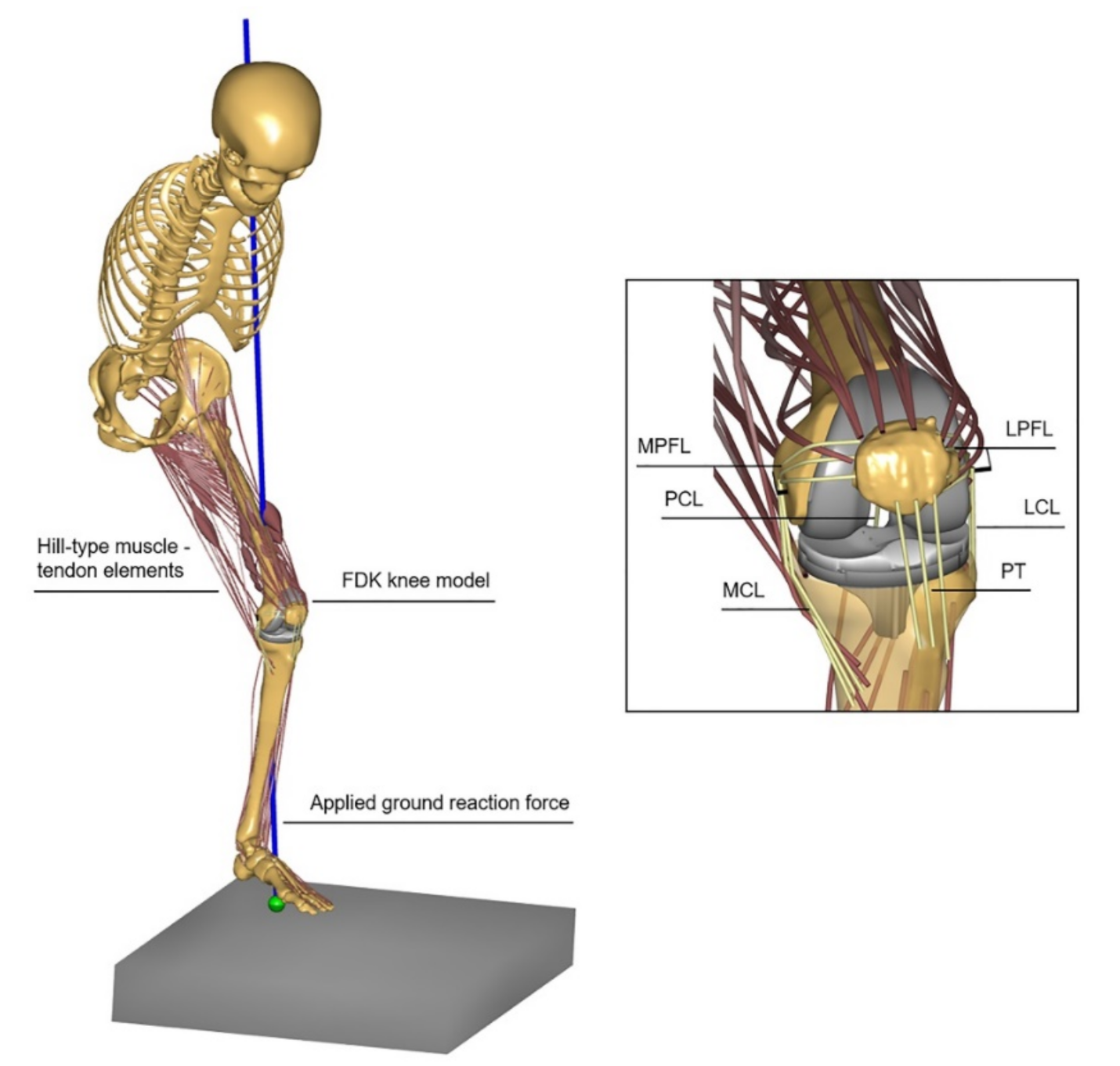
2. Materials and Methods

A previously validated musculoskeletal model of a TKA patient was used in this study [17]. In brief, the model was built upon the Twente Lower Extremity Model 2.0 (TLEM 2.0) template [18] and integrated patient-specific data available from the fifth “Grand Challenge Competition to Predict In Vivo Knee Loads” dataset [19]. This lower-extremity model of the implanted side comprises 11 degrees of freedom in the TF and PF joints. Flexion and extension angle in the TF joint is motion capture driven, whereas the kinematics of the remaining degrees of freedom are solved quasi-statically, using the force-dependent kinematics (FDK) approach, as proposed by Andersen et al. [20]. This methodology enables the concurrent estimation of muscle, ligament, and joint contact forces.
For this study, we introduced some changes with respect to the existing patient-specific model, using the AnyBody Modeling System v. 7.3.1 (AnyBody Technology A/S, Aalborg, Denmark); software for modeling and simulation analysis of the musculoskeletal apparatus [21]. The patellar tendon was modeled as three non-linear spring elements instead of a single rigid element connecting the patella and tibial tuberosity. Stiffness of the patellar tendon was determined by multiplying the median cross-sectional area from the patient-specific computed tomography (CT) with a modulus of elasticity reported in the literature [22]; likewise, stiffness and reference strain of the TF and PF ligament bundles were adopted from the existing musculoskeletal model configuration. In addition, we configured the model to incorporate a size 6 Triathlon (Stryker, Kalamazoo, MI, USA) cruciate-retaining implant (Figure 1), ensuring that the femoral anteroposterior anatomical dimension prevents anterior notching of the femoral cortex. This implant was chosen as it has shown good functional performance and ligamentous stability [23]. The femoral and tibial component geometry was positioned in such a way as to match tangentially the patient-specific distal femoral and proximal tibial bone cuts, applying the well-established mechanical alignment technique [24]. The patella was not resurfaced. Since the cartilage underside of the patella could not be determined from the patient-specific dataset, we generated a 3 mm offset on the dorsal facet of the patellar bone according to the mean patellar cartilage thickness estimation of Cohen et al. [25]. In this model, PE insert thickness was varied with 2 mm increments or decrements (−4 mm, −2 mm, +2 mm, +4 mm) compared to the reference case (9 mm), testing five thickness cases in total: 5 mm, 7 mm, 9 mm, 11 mm, and 13 mm (Figure 2). The effect of PE insert thickness was evaluated by simulating a chair-rising activity based on motion capture data available as part of the open access grand challenge competition dataset.
To allow for an investigation of the model kinetic predictions on the TF joint, the primary outcome measures of the simulations included the medial collateral ligament (MCL), lateral collateral ligament (LCL), and posterior cruciate ligament (PCL) forces and strain, and the TF compressive force. To assess the biomechanical effects on the PF joint, the secondary simulation outcomes were the forces around the knee extensor mechanism, including the quadriceps muscle force, quadriceps tendon-to-femur force, and PF compressive force. The lateral PF ligament bundles were found to remain slack, while the medial PF ligament forces were essentially identical throughout the series of trial simulations regardless of the PE insert thickness and hence excluded from the subsequent analysis. In addition, the PF joint kinematics, including patellar flexion, anterior translation, distal translation, medial rotation, lateral tilt, and lateral shift, were recorded. To quantify the biomechanical effects of PE insert thickness across the joints, we identified the average and peak values in the ligament forces, TF and PF compressive forces throughout the simulated range of motion; and subsequently computed the average and peak percentual change with respect to the reference case.

3. Results

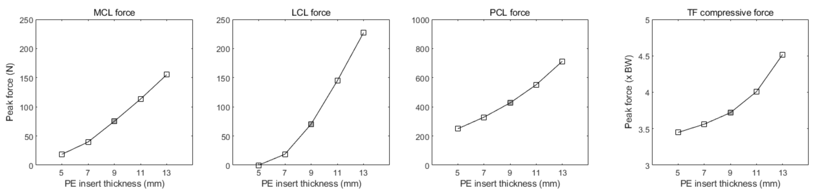
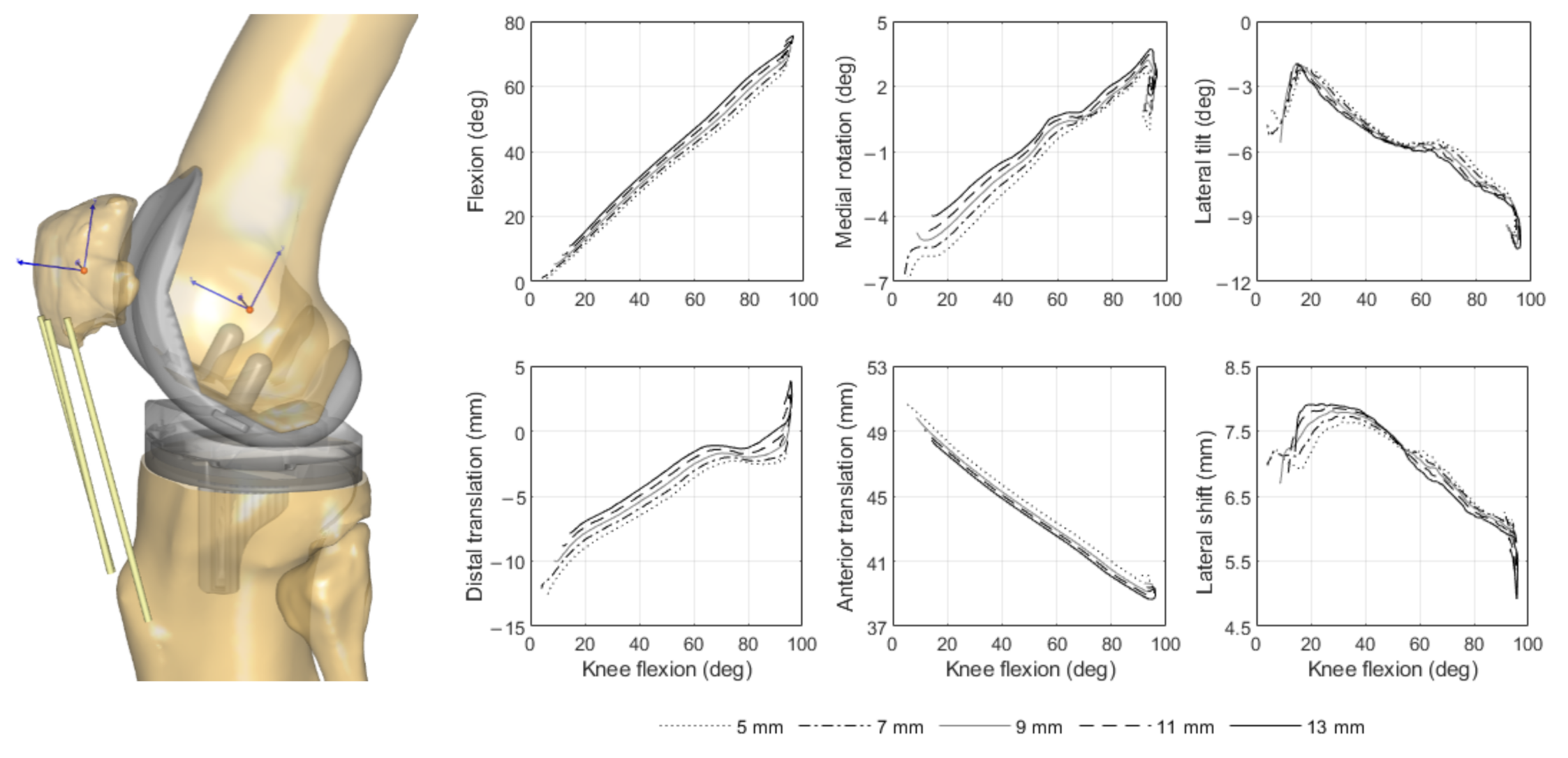
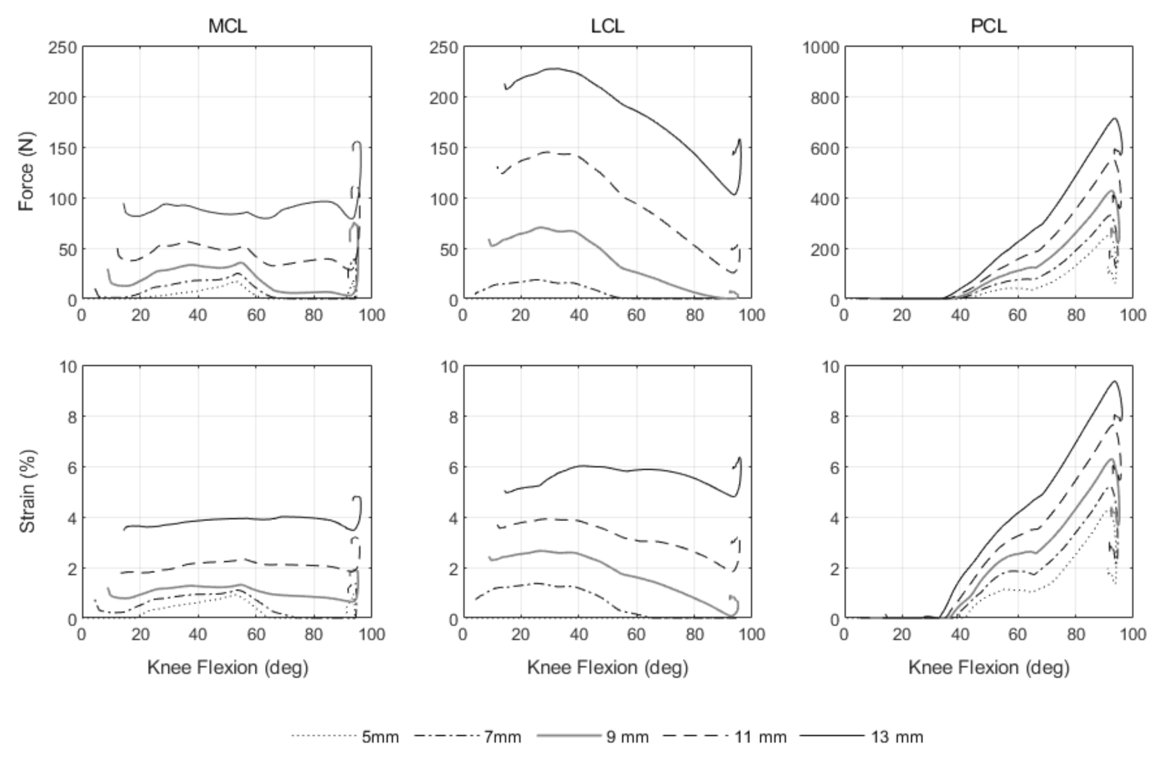
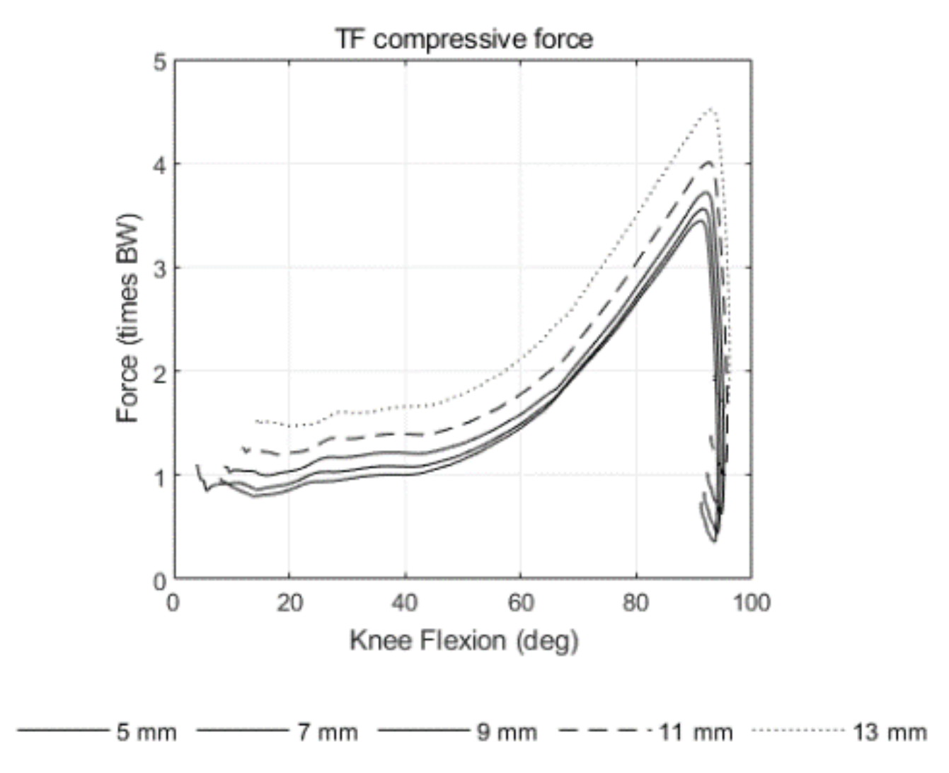
3.1. Tibiofemoral Joint

The peak ligament and compressive forces on the TF joint for all the simulated PE insert thickness cases are depicted in Figure 3. The peak MCL, LCL, and PCL forces increased by 38 N, 74 N, and 125 N, respectively, as PE insert thickness changed from 9 mm to 11 mm; the corresponding peak force increase for a 4 mm greater thickness relative to the reference case was 80 N, 157 N, and 286 N. Conversely, the peak force in the MCL, LCL, and PCL decreased by 36 N, 52 N, and 98 N, respectively, when using a 2 mm thinner PE insert compared to the reference case, while with a 4 mm smaller thickness than the reference, the peak decrease was 57 N, 70 N, 177 N, respectively. Table 1 summarizes the peak as well the average percentage variations relative to the reference case. Varying PE insert thickness simultaneously changed the ligament individual bundle strain. Both medial and lateral collateral ligaments appeared to stretch in the same strain range of about 0–6% for all the simulated thickness cases, whereas the PCL exhibited a maximum strain around 10% at 90° of knee flexion with 13 mm thickness. Force and strain patterns of the MCL, LCL, and PCL at varying thicknesses are provided in Appendix A.
Changes in PE insert thickness affected the TF compressive force, denoting a larger effect at 90° of knee flexion angle at which the highest peak occurred at 4.5 times body weight (BW) with a 13 mm PE insert (Figure 3). Compared to the reference case, the peak TF compressive force increased by 0.3 BW and 0.8 BW with 2 mm and 4 mm greater PE insert thicknesses, respectively. Using 2 mm and 4 mm thinner PE inserts than in the reference case showed a peak decrease of 0.2 BW and 0.3 BW, respectively. Detailed peak and average percentage variations of the TF joint compressive force are also provided in Table 1.

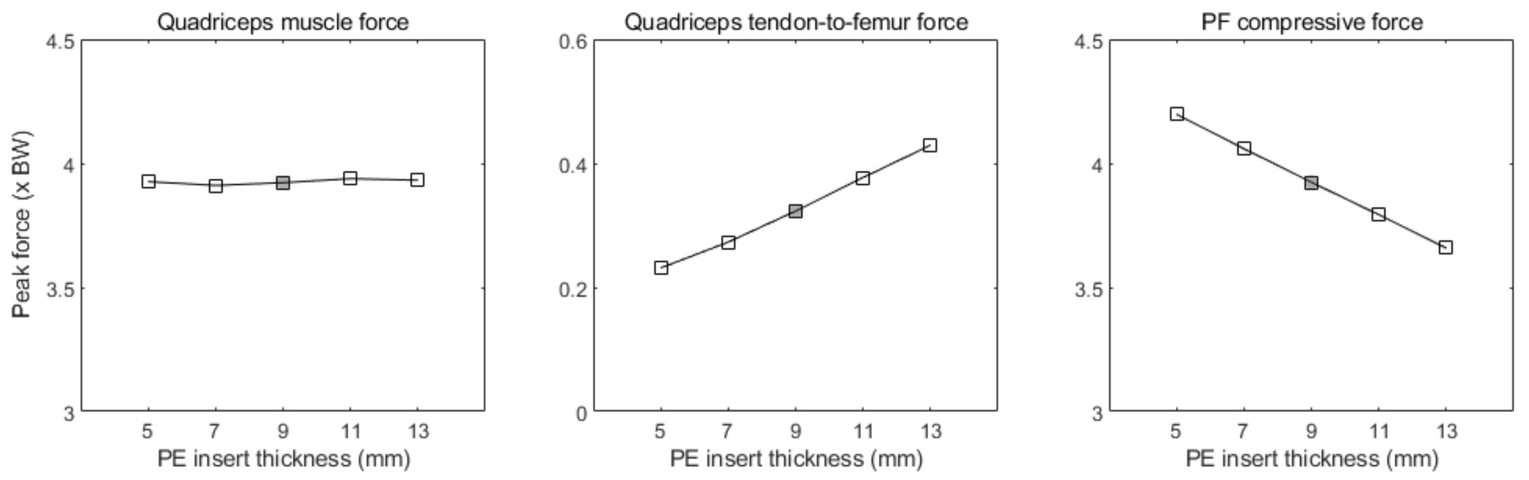
3.2. Patellofemoral Joint

The forces around the knee extensor mechanism exhibited peaks during the momentum transfer phase of the chair-rising movement at about 90° of knee flexion. Peak values of the quadriceps muscle force, quadriceps tendon-to-femur force, and PF compressive force for all the simulated PE insert thickness cases are shown in Figure 4, and their variations with regard to the reference case are summarized in Table 1. Interestingly, the PF compressive force followed an opposite trend than the compressive force on the TF side, indicating a peak decrease of 0.1 BW and 0.3 BW when PE insert thickness was varied from 9 mm to 11 mm or 13 mm, respectively. In contrast, simulation of thinner tibial PE inserts, relative to the reference case, resulted in a slightly increased peak PF joint compressive force by 0.1 BW (−2 mm) and 0.3 BW (−4 mm). The force–angle curves of the knee extensor mechanism structures are provided in Appendix A.
Changes in PE insert thickness affected the patellar proximal–distal translation and medial–lateral rotation, with a minimal effect at full knee flexion (Figure 5). The patella shifted by 0.8 mm distally for every 2 mm incremental change of the PE insert thickness, from 9 mm (reference) up to 13 mm, and about 0.8 more proximally for each 2 mm decrement relative to the reference thickness. Compared to the reference case, the patella was rotated 0.4 and 0.7 mm more medially with 11 mm and 13 mm PE insert thicknesses, respectively and, accordingly, 0.4 and 0.1 mm more laterally with a 7 mm and 5 mm PE insert thickness, respectively.

4. Discussion

The most important findings in the present study were that a larger thickness of the tibial PE insert elevated the ligament forces throughout the range of knee flexion and extension and, consequently, increased the TF compressive force across the mediolateral compartment; in contrast, varying the PE insert thickness had little effect on the PF biomechanics, although it indicated an unexpected slight decrease in the PF joint loading. These results confirm our hypothesis and suggest that there is a trade-off in the kinetic behavior between the TF and PF joint structures with regard to varying PE insert thickness.
Tibial PE insert thickness variation had a marked effect on the ligament forces and strain patterns, as expected because of overstuffing the TF joint space. With thicker PE inserts relative to the reference case, the MCL, LCL, and PCL forces increased considerably both in flexion and extension, although the changes in the PCL were more distinct after mid-flexion. The observed higher LCL forces over the MCL might be related to the larger stiffness value assigned to the MCL individual bundles (3000 N) based on literature experimental evidence [26]. Another fact worth noticing is that the PCL was engaged at lower flexion angles with thicker PE inserts, which may be due to the larger joint distraction in extension. On the other hand, thinner PE inserts (5 mm, 7 mm) slackened the MCL and LCL almost entirely from 60° to 90°, due to the larger flexion and extension gap. This is an unfavorable scenario, as slackening the collateral ligaments may destabilize the knee in flexion. The PCL, unlike the other two ligaments, received more tension in the flexed knee with PE inserts thicker than 9 mm, reaching a peak strain at about 10% when using a 13 mm component. This is, however, well below the yield strain of 14% as reported in the literature, above which there might be a ligament injury as a result of overstretching [22]. Similar to the ligaments’ kinetic behavior, the compressive force in the TF joint increased substantially at varying PE insert thicknesses, particularly at 90° of knee flexion with a 13 mm PE component. Considering that the quadriceps muscle force was roughly the same across PE insert thickness cases, this increase can be solely attributed to the summation of increased joint ligament forces. The magnitude of the TF compressive force found in this study is consistent with earlier observations [27], reporting an average peak of about four times BW during rising from a chair without the aid of arms in a natural knee. This supports our reported values with 9 mm (3.7 BW) or 11 mm (4 BW) thicknesses and raises the concern that using thicker PE inserts, such as 13 mm (4.5 BW) or more, could greatly elevate the joint loads on the PE surface, leading to destructive wear [28]. Surgeons should consider this surgical option relative to the resected bone on the tibial plateau, bearing in mind that a thicker PE insert also requires a lower level of tibial resection, which is associated with posteromedial bone failure and early aseptic loosening [29]. In addition, deeper tibial resections may lead to the removal of a considerable part of the tibial PCL attachment, which can result in a reduced femoral rollback and anteroposterior instability during flexion [30]. A recent cadaveric study proposed a PE insert thickness of 10 mm with a posterior slope of 5° to preserve more than 50% of the tibial PCL attachment site [31]. Our findings highlight the importance of preoperative planning of the appropriate tibial insert thickness, which considers both the thickness value available and the tibial resection depth and, additionally, support the conceptual premise that selection of a tibial insert that is either too thin or too thick can have a negative impact on the TF kinetics, leading to a sub-optimal solution.
Varying the thickness of the PE insert revealed marginal secondary effects on the PF biomechanics. The quadriceps muscle force remained unchanged across PE insert thickness variations in the full arc of the knee range of motion. However, more thickness in the tibial PE component increased the quadriceps tendon-to-femur force in the range of 60°-90° of knee flexion. Contrary to our expectations, the PF compressive force slightly decreased with 2 mm and 4 mm greater PE insert thickness, compared to the reference case, and this change was more perceivable from mid-flexion to 90°. This is in agreement with the findings of a previous study [32] and might be explained by quadriceps–femur load sharing as the quadriceps wraps around the distal femur in a flexed knee position. This mechanism is also reflected in the PF kinematics. The patella was consistently more distal with thicker PE inserts than in the reference case throughout the range of flexion and extension, as expected due to the joint line elevation, which subsequently results in patella baja. From a clinical point of view, unloading the PF joint could reduce patellar complications after TKA, especially anterior knee pain, which strongly correlates with postoperative patient dissatisfaction and impaired quality of life [33,34,35]. On the other hand, patella baja may have serious consequences on the overall function of the knee after surgery, such as limited range of motion because of patellar maltracking. Moreover, the distally displaced patella can impinge on the anterior part of the tibial PE insert or the tibial tray during flexion, potentially increasing wear [36]. Hence, surgeons should carefully assess the concomitant effects on the PF joint when pre-planning the desired PE insert thickness to ensure that the patella slides properly in the trochlear groove.
To the best of our knowledge, this is the first musculoskeletal simulation study which explores the biomechanical consequences of tibial PE insert thickness variations on both the TF and PF joints after cruciate-retaining TKA. A major strength of this study is the use of a computational modeling approach to investigate the effect of tibial PE insert thickness in a highly controlled simulation environment, where all the other surgical variables, such as the tibial resection depth, remained unchanged. This overcomes an important limitation of clinical studies, in which confounding factors, such as the implant design, size, and alignment, are present and can potentially affect the surgical outcomes [9].
This study has several limitations. At first, the modeled ligament mechanical properties, such as stiffness and reference strain, were determined from the literature. Furthermore, the musculoskeletal simulation represented only a mechanically aligned cruciate-retaining TKA, meaning that the results cannot be extrapolated to other surgical techniques or implant designs. It is also noteworthy that the computational model simulated only one patient, disregarding the anatomical variability or other pathological conditions among different patients. Further research to overcome this limitation may be to characterize the effect of variability in the morphological knee joint phenotypes on the postoperative kinetics with varying tibial PE insert thicknesses. A promising methodology to explore this could be the combination of statistical shape modeling and musculoskeletal simulation.

5. Conclusions

Changes in tibial PE insert thickness have considerable influence on the kinetics across the TF articulation and surrounding ligamentous structures, but marginal effects on the PF biomechanics. Increasing the tibial PE insert thickness resulted in higher ligament forces, and subsequently increased loading across the medial and lateral TF compartments in the full arc of knee motion after cruciate-retaining TKA. However, thicker PE inserts resulted in a slightly lower PF force by means of an increased load-sharing between the quadriceps tendon and femur.

Author Contributions

Conceptualization, P.T. and N.V.; methodology, P.T. and M.A.M.; software, P.T., M.A.M., and R.F.; validation, P.T., M.A.M., and R.F.; formal analysis, P.T.; investigation, P.T.; resources, P.T. and M.A.M.; data curation, P.T., M.A.M., and R.F.; writing—original draft preparation, P.T.; writing—review and editing, P.T., M.A.M., R.F., and N.V.; visualization, P.T.; supervision, M.A.M., N.V., and B.K.; project administration, N.V. and B.K.; funding acquisition, M.A.M. and N.V. All authors have read and agreed to the published version of the manuscript.

Funding

This research was funded by Top Technology Twente Connecting Industry program (TKI Top Sector HTSM) and Stryker European Operations Ltd., Ireland.

Institutional Review Board Statement

Not applicable.

Informed Consent Statement

Not applicable.

Data Availability Statement

The data presented in this study are available on request from the corresponding author.

Acknowledgments

We would like to thank Eric Garling (Stryker, Kalamazoo, MI, USA) and Arman Motesharei (Stryker, Newbury, UK) for providing the geometry files of Triathlon prosthesis used in this study.

Conflicts of Interest

The authors declare no conflict of interest. The funders had no role in the design of the study; in the collection, analyses, or interpretation of data; in the writing of the manuscript, or in the decision to publish the results.

Appendix A

Figure A1. Ligament force and strain patterns at varying PE insert thicknesses during a chair-rising simulation. From left to right top and bottom: MCL force and strain, LCL force and strain, PCL force and strain.
Applsci 11 02423 g0a1
Figure A2. TF compressive force at varying PE insert thicknesses during a chair-rising simulation.
Applsci 11 02423 g0a2
Figure A3. Forces around the knee extensor mechanism at varying PE insert thicknesses during a chair-rising simulation. From left to right: quadriceps muscle force, quadriceps tendon-to-femur force, and PF compressive force.
Applsci 11 02423 g0a3

References

  1. Carr, A.J.; Robertsson, O.; Graves, S.; Price, A.J.; Arden, N.K.; Judge, A.; Beard, D.J. Knee replacement. Lancet 2012, 379, 1331–1340. [Google Scholar] [CrossRef]
  2. Hamilton, D.; Burnett, R.; Patton, J.; Howie, C.; Moran, M.; Simpson, A.; Gaston, P. Implant design influences patient outcome after total knee arthroplasty: A prospective double-blind randomised controlled trial. Bone Jt. J. 2015, 97, 64–70. [Google Scholar] [CrossRef] [Green Version]
  3. Bonnin, M.P.; Schmidt, A.; Basiglini, L.; Bossard, N.; Dantony, E. Mediolateral oversizing influences pain, function, and flexion after TKA. Knee Surg. Sports Traumatol. Arthrosc. 2013, 21, 2314–2324. [Google Scholar] [CrossRef] [PubMed] [Green Version]
  4. Longstaff, L.M.; Sloan, K.; Stamp, N.; Scaddan, M.; Beaver, R. Good alignment after total knee arthroplasty leads to faster rehabilitation and better function. J. Arthroplast. 2009, 24, 570–578. [Google Scholar] [CrossRef] [PubMed]
  5. Sharkey, P.F.; Hozack, W.J.; Rothman, R.H.; Shastri, S.; Jacoby, S.M. Why are total knee arthroplasties failing today? Clin. Orthop. Relat. Res. 2002, 404, 7–13. [Google Scholar] [CrossRef]
  6. Edwards, S.; Pandit, H.; Ramos, J.; Grover, M. Analysis of polyethylene thickness of tibial components in total knee replacement. J. Bone Jt. Surg. Am. 2002, 84, 369–371. [Google Scholar] [CrossRef] [PubMed]
  7. Lanting, B.A.; Snider, M.G.; Chess, D.G. Effect of polyethylene component thickness on range of motion and stability in primary total knee arthroplasty. J. Orthop. 2012, 35, e170–e174. [Google Scholar] [CrossRef] [PubMed] [Green Version]
  8. Bartel, D.L.; Bicknell, V.; Wright, T.J.J. The effect of conformity, thickness, and material on stresses in ultra-high molecular weight components for total joint replacement. J. Bone Jt. Surg. Am. 1986, 68, 1041–1051. [Google Scholar] [CrossRef]
  9. Pijls, B.G.; Van der Linden-Van, H.M.; Nelissen, R.G. Polyethylene thickness is a risk factor for wear necessitating insert exchange. Int. Orthop. 2012, 36, 1175–1180. [Google Scholar] [CrossRef] [Green Version]
  10. Yoo, J.Y.; Cai, J.; Chen, A.F.; Austin, M.S.; Sharkey, P.F. Modular Polyethylene Inserts for Total Knee Arthroplasty: Can Surgeons Detect 1-mm Thickness Increments? J. Arthroplast. 2016, 31, 968–970. [Google Scholar] [CrossRef] [PubMed]
  11. Mueller, J.K.P.; Wentorf, F.A.; Moore, R.E. Femoral and tibial insert downsizing increases the laxity envelope in TKA. Knee Surg. Sports Traumatol. Arthrosc. 2014, 22, 3003–3011. [Google Scholar] [CrossRef] [Green Version]
  12. Engh, G.A.; Dwyer, K.A.; Hanes, C.K. Polyethylene wear of metal-backed tibial components in total and unicompartmental knee prostheses. J. Bone Jt. Surg. Br. 1992, 74, 9–17. [Google Scholar] [CrossRef]
  13. Greco, N.J.; Crawford, D.A.; Berend, K.R.; Adams, J.B.; Lombardi, A.V., Jr. “Thicker” polyethylene bearings are not associated with higher failure rates in primary total knee arthroplasty. J. Arthroplast. 2018, 33, 2810–2814. [Google Scholar] [CrossRef] [PubMed]
  14. Berend, M.E.; Davis, P.J.; Ritter, M.A.; Keating, E.M.; Faris, P.M.; Meding, J.B.; Malinzak, R.A. “Thicker” polyethylene bearings are associated with higher failure rates in primary total knee arthroplasty. J. Arthroplast. 2010, 25, 17–20. [Google Scholar] [CrossRef]
  15. Ding, Z.; Nolte, D.; Kit Tsang, C.; Cleather, D.J.; Kedgley, A.E.; Bull, A.M. In vivo knee contact force prediction using patient-specific musculoskeletal geometry in a segment-based computational model. J. Biomech. Eng. 2016, 138. [Google Scholar] [CrossRef] [PubMed] [Green Version]
  16. Peersman, G.; Slane, J.; Dirckx, M.; Vandevyver, A.; Dworschak, P.; Heyse, T.J.; Scheys, L. The influence of polyethylene bearing thickness on the tibiofemoral kinematics of a bicruciate retaining total knee arthroplasty. Knee 2017, 24, 751–760. [Google Scholar] [CrossRef] [PubMed]
  17. Marra, M.A.; Vanheule, V.; Fluit, R.; Koopman, B.; Rasmussen, J.; Verdonschot, N.; Andersen, M.S. A subject-specific musculoskeletal modeling framework to predict in vivo mechanics of total knee arthroplasty. J. Biomech. Eng. 2015, 137. [Google Scholar] [CrossRef] [PubMed]
  18. Carbone, V.; Fluit, R.; Pellikaan, P.; Van Der Krogt, M.; Janssen, D.; Damsgaard, M.; Vigneron, L.; Feilkas, T.; Koopman, H.F.; Verdonschot, N. TLEM 2.0–A comprehensive musculoskeletal geometry dataset for subject-specific modeling of lower extremity. J. Biomech. 2015, 48, 734–741. [Google Scholar] [CrossRef] [Green Version]
  19. Fregly, B.J.; Besier, T.F.; Lloyd, D.G.; Delp, S.L.; Banks, S.A.; Pandy, M.G.; D’lima, D.D. Grand challenge competition to predict in vivo knee loads. J. Orthop. Res. 2012, 30, 503–513. [Google Scholar] [CrossRef] [PubMed] [Green Version]
  20. Skipper Andersen, M.; De Zee, M.; Damsgaard, M.; Nolte, D.; Rasmussen, J. Introduction to force-dependent kinematics: Theory and application to mandible modeling. J. Biomech. Eng. 2017, 139. [Google Scholar] [CrossRef] [PubMed]
  21. Damsgaard, M.; Rasmussen, J.; Christensen, S.T.; Surma, E.; De Zee, M. Analysis of musculoskeletal systems in the AnyBody Modeling System. Simul. Model. Pract. Theory 2006, 14, 1100–1111. [Google Scholar] [CrossRef]
  22. Butler, D.L.; Kay, M.D.; Stouffer, D.C. Comparison of material properties in fascicle-bone units from human patellar tendon and knee ligaments. J. Biomech. 1986, 19, 425–432. [Google Scholar] [CrossRef]
  23. Cook, L.E.; Klika, A.K.; Szubski, C.R.; Rosneck, J.; Molloy, R.; Barsoum, W.K. Functional outcomes used to compare single radius and multiradius of curvature designs in total knee arthroplasty. J. Knee Surg. 2012, 25, 249–254. [Google Scholar] [CrossRef]
  24. Rivière, C.; Iranpour, F.; Auvinet, E.; Howell, S.; Vendittoli, P.-A.; Cobb, J.; Parratte, S. Alignment options for total knee arthroplasty: A systematic review. Orthop. Traumatol. Surg. Res. 2017, 103, 1047–1056. [Google Scholar] [CrossRef] [PubMed]
  25. Cohen, Z.A.; Mccarthy, D.M.; Kwak, S.D.; Legrand, P.; Fogarasi, F.; Ciaccio, E.J.; Ateshian, G.A. Knee cartilage topography, thickness, and contact areas from MRI: In-vitro calibration and in-vivo measurements. Osteoarthr. Cartil. 1999, 7, 95–109. [Google Scholar] [CrossRef] [Green Version]
  26. Blankevoort, L.; Kuiper, J.; Huiskes, R.; Grootenboer, H. Articular contact in a three-dimensional model of the knee. J. Biomech. 1991, 24, 1019–1031. [Google Scholar] [CrossRef] [Green Version]
  27. Ellis, M.; Seedhom, B.; Wright, V. Forces in the knee joint whilst rising from a seated position. J. Biomed. Eng. 1984, 6, 113–120. [Google Scholar] [CrossRef]
  28. Nagura, T.; Matsumoto, H.; Kiriyama, Y.; Chaudhari, A.; Andriacchi, T.P. Tibiofemoral joint contact force in deep knee flexion and its consideration in knee osteoarthritis and joint replacement. J. Appl. Biomech. 2006, 22, 305–313. [Google Scholar] [CrossRef] [Green Version]
  29. Schnurr, C.; Csécsei, G.; Nessler, J.; Eysel, P.; König, D.P. How much tibial resection is required in total knee arthroplasty? Int. Orthop. 2011, 35, 989–994. [Google Scholar] [CrossRef] [Green Version]
  30. Kebbach, M.; Grawe, R.; Geier, A.; Winter, E.; Bergschmidt, P.; Kluess, D.; D’Lima, D.; Woernle, C.; Bader, R. Effect of surgical parameters on the biomechanical behaviour of bicondylar total knee endoprostheses–A robot-assisted test method based on a musculoskeletal model. Sci. Rep. 2019, 9, 1–11. [Google Scholar] [CrossRef] [Green Version]
  31. Onishi, Y.; Hino, K.; Watanabe, S.; Watamori, K.; Kutsuna, T.; Miura, H. The influence of tibial resection on the PCL in PCL-retaining total knee arthroplasty: A clinical and cadaveric study. J. Orthop. Sci. 2016, 21, 798–803. [Google Scholar] [CrossRef] [PubMed]
  32. Marra, M.A.; Strzelczak, M.; Heesterbeek, P.J.; van de Groes, S.A.; Janssen, D.W.; Koopman, B.F.; Wymenga, A.B.; Verdonschot, N.J. Anterior referencing of tibial slope in total knee arthroplasty considerably influences knee kinematics: A musculoskeletal simulation study. Knee Surg. Sports Traumatol. Arthrosc. 2018, 26, 1540–1548. [Google Scholar] [CrossRef] [PubMed]
  33. Scott, C.; Howie, C.; MacDonald, D.; Biant, L. Predicting dissatisfaction following total knee replacement: A prospective study of 1217 patients. J. Bone Jt. Surg. Br. 2010, 92, 1253–1258. [Google Scholar] [CrossRef]
  34. Petersen, W.; Rembitzki, I.V.; Brüggemann, G.-P.; Ellermann, A.; Best, R.; Gösele-Koppenburg, A.; Liebau, C. Anterior knee pain after total knee arthroplasty: A narrative review. Int. Orthop. 2014, 38, 319–328. [Google Scholar] [CrossRef] [Green Version]
  35. Laubach, M.; Hellmann, J.T.; Dirrichs, T.; Gatz, M.; Quack, V.; Tingart, M.; Betsch, M. Anterior knee pain after total knee arthroplasty: A multifactorial analysis. J. Orthop. Surg. 2020, 28. [Google Scholar] [CrossRef]
  36. Chonko, D.J.; Lombardi, A.V., Jr.; Berend, K.R. Patella baja and total knee arthroplasty (TKA): Etiology, diagnosis, and management. Surg. Technol. Int. 2004, 12, 231–238. [Google Scholar] [PubMed]
Figure 1. Illustration of the patient-specific musculoskeletal force-dependent kinematics (FDK) knee model implanted with a Triathlon cruciate-retaining prosthesis. This full-body model includes the head, trunk, pelvis, and the lower extremity of the implanted side (thigh, shank, patella, talus, foot) (left). A close-up anterior view of the knee joint with the medial collateral ligament (MCL), lateral collateral ligament (LCL), posterior cruciate ligament (PCL), medial patellofemoral ligament (MPFL), lateral patellofemoral ligament (LPFL), and patellar tendon (PT) (right).
Applsci 11 02423 g001
Figure 2. Illustration of three custom postoperative cases simulated in this study. The tibial polyethylene (PE) insert thickness cases of 9 mm (reference), 11 mm (+2 mm), and 13 mm (+4 mm) are depicted, plus the tibial metal baseplate fixed to the resected proximal tibia.
Applsci 11 02423 g002
Figure 3. Peak ligament and compressive forces on the tibiofemoral (TF) joint at varying PE insert thicknesses during a chair-rising simulation. From left to right: MCL force, LCL force, PCL force, and TF compressive force. Results are displayed relative to the reference case (gray filled marker).
Applsci 11 02423 g003
Figure 4. Peak forces around the knee extensor mechanism at varying PE insert thicknesses during a chair-rising simulation. From left to right: quadriceps muscle force, quadriceps tendon-to-femur force, and patellofemoral (PF) compressive force.
Applsci 11 02423 g004
Figure 5. Reference frames used to express the PF kinematics (left). Muscles and ligaments of the TF and PF joints are hidden in this model view. Patellar kinematic profiles at varying PE insert thicknesses during a chair-rising simulation (right).
Applsci 11 02423 g005
Table 1. Changes in muscle, ligament, and joint compressive forces due to tibial PE insert thickness variations.
Reference PE Insert Thickness 9 mm
−4 mm−2 mm+2 mm+4 mm
Average DifferencePeak DifferenceAverage DifferencePeak DifferenceAverage DifferencePeak DifferenceAverage DifferencePeak Difference
Tibiofemoral joint
MCL force−89%−75%−59%−47%+100%+51%+251%+106%
LCL force−100%−100%−83%−73%+210%+106%+554%+223%
PCL force−53%−41%−31%−23%+43%+29%+104%+67%
Compressive force−21%−7%−9%%−4%+17%+8%+44%+21%
Patellofemoral joint
Quadriceps muscle force+4%0%+1%0%0%0%−1%0%
Quadriceps tendon-to-femur force−30%−28%−15%−15%+15%+17%+29%+33%
Compressive force+3%+7%+3%+3%−3%−3%−8%−7%
Data are presented as average and peak percentage difference over the chair-rising simulation for each 2 mm PE insert thickness change relative to the reference case.
Publisher’s Note: MDPI stays neutral with regard to jurisdictional claims in published maps and institutional affiliations.

