
Histological and Histomorphometric Effectiveness of the Barrier Membranes for Jawbone Regeneration: An Overview of More Than 30 Years’ Experience of Research Results of the Italian Implant Retrieval Center (1988–2020)

 ,
,  ,
,  , ,
, ,
Abstract
:1. Introduction
2. Materials and Methods
2.1. Inclusion Criteria
2.2. Selection of the Studies
2.3. Data Extraction
3. Results
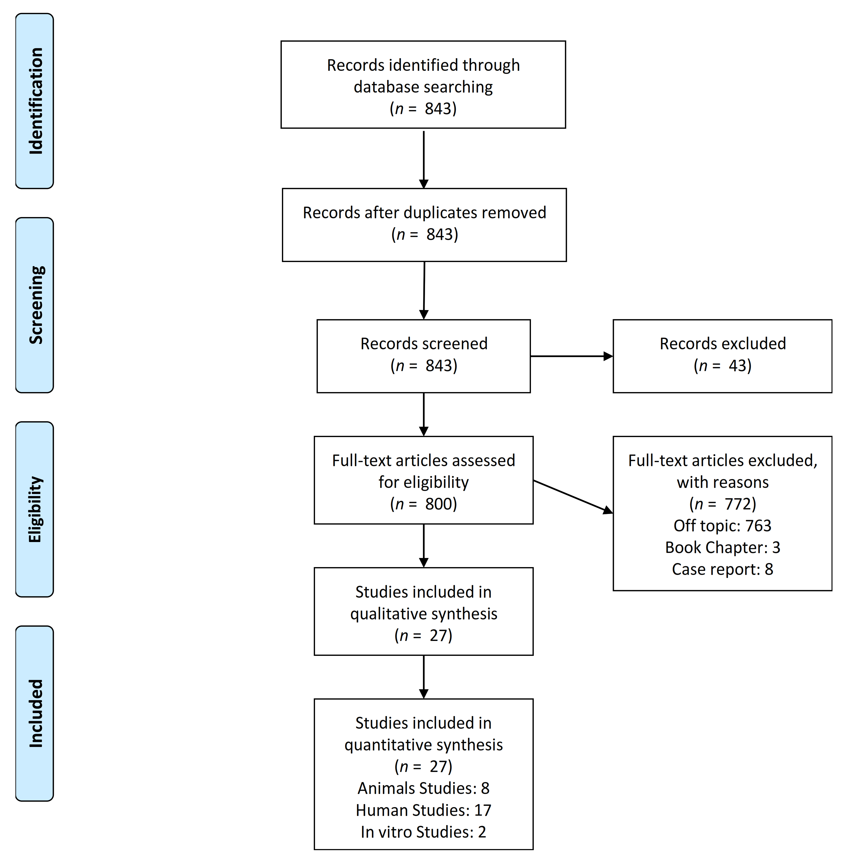
3.1. Papers Selection
3.2. In Vitro Studies
3.3. Animal Studies
3.4. Human Studies
4. Discussion
5. Conclusions
Author Contributions
Funding
Institutional Review Board Statement
Informed Consent Statement
Data Availability Statement
Acknowledgments
Conflicts of Interest
References
- Waechter, J.; Leite, F.R.; Nascimento, G.G.; Carmo Filho, L.C.; Faot, F. The Split Crest Technique and Dental Implants: A Systematic Review and Meta-Analysis. Int. J. Oral Maxillofac. Surg. 2017, 46, 116–128. [Google Scholar] [CrossRef]
- Chavda, S.; Levin, L. Human Studies of Vertical and Horizontal Alveolar Ridge Augmentation Comparing Different Types of Bone Graft Materials: A Systematic Review. J. Oral Implant. 2018, 44, 74–84. [Google Scholar] [CrossRef] [PubMed]
- Chappuis, V.; Rahman, L.; Buser, R.; Janner, S.F.M.; Belser, U.C.; Buser, D. Effectiveness of Contour Augmentation with Guided Bone Regeneration: 10-Year Results. J. Dent. Res. 2018, 97, 266–274. [Google Scholar] [CrossRef]
- Çolpak, H.A.; Gönen, Z.B.; Özdamar, S.; Alkan, A.; Kütük, N. Vertical Ridge Augmentation Using Guided Bone Regeneration Procedure and Dental Pulp Derived Mesenchymal Stem Cells with Simultaneous Dental Implant Placement: A Histologic Study in a Sheep Model. J. Stomatol. Oral Maxillofac. Surg. 2019, 120, 216–223. [Google Scholar] [CrossRef] [PubMed]
- Comuzzi, L.; Tumedei, M.; Pontes, A.E.; Piattelli, A.; Iezzi, G. Primary Stability of Dental Implants in Low-Density (10 and 20 pcf) Polyurethane Foam Blocks: Conical vs Cylindrical Implants. Int. J. Environ. Res. Public Health 2020, 17, 2617. [Google Scholar] [CrossRef] [PubMed] [Green Version]
- Bornstein, M.M.; Halbritter, S.; Harnisch, H.; Weber, H.-P.; Buser, D. A Retrospective Analysis of Patients Referred for Implant Placement to a Specialty Clinic: Indications, Surgical Procedures, and Early Failures. Int. J. Oral Maxillofac. Implant. 2008, 23, 1109–1116. [Google Scholar]
- Buck, D.W.; Dumanian, G.A. Bone Biology and Physiology: Part I. The Fundamentals. Plast. Reconstr. Surg. 2012, 129, 1314–1320. [Google Scholar] [CrossRef]
- Majidinia, M.; Sadeghpour, A.; Yousefi, B. The Roles of Signaling Pathways in Bone Repair and Regeneration. J. Cell. Physiol. 2018, 233, 2937–2948. [Google Scholar] [CrossRef] [PubMed]
- Danza, M.; Zollino, I.; Avantaggiato, A.; Lucchese, A.; Carinci, F. Distance between Implants Has a Potential Impact of Crestal Bone Resorption. Saudi Dent J 2011, 23, 129–133. [Google Scholar] [CrossRef] [Green Version]
- Patianna, A.G.; Ballini, A.; Meneghello, M.; Cantore, S.; Inchingolo, A.M.; Dipalma, G.; Inchingolo, A.D.; Inchingolo, F.; Malcangi, G.; Lucchese, A.; et al. Comparison of Conventional Orthognathic Surgery and “Surgery-First” Protocol: A New Weapon against Time. J. Biol. Regul. Homeost. Agents 2019, 33, 59–67. [Google Scholar]
- Rigo, L.; Viscioni, A.; Franco, M.; Lucchese, A.; Zollino, I.; Brunelli, G.; Carinci, F. Overdentures on Implants Placed in Bone Augmented with Fresh Frozen Bone. Minerva Stomatol. 2011, 60, 5–14. [Google Scholar] [CrossRef]
- Rodriguez y Baena, R.; Pastorino, R.; Gherlone, E.F.; Perillo, L.; Lupi, S.M.; Lucchese, A. Histomorphometric Evaluation of Two Different Bone Substitutes in Sinus Augmentation Procedures: A Randomized Controlled Trial in Humans. Int. J. Oral Maxillofac. Implant. 2017, 32, 188–194. [Google Scholar] [CrossRef] [Green Version]
- Piattelli, A.; Scarano, A.; Paolantonio, M. Bone Formation inside the Material Interstices of E-PTFE Membranes: A Light Microscopical and Histochemical Study in Man. Biomaterials 1996, 17, 1725–1731. [Google Scholar] [CrossRef]
- Al-Hezaimi, K.; Iezzi, G.; Rudek, I.; Al-Daafas, A.; Al-Hamdan, K.; Al-Rasheed, A.; Javed, F.; Piattelli, A.; Wang, H.-L. Histomorphometric Analysis of Bone Regeneration Using a Dual Layer of Membranes (DPTFE Placed over Collagen) in Fresh Extraction Sites: A Canine Model. J. Oral Implant. 2015, 41, 188–195. [Google Scholar] [CrossRef]
- Simion, M.; Trisi, P.; Piattelli, A. GBR with an E-PTFE Membrane Associated with DFDBA: Histologic and Histochemical Analysis in a Human Implant Retrieved after 4 Years of Loading. Int. J. Periodontics Restor. Dent. 1996, 16, 338–347. [Google Scholar] [CrossRef]
- Simion, M.; Baldoni, M.; Rossi, P.; Zaffe, D. A Comparative Study of the Effectiveness of E-PTFE Membranes with and without Early Exposure during the Healing Period. Int. J. Periodontics Restor. Dent. 1994, 14, 166–180. [Google Scholar]
- Bottino, M.C.; Thomas, V.; Schmidt, G.; Vohra, Y.K.; Chu, T.-M.G.; Kowolik, M.J.; Janowski, G.M. Recent Advances in the Development of GTR/GBR Membranes for Periodontal Regeneration--a Materials Perspective. Dent. Mater. 2012, 28, 703–721. [Google Scholar] [CrossRef]
- Elgali, I.; Omar, O.; Dahlin, C.; Thomsen, P. Guided Bone Regeneration: Materials and Biological Mechanisms Revisited. Eur. J. Oral Sci. 2017, 125, 315–337. [Google Scholar] [CrossRef]
- Oh, S.H.; Kim, J.H.; Kim, J.M.; Lee, J.H. Asymmetrically Porous PLGA/Pluronic F127 Membrane for Effective Guided Bone Regeneration. J. Biomater. Sci. Polym. Ed. 2006, 17, 1375–1387. [Google Scholar] [CrossRef]
- Sam, G.; Pillai, B.R.M. Evolution of Barrier Membranes in Periodontal Regeneration-"Are the Third Generation Membranes Really Here?". J. Clin. Diagn. Res. 2014, 8, ZE14–ZE17. [Google Scholar] [CrossRef]
- Diomede, F.; D’Aurora, M.; Gugliandolo, A.; Merciaro, I.; Orsini, T.; Gatta, V.; Piattelli, A.; Trubiani, O.; Mazzon, E. Biofunctionalized Scaffold in Bone Tissue Repair. Int. J. Mol. Sci. 2018, 19, 1022. [Google Scholar] [CrossRef] [Green Version]
- Chierico, A.; Valentini, R.; Majzoub, Z.; Piattelli, A.; Scarano, A.; Okun, L.; Cordioli, G. Electrically Charged GTAM Membranes Stimulate Osteogenesis in Rabbit Calvarial Defects. Clin. Oral Implant. Res. 1999, 10, 415–424. [Google Scholar] [CrossRef]
- Piattelli, A.; Piattelli, M.; Scarano, A. Simultaneous Demonstration of Alkaline and Acid Phosphatase Activity in Bone, at Bone-Implant Interfaces and at the Epiphyseal Growth Plate in Plastic-Embedded Undemineralized Tissues. Biomaterials 1997, 18, 545–549. [Google Scholar] [CrossRef]
- Piattelli, A.; Scarano, A.; Coraggio, F.; Matarasso, S. Early Tissue Reactions to Polylactic Acid Resorbable Membranes: A Histological and Histochemical Study in Rabbit. Biomaterials 1998, 19, 889–896. [Google Scholar] [CrossRef]
- Piattelli, A.; Scarano, A.; Mangano, C. Clinical and Histologic Aspects of Biphasic Calcium Phosphate Ceramic (BCP) Used in Connection with Implant Placement. Biomaterials 1996, 17, 1767–1770. [Google Scholar] [CrossRef]
- Colangelo, P.; Piattelli, A.; Barrucci, S.; Trisi, P.; Formisano, G.; Caiazza, S. Bone Regeneration Guided by Resorbable Collagen Membranes in Rabbits: A Pilot Study. Implant Dent. 1993, 2, 101–105. [Google Scholar] [CrossRef]
- Cerrai, P.; Guerra, G.D.; Tricoli, M.; Krajewski, A.; Ravaglioli, A.; Martinetti, R.; Dolcini, L.; Fini, M.; Scarano, A.; Piattelli, A. Periodontal Membranes from Composites of Hydroxyapatite and Bioresorbable Block Copolymers. J. Mater. Sci. Mater. Med. 1999, 10, 677–682. [Google Scholar] [CrossRef]
- Degidi, M.; Scarano, A.; Piattelli, A. Regeneration of the Alveolar Crest Using Titanium Micromesh with Autologous Bone and a Resorbable Membrane. J. Oral Implant. 2003, 29, 86–90. [Google Scholar] [CrossRef] [Green Version]
- Assenza, B.; Piattelli, M.; Scarano, A.; Lezzi, G.; Petrone, G.; Piattelli, A. Localized Ridge Augmentation Using Titanium Micromesh. J. Oral Implant. 2001, 27, 287–292. [Google Scholar] [CrossRef]
- Majzoub, Z.; Cordioli, G.; Aramouni, P.K.; Vigolo, P.; Piattelli, A. Guided Bone Regeneration Using Demineralized Laminar Bone Sheets versus GTAM Membranes in the Treatment of Implant-Associated Defects. A Clinical and Histological Study. Clin. Oral Implant. Res. 1999, 10, 406–414. [Google Scholar] [CrossRef]
- Malchiodi, L.; Scarano, A.; Quaranta, M.; Piattelli, A. Rigid Fixation by Means of Titanium Mesh in Edentulous Ridge Expansion for Horizontal Ridge Augmentation in the Maxilla. Int. J. Oral Maxillofac. Implant. 1998, 13, 701–705. [Google Scholar]
- Simion, M.; Jovanovic, S.A.; Trisi, P.; Scarano, A.; Piattelli, A. Vertical Ridge Augmentation around Dental Implants Using a Membrane Technique and Autogenous Bone or Allografts in Humans. Int. J. Periodontics Restor. Dent. 1998, 18, 8–23. [Google Scholar]
- Simion, M.; Maglione, M.; Iamoni, F.; Scarano, A.; Piattelli, A.; Salvato, A. Bacterial Penetration through Resolut Resorbable Membrane in Vitro. An Histological and Scanning Electron Microscopic Study. Clin. Oral Implant. Res. 1997, 8, 23–31. [Google Scholar] [CrossRef] [PubMed]
- Simion, M.; Scarano, A.; Gionso, L.; Piattelli, A. Guided Bone Regeneration Using Resorbable and Nonresorbable Membranes: A Comparative Histologic Study in Humans. Int. J. Oral Maxillofac. Implant. 1996, 11, 735–742. [Google Scholar]
- Piattelli, M.; Scarano, A.; Piattelli, A. Vertical Ridge Augmentation Using a Resorbable Membrane: A Case Report. J. Periodontol. 1996, 67, 158–161. [Google Scholar] [CrossRef]
- Donath, K.; Piattelli, A. Bone Tissue Reactions to Demineralized Freeze-Dried Bone in Conjunction with e-PTFE Barrier Membranes in Man. Eur. J. Oral Sci. 1996, 104, 96–101. [Google Scholar] [CrossRef]
- Simion, M.; Trisi, P.; Piattelli, A. Vertical Ridge Augmentation Using a Membrane Technique Associated with Osseointegrated Implants. Int. J. Periodontics Restor. Dent. 1994, 14, 496–511. [Google Scholar]
- Fontana, E.; Trisi, P.; Piattelli, A. Freeze-Dried Dura Mater for Guided Tissue Regeneration in Post-Extraction Dental Implants: A Clinical and Histologic Study. J. Periodontol. 1994, 65, 658–665. [Google Scholar] [CrossRef]
- Simion, M.; Dahlin, C.; Trisi, P.; Piattelli, A. Qualitative and Quantitative Comparative Study on Different Filling Materials Used in Bone Tissue Regeneration: A Controlled Clinical Study. Int. J. Periodontics Restor. Dent. 1994, 14, 198–215. [Google Scholar] [CrossRef]
- De Marco, P.; Zara, S.; De Colli, M.; Radunovic, M.; Lazović, V.; Ettorre, V.; Di Crescenzo, A.; Piattelli, A.; Cataldi, A.; Fontana, A. Graphene Oxide Improves the Biocompatibility of Collagen Membranes in an in Vitro Model of Human Primary Gingival Fibroblasts. Biomed. Mater. 2017, 12, 055005. [Google Scholar] [CrossRef]
- Radunovic, M.; De Colli, M.; De Marco, P.; Di Nisio, C.; Fontana, A.; Piattelli, A.; Cataldi, A.; Zara, S. Graphene Oxide Enrichment of Collagen Membranes Improves DPSCs Differentiation and Controls Inflammation Occurrence. J. Biomed. Mater. Res. A 2017, 105, 2312–2320. [Google Scholar] [CrossRef] [PubMed]
- Piattelli, A.; Scarano, A.; Piattelli, M.; Matarasso, S. Cellular Colonization and Bone Formation into Expanded Polytetrafluoroethylene Membranes: A Light Microscopical and Histochemical Time Course Study in the Rabbit. J. Periodontol. 1996, 67, 720–725. [Google Scholar] [CrossRef] [PubMed]
- Piattelli, A.; Scarano, A.; Russo, P.; Matarasso, S. Evaluation of Guided Bone Regeneration in Rabbit Tibia Using Bioresorbable and Non-Resorbable Membranes. Biomaterials 1996, 17, 791–796. [Google Scholar] [CrossRef]
- Abdulghani, S.; Mitchell, G.R. Biomaterials for In Situ Tissue Regeneration: A Review. Biomolecules 2019, 9, 750. [Google Scholar] [CrossRef] [PubMed] [Green Version]
- Albrektsson, T.; Berglundh, T.; Lindhe, J. Osseointegration: Historic Background and Current Concepts. Clin. Periodontol. Implant Dent. 2003, 4, 809–820. [Google Scholar]
- Gehrke, S.A.; Tumedei, M.; Aramburú Júnior, J.; Treichel, T.L.E.; Kolerman, R.; Lepore, S.; Piattelli, A.; Iezzi, G. Histological and Histomorphometrical Evaluation of a New Implant Macrogeometry. A Sheep Study. Int. J. Environ. Res. Public Health 2020, 17, 3477. [Google Scholar] [CrossRef]
- Tumedei, M.; Savadori, P.; Del Fabbro, M. Synthetic Blocks for Bone Regeneration: A Systematic Review and Meta-Analysis. Int. J. Mol. Sci. 2019, 20, 4221. [Google Scholar] [CrossRef] [PubMed] [Green Version]
- Tumedei, M.; Piattelli, A.; Degidi, M.; Mangano, C.; Iezzi, G. A Narrative Review of the Histological and Histomorphometrical Evaluation of the Peri-Implant Bone in Loaded and Unloaded Dental Implants. A 30-Year Experience (1988–2018). Int. J. Environ. Res. Public Health 2020, 17, 2088. [Google Scholar] [CrossRef] [Green Version]
- Tumedei, M.; Piattelli, A.; Degidi, M.; Mangano, C.; Iezzi, G. A 30-Year (1988–2018) Retrospective Microscopical Evaluation of Dental Implants Retrieved for Different Causes: A Narrative Review. Int. J. Periodontics Restor. Dent. 2020, 40, e211–e227. [Google Scholar] [CrossRef]
- Atsuta, I.; Ayukawa, Y.; Kondo, R.; Oshiro, W.; Matsuura, Y.; Furuhashi, A.; Tsukiyama, Y.; Koyano, K. Soft Tissue Sealing around Dental Implants Based on Histological Interpretation. J. Prosthodont. Res. 2016, 60, 3–11. [Google Scholar] [CrossRef]
- Bhatt, R.A.; Rozental, T.D. Bone Graft Substitutes. Hand. Clin. 2012, 28, 457–468. [Google Scholar] [CrossRef]
- Gazdag, A.R.; Lane, J.M.; Glaser, D.; Forster, R.A. Alternatives to Autogenous Bone Graft: Efficacy and Indications. J. Am. Acad. Orthop. Surg. 1995, 3, 1–8. [Google Scholar] [CrossRef]
- Cornelini, R.; Scarano, A.; Piattelli, M.; Andreana, S.; Covani, U.; Quaranta, A.; Piattelli, A. Effect of Enamel Matrix Derivative (Emdogain) on Bone Defects in Rabbit Tibias. J. Oral Implantol. 2004, 30, 69–73. [Google Scholar] [CrossRef] [PubMed]
- Simion, M.; Misitano, U.; Gionso, L.; Salvato, A. Treatment of Dehiscences and Fenestrations around Dental Implants Using Resorbable and Nonresorbable Membranes Associated with Bone Autografts: A Comparative Clinical Study. Int. J. Oral Maxillofac. Implant. 1997, 12, 159–167. [Google Scholar]
- Lucchese, A.; Carinci, F.; Brunelli, G.; Monguzzi, R. Everstick® and Ribbond® fiber reinforced composites: Scanning Electron Microscope (SEM) comparative analysis. Eur. J. Inflamm. 2011, 9, 73–79. [Google Scholar]
- Manuelli, M. A peaceful man. Prog. Orthod. 2012, 13, 1. [Google Scholar] [CrossRef] [PubMed]


| Authors | Results | Experiment | Ex-Model | N | Defect | Test | Ctr | Time | Membrane Deformation | Genes |
|---|---|---|---|---|---|---|---|---|---|---|
| De Marco, et al. Biomed. Mat. 2017 [40] | Graphene oxide increased the roughness and the total surface exposed to the cells | Fibroblast Activity | In Vitro | ___ | ___ | Collagen Membrane + Graphene Fibroblast Activity (2 ug vs 10 ug) | Collagen Membrane | 1, 3, 7, Days | Control: 1.9 ± 0.6 nm Test: 1.4 ± 0.9 nm | ___ |
| Radunovic, et al. J. Biomed. Mater. Res. A. 2017 [41] | Graphene oxide collagen membranes induce the differentiation of dpscs into osteogenic cells | Dental Pulp Stem Cells activity | In Vitro | ___ | ___ | Graphene + Collagen Membrane + Dental Pulp Stem Cells | Membrane + Stem Cells Without Graphene Oxide | Day 3, 7, 14, 28 | 2–10 μg/mL GO Increased expression of BMP2, RUNX2 and SP7 |
| Authors | Results | Ex-Model | N | Defect | Test | Ctr | Time | New Bone Formation (NBF) |
|---|---|---|---|---|---|---|---|---|
| Diomede, et al. Int. J. Mol. Sci. 2018 [21] | The combination improved the osteogenic differentiation in vitro | Rats | 16 | Calvarial Defect (Scraped) | Human Periodontal Ligament Stem Cells + Conditioned Medium + Pericardium Collagene Membrane | ___ | 6 Weeks | No NBF EVO group, partial NBF EVO + hPDLSCs and EVO + CM groups. Complete NBF EVO + CM + hPDLSCs |
| Al-Hezaimi, et al. J. Oral Implantol. 2015 [14] | No significant difference was found in quantity of nonresorbed bone particles. | Dog | 8 | Post Extractive | Group 1, Control; Group 2, Allograft + With Dptfe Membrane; Group 3, The Buccal Plate Overbuilt With Allograft+Dptfe Membrane; Group 4, Allograft + Dual Layer Membranes | ___ | 16 Weeks | Group 1 (34 ± 19.35%) Group 2 (43 ± 29.41%) Group 3 (56.5 ± 25.01%) Group 4 (92.5 ± 10.4%) |
| Chierico Clin. Oral Implants Res. 1999 [22] | Negatively charged membranes supported new-bone formation | Rabbits | 36 | Calvarial Defect | Electrically Charged Gore-Tex augmentation membranes GTAM | Unfilled | 5 Days, 10 Days, 3 Weeks, 5 Weeks, 10 Weeks and 20 Weeks | Negative charged: 27.95% |
| Piattelli, et al. Biomaterials 1998 [24] | On the outer portion of the membrane, many multinucleated giant cells (mgc) were present, and membrane fragments were present inside the cytoplasm of these cells | Rabbits | ___ | Tibiae | Polylactic Acid Resorbable Membranes | Unfilled | 1–4 Weeks | Some NBF trabeculae near the implant surface 300–400 µm |
| Piattelli, Biomaterials 1997 [23] | No significant adverse soft and hard tissue reaction | Rabbits | ___ | Tibiae | Composite Polymer-Hydroxyapatite Membranes | ___ | 4–6 Months | NBF in direct contact with the implant Surface with cells +ALP |
| Piattelli J. Periodontol. 1996 [42] | All membranes were filled by cells and osteoid tissue: a small percentage of the bone inside the membrane was mineralized | Rabbits | ___ | Calvarial Defect | E-PTFE Membranes | Unfilled | 3, 6, 9, and 12 Weeks | Mature cortical NBF outer membrane surface |
| Piattelli Biomaterials 1996 [43] | Amount of bone was roughly equivalent in all experimental sites | Rabbits | ___ | Knee defects | Guidor, Gore-Tex | ___ | 6, 9, and 12 Weeks | Portions of NBF appeared in close contact with the implant surface. |
| Colangelo Implant Dent. 1993 [26] | The membrane covered cavities were completely filled with regenerated bone. | Rabbits | 12 Sites | Tibiae | Resorbable Collagen Membranes | Unfilled | 30 Days | Collagen membrane group showed a complete recorticalization and NBF compared to the control. |
| Authors | Results | Ex-Model | N | Defect | Test | Ctr | Time | New Bone Formation (NBF) |
|---|---|---|---|---|---|---|---|---|
| Cerrai, et al. J. Mater. Sci. Mater. Med. 1999 [27] | The copolymer presented good biological tolerance, is resorbable under physiological conditions and can promote cell growth. | Human | ___ | Periodontal Defects | Composites Of Hydroxyapatite And Bioresorbable Block Copolymers. | ___ | 6 Months | NBF present in innermost parts of the membranes, with NBF trabeculae closely to the graft. |
| Degidi, et al. J. Oral Implantol. 2003 [28] | No dehiscences were observed. In all cases, the space under the titanium mesh was completely filled by bone. | Human | 18 Patients | Alveolar Defect | Micromesh With Autologous Bone And A Resorbable Membrane | ___ | 7 Years | NBF under the resorbable membrane. |
| Assenza, et al. J. Oral Implantol. 2001 [29] | No residual bone defects were observed, and an increase in the alveolar width or height was observed. No untoward effects on bone regeneration were observed in the cases with membrane exposure. | Human | 22 Patients | Alveolar Defect | Micromesh With Autologous Bone And A Resorbable Membrane | ___ | 6 Months | mature NBF with marrow spaces in contact with the membrane |
| Majzoub, et al. Clin. Oral Implants Res. 1999 [30] | In the laminar bone-treated sites, the membrane maintained its integrity in almost all cases. | Human | 26 Sites | Implant-Associated Defects | Electrically Charged GTAM Membranes | Demineralized Laminar Bone Sheets | 8 Months | |
| Malchiodi, et al. Int. J. Oral Maxillofac. Implants 1998 [31] | At second-stage surgery in all patients, it was possible to see tissue, under the mesh, that had the macroscopic characteristics of | Human | 25 Patients Sites | Alveolar Defect | Titanium Mesh In Edentulous Ridge Expansion | ___ | 8 Months | Mature NBF superficially covered by a thin soft tissue layer |
| Simion, et al. Int. J. Periodontics Restorative Dent. 1998 [32] | Direct correlation between the density of the pre-existing bone and the density of the regenerated bone. The mean percentage of new bone-titanium contact was from 39.1% to 63.2%. | Human | 58 Implant | Jaws | Vertical Ridge Augmentation Around Dental Implants Using A Membrane Technique And Autogenous Bone Or Allografts | ___ | 6 Months | NBF: 75.17 ± 26.72 |
| Simion, et al. Clin. Oral Implants Res. 1997 [44] | The Pla/Pga membranes started to resorb in the early stages: this process concluded itself between the 3rd and 4th weeks of exposure. | Human | 8 Device | Lower Jaw | Pla/Pga Membrane Separated The Composite Chambers | ___ | 4 Weeks | ___ |
| Simion et al. Int. J. Oral Maxillofac. Implants. 1996 [15] | Very little or no bone formation was detected in control specimens. | Human | 21 Implant Defects | Lower Jaw | Seven Defects Were Treated With Pla/Pga Membranes, and Five Were Treated With E-PTFE Membranes, And Four Were Left | Untreated (Control Sites). | 6 months | Higher NBF in membranes is for fresh extraction sockets implants |
| Piattelli et al. Biomaterials 1996 [25] | Defects filled by a newly formed tissue with the macroscopic features of mature bone. | Human | ___ | Alveolar Defect | Granulate Of Biphasic Calcium Phosphate Ceramic (Bcp), E-Ptfe Membranes | ___ | 6 Months | In some regions, the granules appearedto be cemented by the NFB |
| Piattelli, et al. Biomaterials 1996 [13] | E-PTFE membranes showed material interstices of the membranes, in many cases the presence of connective tissue cells and collagen fibres, and in two cases the presence of bone. | Human | 10 Patients | Alveolar Defect | E-PTFE Membranes | ___ | 6 Months | The NBF was locatedin a central portion of E-PTFE Membranes |
| Simion, et al. Int. J. Periodontics Restorative Dent. 1996 [39] | The implant showed an angular bony defect at the smooth collar, but the bone-implant, direct contact rate seemed, to be elevated in the remaining implant surface. | Human | Case Report | Alveolar Defect, Implant Retrieed | E-PTFE Membranes With DFDBA + Implant | ___ | 4 Years | Higher NBF compared to membranes alone after 6 months |
| Donath et al. Eur. J. Oral Sci. 1996 [36] | DFDB with expanded polytetrafluorethylene (e-PTFE) membranes. Was slowly resorbed | Human | Case Report | Bone Defects | Demineralized Freeze-Dried Bone In Conjunction With E-PTFE Barrier | ___ | 6 Months | DFDB particles partial NBF DFDB no NBF. |
| Piattelli, et al. J. Periodontol.1996 [35] | The membrane was filled by a tissue with the macroscopic features of bone, and the newly-formed tissue almost covered the two implants. | Human | Case Report | Vertical Augmentation | Resorbable Freeze-Dried Dura Mater Membrane | ___ | 6 Months | NBF macroscopically in the space under the membrane |
| Simion, et al. Int. J. Periodontics Restorative Dent.1994 [37] | Histologic examination showed that all retrieved miniscrews were in direct contact with bone. Histomorphometric analysis of bone contact gave a mean value of 42.5 +/− 3.6% for five of the six examined miniscrews. | Human | 5 Patients, 15 Sites | Vertical Augmentation Implant | Membrane Technique Associated With Osseointegrated Implants | ___ | 6 Months | NBF of 42.5 ± 3.6% |
| Simion, et al. J. Periodontol. 1994 [16] | The study showed the possibility that oral bacteria may contaminate eptfe membranes exposed to the oral cavity. | Human | 5 Sites | Vertical Augmentation Implant | Polytetrafluoroethylene Membrane | ___ | 4 Weeks | The retrieved samples demonstrated the presence of mature NBF under Polytetrafluoroethylene Membrane |
| Fontana, et al. J. Periodontol. 1994 [38] | There was a partial dehiscence of the membrane in only 4% of the cases. | Human | 69 Patients | Post-Extraction Dental Implants | Freeze-Dried Dura Mater | ___ | 3 To 6 Months | NBF closely adapted to the implants |
| Simion, et al. Int. J. Periodontics Restorative Dent. 1994 [39] | Guided tissue regeneration techniques are capable of producing new bone osseointegrated with titanium dental implants. | Human | ___ | Post-Extraction Sockets | (1) E-PTFE Membranes + Autografts, (2) E-PTFE Membranes + DFDB, (3) E-PTFE Membranes + A Demineralized Allograft (4) E-PTFE Membranes Alone | ___ | 6 Months | Autogenous graft provided the densest and the greatest amount of NBF. |
Publisher’s Note: MDPI stays neutral with regard to jurisdictional claims in published maps and institutional affiliations. |
© 2021 by the authors. Licensee MDPI, Basel, Switzerland. This article is an open access article distributed under the terms and conditions of the Creative Commons Attribution (CC BY) license (http://creativecommons.org/licenses/by/4.0/).
Share and Cite
Tumedei, M.; Mourão, C.F.; D’Agostino, S.; Dolci, M.; Di Cosola, M.; Piattelli, A.; Lucchese, A. Histological and Histomorphometric Effectiveness of the Barrier Membranes for Jawbone Regeneration: An Overview of More Than 30 Years’ Experience of Research Results of the Italian Implant Retrieval Center (1988–2020). Appl. Sci. 2021, 11, 2438. https://0-doi-org.brum.beds.ac.uk/10.3390/app11052438
Tumedei M, Mourão CF, D’Agostino S, Dolci M, Di Cosola M, Piattelli A, Lucchese A. Histological and Histomorphometric Effectiveness of the Barrier Membranes for Jawbone Regeneration: An Overview of More Than 30 Years’ Experience of Research Results of the Italian Implant Retrieval Center (1988–2020). Applied Sciences. 2021; 11(5):2438. https://0-doi-org.brum.beds.ac.uk/10.3390/app11052438
Chicago/Turabian StyleTumedei, Margherita, Carlos Fernando Mourão, Silvia D’Agostino, Marco Dolci, Michele Di Cosola, Adriano Piattelli, and Alessandra Lucchese. 2021. "Histological and Histomorphometric Effectiveness of the Barrier Membranes for Jawbone Regeneration: An Overview of More Than 30 Years’ Experience of Research Results of the Italian Implant Retrieval Center (1988–2020)" Applied Sciences 11, no. 5: 2438. https://0-doi-org.brum.beds.ac.uk/10.3390/app11052438