Review

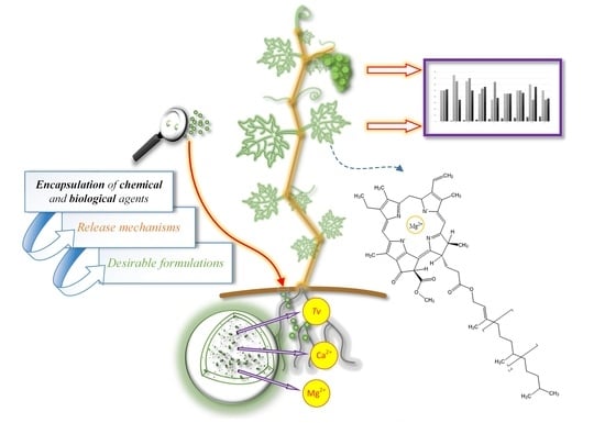
Tailoring Alginate/Chitosan Microparticles Loaded with Chemical and Biological Agents for Agricultural Application and Production of Value-Added Foods

1
Department of Chemistry, Faculty of Agriculture, University of Zagreb, Svetošimunska Cesta 25, 10000 Zagreb, Croatia
2
Department of Pharmacognosy, Faculty of Pharmacy and Biochemistry, University of Zagreb, Ante Kovačića 1, 10000 Zagreb, Croatia
3
Laboratory for Sustainable Development, Faculty of Food Technology and Biotechnology, University of Zagreb, Pierottijeva Ulica 6, 10000 Zagreb, Croatia
*
Author to whom correspondence should be addressed.
Submission received: 26 March 2021 / Revised: 23 April 2021 / Accepted: 28 April 2021 / Published: 29 April 2021

Abstract

:
This work reviews the recent development of biopolymer-based delivery systems for agricultural application. Encapsulation into biopolymer microparticles ensures the protection and targeted delivery of active agents while offering controlled release with higher efficiency and environmental safety for ecological and sustainable plant production. Encapsulation of biological agents provides protection and increases its survivability while providing an environment safe for growth. The application of microparticles loaded with chemical and biological agents presents an innovative way to stimulate plant metabolites synthesis. This enhances plants’ defense against pests and pathogens and results in the production of higher quality food (i.e., higher plant metabolites share). Ionic gelation was presented as a sustainable method in developing biopolymeric microparticles based on the next-generation biopolymers alginate and chitosan. Furthermore, this review highlights the advantages and disadvantages of advanced formulations against conventional ones. The significance of plant metabolites stimulation and their importance in functional food production is also pointed out. This review offers guidelines in developing biopolymeric microparticles loaded with chemical and biological agents and guidelines for the application in plant production, underlining its effect on the plant metabolites synthesis.

Graphical Abstract

1. Introduction

One of the major problems in food production is the overuse of toxic agrochemicals during the production of plants which has a serious negative impact on the environment, food safety, and consequently, on human health. Researchers are increasingly turning to natural ways of treating plants to abandon or at least limit the use of agrochemicals [1]. One of the ways to reduce their overuse is through controlled and targeted delivery of biological (i.e., microorganisms) and chemical agents. Conveniently, controlled, and targeted delivery can be achieved via the encapsulation method, and this has proven to be a suitable way of delivering nutrients for organic and sustainable crop production. Encapsulation into microparticles is an advanced, promising, and fast-developing technology that has significant advantages over other agroformulations in terms of protecting the living organisms from external conditions and the possibility of a higher survival rate. The main benefits of biological and chemical agents’ encapsulation include sustained and controlled release, greater efficiency, and relatively beneficial impact on the environment [2].
As a result of consumer needs and wishes, the production of high-quality, safe, and functional food is becoming increasingly popular. The concept of functional foods refers to foods with a relatively higher content of biologically active compounds, which are thought to have a beneficial effect on human health, reduce the risk of some diseases, and they can even stimulate faster healing of tissues. Although functional foods are not fully defined, the scientific literature considers that these products provide additional benefits beyond the general benefits of nutritional intake and the pure need to satisfy hunger [3,4].
Plant secondary metabolites (PSM) are biologically active compounds often used in healthy nutrition, traditional medicine, and in a wide array of industrial applications [5,6]. Plant secondary metabolites, such as polyphenols, are composed of several groups of structurally distinct natural compounds biogenetically obtained by shikimate-phenylpropanoid-flavonoid biochemical pathways. Plants require these metabolites for pigmentation, growth, reproduction, resistance to pathogens, and many other biochemical processes and functions, while these metabolites represent adaptive characteristics that underwent natural selection during evolution. The effective defense mechanism of plants can be attributed to the wide range of PSM they synthesize [7]. With a wide range of different PSMs, plants can respond to different stressors. Given that the production of specific plant protection features can be extremely costly, new ways of enhancing defense need to be investigated and exploited. Methods involving increased expression of endogenous compounds (i.e., PSM) can significantly affect plant characteristics for resistance to invaders [7]. A high proportion of PSM can also have an important impact on human nutrition and health, by increasing the intake of antioxidants and nutrients [8]. Therefore, not only would the increase of biologically active compounds during plant cultivation have benefits for human consumption, but their primary role would be to increase the plant’s defense mechanisms against pests and pathogens. Furthermore, with significant interest in the increased production of PSM, obtaining high yields can be ideal for commercial exploitation (e.g., functional ingredients). Various strategies, such as screening and selecting high-performance cell lines, cell cultures from different parts of plants, metabolic engineering, media optimization, plant growth regulators, and others, have been used so far to increase PSM production in plant cells [9], but most of these strategies are very expensive and inefficient.
Living microorganisms (e.g., nematodes, bacteria, and fungi) can be applied to the seed, surface of the plant, or soil to colonize the rhizosphere and the interior of the plant to stimulate its growth and production of PSM by increasing the supply and availability of nutrients [10]. Specifically, some studies show that mycorrhizal inoculation can increase PSM content, such as polyphenols, and increase the antioxidant activity in plants [11], but effective formulations require a carrier material that must retain mycorrhizal functional properties after the administration. One way to protect and achieve targeted delivery of microorganisms to a plant is via the encapsulation method. Microorganisms, such as the fungus of the genus Trichoderma, participate in the degradation of plant residues in the soil and act as biocontrol agents against plant pathogens. Trichoderma species synthesize specific compounds and metabolites, such as hydrolytic enzymes, plant growth promoters, antibiotics, siderophores, carbon, and nitrogen permeases. Trichoderma spp., among other things, stimulates plant growth by dissolving otherwise insoluble mineral nutrients, such as calcium, iron, or aluminum phosphates [12]. Strong aggressiveness against plant pathogens and high efficacy in promoting plant growth and defense mechanism have made Trichoderma species an important biocontrol agent [13]. However, biological agents are significantly influenced by detrimental external factors, such as pH, humidity, and ultraviolet radiation, which all impair their action. With encapsulation, a protective barrier around the biological control organism is provided, preserving its activity [14,15].
Compatibility of Trichoderma viride spores with divalent ions, like Cu2+, Ca2+, Mg2+ makes it pragmatic for simultaneous encapsulation into microparticles, that is, simultaneous delivery of chemical and biological agents to a plant. Calcium ions are essential macronutrients, and they have an important function in cell membrane structure and permeability, plant cell division and elongation, carbohydrate translocation, and nitrogen metabolism [16,17]. Ca2+ plays a regulatory role in signal transduction and absorption of nutrients through the cell membrane [17,18,19]. Ca2+ also signals the regulation of genes responsible for polyphenol biosynthesis [20], and binds to the phospholipid membrane, stabilizing the lipid bilayer and maintaining the structure [21]. Furthermore, it was found that in Ca2+-treated plants, malondialdehyde content decreases [18,22,23]. Although the soil is known to be rich in calcium, plants often lack calcium, due to their form and relative insolubility (e.g., CaCO3). In addition to Ca2+, copper ions (Cu2+), as well as magnesium ions (Mg2+), also show a stimulating effect on PSM synthesis. They can stimulate PSM synthesis with a positive effect on alkaloid production, shikonin synthesis [24], digitalin production [25], and betaine [26]. Magnesium ion’s primary role in plants relates to photosynthesis. Mg2+ is an integral part of chlorophyll; it activates some enzymatic processes required for plant growth [27], participates in synthesizing DNA and RNA molecules [28], and is utilized in the plant’s cellular energy source—ATP [27]. Cu2+ plays key roles in photosynthetic and respiratory electron transport chains, on ethylene sensing, cell wall metabolism, oxidative stress protection, and biogenesis of the molybdenum cofactor [29]. Although Cu2+ is an integral part of growth media and is known to be essential for several biochemical and physiological pathways [30], it becomes relatively toxic at high concentrations [31]. Therefore, it is important to control the dose through plant growth and development to minimize excess release into the environment.
Successful delivery of the precisely controlled active substances to the right place and at the right time is a desirable characteristic of all active agent delivery systems, which may aid in precision agriculture [32]. To obtain suitable microparticles (delivery system), effective for simultaneous encapsulation of multiple active agents with appropriate controlled release, it is important to optimize the parameters during their preparation. Suitable selection of formulation variables assists in the design of microparticles with the desired release of biological and chemical for plant nutrition/protection [33,34]. This review involves the procedures of preparation and application of microparticles for the strategic delivery of biological and chemical agents, to make it available to the plant throughout its growth period. Not only to increase the proportion of PSM to protect the plant from predators and pathogens, but also, consequently, to obtain higher quality food with added value, i.e., functional food or a source of functional compounds. Also, the consumption of foods with an increased proportion of these compounds can have a beneficial effect on human health.
This review aims to point to the guidelines for developing biodegradable microparticles simultaneously loaded with a biological agent, specifically a plant growth promoter, Trichoderma viride, and chemical agents (Cu2+, Ca2+, Mg2+). The development, characterization, and optimization of the preparation process are necessary to produce this biotechnological product (microparticles) for plant nutrition/protection in conventional/hydroponic cultivation. Finally, guidelines and tips on the application of biopolymer-based microparticles are highlighted. Recommendations aim to help scientists and the agri-food sector develop procedures and application of microparticles for various purposes. Furthermore, the direction of future research is highlighted. The general discussion of this review gives valuable information on the whole process, from the compatibility of a chemical and biological agent, through the microparticles development and characterization, ending by applying microparticles on model plants with resulting outcomes.

2. Biopolymeric Microparticles—Carrier System Composed of Alginate and Chitosan

Nowadays, research is more focused on developing natural products like, for example, natural carriers for different active agents. The most used biodegradable polymers in the process of producing microparticles are next-generation biopolymers sodium alginate and chitosan. Biodegradable polymers are used to prepare microparticles and are of main interest because they are safe for the environment and are generally non-toxic [35]. Encapsulation in biopolymeric material offers stability to the encapsulated material and can offer a controlled and targeted release of the latter. In the literature, encapsulated material is often labeled as filler, fill, core material, or internal phase, while the material used for encapsulation is called matrix, coating, shell, or external phase. Biopolymeric microparticles are usually spherical, but may have some deviations. The appearance of microparticles varies in size, shape, and composition mainly because of the influence of encapsulated material, biopolymer, and the method used for its preparation [36]. Many microparticle classifications can be found in the literature [37,38,39,40,41,42], while here in Figure 1, a representation of microparticles relevant to this review is presented wherein matrix microsphere, an encapsulated material is homogenized through the biopolymeric matrix (including surface), while shell-matrix particles combine the features of both the matrix and shell materials, where encapsulated material is not found on the particle surface, respectively.
Biopolymeric microparticles have an advantage, due to their controlled and targeted release, but some disadvantages exist, like (i) particle-particle aggregation, (ii) limitation of the storage stability, (iii) difficulty to encapsulate molecules with different degrees of hydrophilicity at the same time, (iv) challenging precise control of the dispersity of the particles. The above-mentioned disadvantages can negatively influence the efficiency of active agent delivery [43]. Even though, biopolymers are considered as great materials to use in the production of microparticles, mainly due to their abundance and affordability alongside their stability and durability throughout the process of encapsulation. Biopolymers can be easily extracted from natural sources or may be prepared with the use of microorganisms. Furthermore, biopolymers can be prepared synthetically with precision and predetermined properties as specific molecular weight, solubility, and permeability [44]. Biopolymers may vary in composition and physicochemical properties, and because of that, their utilization is often dependent exclusively on them. To achieve successful encapsulation, it is necessary to understand the structure of used biopolymers.
Sodium alginate is negatively charged, due to the presence of carboxyl groups from the uronic acid residues [45]. It is composed of two repeating carboxylated monosaccharide units (mannuronic and guluronic acids), and the ratio between them influences the properties of the biopolymer. Chitosan is a partially deacetylated polymer of N-acetylglucosamine obtained after alkaline deacetylation of chitin. The N-deacetylation is seldom complete, and the fraction of N-acetylglucosamine determines the degree of acetylation, which serves as a base to classify the biopolymer either as chitin or chitosan. When the degree of N-acetylation (defined as the average number of N-acetylglucosamine units per 100 monomers expressed as a percentage) is below 50%, chitin becomes soluble in aqueous acidic solutions (pH < 6.0) and is called chitosan [46]. The electrostatic attraction between the cationic amino groups of chitosan and the carboxylic groups of the alginate leads to the formation of the polyelectrolyte complexes of various structures. By controlling the degree of association among the functional groups, the structure and physicochemical properties of these complexes may be adjusted [47]. Immobilization and the sustained release of encapsulated agents are achievable by applying chitosan and alginate complexes in the micro/nanocapsule form [1,45,48,49].

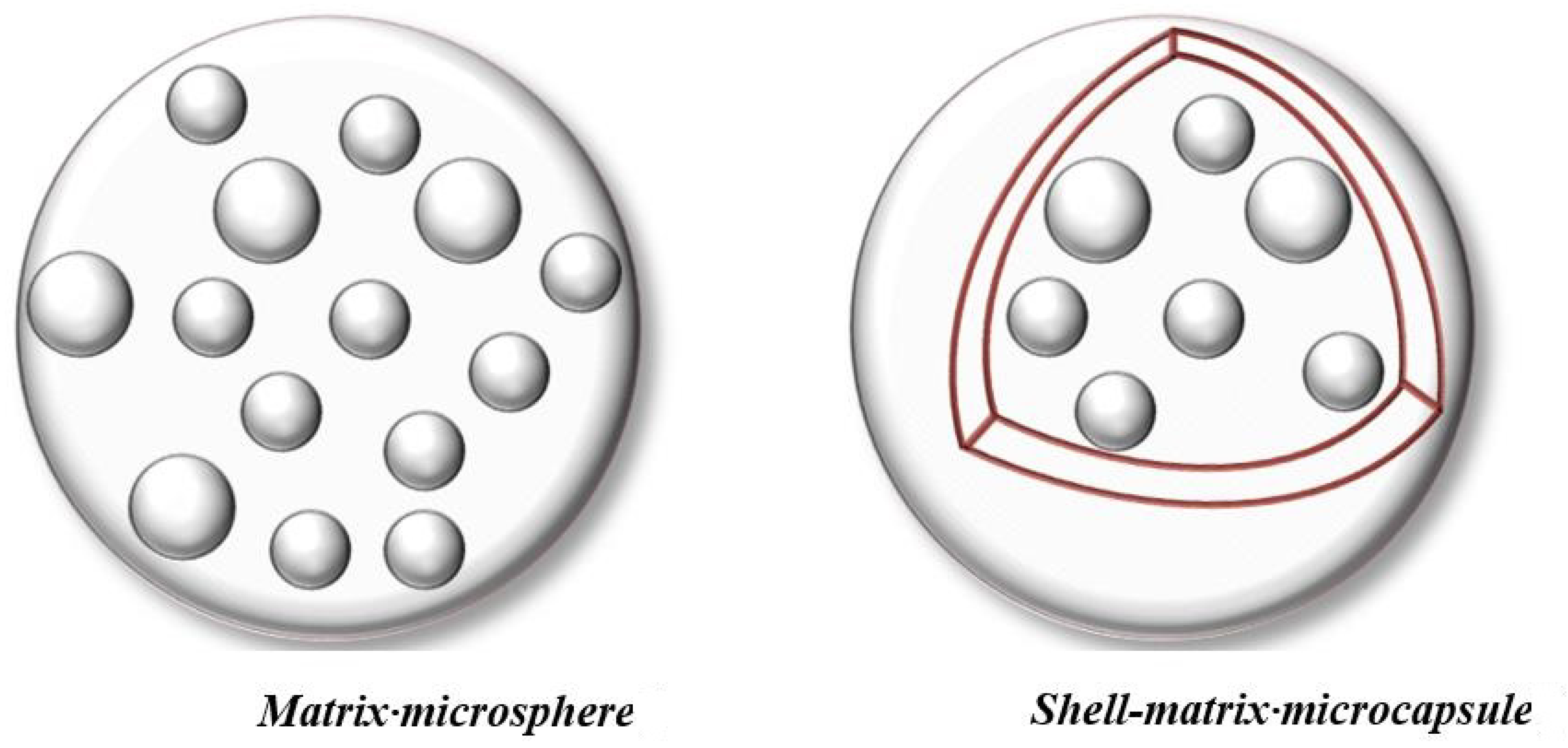
3. A Cost-Efficient Method to Produce Microparticles—Ionic Gelation

Plenty of different techniques may be used for encapsulation in biopolymeric matrices. Hudson and Margaritis [50] comprehensively reviewed biopolymeric particles production, and divided the methods as: (i) Ionotropic gelation or external gelation, (ii) emulsification and internal gelation, (iii) the reverse microemulsion technique, (iv) emulsion crosslinking method, (v) emulsion-solvent extraction, (vi) the emulsification solvent diffusion method, (vii) emulsion-droplet coalescence method, (viii) complex coacervation, (ix) reverse micellar method, (x) self-assembly methods, (xi) water-in-oil emulsification, (xii) desolvation process, (xiii) pH coacervation method, (xiv) emulsification, (xv) nanoparticle albumin-bound (nab) technology, (xvi) self-assembly, (xvii) desolvation method, (xviii) methods involving hydrophobized pullulan derivatives, (xix) reverse micelle synthesis method, and (xx) emulsification-diafiltration. Each of the mentioned methods has its advantages and disadvantages, depending on the targeted application, but one of the most popular and widely used encapsulation methods is ionic gelation.
The ionic gelation method is often utilized in the production of biopolymeric microparticles mainly because it uses mild conditions throughout the encapsulation process and economical production costs [51,52]. Sodium alginate is a common biopolymer used to prepare biodegradable microparticles and the encapsulation procedure, and developed carrier mainly depend on its properties. Alginate-based microparticles are obtained by the dropwise addition of alginate in the bath containing divalent cations. The affinity of sodium alginate for divalent cations mainly depends on its composition. Guluronic acid-based alginate will be more prone to ion binding compared to mannuronic acid-based alginate. The affinity for the metals follows order: Mg2+ < Mn2+ < Ca2+ < Sr2+ < Ba2+ < Cu2+ < Pb2+ [53,54,55]. These cations diffuse in sodium alginate solution forming a gel matrix, due to the cation-binding crosslinks with alginate. Crosslinking density is mainly determined as per the concentration of the cation solution [56]. The binding of the cations is related to the precise chelation process, depending on the distribution of guluronic acid blocks. This has been previously explained with the so-called “egg-box” model. The model is based on the steric configuration of the guluronic acid blocks residues [57,58]. This model describes the gel formation via sodium cation swap with calcium cation from two adjacent guluronic acid blocks and forming a single ion bridge between the chains. Ca2+ ions hold the alginate chains together, and with more bonds, the nature of biopolymer binds more Ca2+ in a stable form. Guluronic groups of alginate correctly distance coordination of Ca2+ between carboxyl and hydroxyl groups. This behavior is ascribed to the self-cooperative process between neighboring elements (Ising model) and is based on a physical bond with unfavorable entropy for the first divalent ion. The bond is favored for all ions to form a one-dimensional “egg-box” (a zipping mechanism) [56]. Kinetics of gelling kinetics are fast and adaptable, but also depend on polymer and cation type and their respective concentrations. With the use of a microdroplet generator (Figure 2) dripping in cation solution, microparticles can be easily prepared. A Microdroplet generator is used for the dropwise addition of sodium alginate into the bath containing divalent cations, where gelling occurs, and spheres are formed (more detail on the process can be found in Section 7.2). Conveniently, Ca2+ has been widely used as a gelling cation, since it is chemical versatile and safe [56,59].
Generally, sodium alginate produces rigid, but relatively porous hydrogels with weak physical and mechanical properties, which are important in delivering active agents [51,60]. Chitosan utilization is limited, due to its limited chain flexibility and poor mechanical strength, but its application has high prospects when coupled with other biopolymers [61]. Properties of these biopolymers (polyelectrolytes) may be amended by combining them, thus, improving their chemical stability and achieving microparticles with improved controlled release of encapsulated material [62]. The coating of alginate microspheres can be achieved with chitosan via polyelectrolyte complexation. The polyelectrolyte complex is dependent on the electrostatic interactions between the two oppositely charged biopolymers. It has to be noted that research on biopolymeric microparticle production is always improving and is directed in the improvement of physicochemical, functional, and release properties of used matrices keeping in mind cost-effectiveness and use of environmentally friendly or “green” material [63].

4. Conventional Formulations vs. Advanced Formulations—A Necessity for Advanced Carrier Systems

Conventional biofertilizers are used in plant applications as powder, liquid, or granulated forms [2]. Inoculants are generally commercialized in solid and liquid forms—while liquid forms are applied to the seeds, granular inoculants are applied to the soil [64]. There are limitations in the use of conventionally immobilized biological agents for agricultural purposes. Unfortunately, conventional formulations encounter several problems like the low viability of microorganisms during storage and field applications, mainly due to the temperature changes and negative impact of other environmental factors [65]. For example, live Trichoderma spores must be stored under refrigeration and applied at temperatures below 28 °C. The field applications must be performed under specific conditions as high relative humidity, and one of the biggest problems is ultraviolet light, which is harmful to the species [38,66].
In the past, seed coated peat-based inoculants were the most popular commercially available inoculants on the market because of the positive effect of peat on storage and application. A significant problem of peat-based products is their negative environmental impact. Even though peat is a good carrier for microorganisms, interests in developing new formulations are of main interest [67]. Liquid inoculants are more approachable, due to the simpler production process, with relatively easy application. Again, there are also problems with liquid inoculants because microorganisms’ survival is inferior, due to the lack of carrier protection [68].
Alternative formulations are always developing, mainly to achieve the advantage of long shelf life and easy transportation while reducing the overall cost of preparation. Encapsulation of microorganisms into biopolymer-based particles mainly tries to enhance efficiency, achieve effective storage and transportation. Encapsulated microorganisms have enhanced survival and controlled release with prolonged effect with the higher performance during storage and applications [2]. Increasing the bioavailability of biological control agents in the field is also possible with the use of encapsulation into biopolymeric microparticles. Especially, since biopolymeric microparticles are high in water content, instantly resolving the problem with the necessity of high relative humidity. Advanced carrier systems would be made via the simple and economic process, using biodegradable and safe material for delivering more than one active ingredient (chemical/biological), with tunable release properties and greater shelf-life compared to the conventional formulations.
Biopolymeric microparticles can engulf microorganisms and protect them with their predefined and fixed microenvironment for the cells’ survival. Thus, the cell metabolic activity can be maintained over an extended period. Except for serving as an energy source for the microorganisms, due to its deterioration, biopolymeric microcapsules allow the controlled release of microorganisms [65,69]. It must be noted that when choosing a relevant biopolymer, its characteristics need to be considered to achieve successful biological control. Important physicochemical parameters to consider when using a biopolymer are molecular weight and distribution, degree of substitution, opposite ions, gelation mechanism, pH, viscoelasticity, primary, secondary, and tertiary structures. Of course, economic factors as cost should always be considered. Safety, quality, and source of biopolymers are also important factors to be considered, as well as degradability and approval from relevant agencies (European Union Commission Regulation, Food and Drug Administration, etc.) [38].
What modern encapsulation science makes advanced is that it considers materials that can add function to the particles while being more than just a carrier, respectively, to their characteristics. Encapsulation science has reached highly sophisticated systems, and still, the gold standard in biological agents’ encapsulation remains the calcium alginate particles. The next step is to formulate several active ingredients in one formulation, such as a biological agent and a chemical agent, enhancing the efficiency of encapsulated material and having a synergistic effect on the plant [38]. According to John et al. [2], it is expected that microencapsulated formulations will dominate marked for ecological and efficient agricultural production, providing all necessary protection of encapsulated agents, while reducing the negative effect of non-degradable conventional chemical pollutants on the environment.

5. Plant Metabolites (Biologically Active Compounds) and Their Importance in Functional Food Production

Plants produce many metabolites of diversified structures and abundance, which play crucial roles in plant growth, development, and response to environments. These metabolites are also valuable nutrition and energy sources for human beings and live stocks [70,71]. Plant metabolites are generally classified into primary and secondary. Plant primary metabolites are indispensable for plant growth and development. But plant secondary metabolites are crucial for plant survival under various environmental conditions. They are responsible for maintaining a delicate balance with the environment so plants can survive under stressful conditions [72]. Plant secondary metabolites also possess biocidal properties making them crucial in the control of various pests and pathogens [73].
Polyphenolic compounds have a photoprotective function, structural support, and protection against attackers, and other significant roles crucial to the plant’s metabolism and survival [74,75]. They are also responsible for the pigmentation and some of the organoleptic characteristics, such as flavor and color [76,77]. Numerous studies confirm the benefits of polyphenolic compound ingestion on human health [78,79]. Antioxidant activity is one of the most important properties of these compounds, since they are efficient proton donors. By accepting electrons, polyphenolic groups form stable radicals [73].
When discussing the content of pigments in plants, such as carotenoids and chlorophylls, their concentration gives us valuable information on the level of stress a plant is experiencing, as well as the extension of stress endurance [80]. The health-beneficial properties of carotenoids are well known, and they are claimed to prevent human diseases like cancer [81,82,83], osteoporosis, neurodegenerative disorders [84], lung diseases [85], cardiovascular diseases, and age-related disorders [86]. Some of the carotenoids are a dietary source of vitamin A which is involved in the regulation of hormone synthesis, immune system, as well as growth and differentiation of skin cells [87,88]. More details on the health benefits of different carotenoids can be found in a review paper by Jurić et al. [89]. Chlorophyll has a similar structure to hemoglobin and may regenerate or act as a substitution for hemoglobin in hemoglobin deficiency conditions. It is important to mention some of the known health benefits of higher chlorophyll intake. Chlorophylls are important for many biological functions, both in plants and animals. They have a significant influence on human health because they are involved in maintaining healthy gut microbiota [90], and they exhibit antioxidant and antimicrobial properties [91]. Some studies revealed that chlorophylls act antimutagenic [92] and antigenotoxic [93], while some studies reveal in vivo [94] and in vitro [95] chemo-preventive effects in humans [96]. The use of chlorophyll-rich juices is recommended for the treatment of thalassemia and hemolytic anemia [97], and has important anticancer functions [98,99]. The use of natural pigments like carotenoids and chlorophylls in food production is becoming increasingly popular. Primarily, due to the consumer’s concerns about the health safety of synthetic food colors. The utilization of natural pigments in foods is gathering more attention from the food industry, and part of that is because they present significant health benefits [100,101].
Plant metabolites are often utilized to increase the functionality of foods [102,103]. The term functional foods refer to foods with a relatively higher content of biologically active compounds, which are thought to exert some benefit upon health. Even though the term for functional foods is not defined, scientists still consider that products with a relatively higher content of bioactive compounds do provide additional benefits to human health [3,4]. Also, some plant metabolites are utilized as colorings in foods. European Union approves carotenoids as food colors with E-numbers E160 and E161 (European Union regulation (EC) No. 1333/2008 and its amendments, European Regulation 1169/2011) [104]. European legislation also permits the use of chlorophyll derivatives in foods, which are labeled E140 and E141 [105].
In the future, plant metabolites will have even greater value, mainly because they can help in the prevention of some chronic diseases and because the general population is starting to grow a health consciousness.

6. Significance of Chemical and Biological Agents Encapsulation in Sustainable Agriculture

When aiming to achieve a high-quality crop, balanced nutrition, and fertilization are essential. Chemical agents (i.e., Ca2+, Cu2+, Mg2+) are essential plant nutrients and play important roles in many biochemical pathways. They are essential in the structure and permeability of cell membranes, plant cell division, and elongation. They act as signaling ions in the regulation of biochemical cascades [17]. For example, Ca2+ has a signaling role in the upregulation of genes responsible for synthesizing polyphenolic compounds [20]. On the other hand, Ca2+ is often found in soil, but the biggest problem is, its prevalent form is relatively insoluble, like CaCO3. Cu2+ also show a stimulating effect on the production of plant secondary metabolites and is essential for some biochemical and physiological pathways, but the problem is that at higher concentration becomes toxic [31]. Mg2+ is tightly connected to the photosynthesis process as it is the central atom of the chlorophyll molecule. Mg2+ becomes extremely phytotoxic when concentrations of Ca2+ in plants are low [106,107] because Ca2+ ions control and modify the uptake of available Mg2+ [108]. It is important to develop products that will have nutrients in bioavailable form. Furthermore, exact concentrations for relatively toxic agents should be regulated more carefully. Thus, control of the dosage to minimize its release into the environment is crucial. Encapsulation into biopolymeric microparticles is one of the ways which can mend these problems.
Trichoderma viride (Figure 3) is a biological control agent used in agriculture as biostimulants and against phytopathogens. Generally, Trichoderma spp. are used in agriculture because of their ability to produce a relatively high quantity of specific enzymes [109], and secondary metabolites to control phytopathogens [110]. Mycoparasitic Trichoderma spp. produce antimicrobial compounds [111], has antibiosis activity, and induces resistance of the host plant [111,112]. As mentioned above, Trichoderma spp. produces hydrolytic enzyme complex, which is made of chitinases, β-glucanases, cellulases, and proteases. These enzymes, especially chitinases, which are the most effective, help in the decomposition of the cell walls of phytopathogens, allowing Trichoderma spp. hyphae penetration, colonization, and mycoparasitic activity to occur [110,113,114]. Trichoderma species are also known to acidify their surrounding environment via the secretion of organic acids and can solubilize some chemical agents [115].
Recently authors [110] reported the protection of encapsulated fungus Trichoderma harzianum against ultraviolet radiation. Furthermore, encapsulation improved chitinolytic and cellulosic activity of the fungus and greater control of Sclerotinia sclerotiorum (white mold), a well-known phytopathogen. Vejan et al. [69] report a simple and innovative way to encapsulate Bacillus salmalaya, a plant growth-promoting rhizobacteria. Successful protection of bacteria was achieved with encapsulation in degradable chitosan/alginate microparticles. The bioactivity of bacteria was maintained in samples that were encapsulated. The application of alginate/chitosan-based particles in agriculture is becoming more popular because of their abundance, relatively low price, and because they are rich in amino and hydroxyl groups. For example, chitosan has been successfully utilized for delivering fertilizers, pesticides, and nutrients, and it found its application as an adsorbent in wastewater treatment [61,116,117,118]. Alginate-based microparticles are lately becoming increasingly popular, and in Table 1, the most recent examples of fungal biocontrol agents encapsulation are presented.
It is important to mention that the synergistic action of chemical and biological (biocontrol) agents may improve their activity, and investigation of their relationship is of crucial importance for developing efficient products [119].

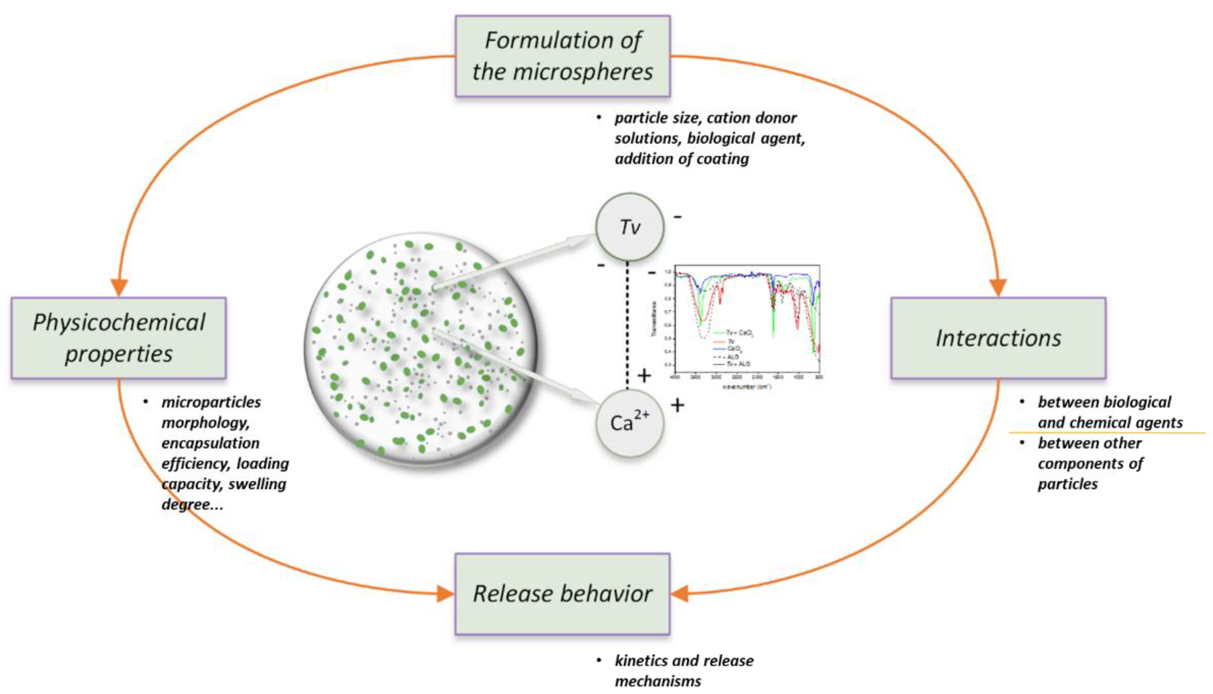
7. Guidelines on Developing Biopolymeric Microparticles

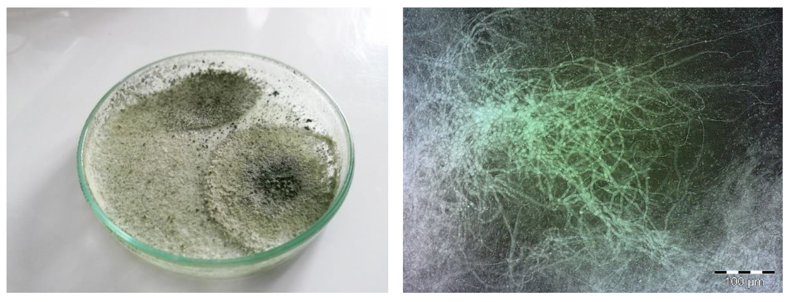
7.1. Effect of Chemical Agents on the Growth and Viability of Biological Agent T. viride

Regarding the encapsulation methods, the focus should be put into adapting already existing methods to produce encapsulated chemical and biological agents. The clever approach in the design of microparticles should include already existing methods and gelation principles to solve some of the main problems regarding the use of biological agents, like shelf life and targeted delivery [38]. The development of guidelines for delivering chemical and biological agents starts before the encapsulation procedure, focusing first on the compatibility of encapsulation material with the material which will be encapsulated.
Throughout this review, we discuss (1) the preparation of biopolymer-based microparticles and (2) the application of tailored microparticles on plant cultures and outcomes. The first part [130,131,132] comprises the preparation of microparticles (microspheres and microcapsules). The development of microparticles loaded both with chemical and biological agents present a challenge to determine conditions of preparation. The focus is, thus, aimed to first observe the influence of each mutual compound used in the encapsulation process. The nature of biological agents is to interact with their environment, thus, molecular interactions with chemical agents, as well as biopolymers used for encapsulation, are important to determine. In the paper of Vinceković et al. [133], it is discussed how Trichoderma viride can survive in the presence of different metals and even can bind them. Vinceković et al. [130] observed that Trichoderma viride survives in the presence of Cu2+, and even at the higher concentrations, the growth was promoted. Encapsulation of more than one component presents a significant problem, mainly because encapsulated material can interact with each other, which may result in a change of its activity and even termination of cells (biological agent). This problem was thoroughly investigated, and it was proved that not only the presence of cations like Cu2+ and Ca2+ inhibits the activity of T. viride, but increases it.
The fungal cell has a very complex chemical composition [134], and is composed of polysaccharides, from which up to 90% fall on glucan and chitin. It is also composed of glycoproteins and lipids as major components and other minor compounds like pigments and inorganic salts. Melanin can be found on the spore wall of Trichoderma species, while chitin was found in mycelial cell walls. Scanning electron images revealed that T. viride spores [131] appear similar in size (around 3.6 μm) and shape to Penicillium [135], and Aspergillus [136] spores. The functional groups detected in the FTIR spectrum relate well with the chemical structure of the cell wall from the literature [133]. The inverse relationship between cell surface electrostatic charge and cell surface hydrophobicity was found, and less hydrophobic cell surfaces were more negatively charged. Detailed information on molecular interactions (FTIR spectra) between Cu2+ or Ca2+ and T. viride spores, as well as between T. viride spores and sodium alginate, is well explained in the papers of Vinceković et al. [52,133] and Jurić et al. [131]. Briefly, the presence of Cu2+ in the alginate matrix causes significant changes in the alginate functional groups region (hydroxyl, ether, and carboxylate). With a higher concentration of Cu2+, intensities of main alginate peaks change and are shifted, implying changes in intensities of hydrogen bonding and Cu2+ interaction with T. viride spores. Respectively, the intensity of hydrogen bonding is weakest with a higher Cu2+ concentration. Similarly, Cu2+ influences the asymmetric and symmetric COO- stretching, indicating complex interactions between all the components necessary to prepare Cu-alginate microparticle. It was further revealed that an increase in Ca2+ concentration causes significant changes in the alginate functional groups region. Regarding the spectrum of T. viride and Ca2+, it is suggested that carboxylate and at least hydroxyl groups are involved in interactions as previously observed for interactions between T. viride and Cu2+. FTIR spectrum of T. viride spores and alginate mixture revealed intermolecular hydrogen bonds. There are indications of at least interactions with hydroxyl, amino, carboxylate, and C-O groups. FTIR spectra of components (gelling cations, sodium alginate, chitosan, and biological agent) can give important information and predictions on how the release from the microparticle will occur. Stronger interactions will slow down the release kinetics, while weaker interactions will have less influence on the diffusion.
In the paper of Vinceković et al. [133], via electron microscopy and cell fractionation studies, it can be observed that Cu2+ is located on the cell wall of T. viride spores, indicating the place of interaction. Zeta potential measurements performed on T. viride spores revealed a negative zeta potential of –35.1 mV, which points to the fact that interactions between Cu2+ are primarily due to the electrostatic attractions. The negative charge of T. viride spores indicates prevailing spore surface hydrophilicity [137]. The negative charge arises from functional groups as carboxyl, hydroxyl, amine, and phosphate [134]. It was found that the increase in copper cations concentration resulted in a less negatively charged spore surface and consequently aggregation [130], thus indicating that electrostatic interactions do occur. FTIR spectrum of T. viride spores and Cu2+ suggest that amine, hydroxyl, carbonyl, and amide bonds are the major sites for cations binding. Cu2+ binding to T. viride is beside electrostatic interactions also associated with possible ion exchange and some physicochemical interactions [133]. Similar to the Cu2+, an increase in Ca2+ concentration results in less negatively charged T. viride spores, due to the electrostatic binding of calcium cations [131]. Also, the diminishing of electrostatic repulsions between spores led to their aggregation. With higher concentrations of Ca2+, a reversal of charge occurs, which suggests that besides the electrostatic interactions, some other mechanisms, like in the case of Cu2+ ions, are responsible for the binding. This is probably due to the ion exchange and/or other physicochemical reactions between cations and functional groups found in the cell walls. Our preliminary tests with Mg2+ and Zn2+ ions reveal the same behavior when introducing cations to the suspension with T. viride spores. Furthermore, some of our tests with the spraying of different solutions containing Cu2+, Ca2+, Mg2+, or Zn2+ revealed a significant increase in the sporulation of T. viride. Also, the presence of Ca2+ in lower concentration does not significantly change aggregate size [131], and an increase in concentration resulted in a less negatively charged spore surface (based on the average zeta potential and the mean hydrodynamic diameter). The diminishing of electrostatic repulsions between spores led to the creation of bigger aggregates. Compared to the Cu2+, the presence of higher concentrations of Ca2+ results in somewhat slower sporulation, but still, it was not prevented, and results are dose-dependent. These results may aid predict kinetics and release mechanisms of encapsulated agents from various microparticles [131].

7.2. Recommended Guidelines to Prepare Microparticles Loaded with Chemical and Biological Agents

This section brings some important aspects to prepare microparticles, not only for agroindustrial purposes, but also for other fields as food engineering or pharmaceutical science. Sodium alginate is used as a carrier biopolymer, and when a solution of the latter encounters divalent or trivalent cations, a rapid, strong, and irreversible formation of the gel matrix occurs, respectively. Residues from sodium alginate blocks react cooperatively with cations forming a gel network. Because of the rapid and irreversible reaction, controlled conditions of preparation should be achieved [138,139]. The procedure of microparticle preparation vary, and the essential need is to achieve high encapsulation efficiency, loading capacity, alongside chemical stability. It is also important to achieve good swelling properties, surface, and core morphology, all of which influence the controlled release of encapsulated agents (Figure 4). Nevertheless, with regards to the preparation of biopolymer-based microparticles, universal microparticles for any purpose do not exist. It is important to tailor microparticles based on the intended use, and therefore, appropriate procedures need to be developed and applied.
To achieve desirable microparticle efficiency for the prolonged release of chemical and biological agents, it is important to choose sodium alginate with a higher content of guluronic acid residues. High guluronic acid content results in more rigid and porous gels and can maintain their integrity for longer periods. One of the most important things to consider when tailoring microparticles is choosing a proper concentration of gelling cation alongside a used biopolymer [133]. The ratio between gelling cations and sodium alginate determines the kinetics of gelation, and in the end, physicochemical characteristics of the formed gel matrix [140]. Research reveals that the decrease in sodium alginate concentration increases mechanical stability, but when the concentration is too low, mechanical stability starts to decrease [138,140,141]. The sodium alginate solution is viscous, depending on the concentration. The minimum viscosity of sodium alginate solution to form microspheres via the ionic gelation method is 0.03 Pa·s [142]. Spherical microparticles may be obtained over a wide range of viscosity of the latter solution. Usually, the viscosity of aqueous sodium alginate solution does not go over 1 Pa·s [133]. Microparticle size mainly depends on the polymer concentration. An increase in the particle size correlates with the higher initial concentrations of biopolymers, because of the functional groups’ proportions. It should be noted that encapsulation efficiency decreases with an increase in polymer concentration, which ends with less space for encapsulated material [46]. The concentration of both sodium alginate and gelling cation significantly affects cation binding and kinetics of gel matrix formation.
This review discusses the impact of Cu2+ or Ca2+ cations in various concentrations on the physicochemical characteristics of formed microparticles with regards to the constant concentration of sodium alginate (1.5% w/v). Respectively, the preparation procedure of microparticles consists of two steps. The first step being ionic gelation, where sodium alginate interacts with cation to form a gel network—microspheres, and the second step involving the additional coating of microspheres with chitosan via polyelectrolyte complexation [143].
The process of preparation starts with the addition of a biological agent (T. viride) to the sodium alginate solution and homogenization of the suspension. Filtration through the muslin cloth is recommended to filter out only spores (to not clog the machine used in the process of preparation—mentioned later in the discussion). The first stage involves the dropwise addition of sodium alginate either with or without biological agent into the bath containing cations (Cu2+, Ca2+, or a mixture of various chemical agents, e.g., Ca2+/Mg2+, since reaction with Mg2+, does not occur instantly as with the above-mentioned cations. It is recommended to combine Mg2+ with other divalent cations that react fast and form a strong gel, like Cu2+, Ca2+, or Zn2+. Since donor solutions always contain anions, e.g., CuSO4x5H2O or CaCl2, the inclusion of the anion into the microparticles also occurs. During the encapsulation process, anions diffuse through the microparticle into the solution, and thus, the encapsulation efficiency for anions is significantly lesser when comparing to the cations involved in the gelling process, which binds irreversibly. After the dropwise addition of sodium alginate into the bath containing divalent cations, gelling occurs, and spheres are formed. Throughout the preparation process, the dropwise addition of carrier solution (sodium alginate) can be achieved using a microdroplet generator (Figure 2). Concerning the size of the nozzle, used for the dropwise addition, the pressure, frequency of vibration (amplitude), and temperature should be considered. Higher temperatures decrease the viscosity of sodium alginate solutions, and smaller spheres may be formed via the usage of nozzles with a small diameter (e.g., 80 µm). When using the bigger nozzle (1000 µm), lower frequency is necessary to separate the droplets in dropwise flow, while the opposite is valid for smaller nozzles. Of course, a smaller nozzle means the need for higher pressure to eject already highly viscosity solution like sodium alginate.
Figure 5 shows a schematic representation of the preparation procedure. As can be seen, in the first stage, a simple method can be used for the preparation of microspheres (gel matrix), while for the second stage of preparation, coating of microspheres, i.e., microcapsules, with chitosan, can be observed. Via the polyelectrolyte complexation, microspheres are dispersed in the acidic chitosan solution, where chitosan rapidly binds onto the surface of alginate-based microspheres by electrostatic interactions between protonated amino groups on chitosan and ionized carboxylic groups on alginate-based microspheres [144]. The main goal of the second step is to reduce the porosity of the gel matrix, improve its stability and efficiency by delaying the release of encapsulated agents. It is well documented in the literature that electrostatic interaction between chitosan and alginate tightens and stabilizes the surface of the microparticle [1,145]. When chitosan binds to the microsphere, competing ions like H+ or Na+ have an insignificant influence on the stability of the polyelectrolyte complex [146], and chitosan diffusion to the inner part of microparticles is very limited [147]. This can be observed in a research paper of Vinceković et al. [133] and Jurić et al. [132] where a distinct chitosan layer was formed on the surface of the microspheres, i.e., microcapsule.

7.3. Characterization of Microparticles Loaded with Chemical and Biological Agents

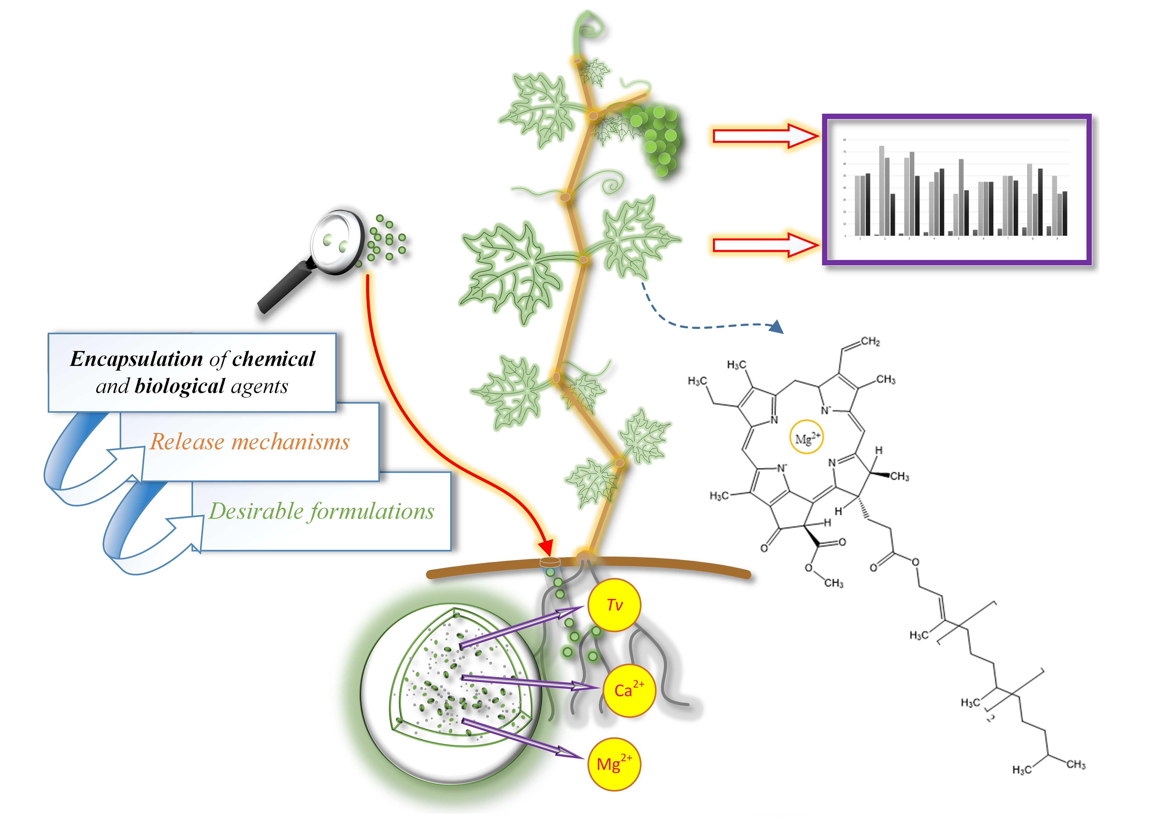
7.3.1. Morphological Characteristics—Microscopical Observations

After the suggested preliminary tests necessary to observe basic interactions between encapsulated material and material which will be used for encapsulation, and after the proper selection of the encapsulation procedure, characterization of microparticles may commence.
Primarily, to observe the success of encapsulation, microscopic observations give valuable information. Not only about the size of microparticles, but the surface texture, proof of chitosan binding, the thickness of the membrane and can even reveal the presence of biological agents either on the surface or in the core. For example, basic optical microscopic observations can also reveal the percentage of shrinkage of microparticles after drying to the constant mass, as well as the collapse of the matrix and even loss of sphericity [133]. In the paper of Jurić et al. [132], a decrease in the size of the microcapsule can be observed with the increase in the Ca2+ ions used in the encapsulation procedure. This is in correlation with other literature data, which revealed the formation of smaller particles with higher Ca2+ concentration, due to the lower percentage of water retention and formation of tougher gel matrix [148].
Confocal laser scanning microscopy (CLSM) may reveal interesting data with regards to the observed microparticle. Vinceković et al. [133] reveal data on chitosan binding over Cu-alginate microparticles. Microphotographs were taken under CLSM in fluorescence mode and transmitted light mode, which clearly shows the existence of the chitosan layer (stained with 0.01% Eosin Y, xanthene dye) on the surface of the microparticle. Eosin dye binds to the amino groups of chitosan and is, therefore, possible to observe the thickness of the coating layer. In this case, the thickness was about 7 μm in the case of the microparticles loaded with only a chemical agent (Cu2+), and in the case of microparticles loaded with Cu2+ and T. viride spores, a layer was about 11 μm thick. The different size in layer thickness over microparticles without T. viride spores was explained as a lower extent of reaction between chitosan and alginate matrix. Electrostatic interactions between protonated amino groups on chitosan and ionized carboxylic acid groups on alginate are enhanced, due to the binding of T. viride on the matrix. Similarly, in the paper of Jurić et al. [132], a clear existence of a thin chitosan layer on the surface of Ca-alginate microparticles was observed.
Optical microscopy and CLSM may reveal data on microparticle size, general morphology, presence of biological agent (even protruding of hyphae from the microparticles) or chitosan layer, but scanning electron microscope (SEM) can shed light on even more details. In the paper of Jurić et al. [131], irregular wrinkles can be observed on dried microspheres as a consequence of biopolymer strain-relaxation processes, which are generally associated with water/humidity loss. Furthermore, SEM images can reveal pores and anomalies on the surfaces. For example, in the paper of Jurić et al. [131], a highly porous surface was observed (size of the pore is about 0.169 μm), as well as numerous spherical blebs (T. viride spores) which are visible as entangled into the matrix of a microsphere. Furthermore, this was confirmed in the publication of Jurić et al. [132], where the surface of microspheres without T. viride spores is highly porous, and where numerous blebs are found on microspheres prepared with T. viride spores. The microcapsule has fibrous and striped surfaces because of the presence of the chitosan layer, and reduced porosity on the surface was confirmed. Microcapsules with T. viride spores have a sleeker surface with the appearance of numerous dimples (protruding hyphae). Some microphotographs reveal structures of assembled holes from which germ tubes penetrate out of the microparticles.
Atomic force microscopy (AFM) can give important data on the texture of microparticles. Jurić et al. [132] covered AFM investigation where we can observe the average size and mean diameters of grains on both surfaces and cross-sections of microparticles, as well as the surface roughness. Results from AFM confirm the change in morphology with a change in the composition of microparticles. Highly porous morphology is detected for microspheres with grainy surface texture and microroughness. Chitosan layer increases surface roughness, but the addition of biological agents (T. viride) reduces the roughness of both microspheres and microcapsules (Figure 6).
The above-mentioned methods for microparticles morphology characterization are important parameters when discussing mechanisms and kinetics of chemical and/or biological agent release. Preparation method, type of active agent, microparticle size, gelling cation type and concentration, additional layers (i.e., chitosan), and the presence of a biological agent significantly affect surface morphology and structure of microparticles and consequently release behavior. All the mentioned factors have a combinatorial effect on the final microparticle properties.

7.3.2. Molecular Interactions between Components in Microparticles

Data on molecular interactions in prepared microparticles can also be obtained using FTIR. FTIR spectra of microparticles prepared with cations of variable concentrations revealed that functional groups of all components interact with each other, and this is well explained in papers by Vinceković et al. [52,131,133] and Jurić et al. [132]. Briefly, complex intermolecular interactions can be seen, which include mainly electrostatic interactions and hydrogen bonds. An increase in the cation concentration increases the crosslinking degree of microparticles, and the presence of T. viride spores diminishes it because of mechanical interactions and electrostatic repulsions between negatively charged T. viride spores and free alginate chains. Herein was reported that changes in FTIR spectra show structural differences in microspheres prepared with and without biological agents. FTIR data on microparticles is in agreement with X-ray diffraction evidence and the proposed so-called “egg-box” model [149]. In the metal-alginate complexes, Papageorgiou et al. [149] proposed a “pseudo bridge” unidentate coordination with intermolecular hydrogen bonds in polyguluronic regions and the bidentate bridging coordination in the polymannuronic region. Both microspheres and microcapsules show that alginate asymmetric and symmetric carboxylate peaks became broader, exhibiting gradual intensity increasing and shifting of carboxylate ions stretching vibrations (asymmetric to a lower and symmetric carboxylate vibrational peak to higher wavenumbers) with increasing cation concentrations. Also, confirmation of chitosan binding to the alginate is presented where some peaks disappear from becoming weaker because of the electrostatic interactions between superposition (two oppositely charged polyelectrolytes) of the functional groups. It is already well known that the (-COO) groups interact with the amino (NH3+) groups of chitosan, forming a polyelectrolyte complex. Changes in the FTIR spectra of microspheres and microcapsules reveal that the presence of the chitosan layer with an increase in cation concentration differently influences the structure of microparticles. FTIR spectra can give interesting information on the behavior of components that are microparticles made from. This information can help predict encapsulated agents release behavior which is of crucial importance when preparing microparticles for various applications.

7.3.3. Encapsulation Efficiency, Loading Capacity, and Swelling Degree

The increase in cation concentration decreases the encapsulation efficiency, but loading with a biological agent (T. viride spores) increases it. This increase is a result of the interaction of negatively charged T. viride with positive ions like Cu2+ or Ca2+. Results for encapsulation efficiency are in correlation with the values of loading capacity [130,131]. Jurić et al. [131], and this data can be related to the investigation of Vinceković et al. [52,133]. The decrease in encapsulation efficiency with higher cation concentration is because maximum loading capacity is reached, so lower concentrations are recommended when preparing microparticles for various applications. If higher values of cations are necessary, this can be achieved with the addition of biological agents. When observing the loading capacity, the presence of T. viride spores decreases free cations, due to the binding, and thus, results may appear somewhat lower. Bespalova et al. [150] also found that T. viride may decrease the amount of Cu2+ bound to organic matter by 1.1—1.2 fold.
Furthermore, regarding the size of microparticles or chitosan presence, respectively, there was no significant effect on changes either on encapsulation efficiency or loading capacity. Some significant decrease in the loading capacity of chitosan-coated microparticles may be observed at lower cation concentrations. This can be explained by the fact that during the addition of the chitosan coat (second step of microparticles preparation), some of the cations from microspheres diffuse into the surrounding solution. Also, it must be noted that the mass of microparticles changes with an additional layer. Using higher cation concentrations during the preparation of microparticles solves this problem. Regarding the encapsulation efficiency of T. viride spores, they are suspended in a viscous solution (sodium alginate) and are dripped in a cation-containing solution. During the formation of spheres, immediate diffusion happens for smaller molecules, but larger particles are being immobilized into the gel matrix. Thus, there were no observed T. viride spores in the filtrate after the encapsulation procedure, revealing the ~100% encapsulation efficiency [133]. The same was noticed in the investigation of Mancera-López et al. [123], where they reveal 100% encapsulation efficiency for Trichoderma harzianum using the same ionic gelation method.
Loading capacity may be used as a method to investigate the storage capabilities of microparticles. Publications of Jurić et al. [131,132] stimulated germination inside of microparticles during the storage period at room temperature. No significant changes in the number of spores were observed after 20 days of storage (based on our experiments even after six months), and this further confirmed that alginate-based microparticles provide an environment that is supportive to the T. viride sporulation. This is in correlation with the literature data (Table 1) where, for example, an investigation of Mancera-López et al. [123] reveals preserved viability of encapsulated Trichoderma spp. of above 70% even after two years of storage. The shelf life of a product is one of the most important factors which is considered when aiming for agricultural application.
When discussing hydrophilic microparticles, one of the most important properties is a swelling degree [151]. When hydrophilic microparticles come in contact with water, they swell, influencing the rate-controlling release mechanism of encapsulated agents. Chitosan-coated microparticles have a higher swelling degree because of the hydrophilic nature of the formed polyelectrolyte complex [1,152,153]. The swelling degree of the complex is associated with electrostatic interactions between functional groups, and since both alginate and chitosan are weak polyelectrolytes, the degree of dissociation depends on the pH of the solution. The swelling degree of the chitosan layer is lower in low pH media because of the strong interactions between protonated amino groups of chitosan and carboxylate anions of alginate. An increase in pH results in more ionization of the functional groups, and close to pH 6.5, chitosan becomes deprotonated, reducing the complexation disrupting the integrity of the structure, allowing a higher swelling degree [154]. In vitro investigation of microcapsules in deionized water (pH ∼6) reveals swelling of the particle and the decrease in pH, due to the ionization of carboxylic groups [145]. Respectively, data from papers of Vinceković et al. [130] and Jurić et al. [131,132] reveal that microparticles prepared at lower cation concentration result in a higher swelling degree, while with higher cation concentration swelling degree decreases. This is also reported in other literature data where higher Ca2+ concentration reduces the swelling degree of Ca-alginate microparticles [130,154,155]. The swelling degree significantly depends on the cation concentration, as well as the presence of the chitosan layer. Higher swelling of chitosan-coated microparticles can be ascribed to its capabilities to uptake more water [156]. More available cations affect the structure and properties of the alginate gel matrix, and more specifically, the size of cavities that can accommodate water [141], causing the formation of a denser network that swells less [157].
Formation of Cu-alginate or Ca-alginate microparticles with the use of high starting concentration of cations form much faster, and thus, are smaller and stiffer mainly because of higher reticulation degree and lower retention of water in the matrix. Lower concentrations mean relatively slower gelation and matrix with compromised integrity. When preparing microparticles, the swelling degree is an important measure for the prediction of rate-controlling release mechanisms of encapsulated agents and should always be part of microparticles properties characterization. The difference in microparticles size also may significantly influence the swelling degree. With the increase in microparticles size, the chitosan layer gets thinner [158], and larger surface area makes bigger microparticles have a higher swelling degree [130]. T. viride spores in microparticles somewhat decrease crosslinking degree, which can be ascribed to the mechanical interactions, as well as to the change in matrix structure because of the electrostatic repulsions between negatively charged spores and negative alginate groups. According to Rokstad et al. [159], the zeta potential of Ca-alginate gel matrix is –10 V. Limitation of hydrogels swelling degree may be ascribed to the crosslinks and may be used as a measure of crosslinking extent [160].

7.3.4. In Vitro Release of Chemical and Biological Agents

One of the most important features of microparticles is the release of encapsulated agents. Their physicochemical characteristics determine the release behavior, and with regard to that, the preparation method is a crucial step. The main factors which determine the properties of microparticles are characteristics of biopolymer (sodium alginate), its concentration and type, and the concentration of crosslinking cation [161]. With this in mind, it is possible to tailor microparticles with desirable release capabilities, which are important when applying them in ecological agriculture.
Some mechanisms need to be considered when observing the release of an active agent from biopolymer-based microparticles. These include (i) surface wetting, (ii) glassy-to-rubbery-phase transition of polymers, (iii) penetration of water molecules into the core of microparticles, (iv) diffusion of agents through the matrix and surface layers, (v) desorption from the surface of the microparticle, (vi) disintegration and (vii) dissolution and/or erosion of microparticle structure [52]. Kinetics and mechanisms of chemical and biological agents released from biopolymer-based microparticles (microspheres and microcapsules) mainly depend on the characteristics of microparticles composition and the type of active agent. The most important rate-controlling release mechanisms from hydrophilic microparticles are diffusion, erosion, and swelling [151]. For optimal development of microparticles, knowledge of mechanisms involved in the release of encapsulated material is crucial. The precise cation concentration used to prepare microparticles loaded with a biological agent is important for synergistic effect (i.e., positive effect on the growth and behavior).
Respectively, cation release from microparticles shows rapid initial release followed by slower release obeying the power-law equation. From the results of Vinceković et al. [130], the number of released cations depends on microparticle size and loaded agent. When microparticles are prepared under the same conditions, but with a variation in size, the thickness of the matrix will be different. With this in mind, it is possible to correlate microparticle release properties with microparticle surface-to-volume ratio [158]. As discussed above in the previous section, with the increase in microparticles diameter, the chitosan layer gets thinner, causing changes in the mechanical and permeability properties of particles. In general, the release of cations from larger microparticles is somewhat slower when comparing to the smaller microparticles. This may be attributed to the fact that cations in the core need more time to diffuse through the matrix and to the surface of the particle. Results of Vinceković et al. [130] indicate that the release process of cations is controlled by the diffusion through the microparticle.
When discussing microspheres loaded with T. viride spores, the fraction of released Cu2+ is diminished [130]. The fraction of released biological agents (T. viride spores) was found to be higher than the chemical agent. Keeping in mind the interactions between the alginate gel matrix and T. viride spores, as well as the physical entanglement of spores in the matrix, the release of spores should be slower. But, because of the supportive environment, T. viride sporulates, and growth can be observed (released fraction is detected to be well above 1). An increase in the fraction of spores may be ascribed to the two facts, (i) the release from microparticles and germination of T. viride and (ii) the formation of germ tube biomass in the surrounding medium. With regards to the Cu-alginate microspheres, the amount of released Cu2+ from microparticles prepared with low Cu2+ concentration was somewhat insufficient to stimulate germination.
Generally speaking, chitosan-coated microparticles (microcapsules) have a slower release rate of both cations and T. viride spores [130]. Microcapsules without T. viride spores have rapid initial release followed by slower release, obeying the power-law equation. Microcapsules with T. viride spores show initial lag time, which is the time equivalent to the time required for the microcapsule to hydrate and reach equilibrium before active agents start to release. There are a couple of processes involved during this lag phase: (i) Penetration of water molecules, (ii) transport of active agents through the gel matrix, (iii) binding of water molecules on chitosan pores without the transport into the surrounding media. Respectively, the release kinetics of active agents from microcapsules is slower in comparison to the microspheres, and with an increase in cation concentration, the release kinetics are faster. The release of Cu2+ is controlled by classical Fickian diffusion, both for small and large microcapsules, respectively. Release of T. viride spores from small microcapsules (Chitosan/Cu-alginate) was found to be Fickian diffusion and release from the large microcapsules a combination of diffusion and the polymer swelling-relaxation. Faster release of T. viride spores from smaller microparticles may be ascribed to the less force necessary to penetrate out of the gel matrix. Faster release of T. viride spores from microparticles prepared at higher cation concentration may be ascribed to form less uniform microcapsules with poorer mechanical properties. The presence of the chitosan layer improved the mechanical properties of microcapsules, but restricted the release properties when comparing them to microspheres. The observed parameters are of crucial concern and should be considered when preparing microparticles for desirable applications.

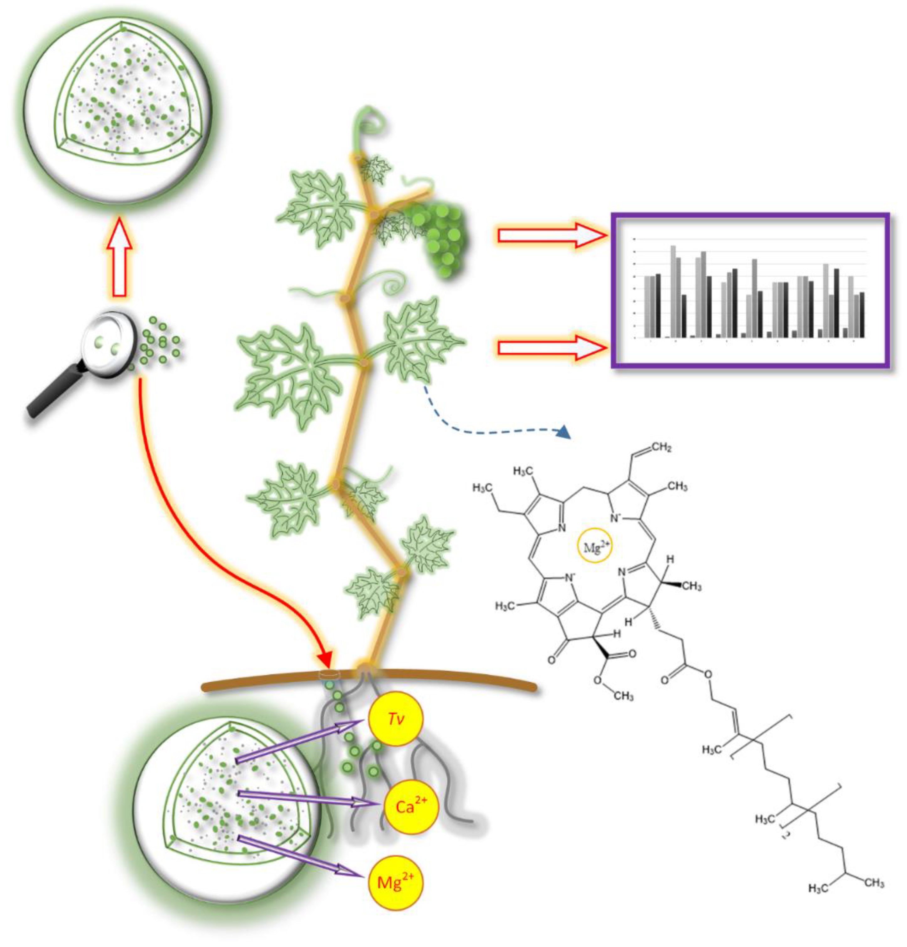
8. Application of Biopolymeric Microparticles and Proposed Guidelines

Keeping in mind the systematics in the production of biopolymeric microparticles loaded with chemical and biological agents, conclusions may apply to diverse possibilities regarding the microparticles application. It is also possible to extrapolate the data with regards to the used models and extend them to almost any type of plant, keeping in mind its needs, maturation time, and harvests.
Encapsulation of chemical and biological agents in biopolymeric microparticles and their application to the plants present an innovative approach to stimulate the production of plant secondary metabolites. This way, it is possible to protect the plant while increasing its nutritive value. Encapsulated agents are protected from the environment, prolonging their lifespan as well as ensuring timely release. Natural polymers have a great advantage when considering their application in food production, where they pose no threat either to the consumer or the environment. The benefits of the micro-delivery systems developed using encapsulation technology are control-released (at a specific target), longer retention time, biosafety (soil friendly), cost-effectiveness (less usage), better stability (against harsh environmental conditions), and easier physical handling [162]. The application of microparticles presented in this review is novel, and there is very limited literature data reported yet, respectively.
When regarding the application of microparticles in agricultural systems, a couple of things need to be considered. The application may be achieved foliar or directly near the root of plants. There are some technical difficulties when applying foliar, where the size of microparticles plays a crucial role. The foliar application requires smaller particles (less than 350 μm), which should be able to pass through the spray nozzle. Clogging of the systems is a significant problem when considering the application of microparticles this way [116].
Application near the root either in some later period of plant maturity or when planting is feasible and relatively easy to achieve. In the paper of Vinceković et al. [163], an explanation of how microspheres were applied to mature Vitis vinifera L. (‘Welschriesling’ cultivar) plants is presented (Figure 7). Briefly, microsphere application was performed manually near the root of a plant, at approximately 20 cm of depth. Plants were treated with microspheres loaded with chemical (Ca2+/Mg2+) and biological (T. viride) agents. Various compositions of microspheres were prepared to test the influence of chemical agents and the synergistic effect of a chemical and biological agent.
In the paper of Jurić et al. [164], microparticles (microspheres or microcapsules) were applied in the planting procedure of lettuce (Lactuca sativa L.) both in conventional (Figure 8) and hydroponic types of cultivation. Alginate-based microspheres containing either only chemical (Ca2+ or Cu2+) or both chemical and biological agents (T. viride spores) and microcapsules containing the aforementioned were prepared. As a side control, suspension of T. viride spores in saline solution was used to observe non-encapsulated vs. encapsulated biological agent influence on plant metabolites synthesis.
Microparticles for the application were prepared based on the results from the previous publications of Vinceković et al. [52,130,133] and Jurić et al. [131,132], fitting the formulation as to the proposed methodology. Respectively, microparticles were prepared with regards to the needs of a plant and economic viability, while keeping in mind the release kinetics and physicochemical properties of microparticles. For a mature vine plant, relatively higher concentrations of cations were used, whilst for lettuce, lower concentrations of cations were used to prepare microparticles. With this in mind, microparticle composition, as well as the final amount, was determined based on the plant’s respective needs. Furthermore, it is very important to control the dosage of copper ions over the plant maturation time and to minimize its release into the environment. Fine-tuning microparticles for different applications may result in a commercially very approachable product. Concerning this review topic, variables for the preparation of microparticles consisted of changes in cation type, its combination, presence of biological agent (T. viride spores) and chitosan coat, as well as the microparticles weight depending on the treated plant type.
The degradability of biopolymers in the soil is also an important factor when considering its application. Our trials revealed that microcapsules degrade slowly over time (Ca-alginate-based microparticles are visible for at least three months), gradually losing volume. A drop in volume is primarily connected to the water loss, but with constant rehydration, microparticles swell, achieving a constant supply (which may be correlated to the release of in vitro conditions) of the encapsulated material. Also, it is important to note that this investigation revealed mycelium growth and sporulation in the soil where vine, lettuce, and tomato plants were used in the experiment. These experiments revealed that sporulation does occur in the soil and that T. viride is present on the root of a plant [116,164].
Of course, the degradability of biopolymeric microparticles depends on their physicochemical parameters. Studies on biodegradation processes of polymers, when applied in agriculture, are scarce, and only a few can be found in the literature [165,166]. Research by Ratajska and Boryniec [167] revealed that the biodegradability of film polymers in soil and water depends on the dimensions of the particles, as well as their distribution in the film. Biopolymers with low degradability in the soil will result in longer protection of encapsulated agents, providing its timely release [38]. The main problem with these types of research is often connected to the fact that soil is of complex composition. The presence of other chemical and biological agents easily omits the predictability of how biopolymeric microparticles will behave. This is an area of interest that still has a lot of potentials to be investigated.

9. Influence of Microparticles Treatments on the Synthesis of Plant Metabolites

It is well known that Ca2+ controls and modifies plant uptake of Mg2+ and increases its availability preventing the negative occurrences of metabolic changes on the plant leaves [108]. It has been shown that microspheres loaded with Ca2+ ions had the most pronounced effect on synthesizing total polyphenols in vine leaves (up to 39%) [163]. Furthermore, it was revealed that treatments with Ca2+/Mg2+ loaded microspheres had a lesser influence on total polyphenols synthesis, signifying the importance of Ca2+ in synthesizing these compounds. Microspheres treatments on the vine (Vitis vinifera L.) have resulted in a significant increase in the antioxidant activity of vine leaves (up to 32%). Ca-alginate microspheres with T. viride spores had the most pronounced effect on antioxidant activity, signifying the synergistic action of chemical and biological agents.
It has been revealed that a significant increase in total carotenoids content can be achieved with microspheres treatments. Ca-alginate microspheres loaded with T. viride spores treatment on Vitis vinifera L. leaves resulted in an impressive 62% increase in total carotenoids content, respectively, to the control samples (untreated). Furthermore, Ca-microspheres (without T. viride spores) had significantly influenced vine leaves with a 24% increase in total carotenoids content. It must be noted that with several harvests, the content of biologically active compounds in plants changes. With time, a gradual decrease in carotenoids content in leaves can be seen [168]. Still, after the second harvest, the positive effect on carotenoid retention is significant, with the highest values achieved with Ca-microspheres treatment [163].
During both harvests of vine leaves, significantly highest chlorophyll content (up to 26%, respectively, to the control) was found in vines treated with Ca/Mg-alginate microspheres with T. viride spores [163]. An increase in the number of total chlorophylls in leaves is due to the synergistic action of Ca2+ and T. viride spores on Mg2+ uptake during the whole plant growth period. The distribution of Ca2+ and Mg2+ in leaves highly depends on the exogenous supply, with the support of T. viride. It is known that Ca2+ influences the uptake of Mg2+ (an important component of the chlorophyll molecule) [108], and treatment with these microspheres eliminates possible competition between Mg2+ and Ca2+, thus increasing the uptake of Mg2+ by the plants. Increased availability of Mg2+ for plants prevents the occurrence of chlorosis, necrotic spots on the leaves, and droop, as well as decreases the level of abiotic and biotic stress.
It has to be noted that, in this research, no statistically significant change was found in treatments of vine plants on grapes, but somewhat higher values (11%) were observed with Ca/Mg-alginate microspheres (with T. viride spores) treatment [163]. Again, antioxidant activity in treated grapes was found to be non-significant. Even though, an increase of 41% (DPPH) and 25% (ABTS) in antioxidant activity was observed on grapes treated with Ca/Mg-alginate microspheres. Cumulatively, results reveal that Ca2+ and T. viride do allow better absorption of Mg2+, stimulating the synthesis of polyphenolic compounds in vine leaves. The positive influence of treatments with microparticles is promising in the vine cultivation process and production of higher quality foods. Future research should be focused on treatments with specific agents to achieve higher stimulation in grapes.
Jurić et al. [164] show that treatments with alginate-based microparticles on lettuce (Lactuca sativa L.) had significantly stimulated the synthesis of plant metabolites, without significant impact on the lettuce morphology. The controlled release was achieved, and passive uptake of ions through the root system was active during the whole period of maturation. Using two types of cultivations (conventional and hydroponics) revealed interesting data. Generally, all treatments had a significant influence on plant metabolites synthesis, relative to the controls. Compared to the Cu-alginate microparticles, Ca-alginate microparticles had a significantly higher effect on synthesizing plant metabolites. Equal treatments in conventional and hydroponics cultivation yielded results with high correlation, which verifies the repeatability and feasibility of the experiment.
Treatment with Ca-alginate microparticles resulted in the highest yield of total chlorophylls in the conventional cultivation of lettuce, with up to an impressive 76% relative to the control [164]. Ca2+ is known to serve as a secondary messenger for cytokinin action in improving the synthesis of chlorophylls [106]. These results are in correlation with research on supplementation with Ca2+, where higher chlorophyll values were also observed in chickpea leaves [19].
Treated lettuce resulted in an increase of up to 48% in total polyphenols content [164]. Generally, Ca-alginate-based microparticles had a stronger effect on synthesizing polyphenolic compounds when comparing them to Cu-alginate-based. Ca-alginate microparticles (with and without T. viride spores) treatments also resulted in a significantly higher accumulation of total flavonoids with an increase of up to 42%. Ca2+ has an important role in polyphenolic metabolism [169]. Application of Ca2+ increases the phenylalanine ammonia-lyase activity, and a higher accumulation of polyphenolic compounds occur. This can significantly increase the plants’ resistance to the infections caused by the pathogens [170]. Regarding the antioxidant activity in treated lettuce plants, Ca-alginate-based microparticles again had the most pronounced effect [164]. Previously, Ahmad et al. [19] revealed that supplementation with Ca2+ significantly boosts antioxidant activity in plants. Cu-based microparticles had a lower effect, but some treatments in hydroponic cultivation were significantly higher than control (untreated). More data on this can be seen in Jurić et al. [164].
In conventional cultivation, non-encapsulated T. viride spores (suspension) had less or even a negative influence on the plant metabolites synthesis when compared to the encapsulated ones. Equal treatment in hydroponic cultivation revealed significant influence, and this can be explained by the fact that this type of cultivation (hydroponic) is sterile compared to the conventional (soil). Furthermore, due to the lower survivability, as well as the need for the organism to uptake some of the nutrients from the surrounding media to survive, there will be fewer available nutrients for the plant. Also, temperatures on the open field and closed hydroponics system are not the same. These facts highlight the importance of biological agent encapsulation, since its survivability increases significantly in an environment that is perhaps not favorable when inoculating the latter.
Increasing the number of plant metabolites as chlorophylls, carotenoids, polyphenols, and an overall increase in antioxidant activity contributes to the quality of food for both humans and animals. Future investigation should focus on the precise agriculture and metabolomics aspect to determine which mechanisms are responsible for synthesis stimulation and to quantify plant needs more precisely. Metabolomics is one of the fastest developing methods for the identification and quantification of changes happening in a sample based on a set of metabolites with low molecular mass [171]. Plant metabolomics aims to investigate plant systems on a molecular level, focusing on a pool of metabolites that respond to different environmental factors [171,172,173]. Plant secondary metabolites are synthesized as a response of plant with the environment, and from one side, they may represent to be final products of gene expression, but also a regulatory system that acts in the organism [174]. An increase in synthesizing plant metabolites via encapsulated chemicals and the biological agent is promising, and, coupled with plant metabolomics, could shed light on complex interactions between plants and stimulants. This can further improve the precision and tailoring of microparticles concerning the plant’s needs.

10. Application of Microparticles in the Cultivation of Other Plants—Unpublished Data

Using the guidelines provided in the previous sections, microparticle treatments on tomatoes and strawberries resulted in success. A total of 120 tomato plants (Solanum lycopersicum L.) (4 different cultivars) were treated in conventional and 120 in hydroponic cultivation. Ca2+ content in Ca-microspheres was from 2.53–2.73 mg g−1, while in Cu-alginate microspheres, Cu2+ content ranged from 2.32–2.50 mg g−1 of microspheres depending on the type of prepared microparticles. The number of T. viride spores was ~104 per gram of microspheres. Tomatoes treated with Ca-alginate microspheres in hydroponic cultivation yielded impressive 230% higher lycopene content (cultivar Vasanta), while conventional cultivation with the same microsphere type had an increase in lycopene content up to 99%, relative to the control (untreated). An increase of up to 164% in lycopene content was observed in the hydroponic cultivation of tomatoes (cultivar Vasanta) after the second harvest. A somewhat lesser influence was observed on other cultivars, e.g., treatments with Ca-alginate microspheres without the T. viride spores on Abellus cultivar had an increase of up to 90% in hydroponic cultivation. The same treatment yielded a significant 63% higher lycopene content in conventional cultivation. Cu-based microspheres had a somewhat lower influence on synthesizing carotenoids, but still, some treatments were significantly (post hoc t-tests with Bonferroni correction (p < 0.05)) higher when compared to the control samples. Also, it should be noted that Ca-microparticles loaded with T. viride spores had a significant influence on synthesizing polyphenols. Generally, relative to the Ca-based microparticles, treatments with Cu-based microparticles had a somewhat lesser influence on synthesizing lycopene, total polyphenols, and antioxidant activity.
Treatments with Zn-alginate microparticles in hydroponic cultivation of strawberries (Fragaria × ananassa Duchesne) on two different cultivars (San Andreas and Albion) revealed significant (post hoc t-tests with Bonferroni correction (p < 0.05)) changes in plant secondary metabolites. A total of 180 plants were used in this research, and the application of 4 grams of microparticles corresponded to 33.64 mg of Zn2+. Prepared Zn-alginate microparticles were spherical with no deformities noticed, with about 1830 μm in size. No significant changes in size and shape were observed in chitosan-coated microparticles (microcapsules). When comparing the microcapsules and microspheres, uncoated Zn-alginate microspheres had a higher impact on the synthesis, probably due to the initial burst release, followed by a slow continuous release of active agents. Relatively to the control, total polyphenolic content (26%), total flavonoid content (21%), antioxidant activity (27%), total anthocyanins (20%), and flavan-3-ols (17%) were significantly increased. Treatments with only chemical agents (Zn2+) result in a significantly higher quality product [175].
Except for Zn-alginate microparticles, treatments with the combination of Zn/Fe-alginate microparticles revealed similar trends. Different varieties of strawberries in conventional cultivation were used as a model for the treatment. Treatment with Zn/Fe-alginate microparticles had a significant influence on the increase of total polyphenols (12%), total flavonoids (17%), total anthocyanins (16%), as well as antioxidant activity (11%). Chitosan-coated Zn/Fe-alginate microcapsules did not significantly affect the synthesis of plant secondary metabolites, probably due to the significantly slower release.
The above-mentioned research on tomatoes and strawberries treated with tailored microparticles is to be published.
There are still a lot of opportunities in this area to be investigated. The importance of biological agent encapsulation to protect it from negative environmental factors is evident. Tailoring microparticles coupled with plant metabolomics may result in precise measurements of plants’ needs. As reported, treatments with microparticles loaded with chemical and biological have a significant effect on the synthesis of plant metabolites. With an increased number of plant metabolites and antioxidant activity, the quality of food is higher, and this could be exploited either to be labeled as functional or as a source of functional components in the production of functional foods.

11. Conclusions and Prospects

The initial carrier system setup determines the chemical and biological agent (T. viride spores) release rates and patterns. By adopting a systematic formulation approach, optimal release can be achieved for prolonged protection and nutritional benefits on the plants via continuous release over an extended period. Knowledge of the molecular interactions between encapsulated agents and delivery systems and mechanisms controlling its release aids in the preparation of microparticles with specifically tailored properties and controllable release kinetics. Simultaneous encapsulation of chemical and biological agents is feasible. Intermolecular interactions between components from which microparticles are necessary to investigate before the encapsulation process. Release from alginate-based microparticles can be controlled by tuning the concentration of cations and with an additional layer, as well as with the presence of a biological agent. This way, a wide array of applications of microparticles with desirable properties, depending on the needs of a plant, is attainable. Proposed preparation methods are applicable for various types of encapsulated cations, as well as biological agents. One should follow the same methodology to successfully characterize microparticles or predict their release behavior over time. Presented guidelines can be universally used for any type of biopolymeric microparticle preparation using the ionic gelation method, regardless of the loaded components. Application guidelines should be followed when applying microparticles for agricultural purposes. Treatments with prepared microparticles loaded with chemical and biological agents have shown a significant influence on the synthesis of plant metabolites. Tailoring microparticles to suit the plant’s needs and to stimulate plant secondary metabolites synthesis is challenging. This area is relatively new, and opportunities are vast. In the future, it can be expected that biopolymer-based formulations will dominate the market, since they can contribute to plant protection and nutrition in ecological production. These formulations are useful in the stimulation of bioactive compounds, which ends in improved nutritional quality and potential dietary sources of natural polyphenolic antioxidants. Microparticles preparation and application methods presented are simple and reliable, friendly to the environment, and economically favorable. Future investigation should focus on the precise agriculture and metabolomics aspect to determine which mechanisms are responsible for synthesis stimulation and quantify plant needs more precisely concerning the tailored microparticles.

Author Contributions

Conceptualization, S.J.; Data curation; Investigation; Methodology; Formal analysis; Validation; Visualization; Writing—original draft, M.J. Visualization, Writing—review & editing, A.R.J. Supervision; Writing—review & editing, M.V. Supervision; Funding acquisition; Writing—review & editing. All authors have read and agreed to the published version of the manuscript.

Funding

This research was funded by the Croatian Science Foundation (UIP-2014-501 09-6462) and Operational Program Competitiveness and Cohesion 2014–2020 (K.K.01.1.1.04.0058) “Potential of microencapsulation in cheese production”.

Data Availability Statement

The data presented in this review are available on request from the corresponding author.

Conflicts of Interest

The authors declare no conflict of interest.

References

  1. Mustafa, I.F.; Hussein, M.Z. Synthesis and Technology of Nanoemulsion-Based Pesticide Formulation. Nanomaterials 2020, 10, 1608. [Google Scholar] [CrossRef] [PubMed]
  2. John, R.P.; Tyagi, R.D.; Brar, S.K.; Surampalli, R.Y.; Prévost, D. Bio-encapsulation of microbial cells for targeted agricultural delivery. Crit. Rev. Biotechnol. 2011, 31, 211–226. [Google Scholar] [CrossRef]
  3. Bigliardi, B.; Galati, F. Innovation trends in the food industry: The case of functional foods. Trends Food Sci. Technol. 2013, 31, 118–129. [Google Scholar] [CrossRef]
  4. Lenssena, K.G.M.; Bast, A.; de Boera, A. Clarifying the health claim assessment procedure of EFSA will benefit functional food innovation. J. Funct. Foods 2018, 47, 386–396. [Google Scholar] [CrossRef]
  5. Tiwari, R.; Rana, C.S. Plant secondary metabolites: A review. Int. J. Eng. Res. Gen. Sci. 2015, 3, 661–670. [Google Scholar]
  6. Guerriero, G.; Berni, R.; Muñoz-Sanchez, J.A.; Apone, F.; Abdel-Salam, E.M.; Qahtan, A.A.; Alatar, A.A.; Cantini, C.; Cai, G.; Hausman, J.F.; et al. Production of plant secondary metabolites: Examples, tips and suggestions for biotechnologists. Genes 2018, 20, 309. [Google Scholar] [CrossRef] [Green Version]
  7. Lattanzio, V.; Lattanzio, V.M.T.; Angela Cardinali, A. Role of phenolics in the resistance mechanisms of plants against fungal pathogens and insects. In Phytochemistry: Advances in Research; Imperato, F., Ed.; Research Signpost: Kerala, India, 2006; pp. 23–67. ISBN 81-308-0034-9. [Google Scholar]
  8. Mampholo, B.M.; Sivakumar, D.; Beukes, M.; van Rensburg, W.J. Effect of modified atmosphere packaging on the quality and bioactive compounds of Chinese cabbage (Brassica rapa L. ssp. chinensis). J. Sci. Food Agric. 2013, 93, 2008–2015. [Google Scholar] [CrossRef] [Green Version]
  9. Anand, S. Various approaches for the secondary metabolite production through plant tissue culture. Pharmacia 2010, 1, 1–7. [Google Scholar]
  10. Banerjee, M.R.; Yesmin, L.; Vessey, J.K. Plant-Growth-Promoting Rhizobacteria as Biofertilizers and Biopesticides. In Handbook of Microbial Biofertilizers; Rai, M., Ed.; Food Products Press: New York, NY, USA, 2006; pp. 140–141. ISBN 9781560222705. [Google Scholar]
  11. Avioa, L.; Sbrana, C.; Giovannetti, M.; Frassin, S. Arbuscular mycorrhizal fungi affect total phenolics content and antioxidant activity in leaves of oak leaf lettuce varieties. Sci. Hortic. 2017, 224, 265–271. [Google Scholar] [CrossRef]
  12. Altomare, C.; Norvell, W.A.; Bjorkman, T.; Harman, G.E. Solubilization of phosphate and micronutrients by the plant-growth promoting and biocontrol fungus Trichoderma harzianum Rifai 1295-22. Appl. Environ. Microbiol. 1999, 65, 2926–2933. [Google Scholar] [CrossRef] [PubMed] [Green Version]
  13. Harman, G.E. Overview of mechanisms and uses of Trichoderma spp. Phytopathology 2006, 96, 190–194. [Google Scholar] [CrossRef] [PubMed] [Green Version]
  14. Paulo, F.; Santos, L. Design of experiments for microencapsulation applications: A review. Mater. Sci. Eng. C 2017, 77, 1327–1340. [Google Scholar] [CrossRef] [PubMed]
  15. Locatelli, G.O.; dos Santos, G.F.; Botelho, P.S.; Finkler, C.L.L.; Bueno, L.A. Development of Trichoderma sp. formulations in encapsulated granules (CG) and evaluation of conidia shelf-life. Biol. Control 2018, 117, 21–29. [Google Scholar] [CrossRef]
  16. White, P.J. Calcium channels in higher plants. Biochim. Biophys. Acta Biomembr. 2000, 1465, 171–189. [Google Scholar] [CrossRef] [Green Version]
  17. El-Beltagi, H.S.; Mohamed, H.I. Alleviation of cadmium toxicity in Pisum sativum L. seedlings by calcium chloride. Not. Bot. Horti. Agrobot. Cluj-Napoca 2013, 41, 157–168. [Google Scholar] [CrossRef] [Green Version]
  18. Talukdar, D. Exogenous calcium alleviates the impact of cadmium induced oxidative stress in Lens culinaris Medic. seedlings through modulation of antioxidant enzyme activities. J. Crop Sci. Biotechnol. 2012, 15, 325–334. [Google Scholar] [CrossRef]
  19. Ahmad, P.; Abdel Latef, A.A.; AbdAllah, E.F.; Hashem, A.; Sarwat, M.; Anjum, N.A.; Gucel, S. Calcium and potassium supplementation enhanced growth, osmolyte secondary metabolite production, and enzymatic antioxidant machinery in cadmium-exposed chickpea (Cicer arietinum L.). Front. Plant Sci. 2016, 7, 513. [Google Scholar] [CrossRef] [Green Version]
  20. Xu, W.; Peng, H.; Yang, T.; Whitaker, B.; Huang, L.; Sun, J.; Chen, P. Effect of calcium on strawberry fruit flavonoid pathway gene expression and anthocyanin accumulation. Plant Physiol. Biochem. 2014, 82, 289–298. [Google Scholar] [CrossRef] [PubMed]
  21. Hirschi, K.D. The calcium conundrum. Both versatile nutrient and specific signal. Plant Physiol. 2004, 136, 2438–2442. [Google Scholar] [CrossRef] [Green Version]
  22. Siddique, M.H.; Al-Whaibi, M.H.; Sakran, A.H.; Basalah, M.O.; Ali, H.M. Effect of calcium and potassium on antioxidant system of Vicia faba L. under cadmium stress. Int. J. Mol. Sci. 2012, 13, 6604–6619. [Google Scholar] [CrossRef] [Green Version]
  23. Li, P.; Zhao, C.; Zhang, Y.; Wang, X.; Wang, X.; Wang, J.; Wang, F.; Bi, Y. Calcium alleviates cadmium-induced inhibition on root growth by maintaining auxin homeostasis in Arabidopsis seedlings. Protoplasma 2016, 253, 185–200. [Google Scholar] [CrossRef]
  24. Fujita, Y.; Hara, Y.; Suga, C.; Morimoto, T. Production of shikonin derivatives by cell suspension cultures of Lithospermum erythrorhizon. II. A new medium for the production of shikonin derivatives. Plant Cell Rep. 1981, 1, 61–63. [Google Scholar] [CrossRef]
  25. Ohlsson, A.B.; Berglund, T. Effect of high MnSO4 levels on cardenolide accumulation by Digitalis lanata tissue cultures in light and darkness. J. Plant Physiol. 1989, 135, 505–507. [Google Scholar] [CrossRef]
  26. Trejo-Tapia, G.; Jimenez-Aparicio, A.; Rodriguez-Monroy, M.; De Jesus-Sanchez, A.; Gutierrez-Lopez, G. Influence of cobalt and other microelements on the production of betalains and the growth of suspension cultures of Beta vulgaris. Plant Cell Tissue Organ. Cult. 2001, 67, 19–23. [Google Scholar] [CrossRef]
  27. Mullins, M.G.; Bouquet, A.; Williams, L.E. Biology of the Grapevine; Cambridge University Press: New York, NY, USA, 1992; ISBN 978-0521305075. [Google Scholar]
  28. Salisbury, F.B.; Ross, C.W. Plant Physiology, Hormones and Plant Regulators: Auxins and Gibberellins, 4th ed.; Wadsworth Publishing Company: Belmont, CA, USA, 1992; ISBN 10: 0534983901. [Google Scholar]
  29. Yruela, I. Copper in plants: Acquisition, transport and interactions. Funct. Plant Biol. 2009, 36, 409–430. [Google Scholar] [CrossRef] [Green Version]
  30. Murashige, T.; Skoog, F. A Revised Medium for Rapid Growth and Bio Assays with Tobacco Tissue Cultures. Physiol. Plant 1962, 15, 473–497. [Google Scholar] [CrossRef]
  31. Narula, A.; Kumar, S.; Srivastava, P.S. Abiotic metal stress enhances diosgenin yield in Dioscorea bulbifera L. cultures. Plant Cell Rep. 2005, 24, 250–254. [Google Scholar] [CrossRef] [PubMed]
  32. Bongiovanni, R.; Lowenberg-Deboer, J. Precision Agriculture and Sustainability. Precis. Agric. 2004, 5, 359–387. [Google Scholar] [CrossRef]
  33. Kudasova, D.; Mutaliyeva, B.; Vlahoviček-Kahlina, K.; Jurić, S.; Marijan, M.; Khalus, S.; Prosyanik, A.; Šegota, S.; Španić, N.; Vinceković, M. Encapsulation of Synthesized Plant Growth Regulator Based on Copper(II) Complex in Chitosan/Alginate Microcapsules. Int. J. Mol. Sci. 2021, 22, 2663. [Google Scholar] [CrossRef] [PubMed]
  34. Vlahoviček-Kahlina, K.; Jurić, S.; Marijan, M.; Mutaliyeva, B.; Khalus, S.; Prosyanik, A.; Vinceković, M. Synthesis, Characterization, and Encapsulation of Novel Plant Growth Regulators (PGRs) in Biopolymer Matrices. Int. J. Mol. Sci. 2021, 22, 1847. [Google Scholar] [CrossRef]
  35. Kumari, A.; Yadav, S.K.; Yadav, S.C. Biodegradable polymeric nanoparticles based drug delivery systems. Colloids Surf. B Biointerfaces 2010, 75, 1–18. [Google Scholar] [CrossRef] [PubMed]
  36. Sinha, N.; Kulshreshtha, N.M.; Dixit, M.; Jadhav, I.; Shrivastava, D.; Bisen, P.S. Nanodentistry: Novel approaches. In Nanostructures for Oral Medicine; Andronescu, E., Grumezescu, A.M., Eds.; Elsevier: Amsterdam, The Netherlands, 2017; pp. 751–776. [Google Scholar]
  37. Singh, M.; Hemant, K.; Ram, M.; Shivakumar, H. Microencapsulation: A promising technique for controlled drug delivery. Res. Pharm. Sci. 2010, 5, 65–77. [Google Scholar]
  38. Vemmer, M.; Patel, A.V. Review of encapsulation methods suitable for microbial biological control agents. Biol. Control 2013, 67, 380–389. [Google Scholar] [CrossRef]
  39. Haffner, F.B.; Diab, R.; Pasc, A. Encapsulation of probiotics: Insights into academic and industrial approaches. AIMS Mater. Sci. 2016, 3, 114–136. [Google Scholar] [CrossRef]
  40. Vinceković, M.; Viskić, M.; Jurić, S.; Giacometti, J.; Kovačević, D.B.; Putnik, P.; Donsì, F.; Barba, F.J.; Jambrak, A.R. Innovative technologies for encapsulation of Mediterranean plants extracts. Trends Food Sci. Technol. 2017, 69, 1–12. [Google Scholar] [CrossRef]
  41. Lengyel, M.; Kállai-Szabó, N.; Antal, V.; Laki, A.J.; Antal, I. Microparticles, Microspheres, and Microcapsules for Advanced Drug Delivery. Sci. Pharm. 2019, 87, 20. [Google Scholar] [CrossRef] [Green Version]
  42. Fathi, M.; Vinceković, M.; Jurić, S.; Viskić, M.; Jambrak, A.R.; Donsì, F. Food-Grade Colloidal Systems for the Delivery of Essential Oils. Food Rev. Int. 2021, 37, 1–45. [Google Scholar] [CrossRef]
  43. Lai, W.-F.; Rogach, A.L. Hydrogel-Based Materials for Delivery of Herbal Medicines. ACS Appl. Mater. Interfaces 2017, 9, 11309–11320. [Google Scholar] [CrossRef]
  44. Chakravarthi, S.S.; Robinson, D.H. Biodegradable nanoparticles. In Pharmaceutical Manufacturing Handbook; Gad, S.C., Ed.; John Wiley & Sons: Hoboken, NJ, USA, 2008; pp. 535–565. ISBN 978-0-470-25958-0. [Google Scholar]
  45. Sundar, S.; Kundu, J.; Kundu, S.C. Biopolymeric nanoparticles. Sci. Technol. Adv. Mater. 2010, 11, 014104. [Google Scholar] [CrossRef]
  46. Motwani, S.K.; Chopra, S.; Talegaonkar, S.; Kohli, K.; Ahmad, F.J.; Khar, R.K. Chitosan–sodium alginate nanoparticles as submicroscopic reservoirs for ocular delivery: Formulation, optimisation and in vitro characterisation. Eur. J. Pharm. Biopharm. 2008, 68, 513–525. [Google Scholar] [CrossRef]
  47. Lawrie, G.; Keen, I.; Drew, B.; Chandler-Temple, A.F.; Rintoul, L.; Fredericks, P.M.; Grøndahl, L. Interactions between Alginate and Chitosan Biopolymers Characterized Using FTIR and XPS. Biomacromolecules 2007, 8, 2533–2541. [Google Scholar] [CrossRef]
  48. Jurić, S.; Tanuwidjaja, I.; Fuka, M.M.; Vlahoviček-Kahlina, K.; Marijan, M.; Boras, A.; Kolić, N.U.; Vinceković, M. Encapsulation of two fermentation agents, Lactobacillus sakei and calcium ions in microspheres. Colloids Surf. B Biointerfaces 2021, 197, 111387. [Google Scholar] [CrossRef]
  49. Fuka, M.M.; Maksimovic, A.Z.; Hulak, N.; Kos, I.; Radovcic, N.M.; Juric, S.; Tanuwidjaja, I.; Karolyi, D.; Vincekovic, M. The survival rate and efficiency of non-encapsulated and encapsulated native starter cultures to improve the quality of artisanal game meat sausages. J. Food Sci. Technol. 2021, 58, 710–719. [Google Scholar] [CrossRef]
  50. Hudson, D.; Margaritis, A. Biopolymer nanoparticle production for controlled release of biopharmaceuticals. Crit. Rev. Biotechnol. 2012, 34, 161–179. [Google Scholar] [CrossRef]
  51. Belščak-Cvitanović, A.; Jurić, S.; Đorđević, V.; Barišić, L.; Komes, D.; Ježek, D.; Bugarski, B.; Nedović, V. Chemometric evaluation of binary mixtures of alginate and polysaccharide biopolymers as carriers for microencapsulation of green tea polyphenols. Int. J. Food Prop. 2017, 20, 1971–1986. [Google Scholar] [CrossRef] [Green Version]
  52. Vinceković, M.; Topolovec Pintarić, S.; Jurić, S.; Viskić, M.; Jalšenjak, N.; Bujan, M.; Đermić, E.; Žutić, I.; Fabek Uher, S. Release of Trichoderma viride spores from microcapsules simultaneously loaded with chemical and biological agents. Agric. Conspec. Sci. 2017, 82, 395–401. [Google Scholar]
  53. Smidsrød, O. Solution properties of alginate. Carbohydr. Res. 1970, 13, 359–372. [Google Scholar] [CrossRef]
  54. Smidsrød, O. Molecular basis for some physical properties of alginates in the gel state. Faraday Discuss. Chem. Soc. 1974, 57, 263–274. [Google Scholar] [CrossRef]
  55. Haug, A.; Smidsrød, O.; Högdahl, B.; Øye, H.A.; Rasmussen, S.E.; Sunde, E.; Sørensen, N.A. Selectivity of Some Anionic Polymers for Divalent Metal Ions. Acta Chem. Scand. 1970, 24, 843–854. [Google Scholar] [CrossRef] [Green Version]
  56. Montanucci, P.; Terenzi, S.; Santi, C.; Pennoni, I.; Bini, V.; Pescara, T.; Basta, G.; Calafiore, R. Insights in Behavior of Variably Formulated Alginate-Based Microcapsules for Cell Transplantation. BioMed Res. Int. 2015, 2015, 965804. [Google Scholar] [CrossRef]
  57. Grant, G.T.; Morris, E.R.; Rees, D.A.; Smith, P.J.; Thom, D. Biological interactions between polysaccharides and divalent cations: The egg-box model. FEBS Lett. 1973, 32, 195–198. [Google Scholar] [CrossRef] [Green Version]
  58. Sikorski, P.; Mo, F.; Skjåk-Bræk, A.G.; Stokke, B.T. Evidence for Egg-Box-Compatible Interactions in Calcium−Alginate Gels from Fiber X-ray Diffraction. Biomacromolecules 2007, 8, 2098–2103. [Google Scholar] [CrossRef]
  59. De Vos, P.; Spasojevic, M.; De Haan, B.J.; Faas, M.M. The association between in vivo physicochemical changes and inflammatory responses against alginate based microcapsules. Biomaterials 2012, 33, 5552–5559. [Google Scholar] [CrossRef] [Green Version]
  60. Eiselt, P.; Yeh, J.; Latvala, R.K.; Shea, L.D.; Mooney, D.J. Porous carriers for biomedical applications based on alginate hydrogels. Biomater. 2000, 21, 1921–1927. [Google Scholar] [CrossRef]
  61. Qu, B.; Luo, Y. Chitosan-based hydrogel beads: Preparations, modifications and applications in food and agriculture sectors—A review. Int. J. Biol. Macromol. 2020, 152, 437–448. [Google Scholar] [CrossRef] [PubMed]
  62. Belščak-Cvitanović, A.; Komes, D.; Karlović, S.; Djaković, S.; Špoljarić, I.; Mršić, G.; Ježek, D. Improving the controlled delivery formulations of caffeine in alginate hydrogel beads combined with pectin, carrageenan, chitosan and psyllium. Food Chem. 2015, 167, 378–386. [Google Scholar] [CrossRef] [PubMed]
  63. Lozano-Vazquez, G.; Lobato-Calleros, C.; Escalona-Buendia, H.; Chavez, G.; Alvarez-Ramirez, J.; Vernon-Carter, E. Effect of the weight ratio of alginate-modified tapioca starch on the physicochemical properties and release kinetics of chlorogenic acid containing beads. Food Hydrocoll. 2015, 48, 301–311. [Google Scholar] [CrossRef]
  64. Benrebah, F.; Prevost, D.; Yezza, A.; Tyagi, R. Agro-industrial waste materials and wastewater sludge for rhizobial inoculant production: A review. Bioresour. Technol. 2007, 98, 3535–3546. [Google Scholar] [CrossRef] [PubMed]
  65. Bashan, Y.; De-Bashan, L.E.; Prabhu, S.R.; Hernandez, J.-P. Advances in plant growth-promoting bacterial inoculant technology: Formulations and practical perspectives (1998–2013). Plant Soil 2014, 378, 1–33. [Google Scholar] [CrossRef] [Green Version]
  66. Paula, A.R.; Carolino, A.T.; Paula, C.O.; Samuels, R.I. The combination of the entomopathogenic fungus Metarhizium anisopliae with the insecticide Imidacloprid increases virulence against the dengue vector Aedes aegypti (Diptera: Culicidae). Parasites Vectors 2011, 4, 8. [Google Scholar] [CrossRef] [Green Version]
  67. Hynes, R.K.; Craig, K.A.; Covert, D.; Rennie, R.J.; Smith, R.S. Liquid Rhizobial Inoculants for Lentil and Field Pea. J. Prod. Agric. 1995, 8, 547–552. [Google Scholar] [CrossRef]
  68. Albareda, M.; Rodríguez-Navarro, D.N.; Camacho, M.; Temprano, F.J. Alternatives to peat as a carrier for rhizobia inoculants: Solid and liquid formulations. Soil Biol. Biochem. 2008, 40, 2771–2779. [Google Scholar] [CrossRef]
  69. Vejan, P.; Abdullah, R.; Khadiran, T.; Ismail, S. Encapsulation of Bacillus salmalaya 139SI using double coating biopolymer technique. Lett. Appl. Microbiol. 2018, 68, 56–63. [Google Scholar] [CrossRef] [Green Version]
  70. Hall, R.D.; Brouwer, I.D.; Fitzgerald, M.A. Plant metabolomics and its potential application for human nutrition. Physiol. Plant 2007, 132, 162–175. [Google Scholar] [CrossRef]
  71. Hong, J.; Yang, L.; Zhang, D.; Shi, J. Plant Metabolomics: An Indispensable System Biology Tool for Plant Science. Int. J. Mol. Sci. 2016, 17, 767. [Google Scholar] [CrossRef]
  72. Scossa, F.; Brotman, Y.; Lima, F.D.A.E.; Willmitzer, L.; Nikoloski, Z.; Tohge, T.; Fernie, A.R. Genomics-based strategies for the use of natural variation in the improvement of crop metabolism. Plant Sci. 2016, 242, 47–64. [Google Scholar] [CrossRef] [PubMed]
  73. Ntalli, N.; Koliopoulos, G.; Giatropoulos, A.; Menkissoglu-Spiroudi, U. Plant secondary metabolites against arthropods of medical importance. Phytochem. Rev. 2019, 18, 1255–1275. [Google Scholar] [CrossRef]
  74. Živković, I.; Jurić, S.; Vinceković, M.; Galešić, M.; Marijan, M.; Vlahovićek-Kahlina, K.; Mikac, K.; Lemic, D. Polyphenol-Based Microencapsulated Extracts as Novel Green Insecticides for Sustainable Management of Polyphagous Brown Marmorated Stink Bug (Halyomorpha halys Stål, 1855). Sustainability 2020, 12, 10079. [Google Scholar] [CrossRef]
  75. Veiga, M.; Costa, E.M.; Silva, S.; Pintado, M. Impact of plant extracts upon human health: A review. Crit. Rev. Food Sci. Nutr. 2018, 60, 873–886. [Google Scholar] [CrossRef]
  76. Djeridane, A.; Yousfi, M.; Nadjemi, B.; Boutassouna, D.; Stocker, P.; Vidal, N. Antioxidant activity of some algerian medicinal plants extracts containing phenolic compounds. Food Chem. 2006, 97, 654–660. [Google Scholar] [CrossRef]
  77. Balasundram, N.; Sundram, K.; Samman, S. Phenolic compounds in plants and agri-industrial by-products: Antioxidant activity, occurrence, and potential uses. Food Chem. 2006, 99, 191–203. [Google Scholar] [CrossRef]
  78. Abuajah, C.I.; Ogbonna, A.C.; Osuji, C.M. Functional components and medicinal properties of food: A review. J. Food Sci. Technol. 2015, 52, 2522–2529. [Google Scholar] [CrossRef] [PubMed] [Green Version]
  79. Dissanayake, A.A.; Zhang, C.-R.; Mills, G.L.; Nair, M.G. Cultivated maitake mushroom demonstrated functional food quality as determined by in vitro bioassays. J. Funct. Foods 2018, 44, 79–85. [Google Scholar] [CrossRef]
  80. Strzałka, K.; Kostecka-Gugała, A.; Latowski, D. Carotenoids and Environmental Stress in Plants: Significance of Carotenoid-Mediated Modulation of Membrane Physical Properties. Russ. J. Plant Physiol. 2003, 50, 168–173. [Google Scholar] [CrossRef]
  81. Kun, Y.; Lule, U.S.; Xiao-Lin, D. Lycopene: Its Properties and Relationship to Human Health. Food Rev. Int. 2006, 22, 309–333. [Google Scholar] [CrossRef]
  82. Kong, K.-W.; Khoo, H.-E.; Prasad, K.N.; Ismail, A.; Tan, C.-P.; Rajab, N.F. Revealing the Power of the Natural Red Pigment Lycopene. Molecules 2010, 15, 959–987. [Google Scholar] [CrossRef] [PubMed] [Green Version]
  83. Jurić, S.; Ferrari, G.; Velikov, K.P.; Donsì, F. High-pressure homogenization treatment to recover bioactive compounds from tomato peels. J. Food Eng. 2019, 262, 170–180. [Google Scholar] [CrossRef]
  84. Viuda-Martos, M.; Sanchez-Zapata, E.; Sayas-Barberá, E.; Sendra, E.; Pérez-Álvarez, J.A.; Fernández-López, J. Tomato and Tomato Byproducts. Human Health Benefits of Lycopene and Its Application to Meat Products: A Review. Crit. Rev. Food Sci. Nutr. 2014, 54, 1032–1049. [Google Scholar] [CrossRef] [PubMed]
  85. Schünemann, H.J.; McCann, S.; Grant, B.J.B.; Trevisan, M.; Muti, P.; Freudenheim, J.L. Lung Function in Relation to Intake of Carotenoids and Other Antioxidant Vitamins in a Population-based Study. Am. J. Epidemiol. 2002, 155, 463–471. [Google Scholar] [CrossRef] [PubMed] [Green Version]
  86. Rao, A.V.; Rao, L.G. Carotenoids and human health. Pharmacol. Res. 2007, 55, 207–216. [Google Scholar] [CrossRef]
  87. Álvarez, R.; Meléndez-Martínez, A.J.; Vicario, I.M.; Alcalde, M.J. Carotenoid and Vitamin A Contents in Biological Fluids and Tissues of Animals as an Effect of the Diet: A Review. Food Rev. Int. 2015, 31, 319–340. [Google Scholar] [CrossRef] [Green Version]
  88. Kim, J.K.; Park, S.U. Current results on the potential health benefits of lutein. EXCLI J. 2016, 15, 308–314. [Google Scholar]
  89. Jurić, S.; Jurić, M.; Król-Kilińska, Ż.; Vlahoviček-Kahlina, K.; Vinceković, M.; Dragović-Uzelac, V.; Donsì, F. Sources, stability, encapsulation and application of natural pigments in foods. Food Rev. Int. 2020, 1–56. [Google Scholar] [CrossRef]
  90. Zepka, L.Q.; Jacob-Lopes, E.; Roca, M. Catabolism and bioactive properties of chlorophylls. Curr. Opin. Food Sci. 2019, 26, 94–100. [Google Scholar] [CrossRef] [Green Version]
  91. Viera, I.; Roca, M. Chemistry in the Bioactivity of Chlorophylls: An Overview. Curr. Med. Chem. 2018, 24, 4515–4536. [Google Scholar] [CrossRef] [Green Version]
  92. Ferruzzi, M.; Bohm, V.; Courtney, P.; Schwartz, S. Antioxidant and Antimutagenic Activity of Dietary Chlorophyll Derivatives Determined by Radical Scavenging and Bacterial Reverse Mutagenesis Assays. J. Food Sci. 2002, 67, 2589–2595. [Google Scholar] [CrossRef]
  93. Jonker, J.W.; Buitelaar, M.; Wagenaar, E.; Van Der Valk, M.A.; Scheffer, G.L.; Scheper, R.J.; Plösch, T.; Kuipers, F.; Elferink, R.P.J.O.; Rosing, H.; et al. Nonlinear partial differential equations and applications: The breast cancer resistance protein protects against a major chlorophyll-derived dietary phototoxin and protoporphyria. Proc. Natl. Acad. Sci. USA 2002, 99, 15649–15654. [Google Scholar] [CrossRef] [PubMed] [Green Version]
  94. De Vogel, J.; Jonker-Termont, D.S.; Van Lieshout, E.M.; Katan, M.B.; Van Der Meer, R. Green vegetables, red meat and colon cancer: Chlorophyll prevents the cytotoxic and hyperproliferative effects of haem in rat colon. Carcinogenesis 2004, 26, 387–393. [Google Scholar] [CrossRef] [Green Version]
  95. Ferruzzi, M.G.; Blakeslee, J. Digestion, absorption, and cancer preventative activity of dietary chlorophyll derivatives. Nutr. Res. 2007, 27, 1–12. [Google Scholar] [CrossRef]
  96. Egner, P.A.; Wang, J.-B.; Zhu, Y.-R.; Zhang, B.-C.; Wu, Y.; Zhang, Q.-N.; Qian, G.-S.; Kuang, S.-Y.; Gange, S.J.; Jacobson, L.P.; et al. Chlorophyllin intervention reduces aflatoxin-DNA adducts in individuals at high risk for liver cancer. Proc. Natl. Acad. Sci. USA 2001, 98, 14601–14606. [Google Scholar] [CrossRef] [Green Version]
  97. Marawaha, R.K.; Bansal, D.; Kaur, S.; Trehan, A. Wheat grass juice reduces transfusion requirement in patients with thalassemia major: A pilot study. Indian Pediatr. 2004, 41, 716–720. [Google Scholar] [PubMed]
  98. Fahey, J.W.; Stephenson, K.K.; Dinkova-Kostova, A.T.; Egner, P.A.; Kensler, T.W.; Talalay, P. Chlorophyll, chlorophyllin and related tetrapyrroles are significant inducers of mammalian phase 2 cytoprotective genes. Carcinogenesis 2005, 26, 1247–1255. [Google Scholar] [CrossRef] [PubMed] [Green Version]
  99. Wangcharoen, W.; Phimphilai, S. Chlorophyll and total phenolic contents, antioxidant activities and consumer acceptance test of processed grass drinks. J. Food Sci. Technol. 2016, 53, 4135–4140. [Google Scholar] [CrossRef]
  100. Kończak, I.; Zhang, W. Anthocyanins—More Than Nature’s Colours. J. Biomed. Biotechnol. 2004, 2004, 239–240. [Google Scholar] [CrossRef] [Green Version]
  101. Pangestuti, R.; Kim, S.-K. Biological activities and health benefit effects of natural pigments derived from marine algae. J. Funct. Foods 2011, 3, 255–266. [Google Scholar] [CrossRef]
  102. Silva, S.; Costa, E.M.; Vicente, S.; Veiga, M.; Calhau, C.; Morais, R.M.; Pintado, M.E. DNA agarose gel electrophoresis for antioxidant analysis: Development of a quantitative approach for phenolic extracts. Food Chem. 2017, 233, 45–51. [Google Scholar] [CrossRef]
  103. Yan, S.; Shao, H.; Zhou, Z.; Wang, Q.; Zhao, L.; Yang, X. Non-extractable polyphenols of green tea and their antioxidant, anti-α-glucosidase capacity, and release during in vitro digestion. J. Funct. Foods 2018, 42, 129–136. [Google Scholar] [CrossRef]
  104. Bogacz-Radomska, L.; Harasym, J.; Piwowar, A. Commercialization aspects of carotenoids. In Carotenoids: Properties, Processing and Applications; Galanakis, C.M., Ed.; Elsevier: Amsterdam, The Netherlands, 2020; pp. 327–357. [Google Scholar] [CrossRef]
  105. Viera, I.; Pérez-Gálvez, A.; Roca, M. Green Natural Colorants. Molecules 2019, 24, 154. [Google Scholar] [CrossRef] [PubMed] [Green Version]
  106. Lechowski, Z.; Białczyk, J. Calcium mediated cytokinin action on chlorophyll synthesis in isolated embryo of Scots pine. Biol. Plant. 1993, 35, 53–62. [Google Scholar] [CrossRef]
  107. Wallace, A.; Mueller, R.T.; Wood, R.A. Arsenic phytotoxicity and interactions in bush bean plants grown in solution culture. J. Plant Nutr. 1980, 2, 111–113. [Google Scholar] [CrossRef]
  108. Pal, R.; Laloraya, M. Effect of Calcium Levels on Chlorophyll Synthesis in Peanut and Linseed Plants. Biochem. Physiol. Pflanz. 1972, 163, 443–449. [Google Scholar] [CrossRef]
  109. Elad, Y. Biological control of foliar pathogens by means of Trichoderma harzianum and potential modes of action. Crop. Prot. 2000, 19, 709–714. [Google Scholar] [CrossRef]
  110. Maruyama, C.R.; Bilesky-José, N.; De Lima, R.; Fraceto, L.F. Encapsulation of Trichoderma harzianum Preserves Enzymatic Activity and Enhances the Potential for Biological Control. Front. Bioeng. Biotechnol. 2020, 8, 225. [Google Scholar] [CrossRef] [Green Version]
  111. Howell, C.R. Mechanisms Employed by Trichoderma Species in the Biological Control of Plant Diseases: The History and Evolution of Current Concepts. Plant Dis. 2003, 87, 4–10. [Google Scholar] [CrossRef] [PubMed] [Green Version]
  112. Keswani, C.; Mishra, S.; Sarma, B.K.; Singh, S.P.; Singh, H.B. Unraveling the efficient applications of secondary metabolites of various Trichoderma spp. Appl. Microbiol. Biotechnol. 2013, 98, 533–544. [Google Scholar] [CrossRef] [PubMed]
  113. Ihrmark, K.; Asmail, N.; Ubhayasekera, W.; Melin, P.; Stenlid, J.; Karlsson, M. Comparative Molecular Evolution of Trichoderma Chitinases in Response to Mycoparasitic Interactions. Evol. Bioinform. 2010, 6, 1–26. [Google Scholar] [CrossRef] [Green Version]
  114. Hermosa, R.; Viterbo, A.; Chet, I.; Monte, E. Plant-beneficial effects of Trichoderma and of its genes. Microbiology 2012, 158, 17–25. [Google Scholar] [CrossRef] [Green Version]
  115. Grondona, I.; Hermosa, R.; Tejada, M.; Gomis, M.D.; Mateos, P.F.; Bridge, P.D.; Monte, E.; Garcia-Acha, I. Physiological and biochemical characterization of Trichoderma harzianum, a biological control agent against soilborne fungal plant pathogens. Appl. Environ. Microbiol. 1997, 63, 3189–3198. [Google Scholar] [CrossRef] [Green Version]
  116. Jurić, S. Bioencapsulation as a Sustainable Delivery of Active Agents for Plant Nutrition/Protection and Production of Functional Foods. Doctoral Thesis, Faculty of Food Technology and Biotechnology, University of Zagreb, Zagreb, Croatia, 2020. [Google Scholar]
  117. Kashyap, P.L.; Xiang, X.; Heiden, P. Chitosan nanoparticle based delivery systems for sustainable agriculture. Int. J. Biol. Macromol. 2015, 77, 36–51. [Google Scholar] [CrossRef]
  118. Wang, J.; Zhuang, S. Removal of various pollutants from water and wastewater by modified chitosan adsorbents. Crit. Rev. Environ. Sci. Technol. 2017, 47, 2331–2386. [Google Scholar] [CrossRef]
  119. El-Mougy, N.S.; Abdel-Kader, M.M.; Aly, M.D.; Lashin, S.M. Application of Fungicides Alternatives as Seed Treatment for Controlling Root Rot of Some Vegetables in Pot Experiments. Adv. Life Sci. 2012, 2, 57–64. [Google Scholar] [CrossRef]
  120. Lopes, A.R.D.O.; Locatelli, G.O.; Barbosa, R.D.M.; Junior, M.L.; Mascarin, G.M.; Finkler, C.L.L. Preparation, characterisation and cell viability of encapsulated Trichoderma asperellum in alginate beads. J. Microencapsul. 2020, 37, 270–282. [Google Scholar] [CrossRef]
  121. Lorenz, S.-C.; Humbert, P.; Wassermann, M.; Mackenstedt, U.; Patel, A.V. A broad approach to screening of Metarhizium spp. blastospores for the control of Ixodes ricinus nymphs. Biol. Control. 2020, 146, 104270. [Google Scholar] [CrossRef]
  122. Feng, J.; Dou, J.; Zhang, Y.; Wu, Z.; Yin, D.; Wu, W. Thermosensitive Hydrogel for Encapsulation and Controlled Release of Biocontrol Agents to Prevent Peanut Aflatoxin Contamination. Polymers 2020, 12, 547. [Google Scholar] [CrossRef] [Green Version]
  123. Mancera-López, M.E.; Izquierdo-Estévez, W.F.; Escalante-Sánchez, A.; Ibarra, J.E.; Barrera-Cortés, J. Encapsulation of Trichoderma harzianum conidia as a method of conidia preservation at room temperature and propagation in submerged culture. Biocontrol Sci. Technol. 2018, 29, 107–130. [Google Scholar] [CrossRef]
  124. Feng, J.; Dou, J.; Wu, Z.; Yin, D.; Wu, W. Controlled Release of Biological Control Agents for Preventing Aflatoxin Contamination from Starch–Alginate Beads. Molecules 2019, 24, 1858. [Google Scholar] [CrossRef] [PubMed] [Green Version]
  125. Humbert, P.; Vemmer, M.; Patel, A.V. Increased neem extract content enhances drying survival of co-encapsulated Saccharomyces cerevisiae and decreases relative release of azadirachtin. Biocontrol Sci. Technol. 2018, 28, 185–191. [Google Scholar] [CrossRef]
  126. Krell, V.; Jakobs-Schoenwandt, D.; Vidal, S.; Patel, A.V. Encapsulation of Metarhizium brunneum enhances endophytism in tomato plants. Biol. Control 2018, 116, 62–73. [Google Scholar] [CrossRef]
  127. Krell, V.; Jakobs-Schoenwandt, D.; Vidal, S.; Patel, A.V. Cellulase enhances endophytism of encapsulated Metarhizium brunneum in potato plants. Fungal Biol. 2018, 122, 373–378. [Google Scholar] [CrossRef] [PubMed]
  128. Krell, V.; Jakobs-Schoenwandt, D.; Persicke, M.; Patel, A.V. Endogenous arabitol and mannitol improve shelf life of encapsulated Metarhizium brunneum. World J. Microbiol. Biotechnol. 2018, 34, 108. [Google Scholar] [CrossRef]
  129. Humbert, P.; Przyklenk, M.; Vemmer, M.; Patel, A.V. Calcium gluconate as cross-linker improves survival and shelf life of encapsulated and dried Metarhizium brunneum and Saccharomyces cerevisiae for the application as biological control agents. J. Microencapsul. 2016, 34, 47–56. [Google Scholar] [CrossRef]
  130. Vinceković, M.; Jurić, S.; Đermić, E.; Topolovec-Pintarić, S. Kinetics and Mechanisms of Chemical and Biological Agents Release from Biopolymeric Microcapsules. J. Agric. Food Chem. 2017, 65, 9608–9617. [Google Scholar] [CrossRef]
  131. Jurić, S.; Đermić, E.; Topolovec-Pintarić, S.; Bedek, M.; Vinceković, M. Physicochemical properties and release characteristics of calcium alginate microspheres loaded with Trichoderma viride spores. J. Integr. Agric. 2019, 18, 2534–2548. [Google Scholar] [CrossRef]
  132. Jurić, S.; Šegota, S.; Vinceković, M. Influence of surface morphology and structure of alginate microparticles on the bioactive agents release behavior. Carbohydr. Polym. 2019, 218, 234–242. [Google Scholar] [CrossRef] [PubMed]
  133. Vinceković, M.; Jalšenjak, N.; Topolovec-Pintarić, S.; Đermić, E.; Bujan, M.; Jurić, S. Encapsulation of Biological and Chemical Agents for Plant Nutrition and Protection: Chitosan/Alginate Microcapsules Loaded with Copper Cations and Trichoderma viride. J. Agric. Food Chem. 2016, 64, 8073–8083. [Google Scholar] [CrossRef] [PubMed]
  134. Gruber, S.; Seidl-Seiboth, V. Self versus non-self: Fungal cell wall degradation in Trichoderma. Microbiology 2012, 158, 26–34. [Google Scholar] [CrossRef] [PubMed] [Green Version]
  135. Ye, S.-Y.; Song, X.-L.; Liang, J.-L.; Zheng, S.-H.; Lin, Y. Disinfection of airborne spores of Penicillium expansum in cold storage using continuous direct current corona discharge. Biosyst. Eng. 2012, 113, 112–119. [Google Scholar] [CrossRef]
  136. Kwon-Chung, K.J.; Sugui, J.A. Aspergillus fumigatus—What Makes the Species a Ubiquitous Human Fungal Pathogen? PLoS Pathog. 2013, 9, e1003743. [Google Scholar] [CrossRef]
  137. Singh, T.; Saikia, R.; Jana, T.; Arora, D.K. Hydrophobicity and surface electrostatic charge of conidia of the mycoparasitic Trichoderma species. Mycol. Prog. 2004, 3, 219–228. [Google Scholar] [CrossRef]
  138. Daly, M.M.; Knorr, D. Chitosan-Alginate Complex Coacervate Capsules: Effects of Calcium Chloride, Plasticizers, and Polyelectrolytes on Mechanical Stability. Basiotechnol. Prog. 1988, 4, 76–81. [Google Scholar] [CrossRef]
  139. Irmanida, B.; Devi, R.; Kusdiantoro, M.; Wahono Esthi, P. Leydig Cells Encapsulation with Alginate-Chitosan: Optimization of Microcapsule Formation. J. Encapsulation Adsorpt. Sci. 2012, 2, 15–20. [Google Scholar] [CrossRef] [Green Version]
  140. Selimoglu, S.M.; Elibol, M. Alginate as an immobilization material for MAb production via encapsulated hybridoma cells. Crit. Rev. Biotechnol. 2010, 30, 145–159. [Google Scholar] [CrossRef] [PubMed]
  141. Rodrigues, J.R.; Lagoa, R. Copper Ions Binding in Cu-Alginate Gelation. J. Carbohydr. Chem. 2006, 25, 219–232. [Google Scholar] [CrossRef]
  142. Goosen, M.F.A.; O’Shea, G.M.; Sun, M.F. Microencapsulation of Living Tissue and Cells. U.S. Patent 4806355A, 16 June 1987. [Google Scholar]
  143. Gåserød, O.; Smidsrød, O.; Skjåk-Bræk, G. Microcapsules of alginate-chitosan. I. A quantitative study of the interaction between alginate and chitosan. Biomaterial 1998, 19, 1815–1825. [Google Scholar] [CrossRef]
  144. Mi, F.-L.; Sung, H.-W.; Shyu, S.-S. Drug release from chitosan–alginate complex beads reinforced by a naturally occurring cross-linking agent. Carbohydr. Polym. 2002, 48, 61–72. [Google Scholar] [CrossRef]
  145. Shi, J.; Alves, N.M.; Mano, J.F. Chitosan coated alginate beads containing poly(N-isopropylacrylamide) for dual-stimuli-responsive drug release. J. Biomed. Mater. Res. Part B Appl. Biomater. 2007, 84, 595–603. [Google Scholar] [CrossRef] [PubMed] [Green Version]
  146. Gasseröd, O.; Sannes, A.; Skjåk-Bræk, G. Microcapsules of alginate–chitosan. II. A study of capsule stability and permeability. Biomaterail 1999, 20, 773–783. [Google Scholar] [CrossRef]
  147. Draget, K.I.; Smidsrød, O.; Skjåk-Bræk, G. Alginates from algae. In Polysaccharides and Polyamides in the Food Industry: Properties, Production, and Patents; Steinbüchel, A.S., Rhee, K., Eds.; Wiley-VCH: Weinheim, Germany, 2005; pp. 1–30. ISBN 978-3-527-31345-7. [Google Scholar]
  148. Daemi, H.; Barikani, M. Synthesis and characterization of calcium alginate nanoparticles, sodium homopolymannuronate salt and its calcium nanoparticles. Sci. Iran. 2012, 19, 2023–2028. [Google Scholar] [CrossRef] [Green Version]
  149. Papageorgiou, S.K.; Kouvelos, E.P.; Favvas, E.P.; Sapalidis, A.A.; Romanos, G.E.; Katsaros, F.K. Metal–carboxylate interactions in metal–alginate complexes studied with FTIR spectroscopy. Carbohydr. Res. 2010, 345, 469–473. [Google Scholar] [CrossRef] [PubMed]
  150. Bespalova, A.Y.; Motuzova, G.V.; Marfenina, O.E. Secondary mobilization of heavy metals in polluted soils under microbial influence (model experiment). Develop. Soil Sci. 2002, 28, 187–193. [Google Scholar] [CrossRef]
  151. Siepmann, J.; Siepmann, F. Modeling of diffusion controlled drug delivery. J. Control Release 2012, 161, 351–362. [Google Scholar] [CrossRef] [PubMed]
  152. Lee, O.-S.; Ha, B.-J.; Park, S.-N.; Lee, Y.-S. Studies on the pH-dependent swelling properties and morphologies of chitosan/calcium-alginate complexed beads. Macromol. Chem. Phys. 1997, 198, 2971–2976. [Google Scholar] [CrossRef]
  153. Rajendran, A.; Basu, S. Alginate-Chitosan Particulate System for Sustained Release of Nimodipine. Trop. J. Pharm. Res. 2009, 8, 433–440. [Google Scholar] [CrossRef] [Green Version]
  154. Liu, X.; Xue, W.; Liu, Q.; Yu, W.; Fu, Y.; Xiong, X.; Ma, X.; Yuan, Q. Swelling behaviour of alginate–chitosan microcapsules prepared by external gelation or internal gelation technology. Carbohydr. Polym. 2004, 56, 459–464. [Google Scholar] [CrossRef]
  155. Davidovich-Pinhas, M.; Bianco-Peled, H. A quantitative analysis of alginate swelling. Carbohydr. Polym. 2010, 79, 1020–1027. [Google Scholar] [CrossRef]
  156. Bhattarai, N.; Gunn, J.; Zhang, M. Chitosan-based hydrogels for controlled, localized drug delivery. Adv. Drug Deliv. Rev. 2010, 62, 83–99. [Google Scholar] [CrossRef]
  157. Bajpai, S.; Sharma, S. Investigation of swelling/degradation behaviour of alginate beads crosslinked with Ca2+ and Ba2+ ions. React. Funct. Polym. 2004, 59, 129–140. [Google Scholar] [CrossRef]
  158. Bartkowiak, A.; Hunkeler, D. Alginate−Oligochitosan Microcapsules. II. Control of Mechanical Resistance and Permeability of the Membrane. Chem. Mater. 2000, 12, 206–212. [Google Scholar] [CrossRef]
  159. Rokstad, A.M.A.; Lacík, I.; De Vos, P.; Strand, B.L. Advances in biocompatibility and physico-chemical characterization of microspheres for cell encapsulation. Adv. Drug Deliv. Rev. 2014, 67–68, 111–130. [Google Scholar] [CrossRef]
  160. Roger, S.; Talbot, D.; Bee, A. Preparation and effect of Ca2+ on water solubility, particle release and swelling properties of magnetic alginate films. J. Magn. Magn. Mater. 2006, 305, 221–227. [Google Scholar] [CrossRef]
  161. Loh, Q.L.; Wong, Y.Y.; Choong, C. Combinatorial effect of different alginate compositions, polycations, and gelling ions on microcapsule properties. Colloid Polym. Sci. 2012, 290, 619–629. [Google Scholar] [CrossRef]
  162. Vejan, P.; Khadiran, T.; Abdullah, R.; Ismail, S.; Dadrasnia, A. Encapsulation of plant growth promoting Rhizobacteria—prospects and potential in agricultural sector: A review. J. Plant Nutr. 2019, 42, 2600–2623. [Google Scholar] [CrossRef]
  163. Vinceković, M.; Bandić, L.M.; Jurić, S.; Jalšenjak, N.; Čaić, A.; Živičnjak, I.; Đermić, E.; Karoglan, M.; Osrečak, M.; Topolovec-Pintarić, S. The enhancement of bioactive potential in Vitis vinifera leaves by application of microspheres loaded with biological and chemical agents. J. Plant Nutr. 2019, 42, 543–558. [Google Scholar] [CrossRef]
  164. Jurić, S.; Stracenski, K.S.; Król-Kilińska, Ż.; Žutić, I.; Uher, S.F.; Đermić, E.; Topolovec-Pintarić, S.; Vinceković, M. The enhancement of plant secondary metabolites content in Lactuca sativa L. by encapsulated bioactive agents. Sci. Rep. 2020, 10, 1–12. [Google Scholar] [CrossRef] [PubMed] [Green Version]
  165. Puoci, F.; Iemma, F.; Spizzirri, U.G.; Cirillo, G.; Curcio, M.; Picci, N. Polymer in Agriculture: A Review. Am. J. Agric. Biol. Sci. 2008, 3, 299–314. [Google Scholar] [CrossRef] [Green Version]
  166. Vroman, I.; Tighzert, L. Biodegradable Polymers. Materials 2009, 2, 307–344. [Google Scholar] [CrossRef]
  167. Ratajska, M.; Boryniec, S. Biodegradation of some natural polymers in blends with polyolefines. Polym. Adv. Technol. 1999, 10, 625–633. [Google Scholar] [CrossRef]
  168. Young, P.R.; Lashbrooke, J.G.; Alexandersson, E.; Jacobson, D.; Moser, C.; Velasco, R.; Vivier, M. The genes and enzymes of the carotenoid metabolic pathway in Vitis vinifera L. BMC Genom. 2012, 13, 243. [Google Scholar] [CrossRef] [Green Version]
  169. Castañeda, P.; Pérez, L.M. Calcium ions promote the response of Citrus limon against fungal elicitors or wounding. Phytochemistry 1996, 42, 595–598. [Google Scholar] [CrossRef]
  170. Penel, C.; Van Cutsem, P.; Greppin, H. Interactions of a plant peroxidase with oligogalacturonides in the presence of calcium ions. Phytochemistry 1999, 51, 193–198. [Google Scholar] [CrossRef]
  171. Fiehn, O. Metabolomics–the link between genotypes and phenotypes. Plant Mol. Biol. 2002, 48, 155–171. [Google Scholar] [CrossRef]
  172. Hall, R.; Beale, M.; Fiehn, O.; Hardy, N.; Sumner, L.; Bino, R. Plant metabolomics: The missing link in functional genomics strategies. Plant Cell 2002, 14, 1437–1440. [Google Scholar] [CrossRef] [Green Version]
  173. Hall, R.D. Plant metabolomics: From holistic hope, to hype, to hot topic. New Phytol. 2006, 169, 453–468. [Google Scholar] [CrossRef]
  174. Heyman, H.M.; Dubery, I.A. The potential of mass spectrometry imaging in plant metabolomics: A review. Phytochem. Rev. 2016, 15, 297–316. [Google Scholar] [CrossRef]
  175. Jurić, S.; Vlahoviček-Kahlina, K.; Duralija, B.; Maslov Bandić, L.; Nekić, P.; Vinceković, M. Stimulation of plant secondary metabolites synthesis in soilless cultivated strawberries (Fragaria × ananassa Duchesne) using zinc- alginate microparticles. Turk. J. Agric. For. 2021, in press. [Google Scholar] [CrossRef]
Figure 1. Representation of microparticles relevant to this review paper, a matrix microsphere, and a coated shell-matrix microcapsule.
Applsci 11 04061 g001
Figure 2. An example of a microdroplet generator used to prepare microparticles, Encapsulator Büchi-B390, BÜCHI Labortechnik AG, Switzerland.
Applsci 11 04061 g002
Figure 3. Macro photograph of Trichoderma viride in a standard Petri dish and microphotograph (optical/fluorescence microscope) of Trichoderma viride stained with Rhodamine 123 mitochondria-specific fluorescent dye.
Applsci 11 04061 g003
Figure 4. Schematic representation of recommended observations when tailoring microparticles for specific purposes, a case of chemical and biological agents.
Applsci 11 04061 g004
Figure 5. Schematic representation of calcium-alginate microparticles preparation (microspheres and microcapsules) in two steps: (I) Ionic gelation (II) microsphere fabrication, and (III) polyelectrolyte complexation.
Applsci 11 04061 g005
Figure 6. Influence of microsphere and microcapsule surface roughness on chemical and biological agents release mechanisms.
Applsci 11 04061 g006
Figure 7. Schematic representation of application and main idea for chemical and biological agents released near the root of the vine (Vitis vinifera L.) plant.
Applsci 11 04061 g007
Figure 8. Example of copper-alginate microparticles (note the blue color) application during the lettuce seedling planting.
Applsci 11 04061 g008
Table 1. Most recent (2017–2020) examples of biocontrol fungal agent encapsulation in biopolymer-based particles and its important outcomes.
Biological AgentParticle TypeEncapsulation Method/MaterialStorageResultsApplication/PurposeLiterature
Trichoderma asperellum
BRM-29104 conidia and
microsclerotia
calcium-alginate beads
size after freeze-drying was 2.5 ± 0.2 mm
ionic gelation
2% sodium alginate
0.2 mol dm−3 CaCl2
fresh beads and freeze-dried beads at 8 °C and in an oven at 25 and 35 °C for 120 days
freeze-drying and cold storage maintains the viability of biocontrol agent
improving the shelf life of biocontrol agent
to fight plant diseases and promote plant growth
[120]
Metarhizium brunneum Ca8II, Cb15III, Cb16III and Cb16IV blastospores and Metarhizium pemphigi X1c blastospores
calcium alginate/starch beads
supplemented with yeast
bead size unknown (syringe diameter 2.1 × 0.8 mm)
ionic gelation
2% sodium alginate, 10% native granular corn starch, 1% inactivated and ground yeast
2.0% CaCl2
drying and storage stability still needs to be determined
calcium-alginate beads function as microfermenter for Metarhizium spp. blastospores
encapsulated blastospores maintained virulence against tick (Ixodes ricinus nymphs)
entomopathogenic fungi in attract-and-kill formulation strategies as an option for arthropod pest control
[121]
Aspergillus flavus H4-5 spores
calcium alginate/starch/poly(N-isopropylacrylamide) hydrogel beads
supplemented with kaolin
dry bead diameter 1.93 ± 0.08 to 2.63 ± 0.12 mm
ionic gelation
2–4% sodium alginate, 10% starch, 1% poly(N-isopropylacrylamide), 1–3% kaolin
2% CaCl2
no shelf-life was investigated
porous honeycomb structure on the surface of the beads
sustainable and controllable release with good thermosensitivity of beads
kaolin supplementation results in a slow-release before peanut flowering and rapid release of biocontrol agents after flowering begins
the beads based on the semi-interpenetrating network hydrogel with kaolin could serve as carriers of biocontrol agents in the early flowering stage of the peanut
[122]
Trichoderma harzianum CDBB-H1-125 conidia
calcium-alginate beads
1.5 ± 0.3 mm and 2.7 ± 0.3 mm (dripping method)
8.6 ± 3 μm (emulsion internal gelation)
dripping method and emulsion internal gelation
2% sodium alginate
0.05 mol dm−3, 0.1 mol dm−3 or 0.15 mol dm−3 CaCl2
dried at 40 °C and stored at room temperature for two years
improved the resistance of the encapsulated fungi to the UV irradiation
preserved viability above 70% for 2 years
the favored size was found to be 1.5 ± 0.3 mm
a reliable formulation for field applications intended to biologically control plant pathogens
[123]
Aspergillus flavus H4-5 spores
calcium-alginate/starch beads
supplemented with kaolin or rice husk powder
co-encapsulation with metalaxyl
dry bead diameter 1.95 ± 0.10 to 2.37 ± 0.09 mm
ionic gelation
1.5% sodium alginate, 10% maize starch, kaolin 1–4%, and rice husk powder 1–4%
0.1 mol dm−3 CaCl2
no shelf-life was investigated
controllable and sustainable release of spores and metalaxyl
increase of kaolin and rice husk powder share in calcium-alginate/starch beads made the release
rate slower
biocontrol of aflatoxin and management of to the environment and harmful ecosystem pesticides
[124]
Saccharomyces cerevisiae Meyen ex E.C. Hansen
calcium-alginate/starch beads
supplemented with neem powder extract
fresh beads size was 3.5 ± 0.2 mm
ionic gelation
2% sodium alginate and 20% native corn starch, and 1, 5, or 10% neem powder extract (NeemAzal® technical) and 0.1 amyloglucosidase/g matrix solution
180 mmol dm−3 CaCl2
no shelf-life was investigated
increase in drying survival with neem powder extract supplementation of co-encapsulated S. cerevisiae
slowed the relative release of azadirachtin (secondary metabolite present in neem seeds)
starch acts as a filler and carbon source while neem powder extract functions as an insecticide and filler
exploitable in integrated pest management approaches
[125]
Metarhizium brunneum strain BIPESCO5 mycelium
calcium-alginate/starch beads
dry bead size ~2.5 mm
ionic gelation
2.0% sodium alginate and 20% sterile native corn starch
drying at 30 °C for 3 days over silica gel (aW ≤ 0.2)
encapsulation of mycelium enhanced drying survival by 31.5%.
encapsulation significantly increased endophytism 3.8-7.0-fold compared to plants treated with non-formulated fungal biomass
protect the fungus during drying, enable growth on different soils and promote endophytism in tomato plants
[126]
Metarhizium brunneum CB15 mycelium
amidated pectin/starch
supplemented with cellulose, baker’s yeast, and cellulase
bead size unknown (syringe diameter 2.1 × 0.8 mm)
dripping method
2% amidated pectin/20% sterile native corn starch
2% cold CaCl2 solution
incubation of beads for 28 days
increased mycelial growth by supplementation of cellulose and inactivated baker’s yeast
improved endophytism in potato plants by 61.2% with co-encapsulated cellulase
enhance endophytism of an endophytic entomopathogenic fungus by enzymatic actions of co-encapsulated cellulase
plant protection strategies against herbivorous pests
[127]
Metarhizium brunneum BIPESCO5 mycelium
calcium-alginate/starch beads
supplemented with polyols
bead size unknown
ionic gelation
2% sodium
alginate and 20% native corn starch
2% CaCl2 solution
drying at 30 °C for 3 days over silica gel (aW ≤ 0.2)
storage in oxygen and moisture impermeable aluminum/polyethylene bags at 5 °C, 18 °C, and 25 °C for 12 months
a significant influence on endogenous arabitol and mannitol contents in the mycelium
high fungal virulence against Tenebrio molitor L. larvae for 12 months
improving the shelf life of encapsulated mycelium and application as encapsulated microbial biocontrol agents
[128]
Metarhizium brunneum ART2825 aero conidia and Saccharomyces cerevisiae H205
calcium-alginate beads (Ca2+ from calcium gluconate)
supplemented with starch
moist bead diameter 3.6 ± 0.1 mm
ionic gelation
2% sodium alginate
180 mmol dm−3 CaCl2, 180 mmol dm−3 or 112.5 mmol dm−3 calcium gluconate
stored in nonoxygen and moisture-permeable aluminum/polyethylene bags at 35 °C for 12 weeks
increased mycelium growth of Metarhizium brunneum
enhanced CO2 release from beads containing Saccharomyces cerevisiae
gluconate has a nutritive effect on encapsulated fungi
increase survival and shelf life of drying-sensitive microbes
[129]
Publisher’s Note: MDPI stays neutral with regard to jurisdictional claims in published maps and institutional affiliations.

