Article

Augmented Reality-Assisted Percutaneous Pedicle Screw Instrumentation: A Cadaveric Feasibility and Accuracy Study

1
Department of Neurosurgery, Neurological Institute, Taipei Veterans General Hospital, Taipei 112201, Taiwan
2
School of Medicine, National Yang Ming Chiao Tung University, Taipei 112304, Taiwan
3
Department of Biomedical Engineering, National Yang Ming Chiao Tung University, Taipei 112304, Taiwan
*
Author to whom correspondence should be addressed.
Submission received: 25 April 2022 / Revised: 13 May 2022 / Accepted: 19 May 2022 / Published: 23 May 2022
(This article belongs to the Special Issue Novel Advances in Computer-Assisted Surgery)

Abstract

:
Percutaneous pedicle screw instrumentation is the keystone of minimally invasive spine surgery. Percutaneous screw placement demands experience and relies greatly on intra-operative image guidance. This study aims to validate the feasibility and accuracy of augmented-reality (AR)-assisted percutaneous pedicle screw instrumentation. One cadaveric torso was prepared for this study. After a pre-operative computed tomography (CT) scan, the images were transferred to an AR station to generate a 3D hologram. The 3D hologram and navigation images were projected to a pair of goggles with a display screen. With registration, the 3D spine hologram was overlayed onto the cadaver. Bilateral instrumentation from T6 to L5 was performed by two surgeons using AR assistance. A post-operative CT scan was obtained. The Gertzbein–Robbins scale (grade 0–3) was used for accuracy assessment. A total of 24 screws were placed. The overall screw accuracy was 87.5%. There were three major medial breaches that occurred on Rt T6/7/8, which were the most distant screws from the iliac reference. The cause of the three major medial breaches appeared to be related to their distance from the iliac reference. AR-assisted percutaneous pedicle screw instrumentation could improve anatomical visualization, facilitate surgical workflow, and provide an intuitive way of performing surgery.

1. Introduction

Percutaneous pedicle screw instrumentation has become the keystone of minimally invasive spine surgery (MISS), which has grown significantly in the past two decades. Compared with conventional open spine surgery, percutaneous pedicle screw instrumentation is challenging and has a steep learning curve. Percutaneous pedicle screw instrumentation demands experience and relies greatly on intra-operative image guidance, such as biplanar fluoroscopy or cone-beam computed tomography (CT), coupled with a navigation system. The use of these image modalities aims to improve screw accuracy, decrease radiation exposure, and optimize the procedural workflow [1]. Although image-guidance pedicle screw instrumentation has been proven to be accurate and efficient, current methods have the common disadvantage that surgeons must move their line of sight repeatedly from the surgical field to a remote screen during percutaneous pedicle screw instrumentation. This could lead to the misplacement of pedicle screws and impede the surgical process. It also generates a gap between the conventional free-hand technique and the current percutaneous technique.
Augmented reality (AR) is an advanced technology that has been widely seen in science fiction movies for decades. With the advancement of technology, it is possible to turn the concept into a viable technology. In most acceptable definitions, AR superimposes digital images onto real-life environments. Recently, AR has been used extensively in the simulator, gaming, medical, and military industries. Most of them are visualized through a head-mounted display, such as goggles with transparent display screens. The use of AR in complex craniotomies has been well reported [2,3,4]. In spine surgery, multiple AR systems are emerging in the market, and some are pending final FDA approval [5,6,7,8,9]. The use of AR in spine surgery could improve the visualization of spinal anatomy. Through a superimposed 3D spine image onto a patient, pedicle orientation could be better recognized, especially in severe rotational scoliosis cases. With AR, surgeons could stay focused on the surgical field without changing their line of sight to a remote screen, thus making the surgery more intuitive. Meanwhile, it also decreases the learning curve and facilitated the surgical workflow by projecting the navigation data directly onto the head-mounted goggles.
Although utilizing AR in spine surgery has many benefits, the current state of AR technology is still in its exceedingly early stages and will require extensive validation before it can be actively applied in real surgical cases. In this study, we present a cadaveric feasibility and accuracy study for applying AR for percutaneous thoracolumbar pedicle screw instrumentation.

2. Materials and Methods

2.1. Study Design

The study was conducted in compliance with the ethical guidelines for human cadaver studies. Two authors (C.C. Chang and T.H. Tu) with experience in thoracolumbar spinal fixation performed all surgical procedures.
One human cadaver with no history of spine surgery was used. The cadaver’s torso was positioned prone. Two pins were inserted onto the right iliac crest, and the reference frame was then mounted on the pin. Multiple skin fiducial markers and reference clamps on the T10 spinous process were set up to enhance the accuracy of navigation (Figure 1).
The torso was transferred to a CT room for a pre-operative scan. The DICOM image was then transferred to the augmented reality system (Caduceus, Taiwan Main Orthopedics Biotechnology Co., Taichung City, Taiwan). The Caduceus is a spine navigation system with a head-mounted display (HMD) using augmented reality technology. The HMD AR Glasses detect the reference markers placed on the torso during the surgical procedure. The pre-surgical planning software converts the DICOM image into a 3D hologram. The torso was then sent back to the lab and positioned prone on a radiolucent table. The surgeon then wore the HMD AR glasses, and the 3D hologram was projected onto the AR glasses. Through reception sensors and the tracker, the 3D hologram was superimposed onto the torso. Initial digital registration was completed from T6 to L5 using a reference frame and surface markers fused with the pre-operative CT scan of the torso. Two attending spine surgeons performed pedicle screw insertion using the AR head goggles. The AR head goggles provided a 3D projection image onto the torso as well as axial, sagittal, and coronal 2D CT images.
Viper cortical fix screws (DePuy Synthes, Raynham, MA, USA) were inserted in the thoracolumbar spine (T6-L5). The diameter and length of the screws were determined intra-operatively, according to the navigation image.

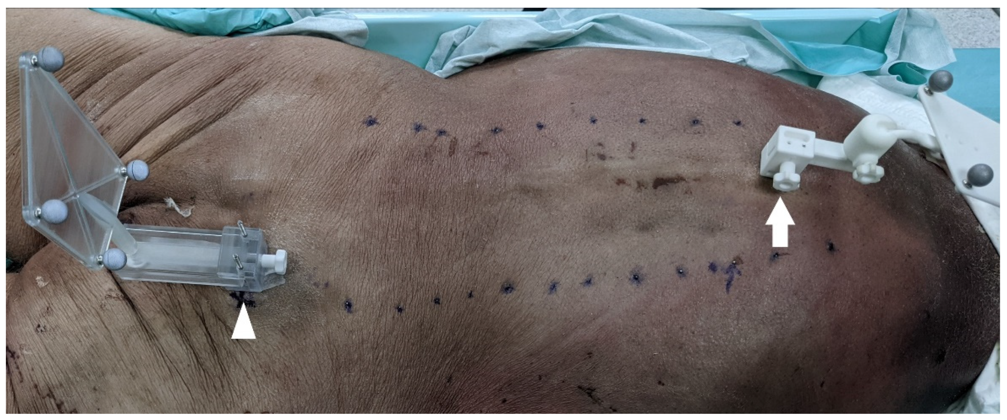
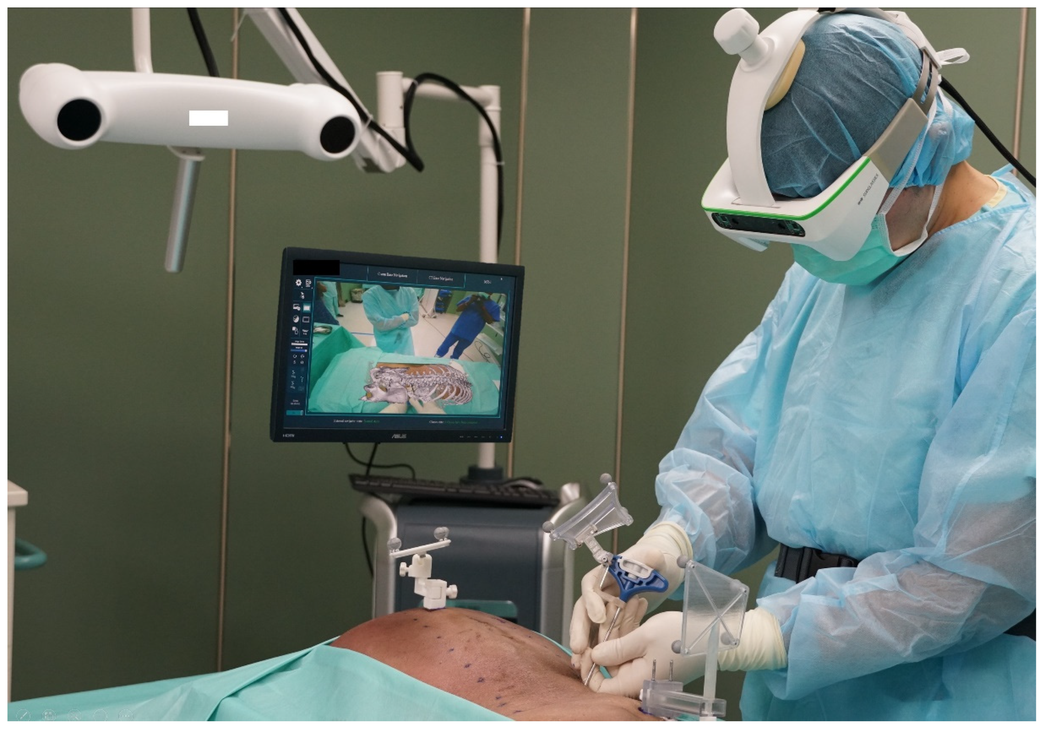
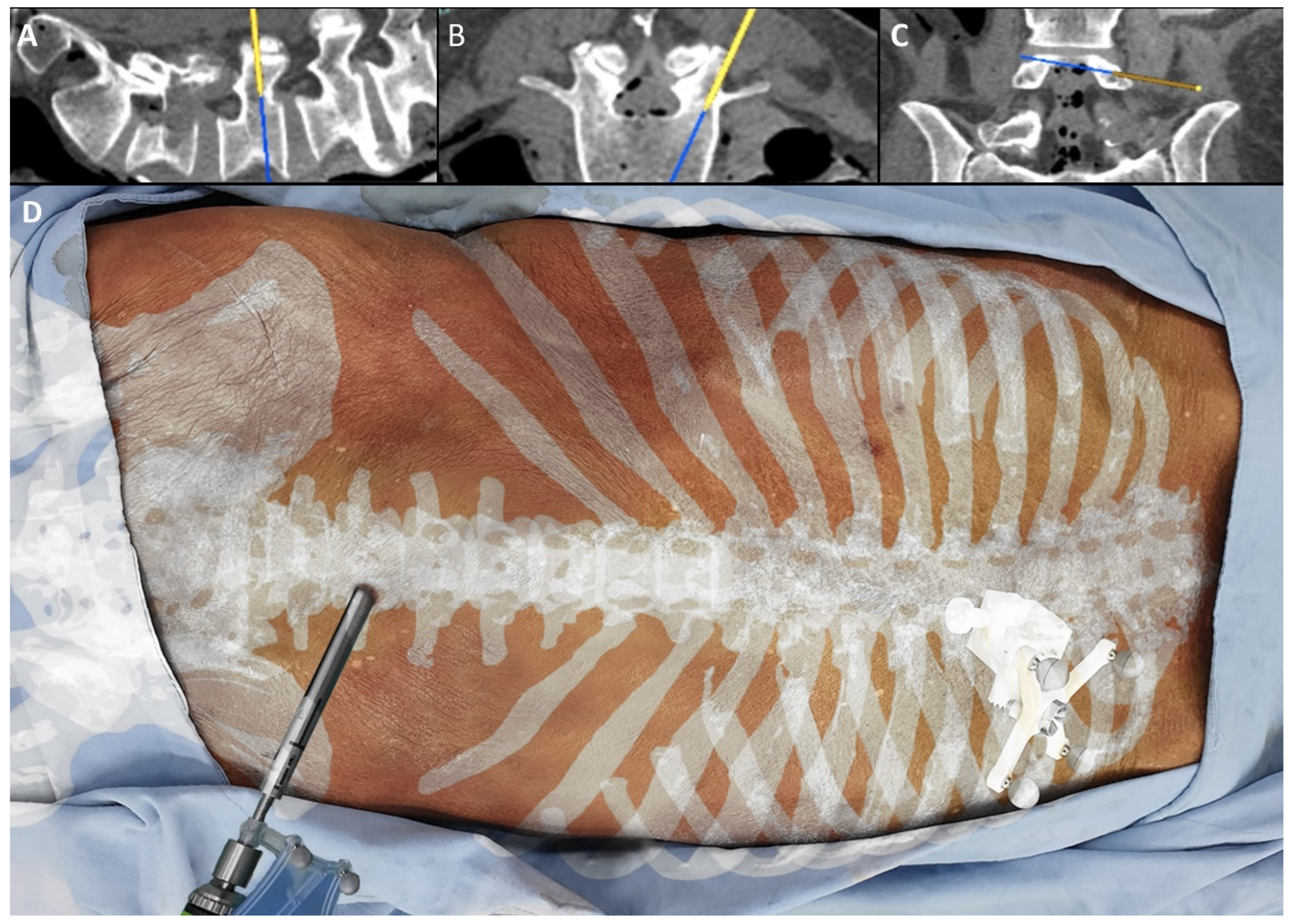
2.2. Instrumentation Method

The surgeons made a 2 cm transverse skin incision based only on the 3D image projection. The 3D image was then used to advance a Jamshidi needle through the soft tissue directly down to the entry point of the pedicle screw (Figure 2).
The AR technique allowed for 3D anatomical visualization of the spine. It also allowed a simultaneous view of standard axial and sagittal reconstruction 2D CT images (Figure 3).
According to the axial and sagittal 2D images, the surgeons determined the craniocaudal and medial-lateral trajectory to advance the Jamshidi needle through the pedicle into the vertebral body. The Jamshidi needle was then removed after placing a k-wire. The cannulated pedicle screws were subsequently placed. All inserted thoracic screws (T6–T12) were 5.0 mm in diameter and 35–40 mm in length. All inserted lumbar screws (L1–L5) were 6.0 or 7.0 mm in diameter and 45–50 mm in length.

2.3. Radiographic Accuracy Analysis

A post-operative CT scan was obtained and sent to two neurosurgeons for an accuracy grading. The grading was performed on axial, coronal, and sagittal reconstruction CT images. The Gertzbein–Robbins grading for clinical accuracy evaluation was used: grade 0 (screw within the pedicle without cortical breach), grade 1 (0–2 mm breach, minor perforation), grade 2 (2–4 mm breach, moderate breach), and grade 3 (more than 4 mm breach, severe displacement) [10]. According to the Gertzbein–Robbins scale, grades 0 and 1 are considered accurate. However, when surgeons place pedicle screws toward an in-out-in trajectory, it is recorded as a lateral breach, but it would be excluded from the final accurate calculation. The Gertzbein–Robbins grading published early in the 1980s required that the trajectory be entirely intrapedicular.

3. Results

A total of 24 pedicle screws were placed in one cadaver. There are 14 screws in the thoracic spine (T6–T12) and 10 screws in the lumbar spine (L1–5). Three screws in the thoracic spine (Rt T6–8) were completely out of the pedicle, of which three screws were a grade 3 medial breach. Another five screws (bilateral L1, L2, and Rt L3) had the in–out–in trajectory and were recorded as a grade 3 lateral breach. As mentioned in Section 2, the five in–out–in screws were considered accurate. The overall accuracy of the study was 87.5% (Table 1).

4. Discussion

In the modern spine surgical field, the development of MISS is important. Combined with tubular decompression and percutaneous instrumentation, MISS features a small wound, less blood loss, less post-operative pain, quick recovery, and a short hospital stay. It has been proven to be efficient in surgery of degenerative lumbar spondylosis, spondylolisthesis, and short segment scoliosis [11,12,13,14,15,16]. However, percutaneous screw instrumentation is an experience-demanding technique, and it relies greatly on image guidance. The traditional method uses biplanar fluoroscopy for guidance. Compared to open surgery, fluoroscopy-guided percutaneous screw instrumentation has a steep learning curve that causes beginners to encounter some difficulties. Firstly, the surgeon must be remarkably familiar with the anatomical structure so that he or she can use 2D images (anteroposterior and lateral fluoroscopy) to reconstruct a 3D image in their mind. If the patient has experienced severe degeneration, it is difficult to find the proper entry point and trajectory. Secondly, surgeons must change their line of sight frequently from the surgical field to a remote screen, which inevitably leads to attention shifts and hand movements. It is an unintuitive way to perform the surgery. Hand–eye coordination is crucial to the success of instrumentation. Recently, anatomical identification experienced great improvement by introducing a computer navigation system. However, the problem of sightline change persisted. This problem has not been solved yet, even using state-of-the-art robots.
With head-mounted goggles, AR could provide an immersive experience through the direct retinal projection of a 3D spine image onto the patient. Meanwhile, the 2D navigation images are also projected onto the AR goggles. The AR goggles could provide the necessary information so that surgeons could stay focused on the surgical field without changing their line of sight. Thus, the AR-assisted instrumentation procedure is an intuitive way to perform surgery, and the whole process is like open pedicle screw placement. This method could effectively reduce surgical time and improve the accuracy of screw placement.
AR-assisted pedicle screw instrumentation is a novel technology. Until now, there are several systems launched and waiting for final FDA approval. The published data are scarce on this technology. Elmi-Terander et al. reported on two cadaveric reports describing AR technology. Although the authors documented the technology as AR, they did not use head-mounted equipment. Instead, they overlaid the navigation data onto a remote screen, which was similar to the current computer navigation system. The first cadaveric study documented the insertion of 47 pedicle screws with an accuracy of 85% [17]. The second report described the placement of 18 pedicle screws with an accuracy of 89% [6].
Molina et al. also published two cadaveric reports using the same AR technology. The first report documented a 94.6% success rate from 120 pedicle screws’ placement [9]. In the second report, the authors inserted 113 instruments, including 93 pedicle screws and 20 Jamshidi needles. Their overall accuracy was 99.1% [8]. Urakov et al. published two AR-assisted pedicle screw studies using different AR systems. In the first study, they described a cadaveric study using the OpenSight AR system (Novarad, American Fork, UT, USA) and Hololens AR glasses (Microsoft, Redmond, WA, USA). They reported that 14 out of 19 AR-assisted pedicle screws had grade 2 or grade 3 breaches. The overall accuracy was 26.3% [5]. The second was a saw bone study using the Caduceus AR system (Caduceus, Taiwan Main Orthopedics Biotechnology Co., Taiwan). The author placed 60 screws on five saw bones with the final accuracy of 98.33% [18]. Liu et al. reported another AR-assisted saw bone study. They documented a 94% accuracy rate from 80 pedicle screws [7]. According to the published series, the accuracy rate varied between different AR systems. In the current cadaveric study, the authors reported an 87.5% accuracy from 24 pedicle screw placements.
Further analysis of those inaccurate screws revealed that there were eight screws with grade 3 breaches in the current study. Among them, five screws had grade 3 lateral breaches over bilateral L1/2 and Rt L3. The torso had a very narrow pedicle over L1 and L2 (<5 mm), and both surgeons utilized the in–out–in technique to place the screws. Due to poor bone quality, there was a bone fracture over the transverse process–facet junction during Rt L3 screw placement that resulted in the lateral displacement of the screw. In the post-operative CT images, the authors could appreciate the original, accurate tract before lateral displacement. All lateral breach screws were recorded, but those screws were considered accurate. There were three screws with grade 3 medial breach, which occurred over Rt T6/7/8. The three inaccurate screws were the most distal screws from the iliac reference, which might yield inaccuracy in both the navigation and the 3D hologram. In the current study, the accuracy limitation of this AR system might be within five to seven spine segments away from the reference. The more distant from the reference, the more inaccurate the system. These results provided important information for the future study of this AR system.
There are limitations to the current study. First, this is a single cadaveric study. The authors are fully aware that this number is not enough. However, as a pilot study, the main purpose of this study is to validate the feasibility and accuracy of AR-assisted percutaneous screw instrumentation. More studies are required before this novel technology can be applied clinically. Meanwhile, there is room for improvement in the AR goggles. The goggles were heavy and became a burden to the surgeons. Surgeons experienced headaches and dizziness after long-time use of the AR system, which might have a negative impact on the study result. Another limitation is that the torso had poor bone quality. Part of the inaccuracy may come from the displacement of the screws or micromovement of the reference. The current study provides preliminary data, and further studies are needed to validate the feasibility of AR systems in spine surgery.

5. Conclusions

AR-assisted percutaneous pedicle screw instrumentation could improve anatomical visualization, facilitate surgical workflow, and provide an intuitive method of surgery.

6. Disclosures

The authors have no financial connection with any of the companies listed in this manuscript. This is an investigator-driven project, and the manuscript was written solely by the authors with no influence from the companies.

Author Contributions

Conceptualization, C.-C.C., C.-H.K. and T.-H.T.; methodology, C.-C.C., C.-H.K. and H.-K.C.; validation, T.-H.T., L.-Y.F. and J.-C.W.; formal analysis, C.-C.C. and L.-Y.F.; investigation, C.-C.C. and C.-H.K.; data curation, C.-C.C.; writing—original draft preparation, C.-C.C.; writing—review and editing, C.-C.C., J.-C.W. and T.-H.T.; visualization, C.-C.C.; supervision, J.-C.W.; project administration, H.C. and W.-C.H. All authors have read and agreed to the published version of the manuscript.

Funding

This research received no external funding.

Institutional Review Board Statement

Not applicable.

Informed Consent Statement

Not applicable.

Conflicts of Interest

The authors declare no conflict of interest.

References

  1. Edström, E.; Burström, G.; Omar, A.; Nachabe, R.; Söderman, M.; Persson, O.; Gerdhem, P.; Elmi-Terander, A. Augmented Reality Surgical Navigation in Spine Surgery to Minimize Staff Radiation Exposure. Spine 2020, 45, E45–E53. [Google Scholar] [CrossRef] [PubMed]
  2. Mishra, R.; Narayanan, M.D.K.; Umana, G.E.; Montemurro, N.; Chaurasia, B.; Deora, H. Virtual Reality in Neurosurgery: Beyond Neurosurgical Planning. Int. J. Environ. Res. Public Health 2022, 19, 1719. [Google Scholar] [CrossRef] [PubMed]
  3. Fiani, B.; De Stefano, F.; Kondilis, A.; Covarrubias, C.; Reier, L.; Sarhadi, K. Virtual Reality in Neurosurgery: “Can You See It?”—A Review of the Current Applications and Future Potential. World Neurosurg. 2020, 141, 291–298. [Google Scholar] [CrossRef] [PubMed]
  4. Condino, S.; Montemurro, N.; Cattari, N.; D’Amato, R.; Thomale, U.; Ferrari, V.; Cutolo, F. Evaluation of a Wearable AR Platform for Guiding Complex Craniotomies in Neurosurgery. Ann. Biomed. Eng. 2021, 49, 2590–2605. [Google Scholar] [CrossRef] [PubMed]
  5. Urakov, T.M.; Wang, M.Y.; Levi, A.D. Workflow Caveats in Augmented Reality-Assisted Pedicle Instrumentation: Cadaver Lab. World Neurosurg. 2019, 126, e1449–e1455. [Google Scholar] [CrossRef] [PubMed]
  6. Elmi-Terander, A.; Nachabe, R.; Skulason, H.; Pedersen, K.; Söderman, M.; Racadio, J.; Babic, D.; Gerdhem, P.; Edström, E. Feasibility and Accuracy of Thoracolumbar Minimally Invasive Pedicle Screw Placement With Augmented Reality Navigation Technology. Spine 2018, 43, 1018–1023. [Google Scholar] [CrossRef] [PubMed]
  7. Liu, H.; Wu, J.; Tang, Y.; Li, H.; Wang, W.; Li, C.; Zhou, Y. Percutaneous placement of lumbar pedicle screws via intraoperative CT image-based augmented reality-guided technology. J. Neurosurg. Spine 2019, 32, 542–547. [Google Scholar] [CrossRef] [PubMed]
  8. Molina, C.A.; Phillips, F.M.; Colman, M.W.; Ray, W.Z.; Khan, M.; Orru, E.; Poelstra, K.; Khoo, L. A cadaveric precision and accuracy analysis of augmented reality-mediated percutaneous pedicle implant insertion. J. Neurosurg. Spine 2020, 34, 316–324. [Google Scholar] [CrossRef] [PubMed]
  9. Molina, C.A.; Theodore, N.; Ahmed, A.K.; Westbroek, E.M.; Mirovsky, Y.; Harel, R.; Orru, E.; Khan, M.; Witham, T.; Sciubba, D.M. Augmented reality-assisted pedicle screw insertion: A cadaveric proof-of-concept study. J. Neurosurg. Spine 2019, 31, 139–146. [Google Scholar] [CrossRef] [PubMed]
  10. Gertzbein, S.D.; Robbins, S.E. Accuracy of pedicular screw placement in vivo. Spine 1990, 15, 11–14. [Google Scholar] [CrossRef] [PubMed]
  11. Choy, W.; Miller, C.A.; Chan, A.K.; Fu, K.M.; Park, P.; Mummaneni, P.V. Evolution of the Minimally Invasive Spinal Deformity Surgery Algorithm: An Evidence-Based Approach to Surgical Strategies for Deformity Correction. Neurosurg. Clin. N. Am. 2018, 29, 399–406. [Google Scholar] [CrossRef] [PubMed]
  12. Mummaneni, P.V.; Park, P.; Shaffrey, C.I.; Wang, M.Y.; Uribe, J.S.; Fessler, R.G.; Chou, D.; Kanter, A.S.; Okonkwo, D.O.; Mundis, G.M.; et al. The MISDEF2 algorithm: An updated algorithm for patient selection in minimally invasive deformity surgery. J. Neurosurg. Spine 2019, 32, 221–228. [Google Scholar] [CrossRef] [PubMed]
  13. Chou, D.; Mundis, G.; Wang, M.; Fu, K.M.; Shaffrey, C.; Okonkwo, D.; Kanter, A.; Eastlack, R.; Nguyen, S.; Deviren, V.; et al. Minimally Invasive Surgery for Mild-to-Moderate Adult Spinal Deformities: Impact on Intensive Care Unit and Hospital Stay. World Neurosurg. 2019, 127, e649–e655. [Google Scholar] [CrossRef] [PubMed]
  14. Hussain, I.; Fu, K.M.; Uribe, J.S.; Chou, D.; Mummaneni, P.V. State of the art advances in minimally invasive surgery for adult spinal deformity. Spine Deform. 2020, 8, 1143–1158. [Google Scholar] [CrossRef] [PubMed]
  15. Park, P.; Than, K.D.; Mummaneni, P.V.; Nunley, P.D.; Eastlack, R.K.; Uribe, J.S.; Wang, M.Y.; Le, V.; Fessler, R.G.; Okonkwo, D.O.; et al. Factors affecting approach selection for minimally invasive versus open surgery in the treatment of adult spinal deformity: Analysis of a prospective, nonrandomized multicenter study. J. Neurosurg. Spine 2020, 33, 601–606. [Google Scholar] [CrossRef] [PubMed]
  16. Wang, M.Y.; Uribe, J.; Mummaneni, P.V.; Tran, S.; Brusko, G.D.; Park, P.; Nunley, P.; Kanter, A.; Okonkwo, D.; Anand, N.; et al. Minimally Invasive Spinal Deformity Surgery: Analysis of Patients Who Fail to Reach Minimal Clinically Important Difference. World Neurosurg. 2020, 137, e499–e505. [Google Scholar] [CrossRef] [PubMed]
  17. Elmi-Terander, A.; Skulason, H.; Söderman, M.; Racadio, J.; Homan, R.; Babic, D.; van der Vaart, N.; Nachabe, R. Surgical Navigation Technology Based on Augmented Reality and Integrated 3D Intraoperative Imaging: A Spine Cadaveric Feasibility and Accuracy Study. Spine 2016, 41, E1303–E1311. [Google Scholar] [CrossRef] [PubMed] [Green Version]
  18. Urakov, T.M. Augmented Reality-assisted Pedicle Instrumentation: Versatility Across Major Instrumentation Sets. Spine 2020, 45, E1622–E1626. [Google Scholar] [CrossRef]
Figure 1. Demonstration of cadaver preparation. Arrowhead: iliac crest reference frame. Arrow: spinous process clamp and registration marker. Blue dots: surface fiducial markers.
Applsci 12 05261 g001
Figure 2. Demonstration of lab settings. The head-mounted goggles have a tracking camera to enhance accuracy and a video camera to share the surgeon’s view with OR staff.
Applsci 12 05261 g002
Figure 3. Demonstration of views through the goggles. (AC) The projected 2D navigation images. (D) The superimposed 3D spine holographic image. The screw entry point was easily identified with the 3D holographic image. The trajectory was then determined by 2D navigation images.
Applsci 12 05261 g003
Table 1. Screw insertion accuracy according to Gertzbein–Robbins grade.
Screw Side
LtRt
T63 (M)0
T73 (M)0
T83 (M)0
T900
T1000
T1100
T1200
L13 (I/O)3 (I/O)
L23 (I/O)3 (I/O)
L33 (I/O)0
L400
L500
Overall accuracy87.5%
M: medial breach; I/O in–out–in trajectory.
Publisher’s Note: MDPI stays neutral with regard to jurisdictional claims in published maps and institutional affiliations.

