Article

Single Puncture TIPS—A 3D Fusion Image-Guided Transjugular Intrahepatic Portosystemic Shunt (TIPS): An Experimental Study

1
Division of Interventional Radiology, Department of Radiology, UCLA Medical Center, David Geffen School of Medicine at UCLA, Los Angeles, CA 90095, USA
2
Division of Liver and Pancreas Transplantation, Department of Surgery, David Geffen School of Medicine at University of California, Los Angeles, CA 90095, USA
3
Dumont-UCLA Transplant Center, University of California, Los Angeles, CA 90095, USA
4
Division of Hepatology, Department of Medicine, UCLA Medical Center, David Geffen School of Medicine at UCLA, Los Angeles, CA 90095, USA
*
Author to whom correspondence should be addressed.
Submission received: 12 April 2022 / Revised: 16 May 2022 / Accepted: 20 May 2022 / Published: 23 May 2022

Abstract

:
Background: The use of a transjugular intrahepatic portosystemic shunt (TIPS) has been established as an effective treatment for portal hypertension. Despite the rapid development of this use, serious peri-procedural complications have been reported in over 10% of cases. This has largely been attributed to the access to the portal vein, also referred to as a “blind puncture”, which often requires multiple attempts. The aim of this study was to demonstrate the safety, reproducibility and accuracy of the use of real-time 3D fusion image-guided (3DFIG) single puncture TIPS to minimize the complications that are related to the “blind puncture” of TIPS procedures. Methods: A 3DFIG TIPS approach was utilized on 22 pigs by combining pre-procedural cross-sectional imaging (CT, MR or CBCT) with intra-procedural cone beam CT or angiogram imaging, which allowed for the improved 3D visual spatial orientation of the portal vein and real-time tracking of the needle in 3D. Results: Thirty-five portosystemic shunts were successfully deployed in all 22 subjects without any peri-procedural complications. Overall, 91% (32/35) of the procedures were carried out using a single puncture. In addition, the mean fluoroscopy time in our study was more than 12 times lower than the proposed reference level that has previously been proposed for TIPS procedures. Conclusion: Multi-modality real-time 3DFIG TIPS can be performed safely using a single puncture, without complications, and can potentially be used in both emergency and non-emergency clinical situations.

1. Introduction

The use of a transjugular intrahepatic portosystemic shunt (TIPS) is an effective treatment for portal hypertension and its associated complications, which has made it a widely used tool worldwide since the 1980s [1,2]. Multiple large clinical trials have confirmed that TIPS procedures play a major role in mitigating the severe consequences of portal hypertension, such as variceal bleeding and intractable ascites [3,4,5,6,7,8]. However, despite the numerous advantages of TIPS procedures, severe peri-procedural complications have been reported to occur in over 10% of cases, including transcapsular puncture, the accidental puncture of non-target structures and the puncture of the extrahepatic portal vein (PV) [9,10,11,12].
The primary source of procedure-related complications is associated with the most technically challenging step of the TIPS procedure: gaining access to the PV, which is often referred to as a “blind puncture” [9,13,14,15]. In this step, the operator has no visualization or real-time information of the spatial relationship between the systemic and portal venous systems. This “blind puncture” can make it more likely that the operator punctures areas other than the PV, such as the liver capsule, bile duct or hepatic artery and the extrahepatic PV, which may result in massive intraperitoneal hemorrhage or death. In rare cases, operators have been reported to make up to 50 attempts before successfully accessing the PV [16], which increases the risk of puncture-related complications. The difficult nature of this procedure can largely depend on the following factors: the malformation/transformation of a chronically diseased liver, the displacement of the liver due to ascites, thrombosed or cavernous portal/hepatic veins and the anatomic and pathologic variants of the portal architecture. In some of these cases, the selection of the correct PV may be critical for the effectiveness of the TIPS procedure [17,18], suggesting that the current standard technique of blind punctures may be inadequate. The use of a direct intrahepatic portosystemic shunt (DIPS), which is a technical variant of the TIPS procedure that relies on intravascular US, has been proposed as being preferable for certain patients, but has the same inherent limitations as current image-guided TIPS procedures [19].
In our study, we developed a new technique for targeting the portal vein during TIPS procedures. By fusing angiographic imaging (cone beam computed tomography; CBCT) and cross-sectional 2D images with CBCT, MR or computed tomography (CT), we aimed to evaluate the safety, reproducibility and accuracy of a real-time 3D fusion image-guided (FIG) TIPS procedure in vivo.

2. Materials and Methods

2.1. Animals

This retrospective study was approved by our institutional animal research committee (ARC#2006-054-33A). Twenty-two female Yorkshire pigs at a weight of 30–50 kg (~15 weeks old) were obtained and maintained by the Division of Laboratory Animal Medicine at our institution. The animals were maintained in group housing, either in pens or individual cages, and were fed a standard laboratory swine diet (LabDiet®, St. Louis, MO, USA). All animals received appropriate humane care from trained professional staff in compliance with the ARRIVE guidelines, the Principals of Laboratory Animal Care and the Guide for the Care and Use of Laboratory Animals, which was approved by the Animal Care and Use Committees of our institution and in accordance with NIH guidelines. All animals were fasted for at least 12 h before the procedure. Of 35 procedures on 22 animals: 10 procedures utilized TIPS with the use of CT-, CBCT- and angiogram-fused image guidance; 10 procedures used MR-, CBCT- and angiogram-fused image guidance; 7 animals underwent only CBCT- and angiogram-fused image guidance; and 8 animals underwent DIPS using pre-procedural CT-, CBCT- and angiogram-fused images.

2.2. General Anesthesia

General anesthesia was induced based on the National Institute of Health (NIH) guidelines for all TIPS procedures and pre-procedural MRI imaging. Each pig was placed in the supine position. For initial sedation, Telazol 4–8 mg/kg (Zoetis, Parisppanany, NJ, USA) was administered intramuscularly (IM). Anesthesia was then maintained by inhaled 1–3.5% isoflurane in oxygen. Pancuronium (0.1 mg/kg) was administered prior to all TIPS procedures to achieve reversible paralysis, which was monitored by evoked motor response. Blood pressure and EKG were also monitored continuously.

2.3. Pre-Procedural Imaging

2.3.1. CT

Pre-procedural contrast-enhanced CT images were taken using a 64-slice multi-detector CT (Somatom Sensation, Siemens, Forscheim, Germany). After the initial unenhanced images of the abdomen were obtained, Iohexol 1.5 mL/kg (Omnipaque 300 mg/mL; GE, Princeton, NJ, USA) was power injected through a peripheral vein in the forelimb or another peripheral vein when a forelimb vein was inaccessible. This was followed by an injection of 40 mL of saline at a rate of 0.4 mL/s and images were subsequently acquired during both the arterial dominant phase (20 s after injection) and in the portal venous dominant phase (60 s after injection). The following CT parameters were used: 250 mA; 120 kVp; 3-mm collimation; and 2:1 pitch. The pigs were not maintained on anesthetic during the CT procedure as it took less than 5 min to complete the study.

2.3.2. MR Imaging

Pre-procedural MR imaging was performed using a 3T MR imaging unit (Siemens Healthcare, Malvern, PA, USA) and consisted of both unenhanced and contrast-enhanced dynamic (arterial and portal venous) fat-saturated T1-weighted gradient-echo imaging (repetition time msec/echo time msec, 257/2.32; thickness, 5 mm; acquisitions, two; field of view, 200 × 300 mm; and matrix, 256 × 256) and unenhanced T2-weighted fat-suppressed turbo spin-echo imaging (TR/TE, 4515/82; 5 mm; flip angle, 140; and field of view, 320 × 272 mm). The dynamic contrast-enhanced T1-weighted imaging was performed after the administration of an intravenous contrast agent (gadodiamide 0.1 mmol/kg, Omniscan; Nycomed, Zürich, Switzerland).

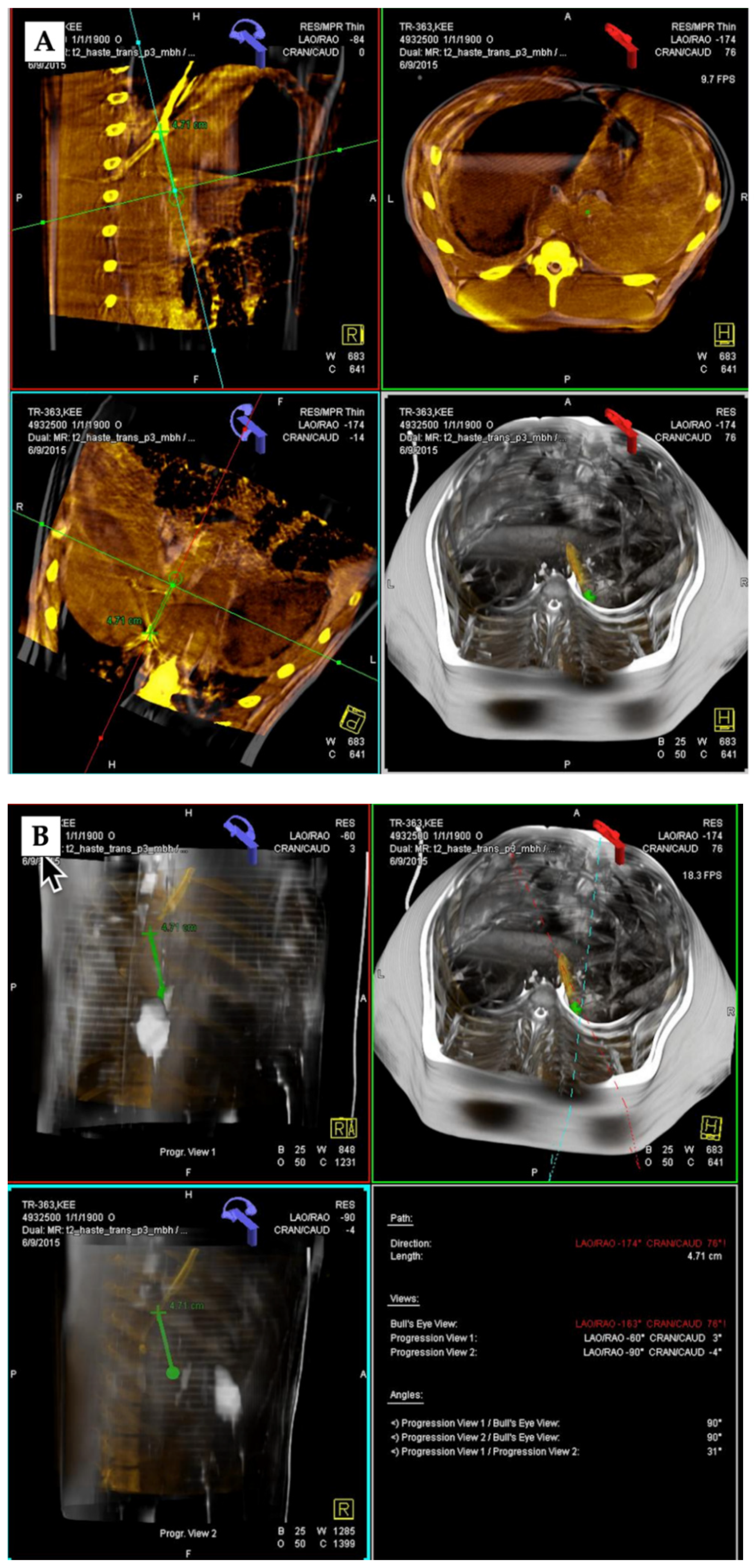
2.4. Cone Beam CT Imaging

Imaging was performed using commercially available flat-panel detectors (Artis Zeego, Siemens, Forcheim, Germany). The 2D and 3D images were acquired using CBCT technology (DynaCT, Siemens, Forcheim, Germany) and volumetric image reconstruction (modified Feldkamp back projection) was subsequently performed at a dedicated workstation. For each cone beam CT scan, 312 projection images (30 frames per second) were acquired, which covered a 200° clockwise arc at a rotation speed of 20° per second. As the images were being acquired, the projections were transferred to the reconstruction workstation. The two-dimensional projection images were reconstructed using modified Feldkamp back projection into three-dimensional volumetric images, which has an isotropic resolution of 0.98 mm for a 250 mm × 250 mm × 194 mm field of view (matrix size, 256 × 256 × 256).

2.5. TIPS and DIPS Creation Using 3D Image Guidance

Following the pre-procedural imaging, the animals were transported to the interventional imaging suite, which was equipped with rotational CBCT angiography and 3D reconstruction-rendered real-time angiography. All procedures were performed using an angiographic C-arm imaging system that utilized a flat-panel x-ray detector (Artis Zeego, Siemens, Forcheim, Germany). The animals were placed under the C-arm and general anesthesia was maintained with 1–3.5% isoflurane. All procedures were carried out by the same interventional radiologist who had 10 years of post-fellowship experience, including over 300 TIPS procedures. Next, the right internal jugular vein (RIJV) was accessed under US guidance. A 10F sheath was then advanced through the RIJV access point and either a glide catheter or multipurpose angiography (MPA) catheter was used to catheterize the right hepatic vein (HV). A Rösch-Uchida TIPS needle (Cook Inc., Bloomington, IN, USA) was then placed over the wire in place of the glide/MPA catheter. Once the Rösch-Ushida needle was placed in the right HV, a CBCT (DynaCT, Siemens, Forcheim, Germany) was obtained using the rotational angiography apparatus.
The pre-procedural CT or MR images were retrieved from a PACS system at the angiographic system workstation (Syngo X-Workplace, Siemens, Forcheim, Germany). The 3D-reconstructed CT or MR images were loaded into the InSpace® application (Siemens, Forcheim, Germany) and subsequently fused with the 3D-reconstructed CBCT images using the iGuide® software (Siemens, Forcheim, Germany). This fused 3D-reconstructed roadmap was overlaid onto the real-time fluoroscopic image. The tip of the Rösch-Ushida needle and the target portal branch were identified on the images and the optimal path from the needle to the target puncture site of the PV was automatically mapped out by the iGuide® software. Under real-time 3D fluoroscopic guidance, the operator then advanced the TIPS needle into the PV following the determined path. Once the iGuide® PV puncture was made, venography was performed to confirm access to the portal system. For the DIPS procedures, the tip of the Rösch-Ushida needle was placed in the intrahepatic IVC instead of the right HV and the imaging fusion, 3D fluoroscopic guidance and portal puncture were performed as in the TIPS procedure.

2.6. Data Collection and Statistical Analysis

The procedural success and complication rates, time required to create the 3D fusion image guidance (3DFIG) images, total procedure time, total fluoroscopy radiation time and the number of PV puncture attempts were determined for all procedures. Continuous data are presented as the means and standard deviations (SDs) or medians and interquartile ranges (IQRs) whereas categorical data are presented as proportions and percentages. The data and statistical analyses were performed using SPSS software (SPSS v. 25.0, IBM, Chicago, IL, USA).

3. Results

3.1. Basic Assessment

Technically successful 3D-fused angiography-guided TIPS (n = 27) or DIPS (n = 8) procedures were performed in 35/35 (100%) cases (Figure 1, Figure 2, Figure 3 and Figure 4). No immediate procedure-related complications were noted during or immediately after the TIPS or DIPS procedures in any of the 22 swine (0/35; 0%).

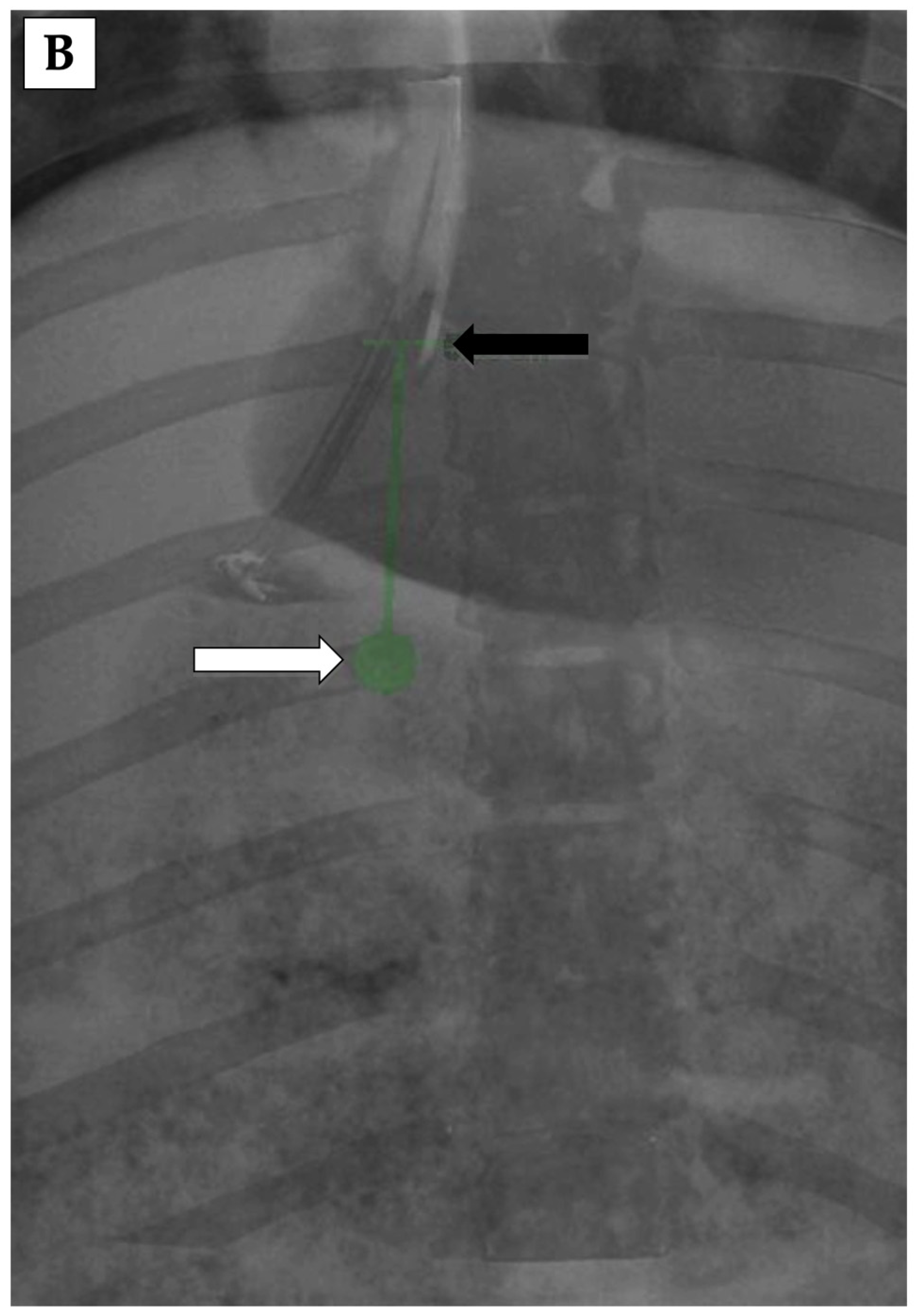
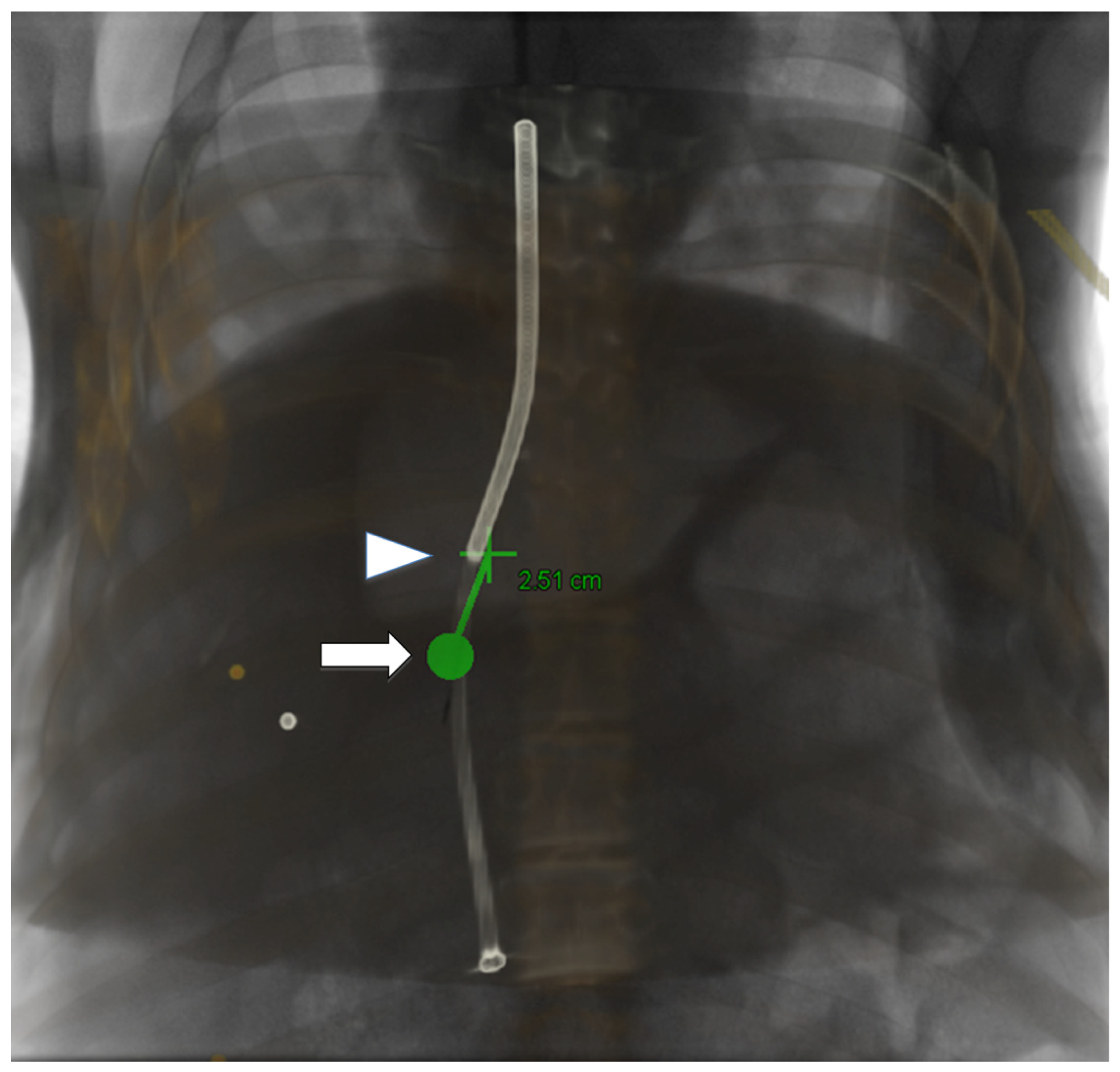
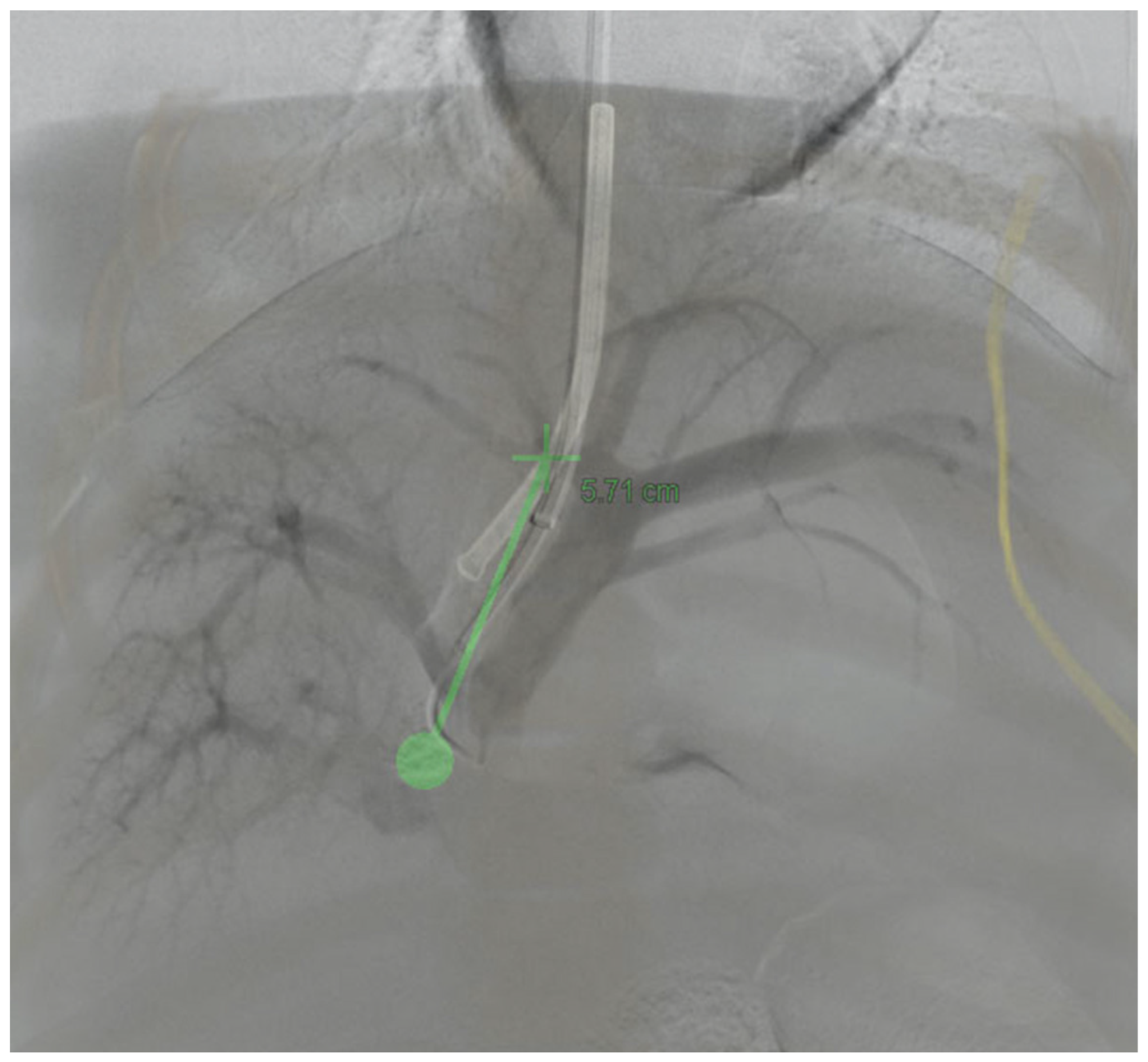
3.2. 3D Angiography-Guided TIPS Procedure Using Pre-Procedural Cross-Sectional Imaging with CBCT/Angiography-Fused Images

The 3DFIG TIPS procedures that utilized CT and MR were successfully performed using a single PV puncture attempt in 90% (9/10) and 90% (9/10) of cases, respectively, and using two attempts in the remaining cases. The mean total procedure time (TPT: time from access to the right internal jugular vein (RIJV) to access to the PV) was 22.4 ± 5.1 and 18.7 ± 3.1 min for the CT and MR 3DFIG procedures, respectively. The median fluoroscopy time (MFT: fluoroscopy time between access to the RIJV to access to the PV) for both CT and MR 3DFIG procedures was 4.0 min (IQR, 3.0–5.3). The total time required to create the 3D images (TTC3D: time from importing images to 3D fusion to the creation of the iGuide® map) was 8.5 ± 3.4 and 7.5 ± 2.7 min for the CT and MR 3DFIG procedures, respectively (Table 1).

3.3. 3D Angiography-Guided TIPS Using CBCT/Angiography-Fused Images

To determine whether the procedure could be carried out without pre-procedural imaging, 3DFIG TIPS procedures were carried out using only CBCT/angiography-fused images. TIPS procedures were successfully carried out using a single stick in 100% (7/7) of cases. The TPT and TTC3D were 14.4 ± 1.0 and 3.0 ± 0.8 min, respectively. The median fluoroscopy time (MFT) was 3.0 min (IQR, 3.0–3.0). All three parameters (TPT, TTC3D and MFT) were significantly shorter during the 3DFIG TIPS procedures, with only CBCT/angiography being comparable to 3DFIG using CT/MR and CBCT/angiography (p < 0.0001).

3.4. 3D Angiography-Guided DIPS Using CT/CBCT/Angiography-Fused Images

DIPS procedures were successfully performed on the first attempt in 88% (7/8) of cases and after two attempts in the remaining case. The TPT and TTC3D were 14.1 ± 1.1 and 3.9 ± 0.7 min, respectively. The median fluoroscopy time was 3.0 min (IQR, 3.0–3.0).

4. Discussion

There is a major need for ancillary systems that are able to enhance the ability of operators to locate and enter the portal vein with relative ease. Many researchers have attempted to solve this problem by introducing various methods, such as guidance by peri-operative ultrasound (US), magnetic resonance (MR) or intraprocedural carbon dioxide (CO2) angiography, but no method has consistently been able to successfully produce single puncture PV access in large cohorts [11,13,20,21,22]. Furthermore, these methods can increase procedure length and the risk of other complications, with little benefit to streamlining the PV puncture step [23].
In this study, we demonstrated the feasibility of pre-procedural 3D fusion imaging for guiding TIPS and DIPS procedures. Specifically, we showed that the 3DFIG approach works by combining pre-procedural cross-sectional imaging (CBCT, CT or MR) with intra-procedural CBCT or angiogram imaging for real-time 3D-guided TIPS placement. The utility of this approach has been demonstrated using pre-procedural CT imaging in patients, in which the efficiency of placing the TIPS was clearly demonstrated [24,25,26]. The potential for the use of pre-procedural cross-sectional imaging for 3DFIG TIPS procedures is vast, as most non-emergency patients have imaging available prior to their planned TIPS procedure. Due to underlying chronic liver disease, these patients are mostly followed by their hepatologists or gastroenterologists and also undergo routine surveillance imaging.
Minimizing procedural times while maintaining accuracy is critical, especially in urgent and emergency TIPS procedures that are used to treat acute variceal bleeding. Although there was a limited number of subjects, we showed that 3DFIG TIPS procedures using CBCT/angiography-fused images alone produced similar outcomes but with significantly shorter procedural times, which has important clinical implications for patients who do not have pre-procedural CT or MR imaging available to them [27]. Finally, we demonstrated the feasibility of using the 3DFIG DIPS procedure, which could prove to be especially relevant in patients for whom the traditional TIPS procedure is contraindicated [28].
The addition of 3DFIG to the TIPS and DIPS procedures led to a 100% success rate, with no complications and short fluoroscopy and procedure times. Most importantly, the number of needle puncture attempts that was required to gain PV access was low compared to the “blind” portosystemic needle punctures that are only guided by fluoroscopy [16,29]. Transvenous intrahepatic access to the PV is the most crucial and also most dangerous step of the TIPS procedure. The typical number of attempts that is required to access the PV has been reported to be between 3–5 attempts in the literature [16,29], although many more attempts may be needed in clinical practice, compared to the single pass success rate of 93% (25/27) for TIPS and 88% (7/8) for DIPS procedures in this study, with the remaining procedures requiring only two passes. Additionally, the mean fluoroscopy time in all procedures was shorter than 5 min, compared to the reference level of 60 min that has been proposed for TIPS procedures, which was derived from a RAD-IR study [30]. These benefits increase the safety profile and accuracy of the procedure and greatly reduce potential radiation exposure. Compared to other existing and proposed methods, the 3DFIG method provides users with the option to track the needle in real time, thereby facilitating the anatomical orientation by projecting fluoroscopic images onto 3D images to visualize the puncture target [11,13,20,21,22,31,32]. The planned puncture path can be adapted to different real-time projections, which allows the operator to make real-time reassessments of the path in 3D by simply adjusting the C-arm.
The 3DFIG method can enhance technical precision and accuracy during technically difficult TIPS procedures by providing: (1) improved 3D visual spatial orientation so the interventional radiologist can successfully puncture the portal vein; (2) 3D visualization of the patient’s unique, and possibly distorted, anatomy; (3) the real-time tracking of the needle in 3D, in both the anteroposterior and lateral planes.
Some potential disadvantages of the 3DFIG method and limitations of our study were also identified. First, with the added steps of CBCT and fusion imaging processing, the procedure time may increase as CBCT and fusion imaging can be time-consuming. This may be especially true for centers that have minimal experience in CBCT and fusion imaging. However, using the current settings in our research angiography unit and an independent fusion imaging station, the process was significantly streamlined. With proper training and an effective protocol set-up, 3DFIG procedures can be easily adapted. Furthermore, the time that could be saved by limiting the number of attempts that is needed using our method could potentially balance out, or even shorten, the overall procedure time. Another limitation of this study was that the 3DFIG TIPS procedures were performed in normal porcine livers, which did not have cirrhosis or anatomical anomalies. Patients with difficult anatomies may produce lower success rates and may require more needle passes to achieve successful PV puncture [9,12,15,18]. Consequently, the risk of peri-procedural complications, morbidity rates, procedure and fluoroscopy times and exposure to radiation also increase. However, this was a feasibility study, which tested the reproducibility of 3DFIG portosystemic shunt placement. Even though the procedure was not performed in cirrhotic livers, it was performed in small porcine livers to mimic the size of a human cirrhotic liver. Potential future studies could employ the 3DFIG TIPS procedure in cirrhotic animal models or in patients with cirrhosis as a clinical safety study. Other potential anatomic and physiologic considerations also remain, however. For example, polycystic liver disease (PCLD) is often listed as a contraindication for TIPS procedures due to risk of cyst rupture and hemorrhage. However, a case of PCLD was reported to have undergone successful TIPS insertion with the aid of a hybrid 2D and 3D imaging system [33]. PV and HV thrombosis have historically been considered to be relative contraindications due to the technical difficulties in placing a shunt in abnormal anatomies of portal and hepatic veins, although recent evidence has suggested that a TIPS, when placed successfully, is in fact crucial for the management of these patients [34]. Future research should evaluate the feasibility of the 3DFIG TIPS procedure for these patients as it could play a particularly useful role when the traditional TIPS procedure has been deemed too high a risk. Another potential limitation was that some of the pigs in this study underwent multiple procedures in order to increase the total number of procedures. In these cases, the knowledge of the previously placed shunts may have confounded any subsequent procedure attempts. For example, when a TIPS catheter was already in place before another was made, it would be revealed on the fluoroscopic images. However, this did not seem to affect the study results as the number of access attempts and the procedure time remained the same for the pigs that had prior procedures and those that did not.
In conclusion, our study showed that the 3DFIG TIPS (or iGuide® TIPS) procedure provides a more efficient and accurate way of performing the PV puncture step of the traditional procedure. The 3DFIG method is compatible with multiple imaging modalities and can potentially be used effectively in both emergency and non-emergency clinical situations. This hybrid technology provides the potential for the TIPS procedure to be used more widely as dependence on technical expertise may be less of a determining factor and it may be a suitable alternative for patients with underlying conditions who have traditionally been considered to be inappropriate candidates for TIPS placement. Regardless of the current limitations of the standard TIPS procedure, it continues to be a viable, and at times the only, method for treating and maintaining patients who are awaiting life-saving liver transplants.

Author Contributions

Conceptualization, E.W.L., S.S., P.-X.D. and S.T.K.; methodology, E.W.L., P.-X.D. and S.T.K.; validation, E.W.L., F.K., J.M. and S.T.K.; formal analysis, E.W.L., P.S. and N.E.; investigation, E.W.L., N.E. and P.-X.D.; writing—original draft preparation, E.W.L.; writing—review and editing, E.W.L., P.S., S.S., F.K., N.E., J.M., P.-X.D. and S.T.K.; supervision, E.W.L.; funding acquisition, E.W.L. All authors have read and agreed to the published version of the manuscript.

Funding

This research was funded by Siemens USA (grant number UCLA-2014-AX-Lee).

Institutional Review Board Statement

The animal study protocol was approved by the Institutional Review Board (or Ethics Committee) of University of California, Los Angeles (ARC#2006-054-33A: approved on 19 August 2014 and renewed on 16 November 2021).

Informed Consent Statement

Not applicable.

Data Availability Statement

Not applicable.

Conflicts of Interest

E.W.L. received research grant funding from Siemens USA. The authors declare no conflict of interest.

References

  1. Rossle, M.; Richter, G.M.; Noldge, G.; Palmaz, J.C.; Wenz, W.; Gerok, W. New non-operative treatment for variceal haemorrhage. Lancet 1989, 2, 153. [Google Scholar] [CrossRef]
  2. D’Amico, G.; Pagliaro, L.; Bosch, J. The treatment of portal hypertension: A meta-analytic review. Hepatology 1995, 22, 332–354. [Google Scholar] [CrossRef] [PubMed]
  3. Coldwell, D.M.; Ring, E.J.; Rees, C.R.; Zemel, G.; Darcy, M.D.; Haskal, Z.J.; McKusick, M.A.; Greenfield, A.J. Multicenter investigation of the role of transjugular intrahepatic portosystemic shunt in management of portal hypertension. Radiology 1995, 196, 335–340. [Google Scholar] [CrossRef] [PubMed]
  4. Ferral, H.; Bjarnason, H.; Wegryn, S.A.; Rengel, G.J.; Nazarian, G.K.; Rank, J.M.; Tadavarthy, S.M.; Hunter, D.W.; Castaneda-Zuniga, W.R. Refractory ascites: Early experience in treatment with transjugular intrahepatic portosystemic shunt. Radiology 1993, 189, 795–801. [Google Scholar] [CrossRef] [PubMed]
  5. LaBerge, J.M.; Ring, E.J.; Gordon, R.L.; Lake, J.R.; Doherty, M.M.; Somberg, K.A.; Roberts, J.P.; Ascher, N.L. Creation of transjugular intrahepatic portosystemic shunts with the wallstent endoprosthesis: Results in 100 patients. Radiology 1993, 187, 413–420. [Google Scholar] [CrossRef]
  6. LaBerge, J.M.; Somberg, K.A.; Lake, J.R.; Gordon, R.L.; Kerlan, R.K., Jr.; Ascher, N.L.; Roberts, J.P.; Simor, M.M.; Doherty, C.A.; Hahn, J.; et al. Two-year outcome following transjugular intrahepatic portosystemic shunt for variceal bleeding: Results in 90 patients. Gastroenterology 1995, 108, 1143–1151. [Google Scholar] [CrossRef]
  7. Ochs, A.; Rossle, M.; Haag, K.; Hauenstein, K.H.; Deibert, P.; Siegerstetter, V.; Huonker, M.; Langer, M.; Blum, H.E. The transjugular intrahepatic portosystemic stent-shunt procedure for refractory ascites. N. Engl. J. Med. 1995, 332, 1192–1197. [Google Scholar] [CrossRef]
  8. Rossle, M.; Haag, K.; Ochs, A.; Sellinger, M.; Noldge, G.; Perarnau, J.M.; Berger, E.; Blum, U.; Gabelmann, A.; Hauenstein, K.; et al. The transjugular intrahepatic portosystemic stent-shunt procedure for variceal bleeding. N. Engl. J. Med. 1994, 330, 165–171. [Google Scholar] [CrossRef]
  9. Freedman, A.M.; Sanyal, A.J.; Tisnado, J.; Cole, P.E.; Shiffman, M.L.; Luketic, V.A.; Purdum, P.P.; Darcy, M.D.; Posner, M.P. Complications of transjugular intrahepatic portosystemic shunt: A comprehensive review. Radiographics 1993, 13, 1185–1210. [Google Scholar] [CrossRef] [Green Version]
  10. Saxon, R.R.; Mendel-Hartvig, J.; Corless, C.L.; Rabkin, J.; Uchida, B.T.; Nishimine, K.; Keller, F.S. Bile duct injury as a major cause of stenosis and occlusion in transjugular intrahepatic portosystemic shunts: Comparative histopathologic analysis in humans and swine. J. Vasc. Interv. Radiol. 1996, 7, 487–497. [Google Scholar] [CrossRef]
  11. Kee, S.T.; Ganguly, A.; Daniel, B.L.; Wen, Z.; Butts, K.; Shimikawa, A.; Pelc, N.J.; Fahrig, R.; Dake, M.D. MR-guided transjugular intrahepatic portosystemic shunt creation with use of a hybrid radiography/MR system. J. Vasc. Interv. Radiol. 2005, 16, 227–234. [Google Scholar] [CrossRef]
  12. Gaba, R.C.; Khiatani, V.L.; Knuttinen, M.G.; Omene, B.O.; Carrillo, T.C.; Bui, J.T.; Owens, C.A. Comprehensive review of TIPS technical complications and how to avoid them. AJR Am. J. Roentgenol. 2011, 196, 675–685. [Google Scholar] [CrossRef]
  13. Rose, S.C.; Pretorius, D.H.; Nelson, T.R.; Kinney, T.B.; Huynh, T.V.; Roberts, A.C.; Valji, K.; D’Agostino, H.R.; Oglevie, S.B.; James, G.M.; et al. Adjunctive 3D US for achieving portal vein access during transjugular intrahepatic portosystemic shunt procedures. J. Vasc. Interv. Radiol. 2000, 11, 611–621. [Google Scholar] [CrossRef]
  14. Sanyal, A.J.; Shiffman, M.L. Transjugular intrahepatic portosystemic shunt: A medical perspective. Dig. Dis. 1995, 13, 153–162. [Google Scholar] [CrossRef]
  15. Saxon, R.R.; Keller, F.S. Technical aspects of accessing the portal vein during the TIPS procedure. J. Vasc. Interv. Radiol. 1997, 8, 733–744. [Google Scholar] [CrossRef]
  16. Funaki, B. Transjugular intrahepatic portosystemic shunt. Semin. Interv. Radiol. 2008, 25, 168–174. [Google Scholar] [CrossRef]
  17. Walser, E.M.; Soloway, R.; Raza, S.A.; Gill, A. Transjugular portosystemic shunt in chronic portal vein occlusion: Importance of segmental portal hypertension in cavernous transformation of the portal vein. J. Vasc. Interv. Radiol. 2006, 17, 373–378. [Google Scholar] [CrossRef]
  18. Jourabchi, N.; McWilliams, J.P.; Lee, E.W.; Sauk, S.; Kee, S.T. TIPS Placement via Combined Transjugular and Transhepatic Approach for Cavernous Portal Vein Occlusion: Targeted Approach. Case Rep. Radiol. 2013, 2013, 635391. [Google Scholar] [CrossRef]
  19. Petersen, B.; Uchida, B.T.; Timmermans, H.; Keller, F.S.; Rosch, J. Intravascular US-guided direct intrahepatic portacaval shunt with a PTFE-covered stent-graft: Feasibility study in swine and initial clinical results. J. Vasc. Interv. Radiol. 2001, 12, 475–486. [Google Scholar] [CrossRef]
  20. Adamus, R.; Pfister, M.; Loose, R.W. Enhancing transjugular intrahepatic portosystemic shunt puncture by using three-dimensional path planning based on the back projection of two two-dimensional portographs. Radiology 2009, 251, 543–547. [Google Scholar] [CrossRef] [Green Version]
  21. Kew, J.; Davies, R.P. Intravascular ultrasound guidance for transjugular intrahepatic portosystemic shunt procedure in a swine model. Cardiovasc. Interv. Radiol. 2004, 27, 38–41. [Google Scholar] [CrossRef]
  22. Roizental, M.; Kane, R.A.; Takahashi, J.; Kruskal, J.; Crenshaw, W.B.; Perry, L.; Stokes, K.; Clouse, M.E. Portal vein: US-guided localization prior to transjugular intrahepatic portosystemic shunt placement. Radiology 1995, 196, 868–870. [Google Scholar] [CrossRef]
  23. Fanelli, F. The Evolution of Transjugular Intrahepatic Portosystemic Shunt: Tips. ISRN Hepatol. 2014, 12, 762096. [Google Scholar] [CrossRef]
  24. Luo, X.; Wang, X.; Zhao, Y.; Ma, H.; Ye, L.; Yang, L.; Tsauo, J.; Jiang, M.; Li, X. Real-Time 3D CT Image Guidance for Transjugular Intrahepatic Portosystemic Shunt Creation Using Preoperative CT: A Prospective Feasibility Study of 20 Patients. AJR Am. J. Roentgenol. 2017, 208, W11–W16. [Google Scholar] [CrossRef]
  25. Rouabah, K.; Varoquaux, A.; Caporossi, J.M.; Louis, G.; Jacquier, A.; Bartoli, J.M.; Moulin, G.; Vidal, V. Image fusion-guided portal vein puncture during transjugular intrahepatic portosystemic shunt placement. Diagn. Interv. Imaging 2016, 97, 1095–1102. [Google Scholar] [CrossRef]
  26. Tacher, V.; Petit, A.; Derbel, H.; Novelli, L.; Vitellius, M.; Ridouani, F.; Luciani, A.; Rahmouni, A.; Duvoux, C.; Salloum, C.; et al. Three-dimensional Image Fusion Guidance for Transjugular Intrahepatic Portosystemic Shunt Placement. Cardiovasc. Interv. Radiol. 2017, 40, 1732–1739. [Google Scholar] [CrossRef]
  27. Loffroy, R.; Estivalet, L.; Cherblanc, V.; Favelier, S.; Pottecher, P.; Hamza, S.; Minello, A.; Hillon, P.; Thouant, P.; Lefevre, P.H.; et al. Transjugular intrahepatic portosystemic shunt for the management of acute variceal hemorrhage. World J. Gastroenterol. 2013, 19, 6131–6143. [Google Scholar] [CrossRef]
  28. Rossle, M. TIPS: 25 years later. J. Hepatol. 2013, 59, 1081–1093. [Google Scholar] [CrossRef] [Green Version]
  29. Farsad, K.; Fuss, C.; Kolbeck, K.J.; Barton, R.E.; Lakin, P.C.; Keller, F.S.; Kaufman, J.A. Transjugular intrahepatic portosystemic shunt creation using intravascular ultrasound guidance. J. Vasc. Interv. Radiol. 2012, 23, 1594–1602. [Google Scholar] [CrossRef]
  30. Miller, D.L.; Kwon, D.; Bonavia, G.H. Reference levels for patient radiation doses in interventional radiology: Proposed initial values for U.S. practice. Radiology 2009, 253, 753–764. [Google Scholar] [CrossRef] [Green Version]
  31. Roeren, T.; Richter, G.M.; Limberg, B.; Jacoby, I.R.; Kauffmann, G.W. Ultrasound guided puncture of the portal vein in transjugular intrahepatic portasystemic stent shunt (TIPSS). Radiologe 1996, 36, 677–682. [Google Scholar] [CrossRef] [PubMed]
  32. Rose, S.C.; Behling, C.; Roberts, A.C.; Pretorius, D.H.; Nelson, T.R.; Kinney, T.B.; Masliah, E.; Hassanein, T.I. Main portal vein access in transjugular intrahepatic portosystemic shunt procedures: Use of three-dimensional ultrasound to ensure safety. J. Vasc. Interv. Radiol. 2002, 13, 267–273. [Google Scholar] [CrossRef]
  33. Sze, D.Y.; Strobel, N.; Fahrig, R.; Moore, T.; Busque, S.; Frisoli, J.K. Transjugular intrahepatic portosystemic shunt creation in a polycystic liver facilitated by hybrid cross-sectional/angiographic imaging. J. Vasc. Interv. Radiol. 2006, 17, 711–715. [Google Scholar] [CrossRef] [PubMed] [Green Version]
  34. Riggio, O.; Ridola, L.; Lucidi, C.; Angeloni, S. Emerging issues in the use of transjugular intrahepatic portosystemic shunt (TIPS) for management of portal hypertension: Time to update the guidelines? Dig. Liver Dis. 2010, 42, 462–467. [Google Scholar] [CrossRef]
Figure 1. CBCT-guided TIPS: (A) procedure for the creation of a fusion image of CBCT, with the green dot (which was derived from CBCT reconstruction images) showing where the target (portal vein) was; (B) a fluoroscopic image of the pre-portal vein puncture for the 3DFIG TIPS procedure, demonstrating the virtual path (green) of the portal vein puncture, the location of the needle tip in the hepatic vein (black arrow) with the expected puncture distance and the expected location of the portal vein (white arrow).
Applsci 12 05267 g001aApplsci 12 05267 g001b
Figure 2. 3D Fusion Processing: (A) screenshots of the image software that was used to produce the three-dimensional fusion images of the pre-procedural MRs (yellow images) and peri-procedural cone beam CTs and angiograms (grey images), including the virtual path (green) of the portal vein TIPS puncture and the direction and distance of the puncture; (B) visualized needle in the hepatic vein, which could be overlayed onto angiograms in real time for portal vein puncture guidance, with the green dot denoting the target (portal vein).
Applsci 12 05267 g002
Figure 3. iGUIDE DIPS: a fluoroscopic image of the pre-portal vein puncture for the 3DFIG TIPS procedure using pre-procedural CT and peri-procedural cone beam CT images, demonstrating the virtual path (green) of the portal vein puncture, the location of the needle tip in the IVC (white arrowhead) with the expected distance of puncture and the expected location of the portal vein (white arrow). As expected, the expected distance from the IVC to the portal vein was very short for the DIPS procedure.
Applsci 12 05267 g003
Figure 4. Portal Venogram: a 3DFIG TIPS procedural angiographic image that demonstrates the path of the needle from the hepatic vein to the portal vein (green) and the successful portal venogram after gaining access to the portal vein with TIPS needle.
Applsci 12 05267 g004
Table 1. Technical outcomes of the 3DFIG TIPS procedures.
3DFIG TIPS with CBCT/Angio Alone3DFIG TIPS with CT/CBCT/Angio3DFIG TIPS with MR/CBCT/Angio3DFIG DIPS with CT/CBCT/Angio
Technical Success7/710/1010/108/8
Complications0000
Single PV Puncture7/7 (100%)9/10 (90%)9/10 (90%)7/8 (88%)
TPT (min)14.4 ± 1.0 *22.4 ± 5.1 18.7 ± 3.1 14.1 ± 1.1
TTC3D (min)3.0 ± 0.8 *8.5 ± 3.47.5 ± 2.7 3.9 ± 0.7
MFT (min)3.0 (IQR, 3.0–3.0) *4.0 (IQR, 3.0–5.3)4.0 (IQR, 3.0–5.3)3.0 (IQR, 3.0–3.0)
TPT, total procedure time (mean ± SD); TTC3D, total time to create 3D fusion images (mean ± SD); MFT, median fluoroscopy time (median with IQRs). * denoted as p < 0.05 compared to CT/CBCT/Angiography or MR/CBCT/Angiography group
Publisher’s Note: MDPI stays neutral with regard to jurisdictional claims in published maps and institutional affiliations.

