Article

Inactivation Kinetics of Salmonella typhimurium and Staphylococcus aureus in Different Media by Dielectric Barrier Discharge Non-Thermal Plasma

1
National Center of Meat Quality and Safety Control, College of Food Science and Technology, Nanjing Agricultural University, Nanjing 210095, China
2
Quality and Safety Assessment Research Unit, U.S. National Poultry Research Center, USDA-ARS, 950 College Station Rd., Athens, GA 30605, USA
3
College of Food Science and Technology, Hainan University, Haikou 570228, China
*
Author to whom correspondence should be addressed.
Submission received: 31 August 2018 / Revised: 22 October 2018 / Accepted: 24 October 2018 / Published: 28 October 2018
(This article belongs to the Special Issue Cold Plasma Treatment for Food Safety and Quality)

Abstract

:
A study was conducted to determine the effect of dielectric barrier discharge non-thermal plasma (DBD-NTP) on Salmonella typhimurium and Staphylococcus aureus populations on solid surfaces and in liquid suspensions. Our results showed that inactivation kinetics of S. typhimurium and S. aureus by DBD-NTP treatments can be well predicted with mathematical models. The survival curves of both S. typhimurium and S. aureus showed a log-linear phase followed by tailing behaviors on solid surfaces, and shoulder behaviors followed by a log-linear phase in liquid suspensions. The D values (decimal reduction time) for S. typhimurium and S. aureus in suspension were higher than those on solid surfaces (p < 0.05). Additionally, the maxima of sublethal injury values under low NaCl concentration and neutral pH condition were higher than those under high NaCl and low pH condition. In addition, mathematical modeling was evaluated to predict the final inactivation result for potential industrial applications. This study indicates that different microbial supporting matrices significantly influence the inactivation effect of DBD-NTP; it also provides useful information for future applications of NTP in enhancing food shelf life and safety.

1. Introduction

Fresh food becomes more prevalent in the global market due to their nutritional benefits. However, fresh food has strict requirements for decontamination, which need to be effective in preservation of fresh food quality and extending shelf-life without causing unacceptable changes in nutritional or organoleptic properties of food. These requirements have spurred scientists to develop novel non-thermal methods to reduce microbial populations in fresh food [1]. Non-thermal plasma (NTP) is an emerging decontaminating technology. Under ambient pressure and temperature conditions, it generates energetic electrons that collide with gas molecules and result in dissociation, excitation and ionization of gas molecule [2]. NTP consists of not only partially ionized gases but also highly bio-reactive components including UV light, free radicals, and oxidative molecules. Dielectric barrier discharge (DBD) is one of the simplest ways for generating NTP.
Numerous studies have been conducted to investigate the effects of NTP on microorganisms on food polymer and medical utensil surfaces, and in liquids and gases [1,3,4]. For example, Perni et al. [5] examined efficiency of NTP at 8 kV in reducing the Escherichia coli type 1, Saccharomyces cerevisiae, Gluconobacter liquefaciens, and Listeria monocytogenes Scott A on the surfaces of filter membranes. Ziuzina et al. [6] investigated the in-package inactivation efficacy of DBD-NTP on Escherichia coli suspended in different media and the diffusion of reactive species into liquid media. Rothrock et al. [7] reported the effects of DBD-NTP on Pseudomonas, Campylobacter, and Salmonella in liquid media. A wide range of microorganisms, including spores, bacterial, fungi, virus, can be inactivated by NTP treatment, and has been reviewed by Scholtz [8]. Technological parameters, such as input power, gas composition and treatment time, influence inactivation efficiency of plasma. However, food intrinsic properties will also affect the resistance of microorganisms towards plasma treatment [2]. Food intrinsic factors such as osmolarity and pH also affect the efficacy of plasma treatment, while lipid and protein content and antioxidant state can diminish the activity of plasma reactive species [9]. Kayes et al. reported that S. Enteritidis cells were more susceptible treated by plasma at pH 5 as compared to pH 7 [10]. S. typhimurium and Listeria monocytogenes cells were more resistant towards plasma treatment at salt concentration 0% (w/v) compared to 6% (w/v) [11]. In addition, these observation also suggest the complexity of NTP treatment effects on microbes and the importance of further investigation of microbial survival curves to elucidate the advantages and limitations of this technology [12]. In food, inactivation kinetics analysis is a useful tool to describe microbial survival curves, since quantitative surviving of microorganisms is essential to maintain food quality [13].
Sublethal injury is a consequence of exposure to a chemical and/or physical antimicrobial process. In sublethal injury, microbes are damaged but not killed by the antimicrobial treatment [14]. The significance of the sublethal injury in foods is twofold and somewhat contradictory. Sublethal injury causes false negative results, since injured cells do not develop in selective media and therefore escape detection. But the potential for hazard is still present because injured cells are still capable of repairing themselves and producing toxins [14]. In order to estimate sublethal injury after an antimicrobial treatment, the difference in plate counts between nonselective media, which support cell recovery and represent both uninjured and injured cells, and the corresponding selective media, to which injured cells become sensitive, is used to indicate a proportion or percentage of sublethal injury in the entire microbial population [14]. To our knowledge, very little literature is available regarding the sublethal injury of microbes by DBD-NTP treatments.
The objectives of this study are: (1) to study the effects of environmental conditions, such as osmotic pressure, pH, and nutrients, in media on survival of gram-positive bacterium S. aureus and gram-negative bacterium S. typhimurium; (2) to investigate the bactericidal mechanisms of DBD plasma against different bacterial type by a kinetics approach; and (3) to estimate sublethal injury of S. aureus and S. typhimurium by DBD-NTP.

2. Materials and Methods

2.1. Bacterial Strains and Culture Conditions

Strains of S. typhimurium (CMCC (B) 50115) and S. aureus (ATCC 6538) were provided by Guangdong Huankai Microbial Sci. and Tech. Co., Ltd. (Guangzhou, China). Freeze-dried stocks were revived in Brain Heart Infusion broth (BHI, Hopebio, Qingdao, China) at 37 °C for 24 h followed by streaking onto BHI agar (Hopebio, Qingdao, China). BHI agar plates were also incubated at 37 °C for 24 h. An isolated colony of S. typhimurium and S. aureus were inoculated into 10 mL tubes containing tryptic soy broth (TSB; Hopebio, Qingdao, China) and incubated overnight at 37 °C. The overnight culture (18 h) was harvested by centrifugation at 8000× g for 10 min.

2.2. Sample Preparation

2.2.1. Preparation of Bacteria Embedded on Solid Media

The bacterial cell pellet was washed twice with sterile phosphate buffered saline (PBS; Oxoid, Basingstoke, UK) and re-suspended in 10 mL of PBS.
With the appropriate amount of NaCl, gelatin at 5% (w/v) (gelatin from bovine skin, type B, Sigma-Aldrich, Saint-Louis, MO, USA) was added to BHI. After gelatin-BHI heating for 20 min at 60 °C in a thermostatic water bath (GR150-S12, Grant, Shepreth, UK), the gelled medium was filter-sterilized using a 0.2 mm filter (0.22 μm, Whatman, TPP, Schaffhausen, Switzerland) and transferred into sterile petri dishes (diameter 5.0 cm). The plates were then surface-inoculated with 100 μL of appropriately diluted cell samples. For glass slide surface (diameter 2.0 cm), 16 μL of the diluted cell sample was transferred and spread on the glass. The cells were allowed to dry for 40 min in the laminar flow cabinet before DBD-NTP treatments.

2.2.2. Preparation of Bacterial Suspensions

The bacterial cell pellet was washed twice with sterile PBS and then re-suspended in 10 mL of physiological saline (0.9, 2% (w/v) NaCl), PBS (pH values 6.0, 7.3), or bovine serum albumin (BSA; Sigma-Aldrich, MO, USA) (1, 12% (w/v)) buffer. A working concentration (3.00 mL) of 1 × 10⁸ CFU mL−1 (CFU stands for colony forming unit) S. typhimurium and S. aureus in NaCl, PBS and BSA were pipetted into 6-well microtitre plates (Sigma-Aldrich, MO, USA).

2.2.3. Sample Packaging

Samples were placed in polypropylene trays (HS-6; Chuo Kagaku, Shanghai, China) with dimension being 178 mm × 126 mm × 35 mm, oxygen transmission rate being 10 cm3/m2/24 h at 23 °C, and water vapor transmission rate being 10 g/m2/24 h at 23 °C. The trays were sealed with laminated barrier film polyamide/polyethylene (oxygen transmission rate of 3 cm3/m2/24 h). A Senrui H 360 modified atmosphere packaging machine was used to pack samples in mixed gas (30% O2 + 70% N2). The gas combination in the sealed packages was checked by a gas analyzer (Check Point-Handheld Gas Analyzer, Dansensor, Ringsted, Denmark). Samples were placed at 25 °C for 2h to let relative humidity reach more than 80% in packages (RH/Temp data logger R-4HC, Elitech, Hangzhou, China) before DBD-NTP treatments.

2.3. Plasma Treatment

The DBD system consisted of an AC Dielectric Test Set (BK-130, Phenix Technologies, Accident, MD, USA), aluminum cyclic annular electrodes (150 mm diameter), and two dielectric barriers layers (polypropylene sheets). We placed the package between two aluminum electrodes with a gap of 40 mm and the two polypropylene layers (upper: 400 × 400 × 2 mm; bottom: 400 × 400 × 3 mm) above and below the package as additional dielectric barriers. The sample was treated at 85 kV (peak-to-peak) for pre-defined durations (0, 20, 40, 60, 80, 100, 120, 140 s). Experiments were repeated 3 times to ensure the stability and accuracy of the equipment. Microbial recovery was conducted after NTP treatment immediately.

2.4. Microbiological Recovery and Analysis

For microbial recovery from glass slides, the inoculated surface with 5 mL of phosphate buffered saline containing 0.05% triton X-100 were repeatedly washed to remove microorganisms. The buffer was then serially diluted in PBS containing 0.1% peptone.
For microbial recovery from a gelatin-BHI surface, the gelatin-BHI was transferred to a stomacher bag, liquefied in a thermostatic water bath at 37 °C, and homogenized in the stomacher for 30 s. 1 mL was taken from the bag, and serial decimal dilutions were prepared with PBS containing 0.1% peptone [11].
For microbial recovery from the liquid media, serial decimal dilutions were prepared with 1 mL of NaCl, PBS or BSA containing microorganism depending upon the bacterial suspensions. The initial suspensions were diluted 10 fold in PBS containing 0.1% peptone.
After series dilutions, 0.1 mL of the initial and diluted suspensions were spread onto BHI-Agar plates (general media) and Salmonella Shigella (SS, S. typhimurium) Agar (selective media) or Baird-Parker (BP, S. aureus) Agar plates (selective media). Plates with general media were incubated at 37 °C for 24 h before counting, while selective plates were incubated up to 48 h at 30 °C. Cell counts for each replication in each treatment were the mean of all countable plates.

2.5. Bacterial Inactivation Model and Sublethal Injury Estimation

Various different models of kinetic were applied to describe plasma inactivation curves, including log-linear model with shoulder or tailing [15], Weibull type model [16], and Biphasic model [17]. The model developed by Geeraerd et al. [18] was used to fit experimental data. This model for a microbial inactivation curve consists of a shoulder, a log-linear inactivation, and a tail phase. It is described as follows:
  N ( t ) = ( N 0 N r e s ) × e x p ( k m a x × t ) × ( exp ( k m a x × t l ) 1 + ( e x p ( k m a x × t l ) 1 ) × exp ( k m a x × t ) ) + N r e s  
where N(t) [CFU/mL] is the cell density at time t [s]; N0 [CFU/mL] is the initial cell density, Nres [CFU/mL] is a resistant subpopulation of cells; kmax [1/s] is the maximum specific inactivation rate; and tl [s] is the length of the shoulder.
In order to calculate the percentage of sublethal injury, actual counts of bacteria obtained from selective and non-selective media were used. The percentage of injured survivors after exposure to cold atmospheric plasma (CAP) treatment was determined using the following equation [19].
  sublethal   injury   % = N n o n s e l e c t i v e ( t ) N s e l e c t i v e ( t ) N n o n s e l e c t i v e ( t ) × 100  
where N n o n s e l e c t i v e ( t ) [CFU/mL] is counts on nonselective medium; and N s e l e c t i v e ( t ) [CFU/mL] is counts on selective medium.

2.6. Statistical Analysis

Statistical significance was assessed with analysis of variance (ANOVA) of SPSS 20.0 (SPSS Inc., Chicago, IL, USA). Significant mean differences were determined with Fisher’s least significant difference (LSD) test at p < 0.05. All the experiments were independently repeated three times with duplicate samples.

3. Results and Discussion

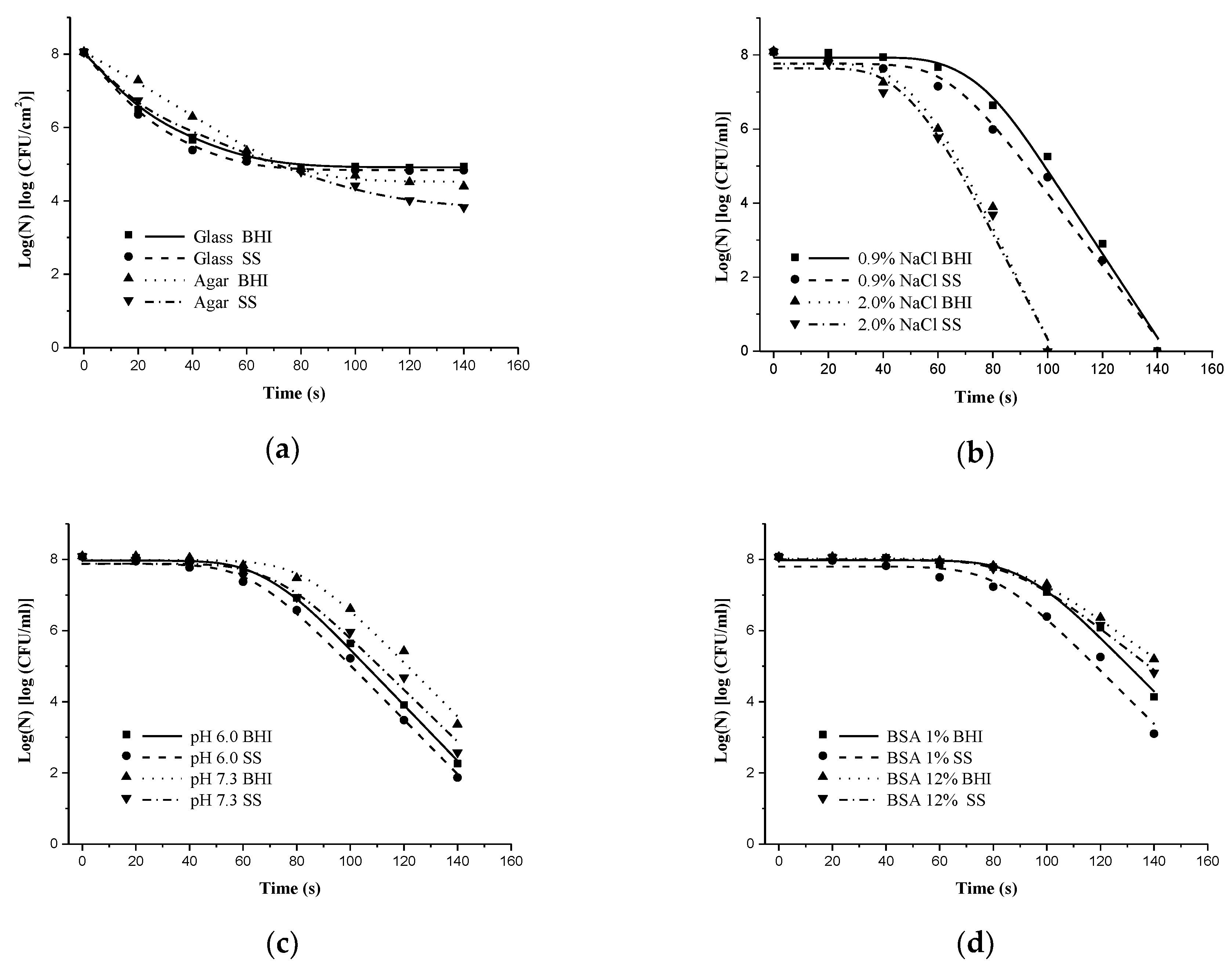
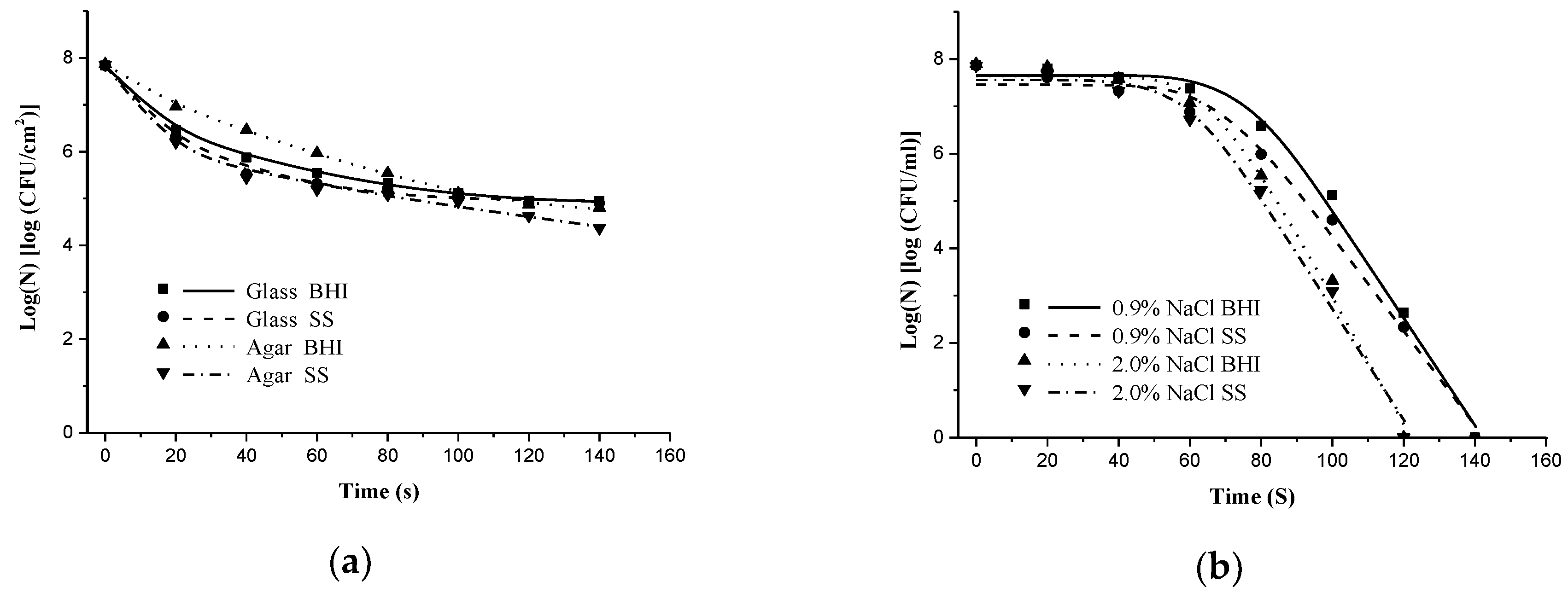
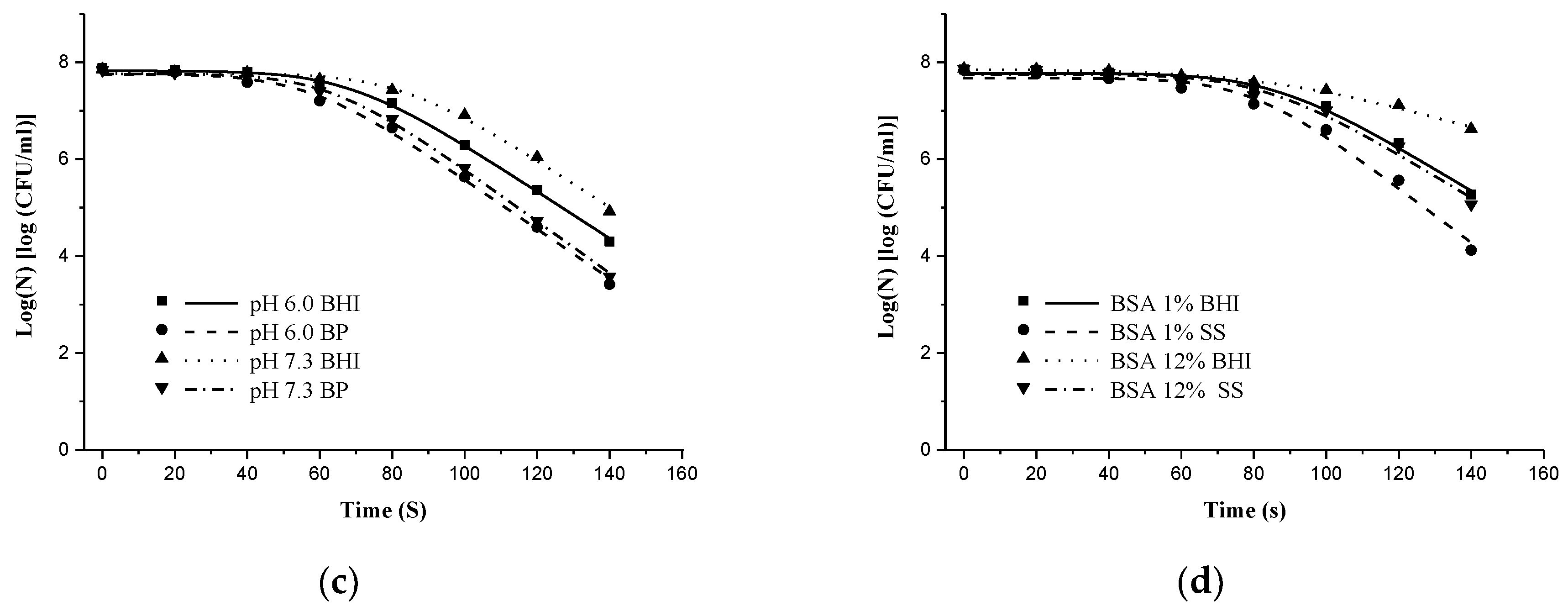
3.1. Bacterial Inactivation Effect and Kinetics Model Analysis

The inactivation effect on S. typhimurium and S. aureus by DBD-NTP are presented in Figure 1 and Figure 2, respectively. All survival curves consist of three phases: shoulder or tailing phase, a log-linear phase, and a transition phase between the shoulder and log-linear phases. As shown in Figure 1a and Table 1, survival curve of S. typhimurium (total population) on the surface of glass showed a log-linear phase (from 8.05 ± 0.07 to 4.91 ± 0.61 log (CFU/cm2)) followed by a long tail. As shown in Figure 1b and Table 1, survival curve of S. typhimurium (total population) in 0.9% NaCl solution showed a shoulder phase (treated time from 0 to 73.3 ± 4.08 s) followed by a log-linear phase (treated time after 73.3 ± 4.08 s). In this study, the inactivation efficiency of DBD plasma was evaluated 5 different parameters. In addition, a log-linear shoulder or tailing model was also used to analyze the plasma-induced inactivation. The analysis was R2 (0.9761–0.9993) and RMSE (0.0393–0.7184), which determined the goodness of fit of the models.

3.1.1. Effect of Solid Surface on Inactivation of S. typhimurium and S. aureus by DBD-NTP

For inactivation of the solid surface (S. typhimurium for Figure 1a and S. aureus for Figure 2a), a steep drop in the slope of inactivation regression lines was noted within the first 20 s. Thereafter, the increment of the pathogen lethality slowed down, following an exponential relationship with treatment time. After 80 s, slopes became mostly flat for most data, indicating that the increase in treatment time does not affect the germicidal efficacy much. Populations of S. typhimurium and S. aureus on the solid surface were reduced to 2.83 and 2.29 log/cm2 in 60 s. However, treatments for an additional 80 s merely reduced them by 0.29 and 0.6 log/cm2, respectively. Similar survival curves were found when low-pressure plasma (1–10 Torr) and UV irradiation were evaluated for their bacteria inactivation kinetics [20]. The kinetics of UV irradiation was ascribed to suppression due to reabsorption by emitting gas from plasma. Similarly, the reduction in inactivation of microbes in kinetic curves with DBD-NTP could be attributed to the “quenching” effects between reactive oxygen/nitrogen species formed in packages during extended treatment time [21,22]. At this power level the UV radiation does not play a significant direct role in the inactivation process. In DBD plasma discharges, reactive species generated through various collisional pathway, commonly, used oxygen and nitrogen gas plasma are excellent sources of reactive oxygen-based and nitrogen-based species, such as O, •OH, NO and NO2 [23]. Firstly, reactive species were regarded as the primary factors in killing microorganisms. They caused oxidation of unsaturated fatty acids and produced unsaturated fatty acid peroxide formation, thereby impeding transport of the biochemical by-products across the membrane. Beyond that, proteins and nucleic acids react with reactive species to formation of 2-oxohistidine and 8-hydroxy-2 deoxyguanosine [24]. Besides, some reports pointed out that charged particles play important roles in inactivation. Electrons and ions can cause surface lesions in membranes by direct bombardment damage. Mendis et al. [25] and Laroussi et al. [26] suggested that electrostatic force caused by charge accumulation on the outer surface of the cell membrane could overcome the tensile strength of the membrane and cause its rupture. This phenomenon is more likely to occur on gram-negative bacteria, whose cell membrane is always irregular. These irregularities offer small radii of curvatures that cause localized high outward electrostatic forces which trigger rupture of gram-negative bacteria [23].
As shown in Table 1 and Table 2, D values for S. typhimurium and S. aureus on the surface of glass slides were 9.14 s and 9.11 s, respectively, and on the surface of gelatin plates were 23.66 s and 25.47 s, respectively. These results indicated that bacteria on the gelling plates were much harder to kill by DBD-NTP than bacteria loaded on glass slides. The differences between glass slides and gelatin plates could be due to the amount of water and chemical composition of medium effect [27]. Dobrynin et al. [27] reported that charged particles generated in plasma catalyzed peroxidation of lipids and polysaccharides on the cell membranes, with reaction rate described above. Bacteria loaded on surface of slides are covered with layer of water molecules bound by van der Waals forces, whereas bacteria loaded on gelatin are covered with thick layer of free water. Charged particles catalyze oxidation and erosion processes of both surface and interior of microorganisms and presence of water and reactive species play a synergistically effect role which contribute to faster inactivation. Cell seems to be less susceptible and slopes of survival curves became flatter at the latter half of inactivation process. Plasma acts only on the surface and charged particle and reactive species cannot penetrate deep into the substrates. It seems plausible that dead bacteria on top layers could form a physical protective layer covering the surviving bacteria. Meanwhile, disruption of membrane integrity could lead to the release of highly oxidative materials (e.g., membrane lipids, free fatty acids, etc.) [28]. The oxidizable materials could serve as a “quenching’’ phase for the highly reactive, oxidizing species produced by plasma, thereby resulting in a decline in microbial inactivation. Although the nature of this tailing effect is not exactly known, it has been attributed to the existence of a fraction of the population more resistant to plasma [2]. Tailing effect is not exactly known and the prospect warrants further investigation.

3.1.2. Embedding S. Typhimurium and S. Aureus in Liquid Media

Survival curves of S. typhimurium and S. aureus in liquid media are shown in Figure 1b–d, and Figure 2b–d. The survival curves exhibited a shoulder region followed by a log-linear phase regardless of liquid medium and bacterial species. The shoulder region has been attributed to the time required by generated germicidal agents in NTP treatments to reach the minimum concentrations on cell membranes for causing damages to bacterial cells [29]. Hence, the differences in shoulder phase length or duration reflect the requirement of limiting levels of germicidal agents to start inactivating bacteria or thresholds for formed germicidal agents to start damaging microbial cells on different media. Once the concentration of germicidal agents passes the threshold, a log-linear phase is triggered, resulting in irreversible damage and lysis of the cells [29].
The kinetics parameters from Table 1 and Table 2 for S. typhimurium and S. aureus, respectively, show that compared with buffer liquid system, a shorter shoulder and a higher maximum specific inactivation rate were found with salt solutions. In addition, no visible colony formation was detected in 2% NaCl solution after samples were treated for as short as 120 s (Figure 1b and Figure 2b) regardless of bacterial species and recovery method. However, it took 140 seconds to finish inactivation in 0.9% NaCl solution. The plasma–liquid interaction likely generated other oxidizing species such as hypochlorous acid (HOCl) [30]. With higher concentration of NaCl in liquids, the more HOCl was produced [31]. However, for the other two media, microbial populations were still countable after 140s’ treatments. These differences could be due to the acidification effect of aqueous solution by DBD-NTP. In our present study, it was noted that the pH values of the salt solution descended to 3.7 (data not shown) after NTP treatments, whereas pH of buffer liquid system did not significantly change. Acidification has been hypothesized to be one of the main mechanisms for DBD-NTP treatments to reduce microbial loads in an aqueous solution. Our results are in agreement with what was reported by Zhou et al. [32]. Oehmigen et al. [33] studied the effect of plasma on properties of water and concluded that the NOx formation, including nitrous HNO2 and nitric acid HNO3, is responsible for acidification of aqueous solution. Bacteria are negatively charged colloidal particles. Their isoelectric points are disturbed in the acidic conditions, which leads to the death of microorganism [34]. Further study with NTP [35] showed that addition of nitrate ions in aqueous solution alone was not able to inactivate microorganisms, but it somehow enhanced antimicrobial effect of nitrate, nitrite, hydrogen peroxide, peroxynitrite, and hydroxyl radicals. In addition, DBD-NTP treatments can also produce hydrogen peroxide in high relative moisture environment. Hydrogen peroxide in acidic conditions is a strong oxidizer and can have a greater impact on the bacterial cell membrane [36]. pH effect on bacterial surviving DBD-NTP treatments was also demonstrated by the inactivation kinetics analyses conducted in this study. D-values for S. typhimurium and S. aureus in pH 7.3 PBS were consistently higher than those in pH 6.0 PBS (Table 1 and Table 2).
Peroxynitrous acid enters the bacterial by passive diffusion. Once inside the cell, ONOOH can damage DNA, lipids, and proteins via direct oxidation reactions or by decomposing to OH and NO2•. Peroxynitrite also reacts with CO2 to form a nitrosoperoxycarbonate anion (ONOOCO2-) followed by decomposing to NO2• and a carbonate radical (CO3-), which also can damage biomolecules [37]. The reaction mentioned above can occur only in acidic conditions. Coinciding with this research, D-values of S. typhimurium and S. aureus planktonic were significantly different between pH = 7.3 PBS and pH = 6.0 PBS. Adverse environmental conditions such as extremes of temperature and pH, osmotic shock, anaerobicity and composition of the growth medium can induce stress responses in S. typhimurium, which in turn affects its ability to withstand antimicrobial treatments [38]. Kayes et al. [10] reported that lower pH rendered Escherichia coli O157:H7, Listeria monocytogenes, S. aureus, more susceptible to the plasma. Along with the number of stressful environmental factors increasing, microorganism became more vulnerable when treated by plasma. D values of S. typhimurium and S. aureus planktonic were higher in 12% BSA compared to 1% BSA (p < 0.05). Log reduction of S. typhimurium and S. aureus (total) were significantly different between 1% BSA and 12% BSA (p < 0.05). The inactivation rate of the mixed culture cocktail was slower by comparison to the respective mono culture preparations, which may be due to the greater potential for organic-based quenching of the reactive species generated by DBD-NTP treatment [39]. Higher resistance of bacteria at 12% BSA to plasma may be attributed to BSA reacting with active species of plasma which are supposed to disinfect microorganisms. Zhou et al. [30] showed that plasma induced the change of the side chains of the amino acids by hydroxylation, nitration, dehydrogenation and dimerization. Scholtz et al. [33] showed the He/O2 plasma jet was used for destruction of BSA proteins and in the case of incomplete protein removal, the treated BSA suffered considerable degradation. This leads to the assumption that existence of BSA could protect microorganisms from being killed by plasma.

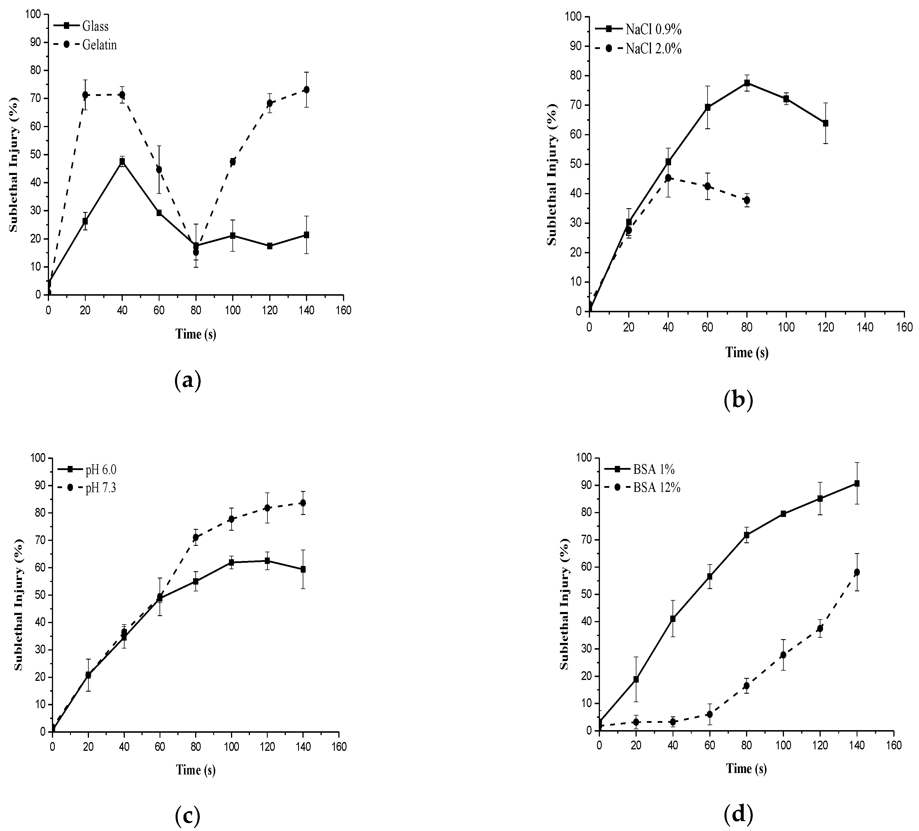
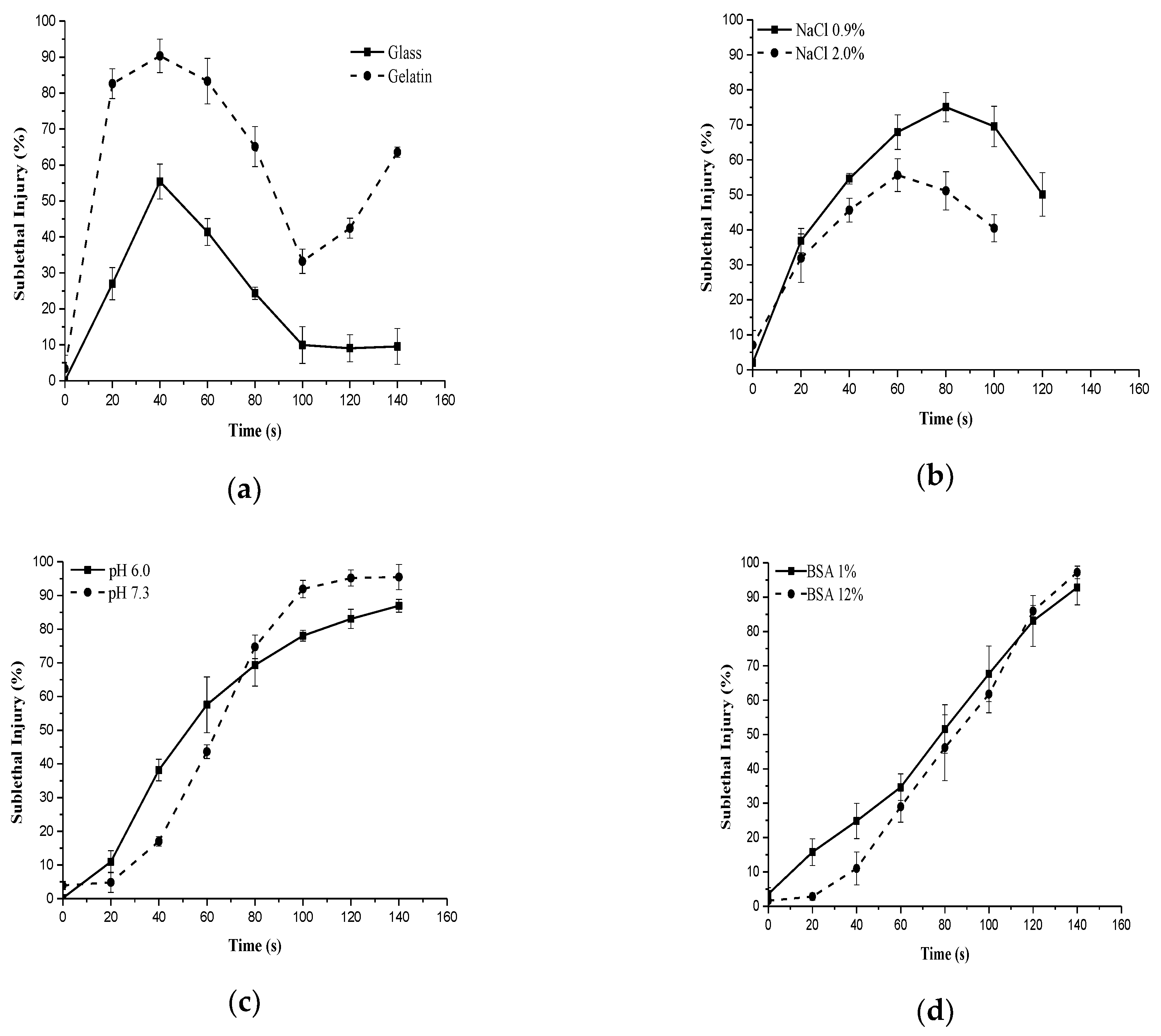
3.2. Effects of NTP on Sublethal Injury

Sublethal injury (SI) is a consequence of exposure to a chemical or physical process that damages but does not kill microorganism cells [40]. Bacteria death can be induced by the effect of one or more sublethal treatments on a microorganism. Sublethal injury implies that cell membrane permeability or functional cell components of microorganisms are damaged. The degree of damage can be estimated by the difference in plate counts between non-selective and selective media as a proportion or percentage of the entire population [23]. Plasma interactions with bacteria are quite complex, the dose-dependent effects range from lethal to sublethal [34].
Figure 3 and Figure 4 show the percentage of sublethal injury as a function of the NTP exposure time for S. typhimurium and S. aureus, respectively. The maximum value and time to reach a maximum peak of sublethal injury varied with medium state, pH, and osmolarity.
The maximum value of SI of S. aureus was consistently higher than that of S. typhimurium during the whole treatment period, both on solid surfaces (glass and gelatin) and in liquid (NaCl and PBS solution) media. This maximum illustrates the phenomenon of injury accumulation finally culminating into cell death [41], and coincides to the start of a new phase in the inactivation kinetics. For example, when S. typhimurium was treated in 2.0% NaCl solution, SI increased rapidly with exposure time and reached the maximum value 45.35% at 40 s. Then it decreased progressively. However, in 0.9% NaCl, SI value increased along with the exposure time and reached the maximum value >70% at 80 s (Figure 3b). When S. typhimurium was treated in 1 % BSA phosphate buffered solution, sublethal damage increased with increasing exposure time and reached the maximum value 90.74% after 140 s. However, in 12% BSA, it increased to only approximate 55% after 140s’ treatment (Figure 3d). SI values for S. typhimurium on gelatin plates were consistently higher than those on glass slides regardless of exposure time (Figure 3a). However, there was no difference in SI values between pH 6.0 and 7.3 bacterial suspensions in the first 60 s. After 60 s, SI values in pH 7.3 medium was higher than those in pH 6.0 medium (Figure 3c). Similar results were also noticed with S. aureus (Figure 4). Smet et al. [11] found that a maximum SI value was usually detected at optimal conditions or moderately stressing environmental conditions. However, our results indicate that in DBD-NTP treatment, maximum SI values for individual bacteria species could be affected by various factors.

4. Conclusions

In conclusion, our data demonstrate that DBD-based NTP can effectively inactivate microbial pathogens both on a solid surface and in liquid suspensions, and inactivation kinetics of S. typhimurium and S. aureus by DBD-NTP treatments on both media can be well predicted with mathematical models. However, both the inactivation effects on microbial pathogens and sublethal injury of microbial pathogens by DBD-NTP treatments could be significantly affected by solid materials and properties of liquid suspensions. In application of DBD-NTP for inactivating solid food surface and liquid food products and extending shelf life and safety, solid materials and properties of liquids should be considered for achieving the optimal results.

Author Contributions

H.Z. and J.Z. (Jianhao Zhang) conceived and designed the experiments. M.H., J.W., W.Y. and J.Z. (Jianying Zhao) performed the experiments. M.H. and J.Z. (Jianhao Zhang) wrote the paper. H.Z. supervised the whole effort. All authors collaborated in the interpretation of the results and the preparation of the manuscript.

Funding

This work was supported financially by the Agricultural Innovation Fund Project of Jiangsu Province (No. CX(18)3041), and the National Natural Science Foundation of China (No. 31601565).

Acknowledgments

The authors would like to thank the organizations or individuals who contributed to this manuscript.

Conflicts of Interest

The authors declare no conflict of interest.

References

  1. Surowsky, B.; Fröhling, A.; Gottschalk, N.; Schlüter, O.; Knorr, D. Impact of cold plasma on Citrobacter freundii in apple juice: Inactivation kinetics and mechanisms. Int. J. Food Microbiol. 2014, 174, 63–71. [Google Scholar] [CrossRef] [PubMed]
  2. Calvo, T.; Álvarez-Ordóñez, A.; Prieto, M.; González-Raurich, M.; López, M. Influence of processing parameters and stress adaptation on the inactivation of Listeria monocytogenes by Non-Thermal Atmospheric Plasma (NTAP). Food Res. Int. 2016, 89, 631–637. [Google Scholar] [CrossRef] [PubMed]
  3. Fröhling, A.; Durek, J.; Schnabel, U.; Ehlbeck, J.; Bolling, J.; Schlüter, O. Indirect plasma treatment of fresh pork: decontamination efficiency and effects on quality attributes. Innov. Food Sci. Emerg. Technol. 2012, 16, 381–390. [Google Scholar] [CrossRef]
  4. Hayashi, N.; Akiyoshi, Y.; Kobayashi, Y.; Kanda, K.; Ohshima, K.; Goto, M. Inactivation characteristics of Bacillus thuringiensis spore in liquid using atmospheric torch plasma using oxygen. Vacuum 2013, 88, 173–176. [Google Scholar] [CrossRef]
  5. Perni, S.; Shama, G.; Kong, M.G. Cold atmospheric plasma disinfection of cut fruit surfaces contaminated with migrating microorganisms. J. Food Prot. 2008, 71, 1619–1625. [Google Scholar] [CrossRef] [PubMed]
  6. Ziuzina, D.; Patil, S.; Cullen, P.J.; Keener, K.M.; Bourke, P. Atmospheric cold plasma inactivation of Escherichia coli in liquid media inside a sealed package. J. Appl. Microbiol. 2013, 114, 778–787. [Google Scholar] [CrossRef] [PubMed]
  7. Rothrock, M.J.; Zhuang, H.; Lawrence, K.C.; Bowker, B.C.; Gamble, G.R.; Hiett, K.L. In-package inactivation of pathogenic and spoilage bacteria associated with poultry using dielectric barrier discharge-cold plasma treatments. Curr. Microbiol. 2017, 74, 149–158. [Google Scholar] [CrossRef] [PubMed]
  8. Scholtz, V.; Pazlarova, J.; Souskova, H.; Khun, J.; Julak, J. Nonthermal plasma–A tool for decontamination and disinfection. Biotechnol. Adv. 2015, 33, 1108–1119. [Google Scholar] [CrossRef] [PubMed]
  9. Bourke, P.; Ziuzina, D.; Boehm, D.; Cullen, P.J.; Keener, K. The potential of cold plasma for safe and sustainable food production. Trends Biotechnol. 2018, 36, 615–626. [Google Scholar] [CrossRef] [PubMed]
  10. Kayes, M.M.; Critzer, F.J.; Kelly-Wintenberg, K.; Roth, J.R.; Montie, T.C.; Golden, D.A. Inactivation of foodborne pathogens using a one atmosphere uniform glow discharge plasma. Foodborne Pathog. Dis. 2007, 4, 50–59. [Google Scholar] [CrossRef] [PubMed]
  11. Smet, C.; Noriega, E.; Rosier, F.; Walsh, J.L.; Valdramidis, V.P.; Van Impe, J.F. Influence of food intrinsic factors on the inactivation efficacy of cold atmospheric plasma: Impact of osmotic stress, suboptimal pH and food structure. Innov. Food Sci. Emerg. 2016, 38, 393–406. [Google Scholar] [CrossRef]
  12. Lee, K.; Paek, K.H.; Ju, W.T.; Lee, Y. Sterilization of bacteria, yeast, and bacterial endospores by atmospheric-pressure cold plasma using helium and oxygen. J. Microbiol. 2006, 44, 269–275. [Google Scholar] [PubMed]
  13. Hertwig, C.; Reineke, K.; Ehlbeck, J.; Knorr, D.; Schlüter, O. Decontamination of whole black pepper using different cold atmospheric pressure plasma applications. Food Control. 2015, 55, 221–229. [Google Scholar] [CrossRef]
  14. Hurst, A. Bacterial injury: A review. Can. J. Microbiol. 1977, 23, 935–944. [Google Scholar] [CrossRef] [PubMed]
  15. Puligundla, P.; Lee, T.; Mok, C. Inactivation effect of dielectric barrier discharge plasma against foodborne pathogens on the surfaces of different packaging materials. Innov. Food Sci. Emerg. Technol. 2016, 36, 221–227. [Google Scholar] [CrossRef]
  16. Jahid, I.K.; Han, N.; Ha, S.-D. Inactivation kinetics of cold oxygen plasma depend on incubation conditions of Aeromonas hydrophila biofilm on lettuce. Food Res. Int. 2014, 55, 181–189. [Google Scholar] [CrossRef]
  17. Noriega, E.; Velliou, E.G.; Van Derlinden, E.; Mertens, L.; Van Impe, J.F.M. Role of growth morphology in the formulation of NaCl-based selective media for injury detection of Escherichia coli, Salmonella Typhimurium and Listeria innocua. Food Res. Int. 2014, 64, 402–411. [Google Scholar] [CrossRef] [PubMed]
  18. Geeraerd, A.; Herremans, C.; Van Impe, J.F. Structural model requirements to describe microbial inactivation during a mild heat treatment. Int. J. Food. Microbiol. 2000, 59, 185–209. [Google Scholar] [CrossRef]
  19. Busch, S.V.; Donnelly, C.W. Development of a repair-enrichment broth for resuscitation of heat-injured Listeria monocytogenes and Listeria innocua. Appl. Environ. Microb. 1992, 58, 14–20. [Google Scholar]
  20. Moisan, M.; Barbeau, J.; Crevier, M.-C.; Pelletier, J.; Philip, N.; Saoudi, B. Plasma sterilization. Methods and mechanisms. Pure Appl. Chem. 2002, 74, 349–358. [Google Scholar] [CrossRef]
  21. Klockow, P.A.; Keener, K.M. Safety and quality assessment of packaged spinach treated with a novel ozone-generation system. LWT–Food Sci. Technol. 2009, 42, 1047–1053. [Google Scholar] [CrossRef]
  22. Kogelschatz, U. Dielectric-Barrier Discharges: Their history, discharge physics, and industrial applications. Plasma Chem. Plasma P. 2003, 23, 1–46. [Google Scholar] [CrossRef]
  23. Noriega, E.; Shama, G.; Laca, A.; Diaz, M.; Kong, M.G. Cold atmospheric gas plasma disinfection of chicken meat and chicken skin contaminated with Listeria innocua. Food Microbiol. 2011, 28, 1293–1300. [Google Scholar] [CrossRef] [PubMed] [Green Version]
  24. Laroussi, M.; Leipold, F. Evaluation of the roles of reactive species, heat, and UV radiation in the inactivation of bacterial cells by air plasmas at atmospheric pressure. Int. J. Mass Spectrom. 2004, 233, 81–86. [Google Scholar] [CrossRef]
  25. Mendis, D.A.; Rosenberg, M.; Azam, F. A note on the possible electrostatic disruption of bacteria. IEEE Trans. Plasma Sci. 2000, 28, 1304–1306. [Google Scholar] [CrossRef]
  26. Laroussi, M.; Mendis, D.A.; Rosenberg, M. Plasma interaction with microbes. New J. Phys. 2003, 5, 41.1–41.10. [Google Scholar] [CrossRef]
  27. Dobrynin, D.; Fridman, G.; Friedman, G.; Fridman, A. Physical and biological mechanisms of direct plasma interaction with living tissue. New J. Phys. 2009, 11, 115020. [Google Scholar] [CrossRef] [Green Version]
  28. Critzer, F.J.; Kelly-Wintenberg, K.; South, S.L.; Golden, D.A. Atmospheric plasma inactivation of foodborne pathogens on fresh produce surfaces. J. Food Prot. 2007, 70, 2290–2296. [Google Scholar] [CrossRef] [PubMed]
  29. Moisan, M.; Barbeau, J.; Moreau, S.; Pelletier, J.; Tabrizian, M.; Yahia, L.H. Low-temperature sterilization using gas plasmas: a review of the experiments and an analysis of the inactivation mechanisms. Int. J. Pharm. 2001, 226, 1–21. [Google Scholar] [CrossRef]
  30. Kim, H.-S.; Wright, K.; Piccioni, J.; Cho, D.J.; Cho, Y.I. Inactivation of bacteria by the application of spark plasma in produced water. Sep. Purif. Technol. 2015, 156, 544–552. [Google Scholar] [CrossRef] [Green Version]
  31. Maheux, S.; Frache, G.; Thomann, J.S.; Clément, F.; Penny, C.; Belmonte, T.; Duday, D. Small unilamellar liposomes as a membrane model for cell inactivation by cold atmospheric plasma treatment. J. Phys. D Appl. Phys. 2016, 49, 344001. [Google Scholar] [CrossRef]
  32. Zhou, R.W.; Zhou, R.S.; Zhuang, J.X.; Zong, Z.C.; Zhang, X.H.; Liu, D.P.; Bazaka, K.; Ostrikov, K. Interaction of atmospheric-pressure air microplasmas with amino acids as fundamental processes in aqueous solution. PLoS ONE 2016, 11, e0155584. [Google Scholar] [CrossRef] [PubMed]
  33. Oehmigen, K.; Hähnel, M.; Brandenburg, R.; Wilke, C.; Weltmann, K.D.; von Woedtke, T. The role of acidifcation for antimicrobial activity of atmospheric pressure plasma in liquid. Plasma Process. Polym. 2010, 7, 250–257. [Google Scholar] [CrossRef]
  34. Stoffels, E.; Sakiyama, Y.; Graves, D.B. Cold atmospheric plasma: Charged species and their interactions with cells and tissues. IEEE T. Plasma Sci. 2008, 36, 1441–1457. [Google Scholar] [CrossRef]
  35. Scholtz, V.; Pazlarová, J.; Soušková, H.; Khun, J.; Julák, J. Nonthermal plasma—a tool for decontamination and disinfection. Biotechnol. Adv. 2015, 33, 1108–1119. [Google Scholar] [CrossRef] [PubMed]
  36. Rashmei, Z.; Bornasi, H.; Ghoranneviss, M. Evaluation of treatment and disinfection of water using cold atmospheric plasma. J. Water Health 2016, 14, 609–616. [Google Scholar] [CrossRef] [PubMed]
  37. Arjunan, K.P.; Obrusnik, A.; Jones, B.T.; Zajickova, L.; Ptasinska, S. Effect of additive oxygen on the reactive species profile and microbicidal property of a helium atmospheric pressure plasma jet. Plasma Process. Polym. 2016, 13, 1089–1105. [Google Scholar] [CrossRef]
  38. Fernández, A.; Thompson, A. The inactivation of Salmonella by cold atmospheric plasma treatment. Food Res. Int. 2012, 45, 678–684. [Google Scholar] [CrossRef]
  39. Patange, A.; Boehm, D.; Giltrap, M.; Lu, P.; Cullen, P.J.; Bourke, P. Assessment of the disinfection capacity and eco-toxicological impact of atmospheric cold plasma for treatment of food industry effluents. Sci. Total Environ. 2018, 631–632, 298–307. [Google Scholar] [CrossRef] [PubMed]
  40. Wesche, A.M.; Gurtler, J.B.; Marks, B.P.; Ryser, E.T. Stress, sublethal injury, resuscitation, and virulence of bacterial foodborne pathogens. J. Food Prot. 2009, 72, 1121–1138. [Google Scholar] [CrossRef] [PubMed]
  41. Noriega, E.; Velliou, E.; Van Derlinden, E.; Mertens, L.; Van Impe, J.F. Effect of cell immobilization on heat-induced sublethal injury of Escherichia coli, Salmonella Typhimurium and Listeria innocua. Food Microbiol. 2013, 36, 355–364. [Google Scholar] [CrossRef] [PubMed]
Figure 1. Survival curves of S. typhimurium after exposure to dielectric barrier discharge (DBD) plasma. Cells were inactivated on solid(like) surface of glass and gelatin (a) and in liquid carrier of NaCl (b), phosphate buffered saline (PBS) (c) and bovine serum albumin (BSA) (d). All points were actual values and all lines were fitted values of the model. BHI: Brain Heart Infusion Agar; SS: Salmonella Shigella Agar.
Applsci 08 02087 g001
Figure 2. Survival curves of S. aureus during DBD-NTP (non-thermal plasma treatment). Cells were inactivated on solid surface of glass and gelatin (a) or in a liquid suspension of NaCl (b), PBS (c) or BSA (d). All points were actual values and all lines were fitted values of the model.
Applsci 08 02087 g002aApplsci 08 02087 g002b
Figure 3. Changes in sublethal injury (%) of S. typhimurium during DBD-NTP treatment. Cells were inactivated on glass and gelatin surfaces (a) and in liquid suspensions of NaCl (b), PBS (c), and BSA (d).
Applsci 08 02087 g003
Figure 4. Changes in sublethal injury (%) of S. aureus during DBD-NTP treatment. Cells were inactivated on glass and gelatin surfaces (a) and in liquid suspensions of NaCl (b), PBS (c), and BSA (d).
Applsci 08 02087 g004
Table 1. Parameters of the kinetics model for the inactivation of S. typhimurium by dielectric barrier discharge non-thermal plasma (DBD-NTP). Cells were inactivated on a solid surface or in a liquid suspension.
Populationlog N0 (log (CFU/mL))/log N0 (log (CFU/cm2))Kmaxtllog Nres (log (CFU/mL))/log Nres (log (CFU/cm2))D value 1Log Reduction 2RMSE 3R 2
NaC l0.9%Total7.91 ± 0.10 aAB0.26 ± 0.011 aB73.3 ± 4.08 aC 81.71 ± 3.72 aC8.07 ± 0.10 aA0.40020.9895
Uninjured7.76 ± 0.15 bBC0.22 ± 0.007 bB63.8 ± 1.08 bC 73.6 ± 0.78 bD8.09 ± 0.07 aA0.43190.9876
NaC l2%Total7.76 ± 0.15 aB0.34 ± 0.011 aA50.2 ± 1.15 aD 56.62 ± 0.94 aD8.09 ± 0.07 aA0.67050.9815
Uninjured7.64 ± 0.16 aC0.32 ± 0.015 aA48.2 ± 1.93 aD 55.02±1.62 aE8.08 ± 0.09 aA0.71840.9783
PBS pH = 6.0Total7.93 ± 0.14 aAB0.21 ± 0.041 aC72.5 ± 1.42 aC 83.2 ± 1.63 aC6.57 ± 1.23 aB0.16180.9968
Uninjured7.82 ± 0.15 aABC0.20 ± 0.031 aBC67.8 ± 3.31 aBC 78.84 ± 2.97 aCD6.83 ± 0.99 aB0.21850.9948
PBS pH = 7.3Total7.97 ± 0.12 aA0.17 ± 0.025 aD82.5 ± 4.18 aB 95.25 ± 5.41 aB4.72 ± 0.88 aC0.26230.9863
Uninjured7.88 ± 0.14 aAB0.16 ± 0.007 aDE71.2 ± 2.85 bB 84.34 ± 2.37 bC5.50 ± 0.03 aC0.32500.9842
BSA 1%Total7.97 ± 0.07 aA0.17 ± 0.004 aD91.4 ± 3.47 aA 104.03 ± 3.48 aA3.93 ± 0.24 aCD0.21260.9867
Uninjured7.78 ± 0.19 aBC0.17 ± 0.033 aCD81.7 ± 4.25 bA 94.49 ± 1.72 bB4.95 ± 0.74 aCD0.35150.9761
BSA 12%Total8.02 ± 0.10 aA0.12 ± 0.009 aE88.3 ± 2.56 aA 106.03 ± 1.73 aA2.87 ± 0.20 aE0.06200.9980
Uninjured7.99 ± 0.08 aAB0.13 ± 0.020 aEF87.6 ± 5.81 aA 104.01 ± 7.28 aA3.24 ± 0.71 aE0.09610.9961
GlassTotal8.05 ± 0.07 bA0.09 ± 0.007 bF 4.91 ± 0.61 aA9.11 ± 3.37 aF3.11 ± 0.58 aDE0.04320.9991
Uninjured8.04 ± 0.07 aA0.10 ± 0.001 aF 4.83 ± 0.59 aA8.11 ± 3.35 aF3.20 ± 0.56 aE0.06650.9980
GelatinTotal8.06 ± 0.07 aA0.10 ± 0.005 aEF 4.52 ± 0.15 aA23.66 ± 2.72 aE3.66 ± 0.02 bDE0.10830.9964
Uninjured8.06 ± 0.07 bA0.06 ± 0.001 bG 3.83 ± 0.18 bB9.02 ± 1.68 bF4.23 ± 0.08 aD0.16290.9928
1D value is defined as time needed for the 1 log reduction of initial target microorganism population. 2 Log reduction is calculated from the difference between log N0 to log N (n = 140 s). 3 RMSE stands for the root mean squared error. a–b Means without common superscripts in each inactivation media (NaCl, PBS, BSA or solid media) within a parameter are different (p < 0.05). A–G Means without common superscripts in each population type (total or uninjured population) within a parameter are different (p < 0.05).
Table 2. Parameters of the kinetics model for the inactivation of S. aureus by DBD-NTP. Cells were inactivated on a solid surface or in a liquid suspension.
Populationlog N0 (log (CFU/mL))/log N0 (log (CFU/cm2))Kmaxtllog Nres (log (CFU/mL))/log Nres (log (CFU/cm2))D value 1Log Reduction 2RMSE 3R 2
NaCl0.9%Total7.65 ± 0.1 aA0.26 ± 0.015 aB74.57 ± 2.78 aC 82.97 ± 2.29 aC7.87 ± 0.12 aA0.33920.9921
Uninjured7.46 ± 0.09 aC0.23 ± 0.013 bB67.27 ± 3.17 bB 76.87 ± 2.61 bCD7.86 ± 0.11 aA0.42180.9874
NaCl2%Total7.65 ± 0.12 aA0.31 ± 0.021 aA64.44 ± 2.97 aD 71.61 ± 2.48 aD7.89 ± 0.14 aA0.42830.9896
Uninjured7.55 ± 0.1 aBC0.27 ± 0.015 aA59.01 ± 2.86 aCD 67.07 ± 2.41 aE7.86 ± 0.11 aA0.47260.9869
PBS pH=6.0Total7.82 ± 0.06 aA0.11 ± 0.002 aC68.26 ± 0.81 aD 87.98 ± 0.74 aC3.57 ± 0.06 bB0.0690.9985
Uninjured7.78 ± 0.1 aA0.12 ± 0.007 aC55.94 ± 0.82 bD 74.28 ± 1.31 bD4.46 ± 0.05 aB0.14640.9955
PBS pH=7.3Total7.78 ± 0.11 aA0.11 ± 0.013 aC82.47 ± 5.77 aB 102.28 ± 5.06 aB2.94 ± 0.34 bCD0.10120.9948
Uninjured7.76 ± 0.1 aA0.12 ± 0.007 aC62.66 ± 2.49 bBC 80.54 ± 1.56 bC4.26 ± 0.08 aB0.0890.9982
BSA 1%Total7.77 ± 0.11 aA0.1 ± 0.004 bC86.22 ± 1.16 aAB 107.39 ± 2.05 aB2.59 ± 0.16 bD0.11760.9907
Uninjured7.67 ± 0.12 aAB0.13 ± 0.009 aC79.66 ± 4.62 aA 96.62 ± 4.2 bB3.71 ± 0.3 aCD0.20960.9854
BSA 12%Total7.85 ± 0.14 aA0.05 ± 0.017 bD88.86 ± 1.51 aA 134.57 ± 15.59 aA1.25 ± 0.43 bE0.04850.993
Uninjured7.75 ± 0.1 aA0.1 ± 0.002 aC83.48 ± 5.41 aA 104.6 ± 5.05 bA2.8 ± 0.24 aD0.18380.9799
GlassTotal7.84 ± 0.12 aA0.04 ± 0.011 aD 4.88 ± 0.09 aA9.14 ± 2.14 aF2.9 ± 0.1 aCD0.04640.9987
Uninjured7.84 ± 0.09 aA0.06 ± 0.035 aD 4.95 ± 0.05 aA7.44 ± 4.18 aF2.94 ± 0.14 aD0.11960.9919
GelatinTotal7.86 ± 0.08 aA0.06 ± 0.011 aD 4.7 ± 0.09 aB25.47 ± 6.38 aE3.06 ± 0.06 aC0.03930.9993
Uninjured7.85 ± 0.08 aA0.02 ± 0.003 bE 3.76 ± 0.7 aB3.44 ± 0.85 bF3.48 ± 0.07 aC0.14650.9901
1D value is defined as time needed for the 1 log reduction of initial target microorganism population. 2 Log reduction is calculated from the difference between log N0 to log N (n = 140 s). 3 RMSE stands for the root mean squared error. a–b Means without common superscripts in each inactivation media (NaCl, PBS, BSA or solid media) within a parameter are different (p < 0.05). A–F Means without common superscripts in each population type (total or uninjured population) within a parameter are different (p < 0.05).

