
TMS-Induced Central Motor Conduction Time at the Non-Infarcted Hemisphere Is Associated with Spontaneous Motor Recovery of the Paretic Upper Limb after Severe Stroke

 ,
,
Abstract
:
1. Introduction
2. Materials and Methods
2.1. Subjects
2.2. Stimulation Procedure
2.3. Measurements
2.4. Data Analysis
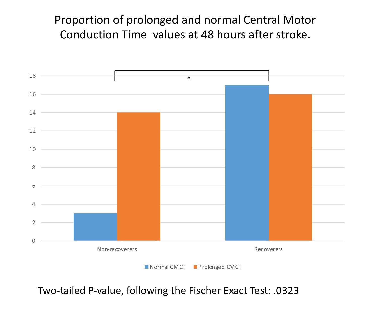
3. Results
4. Discussion
5. Conclusions
Supplementary Materials
Author Contributions
Funding
Institutional Review Board Statement
Informed Consent Statement
Data Availability Statement
Acknowledgments
Conflicts of Interest
References
- Morris, J.H.; Van Wijck, F. Responses of the Less Affected Arm to Bilateral Upper Limb Task Training in Early Rehabilitation After Stroke: A Randomized Controlled Trial. Arch. Phys. Med. Rehabil. 2012, 93, 1129–1137. [Google Scholar] [CrossRef] [PubMed]
- Harrington, R.M.; Chan, E.; Rounds, A.K.; Wutzke, C.J.; Dromerick, A.W.; Turkeltaub, P.E.; Harris-Love, M.L. Roles of Lesioned and Nonlesioned Hemispheres in Reaching Performance Poststroke. Neurorehabilit. Neural Repair 2020, 34, 61–71. [Google Scholar] [CrossRef] [PubMed]
- Maenza, C.; Good, D.C.; Winstein, C.J.; Wagstaff, D.A.; Sainburg, R.L. Functional Deficits in the Less-Impaired Arm of Stroke Survivors Depend on Hemisphere of Damage and Extent of Paretic Arm Impairment. Neurorehabilit. Neural Repair 2020, 34, 39–50. [Google Scholar] [CrossRef] [PubMed]
- Van Dokkum, L.E.H.; Le Bars, E.; Mottet, D.; Bonafé, A.; De Champfleur, N.M.; Laffont, I. Modified Brain Activations of the Nondamaged Hemisphere During Ipsilesional Upper-Limb Movement in Persons with Initial Severe Motor Deficits Poststroke. Neurorehabilit. Neural Repair 2018, 32, 34–45. [Google Scholar] [CrossRef] [PubMed] [Green Version]
- Bustrén, E.-L.; Sunnerhagen, K.S.; Murphy, M.A. Movement Kinematics of the Ipsilesional Upper Extremity in Persons with Moderate or Mild Stroke. Neurorehabilit. Neural Repair 2017, 31, 376–386. [Google Scholar] [CrossRef] [PubMed]
- Lawrence, D.G.; Kuypers, H.G.J.M. Pyramidal and Non-Pyramidal Pathways in Monkeys: Anatomical and Functional Correlation. Science 1965, 148, 973–975. [Google Scholar] [CrossRef]
- Kuypers, H.; Brinkman, J. Precentral projections to different parts of the spinal intermediate zone in the rhesus monkey. Brain Res. 1970, 24, 29–48. [Google Scholar] [CrossRef]
- Lawrence, E.S.; Coshall, C.; Dundas, R.; Stewart, J.; Rudd, A.G.; Howard, R.; Wolfe, C.D.A. Estimates of the Prevalence of Acute Stroke Impairments and Disability in a Multiethnic Population. Stroke 2001, 32, 1279–1284. [Google Scholar] [CrossRef] [PubMed] [Green Version]
- Grefkes, C.; Fink, G.R. Connectivity-based approaches in stroke and recovery of function. Lancet Neurol. 2014, 13, 206–216. [Google Scholar] [CrossRef]
- Nathan, P.W.; Smith, M.C.; Deacon, P. The corticospinal tracts in man. Brain 1990, 113, 303–324. [Google Scholar] [CrossRef]
- Lawrence, D.G.; Kuypers, H.G.J.M. The functional organization of the motor system in the monkey: II. Brain 1968, 91, 15–36. [Google Scholar] [CrossRef]
- Lawrence, D.G.; Kuypers, H.G.J.M. The functional organization of the motor system in the monkey: I. Brain 1968, 91, 1–14. [Google Scholar] [CrossRef] [PubMed]
- Baker, S.N. The primate reticulospinal tract, hand function and functional recovery. J. Physiol. 2011, 589, 5603–5612. [Google Scholar] [CrossRef] [PubMed] [Green Version]
- Baker, S.N.; Zaaimi, B.; Fisher, K.M.; Edgley, S.A.; Soteropoulos, D.S. Pathways mediating functional recovery. Prog. Brain Res. 2015, 218, 389–412. [Google Scholar] [CrossRef] [PubMed]
- Zaaimi, B.; Soteropoulos, D.S.; Fisher, K.M.; Riddle, C.N.; Baker, S.N. Classification of Neurons in the Primate Reticular Formation and Changes after Recovery from Pyramidal Tract Lesion. J. Neurosci. 2018, 38, 6190–6206. [Google Scholar] [CrossRef] [Green Version]
- Benecke, R.; Meyer, B.-U.; Freund, H.-J. Reorganisation of descending motor pathways in patients after hemispherectomy and severe hemispheric lesions demonstrated by magnetic brain stimulation. Exp. Brain Res. 1991, 83, 419–426. [Google Scholar] [CrossRef]
- Feeney, D.M.; Baron, J.C. Diaschisis. Stroke 1986, 17, 817–830. [Google Scholar] [CrossRef] [Green Version]
- Bütefisch, C.M.; Weβling, M.; Netz, J.; Seitz, R.J.; Hömberg, V. Relationship between Interhemispheric Inhibition and Motor Cortex Excitability in Subacute Stroke Patients. Neurorehabilit. Neural Repair 2008, 22, 4–21. [Google Scholar] [CrossRef]
- Carrera, E.; Tononi, G. Diaschisis: Past, present, future. Brain 2014, 137, 2408–2422. [Google Scholar] [CrossRef] [Green Version]
- Grefkes, C.; Fink, G.R. Reorganization of cerebral networks after stroke: New insights from neuroimaging with connectivity approaches. Brain 2011, 134, 1264–1276. [Google Scholar] [CrossRef] [Green Version]
- Van Der Vliet, R.; Selles, R.W.; Andrinopoulou, E.; Nijland, R.; Ribbers, G.M.; Frens, M.A.; Meskers, C.; Kwakkel, G. Predicting Upper Limb Motor Impairment Recovery after Stroke: A Mixture Model. Ann. Neurol. 2020, 87, 383–393. [Google Scholar] [CrossRef] [Green Version]
- Traversa, R.; Cicinelli, P.; Pasqualetti, P.; Filippi, M.; Rossini, P.M. Follow-up of interhemispheric differences of motor evoked potentials from the ‘affected’ and ‘unaffected’ hemispheres in human stroke. Brain Res. 1998, 803, 1–8. [Google Scholar] [CrossRef]
- Barker, R.N.; Brauer, S.G.; Barry, B.K.; Gill, T.J.; Carson, R.G. Training-induced modifications of corticospinal reactivity in severely affected stroke survivors. Exp. Brain Res. 2012, 221, 211–221. [Google Scholar] [CrossRef]
- McDonnell, M.N.; Stinear, C.M. TMS measures of motor cortex function after stroke: A meta-analysis. Brain Stimul. 2017, 10, 721–734. [Google Scholar] [CrossRef]
- Hammerbeck, U.; Hoad, D.; Greenwood, R.; Rothwell, J.C. The unsolved role of heightened connectivity from the unaffected hemisphere to paretic arm muscles in chronic stroke. Clin. Neurophysiol. 2019, 130, 781–788. [Google Scholar] [CrossRef] [PubMed]
- Adams, H.P.; Bendixen, B.H.; Kappelle, L.J.; Biller, J.; Love, B.B.; Gordon, D.L.; Marsh, E. Classification of subtype of acute ischemic stroke. Definitions for use in a multicenter clinical trial. TOAST. Trial of Org 10172 in Acute Stroke Treatment. Stroke 1993, 24, 35–41. [Google Scholar] [CrossRef] [Green Version]
- Rossini, P.; Burke, D.; Chen, R.; Cohen, L.; Daskalakis, Z.; Di Iorio, R.; Di Lazzaro, V.; Ferreri, F.; Fitzgerald, P.; George, M.; et al. Non-invasive electrical and magnetic stimulation of the brain, spinal cord, roots and peripheral nerves: Basic principles and procedures for routine clinical and research application. An updated report from an I.F.C.N. Committee. Clin. Neurophysiol. 2015, 126, 1071–1107. [Google Scholar] [CrossRef]
- Di Lazzaro, V.; Oliviero, A.; Pilato, F.; Saturno, E.; Dileone, M.; Mazzone, P.; Insola, A.; Tonali, P.; Rothwell, J. The physiological basis of transcranial motor cortex stimulation in conscious humans. Clin. Neurophysiol. 2004, 115, 255–266. [Google Scholar] [CrossRef]
- Van Peppen, R.P.S.; Kwakkel, G.; Wood-Dauphinee, S.; Hendriks, H.J.M.; Van Der Wees, P.J.; Dekker, J. The impact of physical therapy on functional outcomes after stroke: What’s the evidence? Clin. Rehabil. 2004, 18, 833–862. [Google Scholar] [CrossRef] [PubMed]
- Rothwell, J. Transcranial Electrical and Magnetic Stimulation of the Brain: Basic Physiological Mechanisms. In Magnetic Stimulation in Clinical Neurophysiology, 2nd ed.; Hallet, M., Chokroverty, S., Eds.; Elsevier: Philadelphia, PA, USA, 2005; pp. 43–60. [Google Scholar]
- Groppa, S.; Oliviero, A.; Eisen, A.; Quartarone, A.; Cohen, L.; Mall, V.; Kaelin-Lang, A.; Mima, T.; Rossi, S.; Thickbroom, G.; et al. A practical guide to diagnostic transcranial magnetic stimulation: Report of an IFCN committee. Clin. Neurophysiol. 2012, 123, 858–882. [Google Scholar] [CrossRef] [PubMed] [Green Version]
- Hoonhorst, M.H.W.J.; Kollen, B.J.; Berg, P.S.P.V.D.; Emmelot, C.H.; Kwakkel, G. How Reproducible Are Transcranial Magnetic Stimulation–Induced MEPs in Subacute Stroke? J. Clin. Neurophysiol. 2014, 31, 556–562. [Google Scholar] [CrossRef]
- Samii, A.; Luciano, C.; Dambrosia, J.; Hallet, M. Central motor conduction time: Reproducibility and discomfort of different methods. Muscle Nerve 1998, 21, 1445–1450. [Google Scholar] [CrossRef]
- Hoonhorst, M.H.J.; Nijland, R.H.M.; Berg, P.J.S.V.D.; Emmelot, C.H.; Kollen, B.J.; Kwakkel, G. Does Transcranial Magnetic Stimulation Have an Added Value to Clinical Assessment in Predicting Upper-Limb Function Very Early After Severe Stroke? Neurorehabilit. Neural Repair 2018, 32, 682–690. [Google Scholar] [CrossRef] [Green Version]
- Collin, C.; Wade, D. Assessing motor impairment after stroke: A pilot reliability study. J. Neurol. Neurosurg. Psychiatry 1990, 53, 576–579. [Google Scholar] [CrossRef] [PubMed] [Green Version]
- Nijland, R.; Van Wegen, E.; Der Wel, B.H.-V.; Kwakkel, G. Presence of Finger Extension and Shoulder Abduction Within 72 Hours After Stroke Predicts Functional Recovery. Stroke 2010, 41, 745–750. [Google Scholar] [CrossRef] [PubMed] [Green Version]
- Collin, C.; Wade, D.T.; Davies, S.; Horne, V. The Barthel ADL Index: A reliability study. Int. Disabil. Stud. 1988, 10, 61–63. [Google Scholar] [CrossRef] [PubMed]
- Osawa, A.; Maeshima, S.; Tanahashi, N. Water-Swallowing Test: Screening for Aspiration in Stroke Patients. Cerebrovasc. Dis. 2013, 35, 276–281. [Google Scholar] [CrossRef]
- Nijboer, T.C.; Kollen, B.J.; Kwakkel, G. Time course of visuospatial neglect early after stroke: A longitudinal cohort study. Cortex 2013, 49, 2021–2027. [Google Scholar] [CrossRef]
- Prabhakaran, S.; Zarahn, E.; Riley, C.; Speizer, A.; Chong, J.Y.; Lazar, R.M.; Marshall, R.S.; Krakauer, J.W. Inter-individual Variability in the Capacity for Motor Recovery After Ischemic Stroke. Neurorehabilit. Neural Repair 2007, 22, 64–71. [Google Scholar] [CrossRef] [PubMed]
- Winters, C.; Van Wegen, E.E.H.; Daffertshofer, A.; Kwakkel, G. Generalizability of the Proportional Recovery Model for the Upper Extremity After an Ischemic Stroke. Neurorehabilit. Neural Repair 2014, 29, 614–622. [Google Scholar] [CrossRef]
- Sanford, J.; Moreland, J.; Swanson, L.R.; Stratford, P.W.; Gowland, C. Reliability of the Fugl-Meyer Assessment for Testing Motor Performance in Patients Following Stroke. Phys. Ther. 1993, 73, 447–454. [Google Scholar] [CrossRef]
- Gladstone, D.J.; Danells, C.J.; Black, S.E. The Fugl-Meyer Assessment of Motor Recovery after Stroke: A Critical Review of Its Measurement Properties. Neurorehabilit. Neural Repair 2002, 16, 232–240. [Google Scholar] [CrossRef] [PubMed]
- Byblow, W.D.; Stinear, C.M.; Barber, P.A.; Petoe, M.A.; Ackerley, S.J. Proportional recovery after stroke depends on corticomotor integrity. Ann. Neurol. 2015, 78, 848–859. [Google Scholar] [CrossRef] [PubMed]
- Nudo, R.J.; Wise, B.M.; Sifuentes, F.; Milliken, G.W. Neural Substrates for the Effects of Rehabilitative Training on Motor Recovery after Ischemic Infarct. Science 1996, 272, 1791–1794. [Google Scholar] [CrossRef] [Green Version]
- von Monakow, C. Die Lokalisation im Grosshirn und der Abbau der Funktion Durch Kortikale Herde; Bergmann: Wiesbaden, Germany, 1914. [Google Scholar]
- Block, F.; Dihné, M.; Loos, M. Inflammation in areas of remote changes following focal brain lesion. Prog. Neurobiol. 2005, 75, 342–365. [Google Scholar] [CrossRef] [PubMed]
- Jones, K.A.; Zouikr, I.; Patience, M.; Clarkson, A.N.; Isgaard, J.; Johnson, S.J.; Spratt, N.; Nilsson, M.; Walker, F.R. Chronic stress exacerbates neuronal loss associated with secondary neurodegeneration and suppresses microglial-like cells following focal motor cortex ischemia in the mouse. Brain Behav. Immun. 2015, 48, 57–67. [Google Scholar] [CrossRef]
- Weishaupt, N.; Zhang, A.; DeZiel, R.A.; Tasker, R.A.; Whitehead, S.N. Prefrontal Ischemia in the Rat Leads to Secondary Damage and Inflammation in Remote Gray and White Matter Regions. Front. Neurosci. 2016, 10, 81. [Google Scholar] [CrossRef] [Green Version]
- Visser, M.M.; Yassi, N.; Campbell, B.C.; Desmond, P.M.; Davis, S.M.; Spratt, N.; Parsons, M.; Bivard, A. White Matter Degeneration after Ischemic Stroke: A Longitudinal Diffusion Tensor Imaging Study. J. Neuroimaging 2018, 29, 111–118. [Google Scholar] [CrossRef] [PubMed]
- Volz, L.J.; Rehme, A.K.; Michely, J.; Nettekoven, C.; Eickhoff, S.B.; Fink, G.R.; Grefkes, C. Shaping Early Reorganization of Neural Networks Promotes Motor Function after Stroke. Cereb. Cortex 2016, 26, 2882–2894. [Google Scholar] [CrossRef] [Green Version]
- Soteropoulos, D.S.; Williams, E.R.; Baker, S.N. Cells in the monkey ponto-medullary reticular formation modulate their activity with slow finger movements. J. Physiol. 2012, 590, 4011–4027. [Google Scholar] [CrossRef] [Green Version]
- Ziemann, U.; Ishii, K.; Borgheresi, A.; Yaseen, Z.; Battaglia, F.; Hallett, M.; Cincotta, M.; Wassermann, E.M. Dissociation of the pathways mediating ipsilateral and contralateral motor-evoked potentials in human hand and arm muscles. J. Physiol. 1999, 518, 895–906. [Google Scholar] [CrossRef] [PubMed]
- Lui, Y.; Tang, E.; Allmendinger, A.; Spektor, V. Evaluation of CT Perfusion in the Setting of Cerebral Ischemia: Patterns and Pitfalls. Am. J. Neuroradiol. 2010, 31, 1552–1563. [Google Scholar] [CrossRef] [Green Version]
- Nakano, S.; Iseda, T.; Kawano, H.; Yoneyama, T.; Ikeda, T.; Wakisaka, S. Correlation of early CT signs in the deep middle cerebral artery territories with angiographically confirmed site of arterial occlusion. Am. J. Neuroradiol. 2001, 22, 654–659. [Google Scholar] [PubMed]
- Kwakkel, G.; Winters, C.; Van Wegen, E.E.H.; Nijland, R.H.M.; Van Kuijk, A.A.A.; Visser-Meily, A.; De Groot, J.; De Vlugt, E.; Arendzen, J.H.; Geurts, A.C.H.; et al. Effects of Unilateral Upper Limb Training in Two Distinct Prognostic Groups Early After Stroke. Neurorehabilit. Neural Repair 2016, 30, 804–816. [Google Scholar] [CrossRef] [PubMed] [Green Version]


| Total Group | Recoverers (N = 33) | Non-Recoverers (N = 17) | p | |
|---|---|---|---|---|
| Gender, F/M | 29/21 | 17/16 | 12/5 | 0.571 |
| Age, mean (SD), yr | 70.3 (12.3) | 69.7 (12.8) | 71.4 (11.4) | <0.0001 * |
| Hemisphere of stroke, L/R | 25/25 | 16/17 | 9/8 | 0.170 |
| Length of hospital stay, mean (range), d | 4.9 (6–38) | 13.1 (6–25) | 15.7 (6–38) | <0.0001 * |
| Type of Stroke (TOAST): LVD/SVD/undetermined | 34/14/2 | 22/10/1 | 12/4/1 | 0.804 |
| Dysphagia at 48 h, yes/no | 33/17 | 20/13 | 13/4 | 1.00 |
| VSN at 48 h, yes/no | 12/38 | 7/26 | 5/12 | <0.0001 * |
| Barthel index score (0–20) at 48 h, median (IQR) | 5.0 (5.5) | 7 (5) | 2 (4) | <0.0001 * |
| Total Group | Recoverers (N = 33) | Non-Recoverers (N = 17) | p | |
|---|---|---|---|---|
| CMCT at 48 h, mean (SD), ms | 10.27 (4.35) | 9.54 (2.95) | 11.7 (6.10) | <0.0001 * |
| CMCT at 11 days, mean (SD), ms | 9.44 (3.99) | 8.81 (3.17) | 10.7 (5.18) | <0.0001 * |
| FM-UE finger extension at 48 h, yes/no | 18/32 | 18/15 | 0/17 | <0.0001 * |
| FM-UE finger extension at 11 days, yes/no | 26/23 § | 24/9 | 2/14 | 0.065 |
| FM-UE total score (0–66), median (IQR) at 48 h | 7.5 (43) | 35 (53.5) | 3 (2.5) | <0.0001 * |
| FM-UE total score (0–66), median (IQR) at 6 months | 54 (58) § | 61 (12) | 5 (3.8) | <0.0001 * |
Publisher’s Note: MDPI stays neutral with regard to jurisdictional claims in published maps and institutional affiliations. |
© 2021 by the authors. Licensee MDPI, Basel, Switzerland. This article is an open access article distributed under the terms and conditions of the Creative Commons Attribution (CC BY) license (https://creativecommons.org/licenses/by/4.0/).
Share and Cite
Hoonhorst, M.H.J.; Nijland, R.H.M.; Emmelot, C.H.; Kollen, B.J.; Kwakkel, G. TMS-Induced Central Motor Conduction Time at the Non-Infarcted Hemisphere Is Associated with Spontaneous Motor Recovery of the Paretic Upper Limb after Severe Stroke. Brain Sci. 2021, 11, 648. https://0-doi-org.brum.beds.ac.uk/10.3390/brainsci11050648
Hoonhorst MHJ, Nijland RHM, Emmelot CH, Kollen BJ, Kwakkel G. TMS-Induced Central Motor Conduction Time at the Non-Infarcted Hemisphere Is Associated with Spontaneous Motor Recovery of the Paretic Upper Limb after Severe Stroke. Brain Sciences. 2021; 11(5):648. https://0-doi-org.brum.beds.ac.uk/10.3390/brainsci11050648
Chicago/Turabian StyleHoonhorst, Maurits H. J., Rinske H. M. Nijland, Cornelis H. Emmelot, Boudewijn J. Kollen, and Gert Kwakkel. 2021. "TMS-Induced Central Motor Conduction Time at the Non-Infarcted Hemisphere Is Associated with Spontaneous Motor Recovery of the Paretic Upper Limb after Severe Stroke" Brain Sciences 11, no. 5: 648. https://0-doi-org.brum.beds.ac.uk/10.3390/brainsci11050648