
Distinct Cellular Profiles of Hif1a and Vegf mRNA Localization in Microglia, Astrocytes and Neurons during a Period of Vascular Maturation in the Auditory Brainstem of Neonate Rats


 , and
, and
Abstract
:
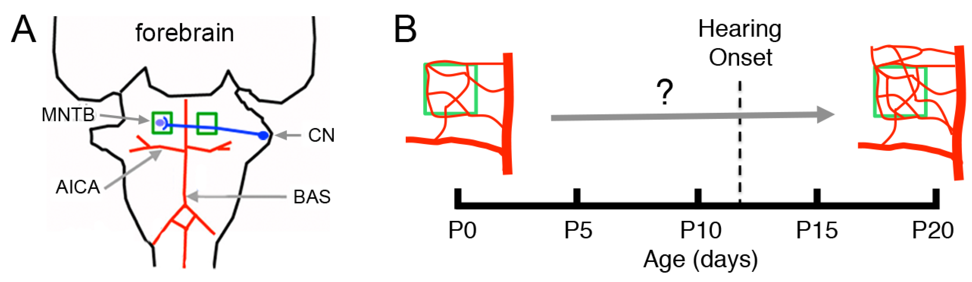
1. Introduction
2. Materials and Methods
2.1. Animals
2.2. Histology
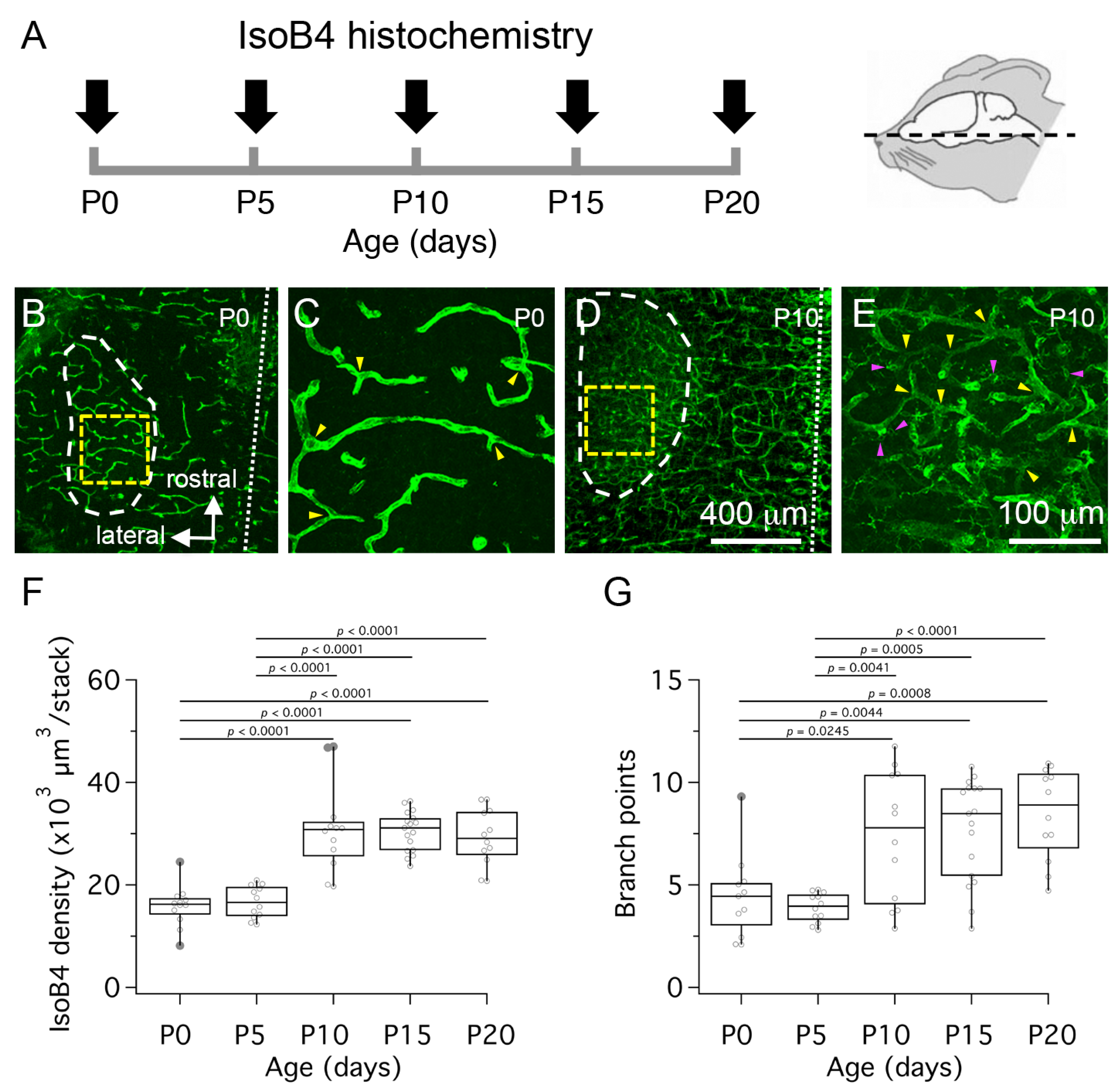
2.3. Isolectin-B4 (IsoB4) Histochemistry
2.4. 5-Ethynyl-2’-deoyuridine (EdU) Tissue Labeling
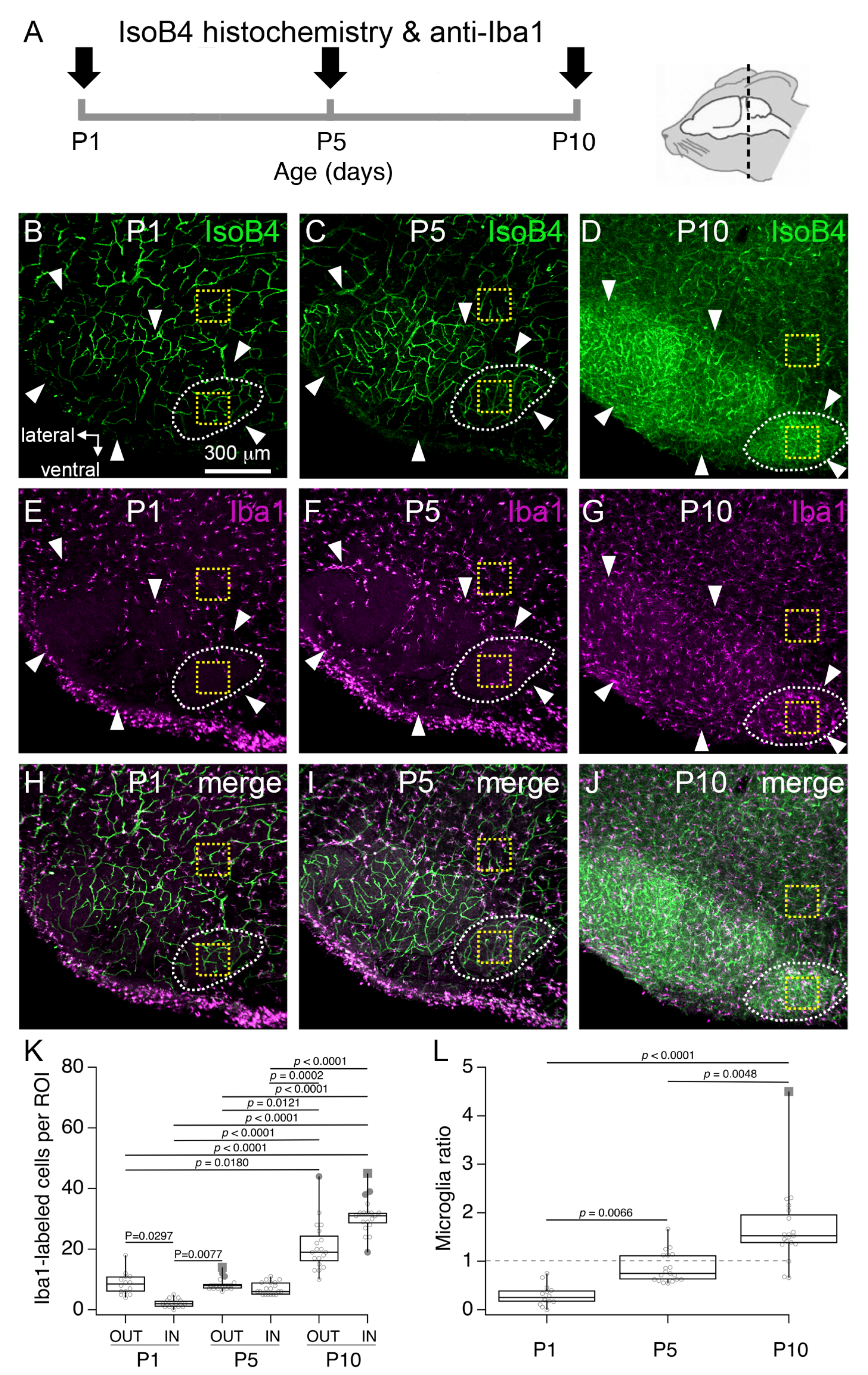
2.5. Multiple Labeling Experiments
2.6. RNAscope Fluorescence in situ Hybridization and Immunohistochemistry
2.7. Confocal Microscopy and Data Analysis
2.8. Statistics
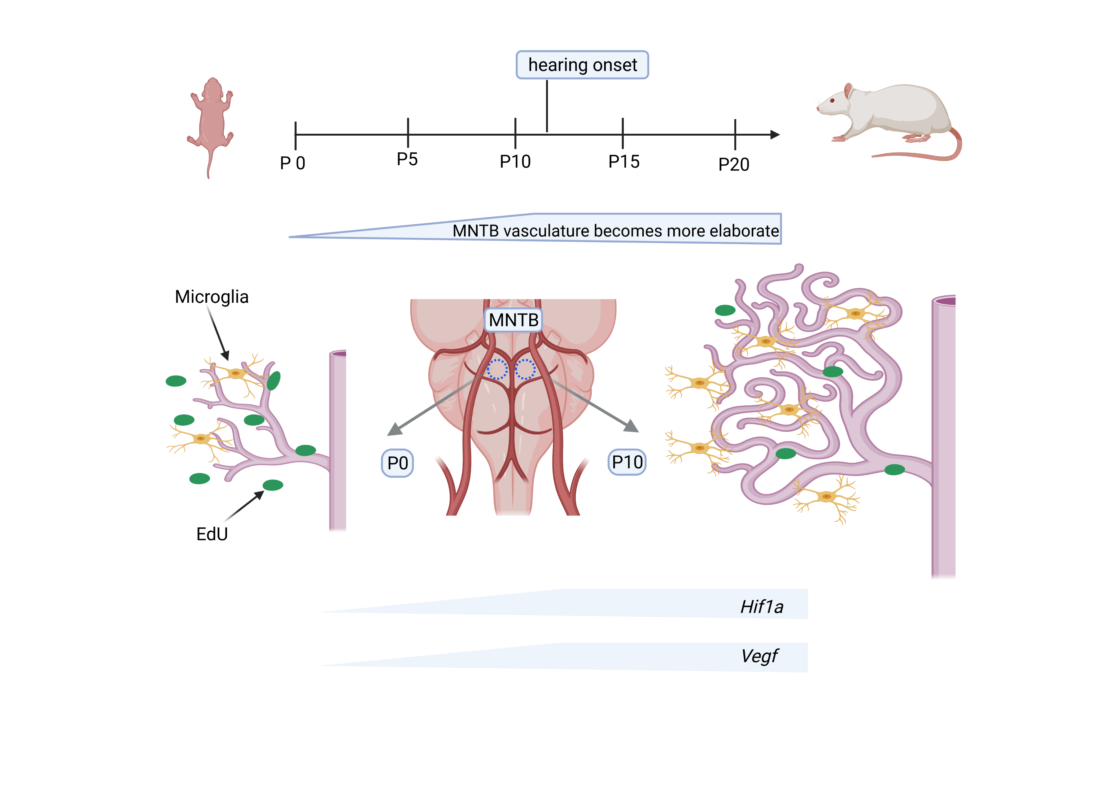
3. Results
3.1. Postnatal Development of the Vascular Bed in the Medial Nucleus of the Trapezoid Body (MNTB)
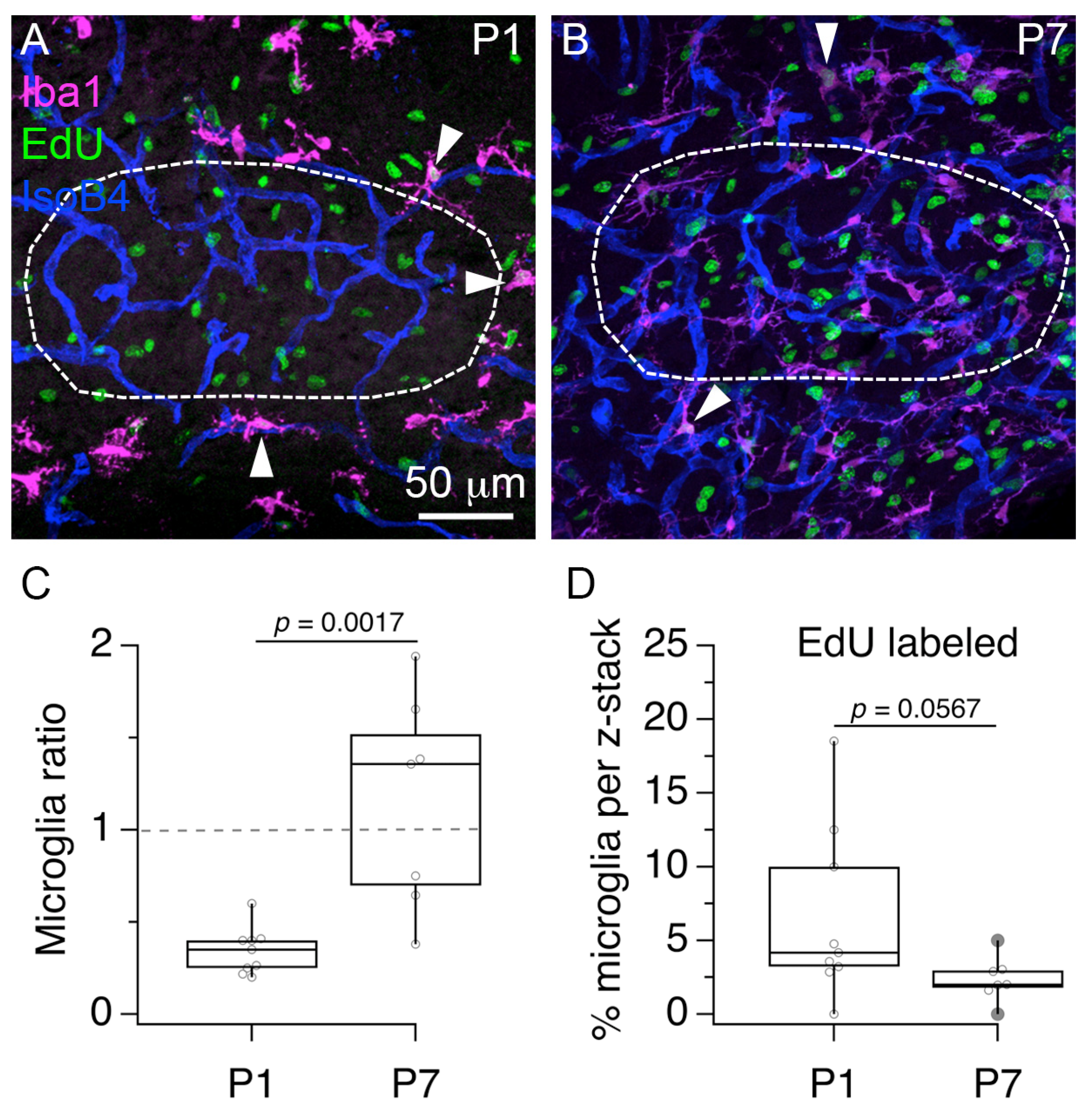
3.2. Postnatal Increase of Microglia Cells in the Rat MNTB
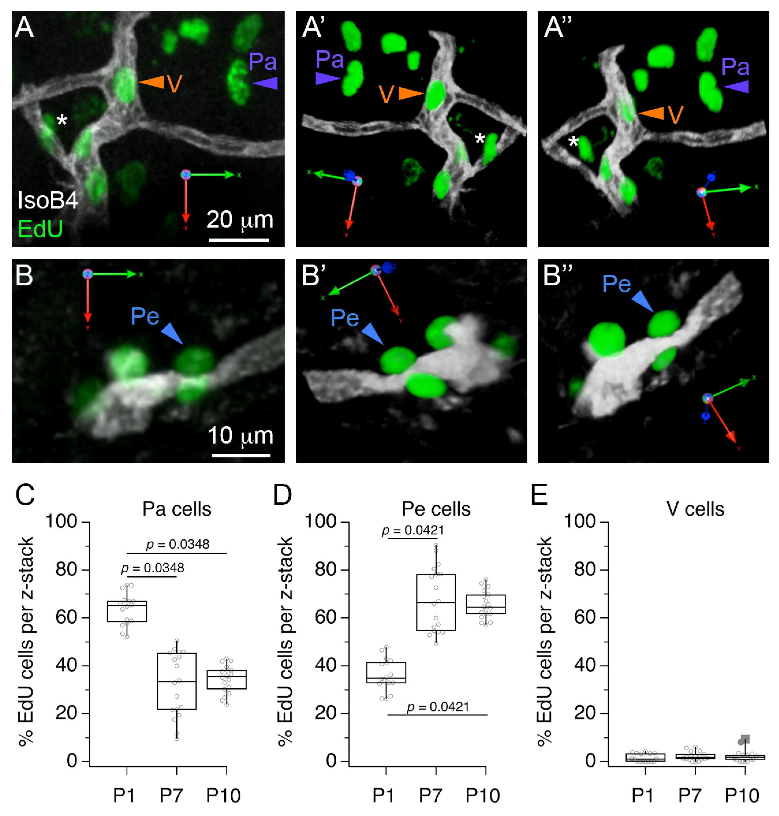
3.3. Postnatal Change in the Perivascular Localization of Proliferating Cells in the MNTB
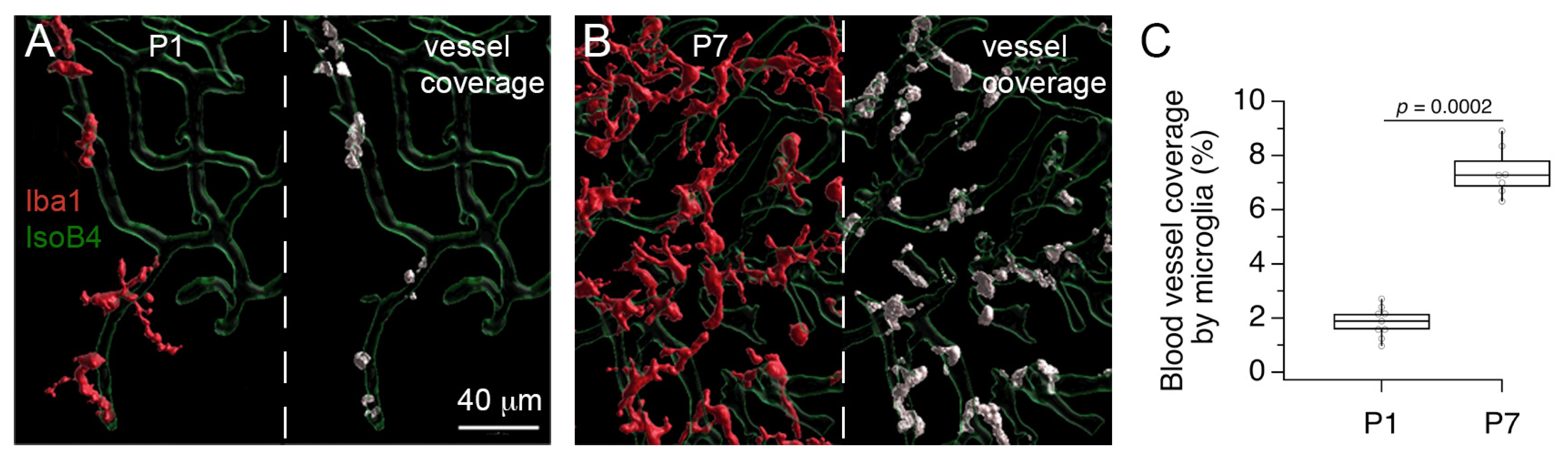
3.4. Microglia Are Perivascular Cells with Proliferative Activity
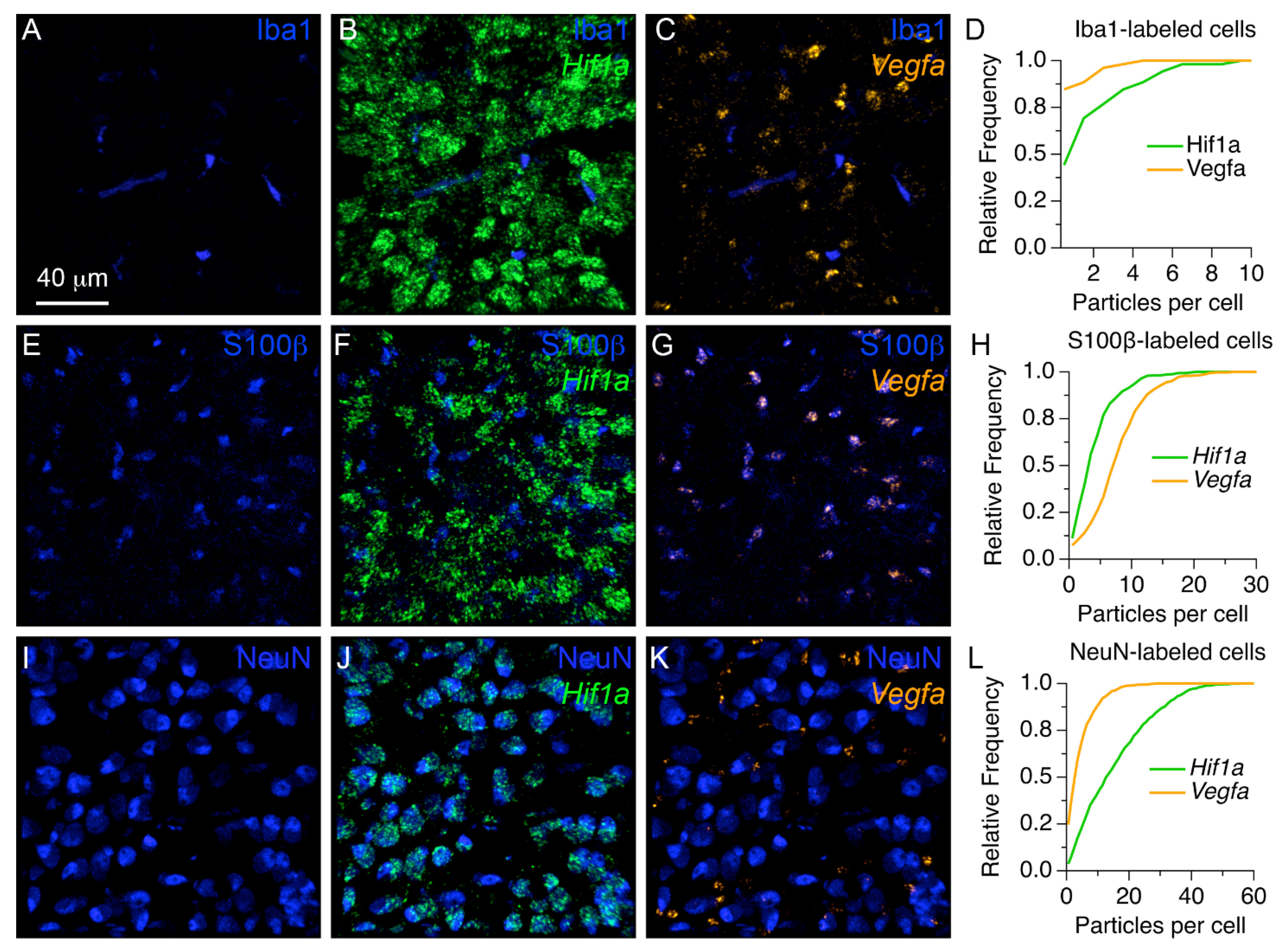
3.5. Localization Profiles of Hif1a and Vegfa mRNAs in the MNTB of Neonate Rats
4. Discussion
5. Conclusions
Author Contributions
Funding
Institutional Review Board Statement
Data Availability Statement
Acknowledgments
Conflicts of Interest
References
- Pham, N.S. The management of pediatric hearing loss caused by auditory neuropathy spectrum disorder. Curr. Opin. Otolaryngol. Head Neck Surg. 2017, 25, 396–399. [Google Scholar] [CrossRef]
- Strata, F.; Stoianov, I.P.; de Villers-Sidani, E.; Bonham, B.; Martone, T.; Kenet, T.; Chang, E.F.; Vincnti, V.; Merzenich, M.M. Perinatal asphyxia affects rat auditory processing: Implications for auditory perceptual impairments in neurodevelopment disorders. PLoS ONE 2010, 5, e15326. [Google Scholar] [CrossRef] [PubMed] [Green Version]
- Truskey, G.A.; Yuan, F.; Katz, D.F. Transport Phenomena in Biological Systems; Pearson Prentice Hall Bioengineering: Upper Saddle River, NJ, USA, 2009; pp. 23–29. [Google Scholar]
- Semenza, G.L. Regulation of mammalian O2 homeostasis by hypoxia-inducible factor 1. Ann. Rev. Cell Dev. Biol. 1999, 15, 551–578. [Google Scholar] [CrossRef] [PubMed]
- Yuen, T.J.; Silberesis, J.C.; Griveau, A.; Chang, S.M.; Daneman, R.; Fancy, S.P.J.; Zahed, H.; Maltepe, E.; Rowitch, D.H. Oligodendrocyte-encoded HIF function couples postnatal myelination and white matter angiogenesis. Cell 2014, 158, 383–396. [Google Scholar] [CrossRef] [Green Version]
- Zimna, A.; Kurpisz, M. Hypoxia-inducible factor-1 in physiological and pathophysiological angiogenesis: Applications and therapies. Biomed. Res. Int 2015, 2015, 549412. [Google Scholar] [CrossRef] [PubMed] [Green Version]
- Rattner, A.; Williams, J.; Nathans, J. Roles of HIFs and VEGF in angiogenesis in the retina and brain. J. Clin. Investig. 2019, 129, 3807–3820. [Google Scholar] [CrossRef]
- Borst, J.G.G.; Soria van Hoeve, J. The calyx of Hed synapse: From model synapse to auditory relay. Annu. Rev. Physiol. 2012, 74, 199–224. [Google Scholar] [CrossRef]
- Kulesza, R.J., Jr.; Grothe, B. Yes, there is a medial nucleus of the trapezoid body in humans. Front. Neuroanat. 2015, 9, 35. [Google Scholar] [CrossRef] [Green Version]
- Dinh, M.L.; Koppel, S.J.; Korn, M.J.; Cramer, K.S. Distribution of glial cells in the auditory brainstem: Normal development and effects of unilateral lesion. Neuroscience 2014, 278, 237–252. [Google Scholar] [CrossRef] [Green Version]
- Saliu, A.; Adise, S.; Xian, S.; Kudelska, K.; Rodríguez-Contreras, A. Natural and lesion-induced decrease in cell proliferation in the medial nucleus of the trapezoid body during hearing development. J. Comp. Neurol. 2014, 522, 971–985. [Google Scholar] [CrossRef] [Green Version]
- Adise, S.; Saliu, A.; Maldonado, N.; Khatri, V.; Cardoso, L.; Rodríguez-Contreras, A. Effect of maternal care on hearing onset induced by developmental changes in the auditory periphery. J. Neurosci. 2014, 34, 4528–4533. [Google Scholar] [CrossRef] [Green Version]
- Cramer, K.; Rubel, E.W. Glial cell contributions to auditory brainstem development. Front. Neural Circuits 2016, 10, 83. [Google Scholar] [CrossRef] [Green Version]
- Brandebura, A.N.; Morehead, M.; Heller, D.T.; Holcomb, P.; Kolson, D.R.; Jones, G.; Mathers, P.H.; Spirou, G.A. Glial cell expansion coincides with neural circuit formation in the developing auditory brainstem. Dev. Neurobiol. 2018, 78, 1097–1116. [Google Scholar] [CrossRef]
- Qiu, J.; Singh, P.; Pan, G.; de Paolis, A.; Champagne, F.A.; Liu, J.; Cardoso, L.; Rodríguez-Contreras, A. Defining the relationship between maternal care behavior and sensory development in Wistar rats: Auditory periphery development, eye opening and brain gene expression. PLoS ONE 2020, 15, e0237933. [Google Scholar] [CrossRef] [PubMed]
- Miraux, S.; Franconi, J.M.; Thiaudiere, E. Blood velocity assessment using 3D bright-blood time-resolved magnetic resonance angiography. Magn. Res. Med. 2006, 56, 469–473. [Google Scholar] [CrossRef] [PubMed]
- Sinha, A.K.; Cane, C.; Kempley, S.T. Blood flow in the common carotid artery in term and preterm infants: Reproducibility and relation to cardiac output. Arch. Dis. Child. Fetal Neonatal Ed. 2006, 91, F31–F35. [Google Scholar] [CrossRef] [PubMed] [Green Version]
- Rodríguez-Contreras, A.; Liu, X.-B.; DeBello, W.M. Axodendritic contacts onto calcium/calmodulin-dependent protein kinase type II-expressing neurons in the barn owl auditory space map. J. Neurosci. 2005, 25, 5611–5622. [Google Scholar] [CrossRef] [PubMed] [Green Version]
- Rodríguez-Contreras, A.; van Hoeve, J.S.; Habets, R.P.; Locher, H.; Borst, J.G.G. Dynamic development of the calyx of Held synapse. Proc. Natl. Acad. Sci. USA 2008, 105, 5603–5608. [Google Scholar] [CrossRef] [Green Version]
- Beach, J.R.; Bruun, K.S.; Shao, L.; Li, D.; Swider, Z.; Remmert, K.; Zhang, Y.; Conti, M.A.; Adelstein, R.S.; Rusan, N.M.; et al. Actin dynamics and competition for myosin monomer govern the sequential amplification of myosin filaments. Nat. Cell Biol. 2017, 19, 85–93. [Google Scholar] [CrossRef] [Green Version]
- Madry, C.; Kyrargyri, V.; Arancibia-Cárcamo, I.L.; Jolivet, R.; Kohsaka, S.; Bryan, R.; Attwell, D. Microglial ramification, surveillance, and interleukin-1β release are regulated by the two-pore domain K+ channel THIK-1. Neuron 2018, 97, 299–312. [Google Scholar] [CrossRef]
- Arnoux, I.; Hoshiko, M.; Mandavy, L.; Avignone, E.; Yamamoto, N.; Audinat, E. Adaptive phenotype of microglial cells during the normal postnatal development of the somatosensory “Barrel” cortex. Glia 2013, 61, 1582–1594. [Google Scholar] [CrossRef] [PubMed]
- Lacoste, B.; Comin, C.H.; Ben-Zvi, A.; Kaeser, P.S.; Xu, X.; da Costa, L.; Gu, C. Sensory-related neural activity regulates the structure of vascular networks in the cerebral cortex. Neuron 2014, 83, 1117–1130. [Google Scholar] [CrossRef] [PubMed] [Green Version]
- Whiteus, C.; Freitas, C.; Grutzendler, J. Perturbed neural activity disrupts cerebral angiogenesis during a postnatal critical period. Nature 2014, 505, 407–411. [Google Scholar] [CrossRef] [PubMed]
- Chung, A.S.; Ferrara, N. Developmental and pathological angiogenesis. Annu. Rev. Cell Dev. Biol. 2011, 27, 563–584. [Google Scholar] [CrossRef] [PubMed]
- Shi, L.; Rodríguez-Contreras, A. In vivo two-photon imaging measuring the blood-brain barrier permeability during early postnatal development in rodent. In Proceedings of the SPIE 9712, Multiphoton Microscopy in the Biomedical Sciences XVI, 97121Z, San Francisco, CA, USA, 14 March 2016. [Google Scholar]
- Gross, J.; Rheinländer, C.; Fuchs, J.; Mazurek, B.; Machulik, A.; Andreeva, N.; Kietzman, T. Expression of hypoxia-inducible factor-1 in the cochlea of newborn rats. Hear. Res. 2003, 183, 73–83. [Google Scholar] [CrossRef]
- Kitchen, P.; Salman, M.M.; Halsey, A.M.; Clarke-Bland, C.; MacDonald, J.A.; Ishida, H.; Vogel, H.J.; Almutiri, S.; Logan, A.; Kreida, S.; et al. Targeting aquaporin-4 subcellular localization to treat central nervous system edema. Cell 2020, 181, 784–799. [Google Scholar] [CrossRef] [PubMed]
- Bennett, M.L.; Bennett, F.C.; Liddelow, S.A.; Ajami, B.; Zamanian, J.L.; Fernhoff, N.B.; Mulinyawe, S.B.; Bohlen, C.J.; Adil, A.; Tucker, A.; et al. New tools for studying microgia in the mouse and human CNS. Proc. Natl. Acad. Sci. USA 2016, 113, E1738–E1746. [Google Scholar] [CrossRef] [Green Version]
- Satoh, J.I.; Kino, Y.; Asahina, N.; Takitani, M.; Miyoshi, J.; Ishida, T.; Saito, Y. TMEM119 marks a subset of microglia in the human brain. Neuropathology 2016, 36, 39–49. [Google Scholar] [CrossRef]
- Zhao, X.; Eyo, U.B.; Murugan, M.; Wu, L.-J. Microglial interactions with the neurovascular system in physiology and pathology. Dev. Neurobiol. 2018, 78, 604–617. [Google Scholar] [CrossRef]
- Eyo, U.B.; Dailey, M.E. Effects of oxygen-glucose deprivation on microglial mobility and viability in developing mouse hippocampal tissues. Glia 2012, 60, 1747–1760. [Google Scholar] [CrossRef] [Green Version]
- Savaki, H.E.; Kadekaro, M.; Jehle, J.; Sokoloff, L. Alpha- and beta-adrenoreceptor blockers have opposite effects on energy metabolism of the central auditory system. Nature 1978, 276, 521–523. [Google Scholar] [CrossRef]
- Sokoloff, L. Localization of functional activity in the central nervous system by measurement of glucose utilization with radioactive deoxyglucose. J. Cereb. Blood Flow Metab. 1981, 1, 7–36. [Google Scholar] [CrossRef] [PubMed] [Green Version]
- Chen, G.-D.; Liu, Y. Mechanisms of noise-induced hearing loss potentiation by hypoxia. Hear. Res. 2005, 200, 1–9. [Google Scholar] [CrossRef]
- Trattner, B.; Gravot, C.M.; Grothe, B.; Kunz, L. Metabolic maturation of auditory neurons in the superior olivary complex. PLoS ONE 2013, 8, e67351. [Google Scholar] [CrossRef] [Green Version]
- Thomas, C.I.; Keine, C.; Okayama, S.; Satterfield, R.; Musgrove, M.; Guerrero-Given, D.; Kamasawa, N.; Young, S.M., Jr. Presynaptic mitochondria volume and abundance increase during development of a high-fidelity synapse. J. Neurosci. 2019, 39, 7994–8012. [Google Scholar] [CrossRef] [Green Version]
- Velasco, S.; Paulsen, B.; Arlotta, P. 3D brain organoids: Studying brain development and disease outside the embryo. Annu. Rev. Neurosci. 2020, 43, 375–389. [Google Scholar] [CrossRef] [PubMed]
- Salman, M.M.; Marsh, G.; Kusters, I.; Delincé, M.; Di Caprio, G.; Upadhyayula, S.; de Nola, G.; Hunt, R.; Ohashi, K.G.; Gray, T.; et al. Design and validation of a human brain endothelial microvessel-on-a-chip open microfluidic model enabling advanced optical imaging. Front. Bioeng. Biotechnol. 2020, 8, 573775. [Google Scholar] [CrossRef] [PubMed]
- Sahu, S.; Sharan, S.K. Translating embryogenesis to generate organoids: Novel approaches to personalized medicine. iScience 2020, 23, 101485. [Google Scholar] [CrossRef] [PubMed]


Publisher’s Note: MDPI stays neutral with regard to jurisdictional claims in published maps and institutional affiliations. |
© 2021 by the authors. Licensee MDPI, Basel, Switzerland. This article is an open access article distributed under the terms and conditions of the Creative Commons Attribution (CC BY) license (https://creativecommons.org/licenses/by/4.0/).
Share and Cite
Chang, D.; Brown, Q.; Tsui, G.; He, Y.; Liu, J.; Shi, L.; Rodríguez-Contreras, A. Distinct Cellular Profiles of Hif1a and Vegf mRNA Localization in Microglia, Astrocytes and Neurons during a Period of Vascular Maturation in the Auditory Brainstem of Neonate Rats. Brain Sci. 2021, 11, 944. https://0-doi-org.brum.beds.ac.uk/10.3390/brainsci11070944
Chang D, Brown Q, Tsui G, He Y, Liu J, Shi L, Rodríguez-Contreras A. Distinct Cellular Profiles of Hif1a and Vegf mRNA Localization in Microglia, Astrocytes and Neurons during a Period of Vascular Maturation in the Auditory Brainstem of Neonate Rats. Brain Sciences. 2021; 11(7):944. https://0-doi-org.brum.beds.ac.uk/10.3390/brainsci11070944
Chicago/Turabian StyleChang, Daphne, Quetanya Brown, Grace Tsui, Ye He, Jia Liu, Lingyan Shi, and Adrián Rodríguez-Contreras. 2021. "Distinct Cellular Profiles of Hif1a and Vegf mRNA Localization in Microglia, Astrocytes and Neurons during a Period of Vascular Maturation in the Auditory Brainstem of Neonate Rats" Brain Sciences 11, no. 7: 944. https://0-doi-org.brum.beds.ac.uk/10.3390/brainsci11070944