Next Issue
Volume 9, October
Previous Issue
Volume 9, August


Brain Sci., Volume 9, Issue 9 (September 2019) – 33 articles

Cover Story (view full-size image): 
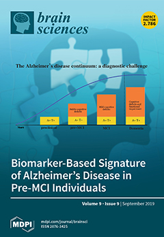
The possibility to detect the earliest clinical manifestations of Alzheimer’s disease, in order to proceed to a biomarker based diagnosis even before the stage of mild cognitive impairment (MCI), represents a major goal, since it would offer the opportunity to have a timely access disease-modifying drugs. In a recent systematic review and meta-analysis, it has been shown that individuals with subtle cognitive decline (“pre-MCI”) with AD biomarker positivity - pre-MCI due to AD - have a high risk of progression to MCI or dementia, similar to what observed for MCI due to AD (Parnetti et al., 2019). In this review, we further focus on this issue, giving more details about neuropsychological profile, its association with biomarkers and neuropathological picture. View this paper.
  • Issues are regarded as officially published after their release is announced to the table of contents alert mailing list.
  • You may sign up for e-mail alerts to receive table of contents of newly released issues.
  • PDF is the official format for papers published in both, html and pdf forms. To view the papers in pdf format, click on the "PDF Full-text" link, and use the free Adobe Reader to open them.
Order results
Result details
Section
Select all
Export citation of selected articles as:
13 pages, 697 KiB  
Article
Using an Overlapping Time Interval Strategy to Study Diagnostic Instability in Mild Cognitive Impairment Subtypes
by David Facal, Joan Guàrdia-Olmos, Arturo X. Pereiro, Cristina Lojo-Seoane, Maribel Peró and Onésimo Juncos-Rabadán
Brain Sci. 2019, 9(9), 242; https://0-doi-org.brum.beds.ac.uk/10.3390/brainsci9090242 - 19 Sep 2019
Cited by 6 | Viewed by 2712
Abstract
(1) Background: Mild cognitive impairment (MCI) is a diagnostic label in which stability is typically low. The aim of this study was to examine temporal changes in the diagnosis of MCI subtypes by using an overlapping-time strategy; (2) Methods: The study included 435 [...] Read more.
(1) Background: Mild cognitive impairment (MCI) is a diagnostic label in which stability is typically low. The aim of this study was to examine temporal changes in the diagnosis of MCI subtypes by using an overlapping-time strategy; (2) Methods: The study included 435 participants aged over 50 years with subjective cognitive complaints and who completed at least one follow-up evaluation. The probability of transition was estimated using Bayesian odds ratios; (3) Results: Within the different time intervals, the controls with subjective cognitive complaints represented the largest proportion of participants, followed by sda-MCI at baseline and in the first five intervals of the follow-up, but not in the last eight intervals. The odds ratios indicated higher odds of conversion to dementia in sda-MCI and mda-MCI groups relative to na-MCI (e.g., interval 9–15 months—sda-MCI OR = 9 and mda-MCI OR = 3.36; interval 27–33—sda-MCI OR = 16 and mda-MCI = 5.06; interval 42–48—sda-MCI OR = 8.16 and mda-MCI = 3.45; interval 45–51—sda-MCI OR = 3.31 and mda-MCI = 1); (4) Conclusions: Notable patterns of instability consistent with the current literature were observed. The limitations of a prospective approach in the study of MCI transitions are discussed. Full article
(This article belongs to the Special Issue Cognitive Aging)
Show Figures

Figure 1

13 pages, 1037 KiB  
Article
Fumaric Acids Do Not Directly Influence Gene Expression of Neuroprotective Factors in Highly Purified Rodent Astrocytes
by Kaweh Pars, Marina Gingele, Jessica Kronenberg, Chittappen K Prajeeth, Thomas Skripuletz, Refik Pul, Roland Jacobs, Viktoria Gudi and Martin Stangel
Brain Sci. 2019, 9(9), 241; https://0-doi-org.brum.beds.ac.uk/10.3390/brainsci9090241 - 19 Sep 2019
Cited by 5 | Viewed by 2772
Abstract
(1) Background: Dimethylfumarate (DMF) has been approved for the treatment of relapsing remitting multiple sclerosis. However, the mode of action of DMF and its assumed active primary metabolite monomethylfumarate (MMF) is still not fully understood. Former reports suggest a neuroprotective effect of DMF [...] Read more.
(1) Background: Dimethylfumarate (DMF) has been approved for the treatment of relapsing remitting multiple sclerosis. However, the mode of action of DMF and its assumed active primary metabolite monomethylfumarate (MMF) is still not fully understood. Former reports suggest a neuroprotective effect of DMF mediated via astrocytes by reducing pro-inflammatory activation of these glial cells. We investigated potential direct effects of DMF and MMF on neuroprotective factors like neurotrophic factors and growth factors in astrocytes to elucidate further possible mechanisms of the mode of action of fumaric acids; (2) Methods: highly purified cultures of primary rat astrocytes were pre-treated in vitro with DMF or MMF and incubated with lipopolysaccharides (LPS) or a mixture of interferon gamma (IFN-γ) plus interleukin 1 beta (IL-1β) in order to simulate an inflammatory environment. The gene expression of neuroprotective factors such as neurotrophic factors (nuclear factor E2-related factor 2 (NGF), brain-derived neurotrophic factor (BDNF), glial cell-derived neurotrophic factor (GDNF)) and growth factors (fibroblast growth factor 2 (FGF2), platelet-derived growth factor subunit A (PDGFa), ciliary neurotrophic factor (CNTF)) as well as cytokines (tumor necrosis factor alpha (TNFα), interleukin 6 (IL-6), IL-1β, inducible nitric oxide synthase (iNOS)) was examined by determining the transcription level with real-time quantitative polymerase chain reaction (qPCR); (3) Results: The stimulation of highly purified astrocytes with either LPS or cytokines changed the expression profile of growth factors and pro- inflammatory factors. However, the expression was not altered by either DMF nor MMF in unstimulated or stimulated astrocytes; (4) Conclusions: There was no direct influence of fumaric acids on neuroprotective factors in highly purified primary rat astrocytes. This suggests that the proposed potential neuroprotective effect of fumaric acid is not mediated by direct stimulation of neurotrophic factors in astrocytes but is rather mediated by other pathways or indirect mechanisms via other glial cells like microglia as previously demonstrated. Full article
(This article belongs to the Special Issue Understanding the Molecular Diversity of Astrocytes)
Show Figures

Figure 1

15 pages, 1618 KiB  
Article
Genetic Predisposition and Disease Expression of Bipolar Disorder Reflected in Shape Changes of the Anterior Limbic Network
by Chia-Feng Lu, Yu-Te Wu, Shin Teng, Po-Shan Wang, Pei-Chi Tu, Tung-Ping Su, Chi-Wen Jao and Cheng-Ta Li
Brain Sci. 2019, 9(9), 240; https://0-doi-org.brum.beds.ac.uk/10.3390/brainsci9090240 - 19 Sep 2019
Cited by 2 | Viewed by 3076
Abstract
Bipolar disorder (BD) is a genetically and phenotypically complex psychiatric disease. Although previous studies have suggested that the relatives of BD patients have an increased risk of experiencing affective disturbances, most relatives who have similar genotypes may not manifest the disorder. We aim [...] Read more.
Bipolar disorder (BD) is a genetically and phenotypically complex psychiatric disease. Although previous studies have suggested that the relatives of BD patients have an increased risk of experiencing affective disturbances, most relatives who have similar genotypes may not manifest the disorder. We aim to identify the neuroimaging alterations—specifically, the cortical folding structures of the anterior limbic network (ALN)—in BD patients and their siblings, compared to healthy controls. The shared alterations in patients and their siblings may indicate the hereditary predisposition of BD, and the altered cortical structures unique to BD patients may be a probe of BD expression. High-resolution, T1-weighted magnetic resonance images for 17 euthymic patients with BD, 17 unaffected siblings of BD patients, and 22 healthy controls were acquired. We categorized the cortical regions within the ALN into sulcal and gyral areas, based on the shape index, followed by the measurement of the folding degree, using the curvedness. Our results revealed that the changes in cortical folding in the orbitofrontal and temporal regions were associated with a hereditary predisposition to BD. Cortical folding structures in multiple regions of the ALN, particularly in the striatal–thalamic circuit and anterior cingulate cortex, could be used to differentiate BD patients from healthy controls and unaffected siblings. We concluded that the cortical folding structures of ALN can provide potential biomarkers for clinical diagnosis of BD and differentiation from the unaffected siblings. Full article
Show Figures

Figure 1

16 pages, 452 KiB  
Review
Postural Instability in Parkinson’s Disease: A Review
by Bhavana Palakurthi and Sindhu Preetham Burugupally
Brain Sci. 2019, 9(9), 239; https://0-doi-org.brum.beds.ac.uk/10.3390/brainsci9090239 - 18 Sep 2019
Cited by 57 | Viewed by 8434
Abstract
Parkinson’s disease (PD) is a heterogeneous progressive neurodegenerative disorder, which typically affects older adults; it is predicted that by 2030 about 3% of the world population above 65 years of age is likely to be affected. At present, the diagnosis of PD is [...] Read more.
Parkinson’s disease (PD) is a heterogeneous progressive neurodegenerative disorder, which typically affects older adults; it is predicted that by 2030 about 3% of the world population above 65 years of age is likely to be affected. At present, the diagnosis of PD is clinical, subjective, nonspecific, and often inadequate. There is a need to quantify the PD factors for an objective disease assessment. Among the various factors, postural instability (PI) is unresponsive to the existing treatment strategies resulting in morbidity. In this work, we review the physiology and pathophysiology of postural balance that is essential to treat PI among PD patients. Specifically, we discuss some of the reported factors for an early PI diagnosis, including age, nervous system lesions, genetic mutations, abnormal proprioception, impaired reflexes, and altered biomechanics. Though the contributing factors to PI have been identified, how their quantification to grade PI severity in a patient can help in treatment is not fully understood. By contextualizing the contributing factors, we aim to assist the future research efforts that underpin posturographical and histopathological studies to measure PI in PD. Once the pathology of PI is established, effective diagnostic tools and treatment strategies could be developed to curtail patient falls. Full article
(This article belongs to the Special Issue Frontiers in Parkinson’s Disease (PD))
Show Figures

Figure 1

15 pages, 765 KiB  
Review
Targeting the Mitochondrial Pyruvate Carrier for Neuroprotection
by Bor Luen Tang
Brain Sci. 2019, 9(9), 238; https://0-doi-org.brum.beds.ac.uk/10.3390/brainsci9090238 - 18 Sep 2019
Cited by 11 | Viewed by 9000
Abstract
The mitochondrial pyruvate carriers mediate pyruvate import into the mitochondria, which is key to the sustenance of the tricarboxylic cycle and oxidative phosphorylation. However, inhibition of mitochondria pyruvate carrier-mediated pyruvate transport was recently shown to be beneficial in experimental models of neurotoxicity pertaining [...] Read more.
The mitochondrial pyruvate carriers mediate pyruvate import into the mitochondria, which is key to the sustenance of the tricarboxylic cycle and oxidative phosphorylation. However, inhibition of mitochondria pyruvate carrier-mediated pyruvate transport was recently shown to be beneficial in experimental models of neurotoxicity pertaining to the context of Parkinson’s disease, and is also protective against excitotoxic neuronal death. These findings attested to the metabolic adaptability of neurons resulting from MPC inhibition, a phenomenon that has also been shown in other tissue types. In this short review, I discuss the mechanism and potential feasibility of mitochondrial pyruvate carrier inhibition as a neuroprotective strategy in neuronal injury and neurodegenerative diseases. Full article
(This article belongs to the Section Molecular and Cellular Neuroscience)
Show Figures

Figure 1

8 pages, 203 KiB  
Brief Report
Mental Imagery and Acute Exercise on Episodic Memory Function
by Lauren Johnson, Jie Yao, Liye Zou, Tao Xiao and Paul D. Loprinzi
Brain Sci. 2019, 9(9), 237; https://0-doi-org.brum.beds.ac.uk/10.3390/brainsci9090237 - 18 Sep 2019
Cited by 2 | Viewed by 3344
Abstract
Mental imagery is used extensively in the sporting domain. It is used for performance-enhancement purposes, arousal regulation, affective and cognitive modification, and rehabilitation purposes. The purpose of this experiment was to evaluate whether acute exercise and mental imagery of acute exercise have similar [...] Read more.
Mental imagery is used extensively in the sporting domain. It is used for performance-enhancement purposes, arousal regulation, affective and cognitive modification, and rehabilitation purposes. The purpose of this experiment was to evaluate whether acute exercise and mental imagery of acute exercise have similar effects on cognitive performance, specifically memory function. A within-subject randomized controlled experiment was employed. Participants (N = 24; Mage = 21.5 years) completed two exercise-related visits (i.e., actual exercise and mental imagery of exercise), in a counterbalanced order. The acute-exercise session involved 10 min of intermittent sprints. The mental-imagery session involved a time-matched period of mental imagery. After each manipulation (i.e., acute exercise or mental imagery of acute exercise), memory was evaluated from a paired-associative learning task and a comprehensive evaluation of memory, involving spatial–temporal integration (i.e., what, where, and when aspects of memory). Bayesian analyses were computed to evaluate the effects of actual exercise and mental imagery of exercise on memory function. For the paired-associative learning task, there was moderate evidence in favor of the null hypothesis for a main effect for condition (BF01 = 2.85) and time by condition interaction (BF01 = 3.30). Similarly, there was moderate evidence in favor of the null hypothesis for overall (what-where-when) memory integration (BF01 = 3.37), what-loop (BF01 = 2.34), where-loop (BF01 = 3.45), and when-loop (BF01 = 3.46). This experiment provides moderate evidence in support of the null hypothesis. That is, there was moderate evidence to support a non-differential effect of acute exercise and mental imagery of acute exercise on memory function. Full article
(This article belongs to the Special Issue Exercising against Age-Effects on the Brain)
14 pages, 3295 KiB  
Article
Timing-Dependent Protection of Swimming Exercise against d-Galactose-Induced Aging-Like Impairments in Spatial Learning/Memory in Rats
by Xue Li, Lu Wang, Shuling Zhang, Xiang Hu, Huijun Yang and Lei Xi
Brain Sci. 2019, 9(9), 236; https://0-doi-org.brum.beds.ac.uk/10.3390/brainsci9090236 - 14 Sep 2019
Cited by 10 | Viewed by 3262
Abstract
This study was designed to investigate beneficial effects of swimming exercise training on learning/memory, synaptic plasticity and CREB (cAMP response element binding protein) expression in hippocampus in a rat model of d-galactose-induced aging (DGA). Eighty adult male rats were randomly divided into [...] Read more.
This study was designed to investigate beneficial effects of swimming exercise training on learning/memory, synaptic plasticity and CREB (cAMP response element binding protein) expression in hippocampus in a rat model of d-galactose-induced aging (DGA). Eighty adult male rats were randomly divided into four groups: Saline Control (group C), DGA (group A), Swimming exercise before DGA (group S1), and Swimming during DGA (group S2). These four groups of animals were further divided into Morris water maze training group (M subgroup) and sedentary control group (N subgroup). Spatial learning/memory was tested using Morris water maze training. The number and density of synaptophysin (Syp) and metabotropic glutamate receptor 1 (mGluR1) in hippocampal dentate gyrus area, CREB mRNA and protein expression and DNA methylation levels were determined respectively with immunohistochemistry, western blot, real-time PCR, and MassArray methylation detection platform. We found that compared with group C, DGA rats showed aging-like poor health and weight loss as well as hippocampal neurodegenerative characteristics. Exercise training led to a time-dependent decrease in average escape latency and improved spatial memory. Exercise training group (S2M) had significantly increased swim distance as compared with controls. These functional improvements in S2M group were associated with higher Syp and mGluR1 values in hippocampus (p < 0.01) as well as higher levels of hippocampal CREB protein/mRNA expression and gene methylation. In conclusion, swimming exercise training selectively during drug-induced aging process protected hippocampal neurons against DGA-elicited degenerative changes and in turn maintained neuronal synaptic plasticity and learning/memory function, possibly through upregulation of hippocampal CREB protein/mRNA and reduction of DGA-induced methylation of CREB. Full article
(This article belongs to the Special Issue Exercising against Age-Effects on the Brain)
Show Figures

Figure 1

42 pages, 557 KiB  
Review
Use of Prescribed Psychotropics during Pregnancy: A Systematic Review of Pregnancy, Neonatal, and Childhood Outcomes
by Catherine E. Creeley and Lisa K. Denton
Brain Sci. 2019, 9(9), 235; https://0-doi-org.brum.beds.ac.uk/10.3390/brainsci9090235 - 14 Sep 2019
Cited by 24 | Viewed by 11935
Abstract
This paper reviews the findings from preclinical animal and human clinical research investigating maternal/fetal, neonatal, and child neurodevelopmental outcomes following prenatal exposure to psychotropic drugs. Evidence for the risks associated with prenatal exposure was examined, including teratogenicity, neurodevelopmental effects, neonatal toxicity, and long-term [...] Read more.
This paper reviews the findings from preclinical animal and human clinical research investigating maternal/fetal, neonatal, and child neurodevelopmental outcomes following prenatal exposure to psychotropic drugs. Evidence for the risks associated with prenatal exposure was examined, including teratogenicity, neurodevelopmental effects, neonatal toxicity, and long-term neurobehavioral consequences (i.e., behavioral teratogenicity). We conducted a comprehensive review of the recent results and conclusions of original research and reviews, respectively, which have investigated the short- and long-term impact of drugs commonly prescribed to pregnant women for psychological disorders, including mood, anxiety, and sleep disorders. Because mental illness in the mother is not a benign event, and may itself pose significant risks to both mother and child, simply discontinuing or avoiding medication use during pregnancy may not be possible. Therefore, prenatal exposure to psychotropic drugs is a major public health concern. Decisions regarding drug choice, dose, and duration should be made carefully, by balancing severity, chronicity, and co-morbidity of the mental illness, disorder, or condition against the potential risk for adverse outcomes due to drug exposure. Globally, maternal mental health problems are considered as a major public health challenge, which requires a stronger focus on mental health services that will benefit both mother and child. More preclinical and clinical research is needed in order to make well-informed decisions, understanding the risks associated with the use of psychotropic medications during pregnancy. Full article
(This article belongs to the Collection Collection on Developmental Neuroscience)
3 pages, 185 KiB  
Editorial
Diet in Brain Health and Neurological Disorders: Risk Factors and Treatments
by Jason Brandt
Brain Sci. 2019, 9(9), 234; https://0-doi-org.brum.beds.ac.uk/10.3390/brainsci9090234 - 13 Sep 2019
Cited by 10 | Viewed by 4010
Abstract
The role of nutrition in health and disease has been appreciated from time immemorial [...] Full article
13 pages, 2903 KiB  
Article
Different Representation Procedures Originated from Multivariate Temporal Pattern Analysis of the Behavioral Response to Pain in Wistar Rats Tested in a Hot-Plate under Morphine
by Maurizio Casarrubea, Stefania Aiello, Andrea Santangelo, Giuseppe Di Giovanni and Giuseppe Crescimanno
Brain Sci. 2019, 9(9), 233; https://0-doi-org.brum.beds.ac.uk/10.3390/brainsci9090233 - 12 Sep 2019
Cited by 2 | Viewed by 2972
Abstract
Temporal pattern analysis is an advanced multivariate technique able to investigate the structure of behavior by unveiling the existence of statistically significant constraints among the interval length separating events in sequence. If on the one hand, such an approach allows investigating the behavioral [...] Read more.
Temporal pattern analysis is an advanced multivariate technique able to investigate the structure of behavior by unveiling the existence of statistically significant constraints among the interval length separating events in sequence. If on the one hand, such an approach allows investigating the behavioral response to pain in its most intimate and inner features, on the other hand, due to the meaning of the studies on pain, it is of relevant importance that the results utilize intuitive and easily comprehensible ways of representation. The aim of this paper is to show various procedures useful to represent the results originating from the multivariate T-pattern analysis of the behavioral response to pain in Wistar rats tested in a hot-plate and IP injected morphine or saline as a control. Full article
(This article belongs to the Collection Collection on Systems Neuroscience)
Show Figures

Figure 1

19 pages, 347 KiB  
Review
The Use of Neuromodulation for Symptom Management
by Sarah Marie Farrell, Alexander Green and Tipu Aziz
Brain Sci. 2019, 9(9), 232; https://0-doi-org.brum.beds.ac.uk/10.3390/brainsci9090232 - 12 Sep 2019
Cited by 4 | Viewed by 3101
Abstract
Pain and other symptoms of autonomic dysregulation such as hypertension, dyspnoea and bladder instability can lead to intractable suffering. Incorporation of neuromodulation into symptom management, including palliative care treatment protocols, is becoming a viable option scientifically, ethically, and economically in order to relieve [...] Read more.
Pain and other symptoms of autonomic dysregulation such as hypertension, dyspnoea and bladder instability can lead to intractable suffering. Incorporation of neuromodulation into symptom management, including palliative care treatment protocols, is becoming a viable option scientifically, ethically, and economically in order to relieve suffering. It provides further opportunity for symptom control that cannot otherwise be provided by pharmacology and other conventional methods. Full article
(This article belongs to the Special Issue Neuromodulation for Intractable Pain)
22 pages, 2449 KiB  
Article
Fetal Brain Abnormality Classification from MRI Images of Different Gestational Age
by Omneya Attallah, Maha A. Sharkas and Heba Gadelkarim
Brain Sci. 2019, 9(9), 231; https://0-doi-org.brum.beds.ac.uk/10.3390/brainsci9090231 - 12 Sep 2019
Cited by 55 | Viewed by 5467
Abstract
Magnetic resonance imaging (MRI) is a common imaging technique used extensively to study human brain activities. Recently, it has been used for scanning the fetal brain. Amongst 1000 pregnant women, 3 of them have fetuses with brain abnormality. Hence, the primary detection and [...] Read more.
Magnetic resonance imaging (MRI) is a common imaging technique used extensively to study human brain activities. Recently, it has been used for scanning the fetal brain. Amongst 1000 pregnant women, 3 of them have fetuses with brain abnormality. Hence, the primary detection and classification are important. Machine learning techniques have a large potential in aiding the early detection of these abnormalities, which correspondingly could enhance the diagnosis process and follow up plans. Most research focused on the classification of abnormal brains in a primary age has been for newborns and premature infants, with fewer studies focusing on images for fetuses. These studies associated fetal scans to scans after birth for the detection and classification of brain defects early in the neonatal age. This type of brain abnormality is named small for gestational age (SGA). This article proposes a novel framework for the classification of fetal brains at an early age (before the fetus is born). As far as we could know, this is the first study to classify brain abnormalities of fetuses of widespread gestational ages (GAs). The study incorporates several machine learning classifiers, such as diagonal quadratic discriminates analysis (DQDA), K-nearest neighbour (K-NN), random forest, naïve Bayes, and radial basis function (RBF) neural network classifiers. Moreover, several bagging and Adaboosting ensembles models have been constructed using random forest, naïve Bayes, and RBF network classifiers. The performances of these ensembles have been compared with their individual models. Our results show that our novel approach can successfully identify and classify numerous types of defects within MRI images of the fetal brain of various GAs. Using the KNN classifier, we were able to achieve the highest classification accuracy and area under receiving operating characteristics of 95.6% and 99% respectively. In addition, ensemble classifiers improved the results of their respective individual models. Full article
(This article belongs to the Section Neurotechnology and Neuroimaging)
Show Figures

Figure 1

11 pages, 485 KiB  
Article
Effects of High Frequency Repetitive Transcranial Magnetic Stimulation (HF-rTMS) on Delay Discounting in Major Depressive Disorder: An Open-Label Uncontrolled Pilot Study
by Juliana Teti Mayer, Magali Nicolier, Grégory Tio, Stephane Mouchabac, Emmanuel Haffen and Djamila Bennabi
Brain Sci. 2019, 9(9), 230; https://0-doi-org.brum.beds.ac.uk/10.3390/brainsci9090230 - 11 Sep 2019
Cited by 8 | Viewed by 3562
Abstract
Background: Delay discounting (DD) refers to the decrease of a present subjective value of a future reward as the delay of its delivery increases. Major depressive disorder (MDD), besides core emotional and physical symptoms, involves difficulties in reward processing. Depressed patients often display [...] Read more.
Background: Delay discounting (DD) refers to the decrease of a present subjective value of a future reward as the delay of its delivery increases. Major depressive disorder (MDD), besides core emotional and physical symptoms, involves difficulties in reward processing. Depressed patients often display greater temporal discounting rates than healthy subjects. Repetitive transcranial magnetic stimulation (rTMS) is a non-invasive brain stimulation technique applied in several countries to adult patients with treatment resistant depression. Studies suggest that this technique can be used to modulate DD, but no trial has assessed its effects on depressed patients. Methods: In this open-label uncontrolled trial, 20 patients diagnosed with MDD and at least stage II treatment resistance criteria underwent 20 HF-rTMS sessions over the dorsolateral prefrontal cortex (dlPFC; 10 Hz, 110% MT, 20 min). Pre-post treatment DD rates were compared. Effects on impulsivity, personality factors, and depressive symptoms were also evaluated. Results: No significant effect of HF-rTMS over the left dlPFC on DD of depressed individuals was observed, although rates seemed to increase after sessions. However, treatment resulted in significant improvement on cognitive impulsivity and depressive symptoms, and was well-tolerated. Conclusion: Despite the limitations involved, this pilot study allows preliminary evaluation of HF-rTMS effects on DD in MDD, providing substrate for further research. Full article
(This article belongs to the Special Issue Collection on Clinical Neuroscience)
Show Figures

Figure 1

9 pages, 752 KiB  
Article
Physiological Correlates of Moral Decision-Making in the Professional Domain
by Michela Balconi and Giulia Fronda
Brain Sci. 2019, 9(9), 229; https://0-doi-org.brum.beds.ac.uk/10.3390/brainsci9090229 - 11 Sep 2019
Cited by 17 | Viewed by 2841
Abstract
Moral decision-making is central to guide our social behavior, and it is based on emotional and cognitive reasoning processes. In the present research, we investigated the moral decision-making in a company context by the recording of autonomic responses (skin conductance response, heart rate [...] Read more.
Moral decision-making is central to guide our social behavior, and it is based on emotional and cognitive reasoning processes. In the present research, we investigated the moral decision-making in a company context by the recording of autonomic responses (skin conductance response, heart rate frequency, and variability), in three different moral conditions (professional fit, company fit, social fit) and three different offers (fair, unfair, neutral). In particular, the first professional fit condition required participants to accept or reject some offers proposing the money subdivision for a work done together with a colleague. The second company fit condition required participants to evaluate offers regarding the investment of a part of the money in the introduction of some company’s benefits. Finally, the third social fit condition required participants to accept or refuse a money subdivision to support a colleague’s relative with health problems financially. Results underlined the significant effect of both the condition, with increased autonomic effects more for personal and social than company fit, and the offer type, with differences for fair and neutral offers compared to unfair ones. This research shows how individual, situational, and contextual factors influence moral decision-making in a company context. Full article
(This article belongs to the Special Issue Collection on Cognitive Neuroscience)
Show Figures

Figure 1

20 pages, 2693 KiB  
Article
Ethanol Induction of Innate Immune Signals Across BV2 Microglia and SH-SY5Y Neuroblastoma Involves Induction of IL-4 and IL-13
by Colleen J. Lawrimore, Leon G. Coleman, Jian Zou and Fulton T. Crews
Brain Sci. 2019, 9(9), 228; https://0-doi-org.brum.beds.ac.uk/10.3390/brainsci9090228 - 10 Sep 2019
Cited by 9 | Viewed by 4533
Abstract
Innate immune signaling molecules, such as Toll-like receptors (TLRs), cytokines and transcription factor NFκB, are increased in post-mortem human alcoholic brain and may play roles in alcohol dependence and neurodegeneration. Innate immune signaling involves microglia -neuronal signaling which while poorly understood, may impact [...] Read more.
Innate immune signaling molecules, such as Toll-like receptors (TLRs), cytokines and transcription factor NFκB, are increased in post-mortem human alcoholic brain and may play roles in alcohol dependence and neurodegeneration. Innate immune signaling involves microglia -neuronal signaling which while poorly understood, may impact learning and memory. To investigate mechanisms of ethanol induction of innate immune signaling within and between brain cells, we studied immortalized BV2 microglia and SH-SY5Y human neuroblastoma to model microglial and neuronal signaling. Cells were treated alone or in co-culture using a Transwell system, which allows transfer of soluble mediators. We determined immune signaling mRNA using real-time polymerase chain reaction. Ethanol induced innate immune genes in both BV2 and SH-SY5Y cultured alone, with co-culture altering gene expression at baseline and following ethanol exposure. Co-culture blunted ethanol-induced high mobility group box protein 1 (HMGB1)-TLR responses, corresponding with reduced ethanol induction of several proinflammatory NFκB target genes. In contrast, co-culture resulted in ethanol upregulation of cytokines IL-4 and IL-13 in BV2 and corresponding receptors, that is, IL-4 and IL-13 receptors, in SH-SY5Y, suggesting induction of a novel signaling pathway. Co-culture reduction in HMGB1-TLR levels occurs in parallel with reduced proinflammatory gene induction and increased IL-4 and IL-13 ligands and receptors. Findings from these immortalized and tumor-derived cell lines could provide insight into microglial-neuronal interactions via release of soluble mediators in vivo. Full article
Show Figures

Figure 1

21 pages, 1217 KiB  
Article
Infant Understanding of Different Forms of Social Exclusion
by Claire Nicole Prendergast
Brain Sci. 2019, 9(9), 227; https://0-doi-org.brum.beds.ac.uk/10.3390/brainsci9090227 - 07 Sep 2019
Viewed by 3732
Abstract
In a series of eye-tracking studies, we investigated preverbal infants’ understanding of social exclusion by analyzing their gaze behaviors as they were familiarized with animations depicting social acceptance and explicit or implicit social exclusion. In addition, we implemented preferential reaching and anticipatory looking [...] Read more.
In a series of eye-tracking studies, we investigated preverbal infants’ understanding of social exclusion by analyzing their gaze behaviors as they were familiarized with animations depicting social acceptance and explicit or implicit social exclusion. In addition, we implemented preferential reaching and anticipatory looking paradigms to further assess understanding of outcomes. Across all experiments (n = 81), it was found that 7–9 month-old infants exhibited non-random visual scanning and gaze behaviors and responded systematically and above random chance in their choice of character and, to some extent, in their anticipation of the movement of a neutral character during a test trial. Together, the results suggest that not only do preverbal infants follow and understand third party social events, such as acceptance and exclusion, but that they also update their representations of particular characters as events unfold and evaluate characters on the basis of their actions, as well as the consequences of those actions. Full article
(This article belongs to the Special Issue The Study of Eye Movements in Infancy)
Show Figures

Figure 1

23 pages, 1173 KiB  
Article
On Variation in Mindfulness Training: A Multimodal Study of Brief Open Monitoring Meditation on Error Monitoring
by Yanli Lin, William D. Eckerle, Ling W. Peng and Jason S. Moser
Brain Sci. 2019, 9(9), 226; https://0-doi-org.brum.beds.ac.uk/10.3390/brainsci9090226 - 06 Sep 2019
Cited by 12 | Viewed by 704394
Abstract
A nascent line of research aimed at elucidating the neurocognitive mechanisms of mindfulness has consistently identified a relationship between mindfulness and error monitoring. However, the exact nature of this relationship is unclear, with studies reporting divergent outcomes. The current study sought to clarify [...] Read more.
A nascent line of research aimed at elucidating the neurocognitive mechanisms of mindfulness has consistently identified a relationship between mindfulness and error monitoring. However, the exact nature of this relationship is unclear, with studies reporting divergent outcomes. The current study sought to clarify the ambiguity by addressing issues related to construct heterogeneity and technical variation in mindfulness training. Specifically, we examined the effects of a brief open monitoring (OM) meditation on neural (error-related negativity (ERN) and error positivity (Pe)) and behavioral indices of error monitoring in one of the largest novice non-meditating samples to date (N = 212). Results revealed that the OM meditation enhanced Pe amplitude relative to active controls but did not modulate the ERN or behavioral performance. Moreover, exploratory analyses yielded no relationships between trait mindfulness and the ERN or Pe across either group. Broadly, our findings suggest that technical variation in scope and object of awareness during mindfulness training may differentially modulate the ERN and Pe. Conceptual and methodological implications pertaining to the operationalization of mindfulness and its training are discussed. Full article
(This article belongs to the Special Issue The Neuroscience of Mindfulness)
Show Figures

Figure 1

14 pages, 1251 KiB  
Article
Effects of Differential Strategies of Emotion Regulation
by Stephanie Boehme, Stefanie C. Biehl and Andreas Mühlberger
Brain Sci. 2019, 9(9), 225; https://0-doi-org.brum.beds.ac.uk/10.3390/brainsci9090225 - 05 Sep 2019
Cited by 23 | Viewed by 5598
Abstract
Patients suffering from mental disorders, especially anxiety disorders, are often impaired by inadequate emotional reactions. Specific aspects are the insufficient perception of their own emotional states and the use of dysfunctional emotion regulation strategies. Both aspects are interdependent. Thus, Cognitive Behavioral Therapy (CBT) [...] Read more.
Patients suffering from mental disorders, especially anxiety disorders, are often impaired by inadequate emotional reactions. Specific aspects are the insufficient perception of their own emotional states and the use of dysfunctional emotion regulation strategies. Both aspects are interdependent. Thus, Cognitive Behavioral Therapy (CBT) comprises the development and training of adequate emotion regulation strategies. Traditionally, reappraisal is the most common strategy, but strategies of acceptance are becoming more important in the course of advancing CBT. Indeed, there is evidence that emotion regulation strategies differ in self-reported effectiveness, psychophysiological reactions, and underlying neural correlates. However, comprehensive comparisons of different emotion regulation strategies are sparse. The present study, therefore, compared the effect of three common emotion regulation strategies (reappraisal, acceptance, and suppression) on self-reported effectiveness, recollection, and psychophysiological as well as electroencephalographic dimensions. Twenty-nine healthy participants were instructed to either reappraise, accept, suppress, or passively observe their upcoming emotional reactions while anxiety- and sadness-inducing pictures were presented. Results showed a compelling effect of reappraisal on emotional experience, skin conductance response, and P300 amplitude. Acceptance was almost as effective as reappraisal, but led to increased emotional experience. Combining all results, suppression was shown to be the least effective but significantly decreased emotional experience when thoughts and feelings had to be suppressed. Moreover, results show that greater propensity for rumination differentially impairs strategies of emotion regulation. Full article
(This article belongs to the Special Issue Neurobiology of Fear: From Basic Mechanisms to Therapeutic Approaches)
Show Figures

Figure 1

15 pages, 1564 KiB  
Review
Cerebral Blood Flow Regulation in Pregnancy, Hypertension, and Hypertensive Disorders of Pregnancy
by Maria Jones-Muhammad and Junie P. Warrington
Brain Sci. 2019, 9(9), 224; https://0-doi-org.brum.beds.ac.uk/10.3390/brainsci9090224 - 04 Sep 2019
Cited by 29 | Viewed by 12565
Abstract
The regulation of cerebral blood flow (CBF) allows for the metabolic demands of the brain to be met and for normal brain function including cognition (learning and memory). Regulation of CBF ensures relatively constant blood flow to the brain despite changes in systemic [...] Read more.
The regulation of cerebral blood flow (CBF) allows for the metabolic demands of the brain to be met and for normal brain function including cognition (learning and memory). Regulation of CBF ensures relatively constant blood flow to the brain despite changes in systemic blood pressure, protecting the fragile micro-vessels from damage. CBF regulation is altered in pregnancy and is further altered by hypertension and hypertensive disorders of pregnancy including preeclampsia. The mechanisms contributing to changes in CBF in normal pregnancy, hypertension, and preeclampsia have not been fully elucidated. This review summarizes what is known about changes in CBF regulation during pregnancy, hypertension, and preeclampsia. Full article
Show Figures

Figure 1

14 pages, 1969 KiB  
Article
A Comprehensive Examination of Percutaneous Endoscopic Gastrostomy and Its Association with Amyotrophic Lateral Sclerosis Patient Outcomes
by Leila Bond, Paulamy Ganguly, Nishad Khamankar, Nolan Mallet, Gloria Bowen, Braden Green and Cassie S. Mitchell
Brain Sci. 2019, 9(9), 223; https://0-doi-org.brum.beds.ac.uk/10.3390/brainsci9090223 - 04 Sep 2019
Cited by 28 | Viewed by 4462
Abstract
There is literature discord regarding the impact of percutaneous endoscopic gastrostomy (PEG), or “feeding tube”, on amyotrophic lateral sclerosis (ALS) outcomes. We assess one of the largest retrospective ALS cohorts to date (278 PEG users, 679 non-users). Kruskal–Wallis and Kaplan–Meier analysis compared cohort [...] Read more.
There is literature discord regarding the impact of percutaneous endoscopic gastrostomy (PEG), or “feeding tube”, on amyotrophic lateral sclerosis (ALS) outcomes. We assess one of the largest retrospective ALS cohorts to date (278 PEG users, 679 non-users). Kruskal–Wallis and Kaplan–Meier analysis compared cohort medians and survival duration trends. A meta-analysis determined the aggregate associative effect of PEG on survival duration by combining primary results with 7 published studies. Primary results (p < 0.001) and meta-analysis (p < 0.05) showed PEG usage is associated with an overall significant increase in ALS survival duration, regardless of onset type. Percent predicted forced vital capacity (FVC %predict) ≥50 at PEG insertion significantly increases survival duration (p < 0.001); FVC %predict ≥60 has the largest associative benefit (+6.7 months, p < 0.05). Time elapsed from ALS onset until PEG placement is not predictive (p > 0.05). ALSFRS-R survey assessment illustrates PEG usage does not slow functional ALS pathology (p > 0.05), but does stabilize weight and/or body mass index (BMI) (p < 0.05). Observed clinical impression of mood (CIM), was not impacted by PEG usage (p > 0.05). Overall results support PEG as a palliative intervention for ALS patients with ≥50 FVC %predict at PEG insertion. Full article
(This article belongs to the Special Issue Collection on Clinical Neuroscience)
Show Figures

Graphical abstract

7 pages, 2276 KiB  
Article
Effects of Transpulmonary Administration of Caffeine on Brain Activity in Healthy Men
by Kazutaka Ueda and Masayuki Nakao
Brain Sci. 2019, 9(9), 222; https://0-doi-org.brum.beds.ac.uk/10.3390/brainsci9090222 - 03 Sep 2019
Cited by 6 | Viewed by 3468
Abstract
The present study aimed to examine the effect of transpulmonary administration of caffeine on working memory and related brain functions by electroencephalography measurement. The participants performed working memory tasks before and after vaporizer-assisted aspiration with inhalation of caffeinated- and non-caffeinated liquids in the [...] Read more.
The present study aimed to examine the effect of transpulmonary administration of caffeine on working memory and related brain functions by electroencephalography measurement. The participants performed working memory tasks before and after vaporizer-assisted aspiration with inhalation of caffeinated- and non-caffeinated liquids in the caffeine and sham conditions, respectively. Transpulmonary administration of caffeine tended to increase the rate of correct answers. Moreover, our findings suggest that transpulmonary administration of caffeine increases the theta-band activity in the right prefrontal, central, and temporal areas during the task assigned post-aspiration. Our results may indicate an efficient and fast means of eliciting the stimulatory effects of transpulmonary administration of caffeine. Full article
Show Figures

Figure 1

11 pages, 2028 KiB  
Article
Prediction of PD-L1 Expression in Neuroblastoma via Computational Modeling
by Salvo Danilo Lombardo, Mario Presti, Katia Mangano, Maria Cristina Petralia, Maria Sofia Basile, Massimo Libra, Saverio Candido, Paolo Fagone, Emanuela Mazzon, Ferdinando Nicoletti and Alessia Bramanti
Brain Sci. 2019, 9(9), 221; https://0-doi-org.brum.beds.ac.uk/10.3390/brainsci9090221 - 31 Aug 2019
Cited by 22 | Viewed by 3785
Abstract
Immunotherapy is a promising new therapeutic approach for neuroblastoma (NBM): an anti-GD2 vaccine combined with orally administered soluble beta-glucan is undergoing a phase II clinical trial and nivolumab and ipilimumab are being tested in recurrent and refractory tumors. Unfortunately, predictive biomarkers of response [...] Read more.
Immunotherapy is a promising new therapeutic approach for neuroblastoma (NBM): an anti-GD2 vaccine combined with orally administered soluble beta-glucan is undergoing a phase II clinical trial and nivolumab and ipilimumab are being tested in recurrent and refractory tumors. Unfortunately, predictive biomarkers of response to immunotherapy are currently not available for NBM patients. The aim of this study was to create a computational network model simulating the different intracellular pathways involved in NBM, in order to predict how the tumor phenotype may be influenced to increase the sensitivity to anti-programmed cell death-ligand-1 (PD-L1)/programmed cell death-1 (PD-1) immunotherapy. The model runs on COPASI software. In order to determine the influence of intracellular signaling pathways on the expression of PD-L1 in NBM, we first developed an integrated network of protein kinase cascades. Michaelis–Menten kinetics were associated to each reaction in order to tailor the different enzymes kinetics, creating a system of ordinary differential equations (ODEs). The data of this study offers a first tool to be considered in the therapeutic management of the NBM patient undergoing immunotherapeutic treatment. Full article
Show Figures

Figure 1

10 pages, 1865 KiB  
Article
Local and Global Changes in Brain Metabolism during Deep Brain Stimulation for Obsessive-Compulsive Disorder
by Juan Carlos Baldermann, Karl Peter Bohn, Jochen Hammes, Canan Beate Schüller, Veerle Visser-Vandewalle, Alexander Drzezga and Jens Kuhn
Brain Sci. 2019, 9(9), 220; https://0-doi-org.brum.beds.ac.uk/10.3390/brainsci9090220 - 30 Aug 2019
Cited by 7 | Viewed by 3237
Abstract
Recent approaches have suggested that deep brain stimulation (DBS) for obsessive-compulsive disorder relies on distributed networks rather than local brain modulation. However, there is insufficient data on how DBS affects brain metabolism both locally and globally. We enrolled three patients with treatment-refractory obsessive-compulsive [...] Read more.
Recent approaches have suggested that deep brain stimulation (DBS) for obsessive-compulsive disorder relies on distributed networks rather than local brain modulation. However, there is insufficient data on how DBS affects brain metabolism both locally and globally. We enrolled three patients with treatment-refractory obsessive-compulsive disorder with ongoing DBS of the bilateral ventral capsule/ventral striatum. Patients underwent resting-state 18F-fluorodeoxyglucose and positron emission tomography in both stimulation ON and OFF conditions. All subjects showed relative hypometabolism in prefronto-basal ganglia-thalamic networks compared to a healthy control cohort when stimulation was switched OFF. Switching the stimulation ON resulted in differential changes in brain metabolism. Locally, volumes of activated tissue at stimulation sites (n = 6) showed a significant increase in metabolism during DBS ON compared to DBS OFF (Mean difference 4.5% ± SD 2.8; p = 0.012). Globally, differential changes were observed across patients encompassing prefrontal increase in metabolism in ON vs. OFF condition. Bearing in mind limitations of the small sample size, we conclude that DBS of the ventral capsule/ventral striatum for obsessive-compulsive disorder increases brain metabolism locally. Across distributed global networks, DBS appears to exert differential effects, possibly depending on localization of stimulation sites and response to the intervention. Full article
Show Figures

Figure 1

13 pages, 933 KiB  
Article
Frontal Alpha Asymmetry and Inhibitory Control among Individuals with Cannabis Use Disorders
by Alina Shevorykin, Lesia M. Ruglass and Robert D. Melara
Brain Sci. 2019, 9(9), 219; https://0-doi-org.brum.beds.ac.uk/10.3390/brainsci9090219 - 29 Aug 2019
Cited by 7 | Viewed by 3504
Abstract
To better understand the biopsychosocial mechanisms associated with development and maintenance of cannabis use disorder (CUD), we examined frontal alpha asymmetry (FAA) as a measure of approach bias and inhibitory control in cannabis users versus healthy nonusers. We investigated: (1) whether FAA could [...] Read more.
To better understand the biopsychosocial mechanisms associated with development and maintenance of cannabis use disorder (CUD), we examined frontal alpha asymmetry (FAA) as a measure of approach bias and inhibitory control in cannabis users versus healthy nonusers. We investigated: (1) whether FAA could distinguish cannabis users from healthy controls; (2) whether there are cue-specific FAA effects in cannabis users versus controls; and (3) the time course of cue-specific approach motivation and inhibitory control processes. EEG data were analyzed from forty participants (CUD (n = 20) and controls (n = 20)) who completed a modified visual attention task. Results showed controls exhibited greater relative right hemisphere activation (indicating avoidance/withdrawal motivation) when exposed to cannabis cues during the filtering task. By contrast, cannabis users exhibited greater relative left activation (approach) to all cues (cannabis, positive, negative, and neutral), reflecting a generalized approach motivational tendency, particularly during later stages of inhibitory control processes. The difference between cannabis users and controls in FAA was largest during mid- to late processing stages of all cues, indicating greater approach motivation during later stages of information processing among cannabis users. Findings suggest FAA may distinguish cannabis users from healthy controls and shows promise as a measure of inhibitory control processes in cannabis users. Full article
(This article belongs to the Special Issue Cannabis: Neuropsychiatry and Its Effects on Brain and Behavior)
Show Figures

Figure 1

12 pages, 3097 KiB  
Article
New Design of the Electrophoretic Part of CLARITY Technology for Confocal Light Microscopy of Rat and Human Brains
by Petr Zach, Jana Mrzílková, Jan Pala, Libor Uttl, Viera Kútna, Vladimír Musil, Blanka Sommerová and Petr Tůma
Brain Sci. 2019, 9(9), 218; https://0-doi-org.brum.beds.ac.uk/10.3390/brainsci9090218 - 29 Aug 2019
Cited by 1 | Viewed by 3050
Abstract
Background: CLARITY is a method of rendering postmortem brain tissue transparent using acrylamide-based hydrogels so that this tissue could be further used for immunohistochemistry, molecular biology, or gross anatomical studies. Published papers using the CLARITY method have included studies on human brains suffering [...] Read more.
Background: CLARITY is a method of rendering postmortem brain tissue transparent using acrylamide-based hydrogels so that this tissue could be further used for immunohistochemistry, molecular biology, or gross anatomical studies. Published papers using the CLARITY method have included studies on human brains suffering from Alzheimer’s disease using mouse spinal cords as animal models for multiple sclerosis. Methods: We modified the original design of the Chung CLARITY system by altering the electrophoretic flow-through cell, the shape of the platinum electrophoresis electrodes and their positions, as well as the cooling and recirculation system, so that it provided a greater effect and can be used in any laboratory. Results: The adapted CLARITY system is assembled from basic laboratory components, in contrast to the original design. The modified CLARITY system was tested both on rat brain stained with a rabbit polyclonal anti-Iba-1 for microglial cells and on human nucleus accumbens stained with parvalbumin and tyrosine hydroxylase for visualization of specific neurons by confocal laser scanning microscopy. Conclusions: Our design has the advantage of simplicity, functional robustness, and minimal requirement for specialized additional items for the construction of the CLARITY apparatus. Full article
(This article belongs to the Section Neurotechnology and Neuroimaging)
Show Figures

Figure 1

14 pages, 1568 KiB  
Article
A Deep Learning approach for Diagnosis of Mild Cognitive Impairment Based on MRI Images
by Hamed Taheri Gorji and Naima Kaabouch
Brain Sci. 2019, 9(9), 217; https://0-doi-org.brum.beds.ac.uk/10.3390/brainsci9090217 - 28 Aug 2019
Cited by 59 | Viewed by 4937
Abstract
Mild cognitive impairment (MCI) is an intermediary stage condition between healthy people and Alzheimer’s disease (AD) patients and other dementias. AD is a progressive and irreversible neurodegenerative disorder, which is a significant threat to people, age 65 and older. Although MCI does not [...] Read more.
Mild cognitive impairment (MCI) is an intermediary stage condition between healthy people and Alzheimer’s disease (AD) patients and other dementias. AD is a progressive and irreversible neurodegenerative disorder, which is a significant threat to people, age 65 and older. Although MCI does not always lead to AD, an early diagnosis at the stage of MCI can be very helpful in identifying people who are at risk of AD. Moreover, the early diagnosis of MCI can lead to more effective treatment, or at least, significantly delay the disease’s progress, and can lead to social and financial benefits. Magnetic resonance imaging (MRI), which has become a significant tool for the diagnosis of MCI and AD, can provide neuropsychological data for analyzing the variance in brain structure and function. MCI is divided into early and late MCI (EMCI and LMCI) and sadly, there is no clear differentiation between the brain structure of healthy people and MCI patients, especially in the EMCI stage. This paper aims to use a deep learning approach, which is one of the most powerful branches of machine learning, to discriminate between healthy people and the two types of MCI groups based on MRI results. The convolutional neural network (CNN) with an efficient architecture was used to extract high-quality features from MRIs to classify people into healthy, EMCI, or LMCI groups. The MRIs of 600 individuals used in this study included 200 control normal (CN) people, 200 EMCI patients, and 200 LMCI patients. This study randomly selected 70 percent of the data to train our model and 30 percent for the test set. The results showed the best overall classification between CN and LMCI groups in the sagittal view with an accuracy of 94.54 percent. In addition, 93.96 percent and 93.00 percent accuracy were reached for the pairs of EMCI/LMCI and CN/EMCI, respectively. Full article
(This article belongs to the Special Issue Dementia and Cognitive Ageing)
Show Figures

Figure 1

20 pages, 4787 KiB  
Article
Modulation of the Visual to Auditory Human Inhibitory Brain Network: An EEG Dipole Source Localization Study
by Rupesh Kumar Chikara and Li-Wei Ko
Brain Sci. 2019, 9(9), 216; https://0-doi-org.brum.beds.ac.uk/10.3390/brainsci9090216 - 27 Aug 2019
Cited by 18 | Viewed by 4300
Abstract
Auditory alarms are used to direct people’s attention to critical events in complicated environments. The capacity for identifying the auditory alarms in order to take the right action in our daily life is critical. In this work, we investigate how auditory alarms affect [...] Read more.
Auditory alarms are used to direct people’s attention to critical events in complicated environments. The capacity for identifying the auditory alarms in order to take the right action in our daily life is critical. In this work, we investigate how auditory alarms affect the neural networks of human inhibition. We used a famous stop-signal or go/no-go task to measure the effect of visual stimuli and auditory alarms on the human brain. In this experiment, go-trials used visual stimulation, via a square or circle symbol, and stop trials used auditory stimulation, via an auditory alarm. Electroencephalography (EEG) signals from twelve subjects were acquired and analyzed using an advanced EEG dipole source localization method via independent component analysis (ICA) and EEG-coherence analysis. Behaviorally, the visual stimulus elicited a significantly higher accuracy rate (96.35%) than the auditory stimulus (57.07%) during inhibitory control. EEG theta and beta band power increases in the right middle frontal gyrus (rMFG) were associated with human inhibitory control. In addition, delta, theta, alpha, and beta band increases in the right cingulate gyrus (rCG) and delta band increases in both right superior temporal gyrus (rSTG) and left superior temporal gyrus (lSTG) were associated with the network changes induced by auditory alarms. We further observed that theta-alpha and beta bands between lSTG-rMFG and lSTG-rSTG pathways had higher connectivity magnitudes in the brain network when performing the visual tasks changed to receiving the auditory alarms. These findings could be useful for further understanding the human brain in realistic environments. Full article
(This article belongs to the Special Issue Advances in EEG/ MEG Source Imaging )
Show Figures

Figure 1

17 pages, 4045 KiB  
Article
Effects of Stathmin 1 Gene Knockout on Behaviors and Dopaminergic Markers in Mice Exposed to Social Defeat Stress
by Thong Ba Nguyen, Vishwanath Vasudev Prabhu, Yan Hong Piao, Young Eun Oh, Rami Fatima Zahra and Young-Chul Chung
Brain Sci. 2019, 9(9), 215; https://0-doi-org.brum.beds.ac.uk/10.3390/brainsci9090215 - 26 Aug 2019
Cited by 9 | Viewed by 4499
Abstract
Stathmin (STMN), a microtubule-destabilizing factor, can regulate fear, anxiety, and learning. Social defeat stress (SDS) has detrimental effects on mental health and increases the risk of various psychiatric diseases. This study investigated the effects of STMN1 gene knockout (KO) on behavioral [...] Read more.
Stathmin (STMN), a microtubule-destabilizing factor, can regulate fear, anxiety, and learning. Social defeat stress (SDS) has detrimental effects on mental health and increases the risk of various psychiatric diseases. This study investigated the effects of STMN1 gene knockout (KO) on behavioral parameters and dopaminergic markers using an SDS mouse model. The STMN1 KO mice showed anxious hyperactivity, impaired object recognition, and decreased levels of neutral and social investigating behaviors at baseline compared to wild-type (WT) mice. The impact of SDS on neutral, social investigating and dominant behaviors differed markedly between the STMN1 WT and KO mice. In addition, different levels of total DARPP-32 and pDARPP-32 Thr75 expression were observed among the control, unsusceptible, and susceptible groups of STMN1 KO mice. Our results show that STMN1 has specific roles in locomotion, object recognition, and social interactions. Moreover, SDS had differential impacts on social interactions and dopaminergic markers between STMN1 WT and KO mice. Full article
(This article belongs to the Collection Collection on Molecular and Cellular Neuroscience)
Show Figures

Figure 1

9 pages, 259 KiB  
Article
Cerebral Vascular Reactivity in Frail Older Adults with Vascular Cognitive Impairment
by Sara G. Aguilar-Navarro, Alberto José Mimenza-Alvarado, Isaac Corona-Sevilla, Gilberto A. Jiménez-Castillo, Teresa Juárez-Cedillo, José Alberto Ávila-Funes and Gustavo C. Román
Brain Sci. 2019, 9(9), 214; https://0-doi-org.brum.beds.ac.uk/10.3390/brainsci9090214 - 24 Aug 2019
Cited by 11 | Viewed by 3195
Abstract
Background: Frailty, a state of increased vulnerability, could play a role in the progression of vascular dementia. We aim to describe the changes in cerebrovascular reactivity of older adults with frailty and vascular-type mild cognitive impairment (MCIv). Methods: This was a [...] Read more.
Background: Frailty, a state of increased vulnerability, could play a role in the progression of vascular dementia. We aim to describe the changes in cerebrovascular reactivity of older adults with frailty and vascular-type mild cognitive impairment (MCIv). Methods: This was a cross-sectional study. A comprehensive geriatric assessment, neuropsychological evaluation, and transcranial Doppler ultrasound (TCD) was performed on 180 participants who were allocated into four groups: healthy (n = 74), frail (n = 40), MCIv (n = 35), and mixed (frail + MCIv) (n = 31). ANOVA and Kruskal–Wallis tests were used for the analysis of continuous variables with and without normal distribution. Multinomial logistic regression was constructed to identify associated covariates. Results: Subjects in the mixed group, compared to healthy group, were older (75.0 ± 5.9 vs 70.3 ± 5.9 years; p < 0.001), showed lower education (9.3 ± 6.4 vs 12.2 ± 4.0 years; p = 0.054), greater frequency of diabetes (42% vs 12%; p = 0.005), worse cognitive performance (z = −0.81 ± 0.94), and reduced left medial-cerebral artery cerebrovascular reactivity (0.43 ± 0.42 cm/s). The mixed group was associated with age (odds ratio (OR) 1.16, 95% Confidence Interval (CI) = 1.06–1.27; p < 0.001), diabetes (OR 6.28, 1.81–21.84; p = 0.004), and Geriatric Depression Scale (GDS) score (OR 1.34, 95% CI = 1.09–1.67; p = 0.007). Conclusions: Frailty among older adults was associated with worse cognitive performance, diabetes, and decreased cerebral blood flow. Full article
22 pages, 399 KiB  
Review
Biomarker-Based Signature of Alzheimer’s Disease in Pre-MCI Individuals
by Elena Chipi, Nicola Salvadori, Lucia Farotti and Lucilla Parnetti
Brain Sci. 2019, 9(9), 213; https://0-doi-org.brum.beds.ac.uk/10.3390/brainsci9090213 - 23 Aug 2019
Cited by 15 | Viewed by 3994
Abstract
Alzheimer’s disease (AD) pathology begins decades before the onset of clinical symptoms. It is recognized as a clinicobiological entity, being detectable in vivo independently of the clinical stage by means of pathophysiological biomarkers. Accordingly, neuropathological studies that were carried out on healthy elderly [...] Read more.
Alzheimer’s disease (AD) pathology begins decades before the onset of clinical symptoms. It is recognized as a clinicobiological entity, being detectable in vivo independently of the clinical stage by means of pathophysiological biomarkers. Accordingly, neuropathological studies that were carried out on healthy elderly subjects, with or without subjective experience of cognitive decline, reported evidence of AD pathology in a high proportion of cases. At present, mild cognitive impairment (MCI) represents the only clinically diagnosed pre-dementia stage. Several attempts have been carried out to detect AD as early as possible, when subtle cognitive alterations, still not fulfilling MCI criteria, appear. Importantly, pre-MCI individuals showing the positivity of pathophysiological AD biomarkers show a risk of progression similar to MCI patients. In view of successful treatment with disease modifying agents, in a clinical setting, a timely diagnosis is mandatory. In clinical routine, biomarkers assessment should be taken into consideration whenever a subject with subtle cognitive deficits (pre-MCI), who is aware of his/her decline, requests to know the cause of such disturbances. In this review, we report the available neuropsychological and biomarkers data that characterize the pre-MCI patients, thus proposing pre-MCI as the first clinical manifestation of AD. Full article
(This article belongs to the Special Issue Biomarkers for Early Diagnosis of Dementia)
Previous Issue
Next Issue
Back to TopTop