
The Importance of Developing Electrochemical Sensors Based on Molecularly Imprinted Polymers for a Rapid Detection of Antioxidants


 ,
, 
 and
and
Abstract
:1. Introduction
2. Antioxidants
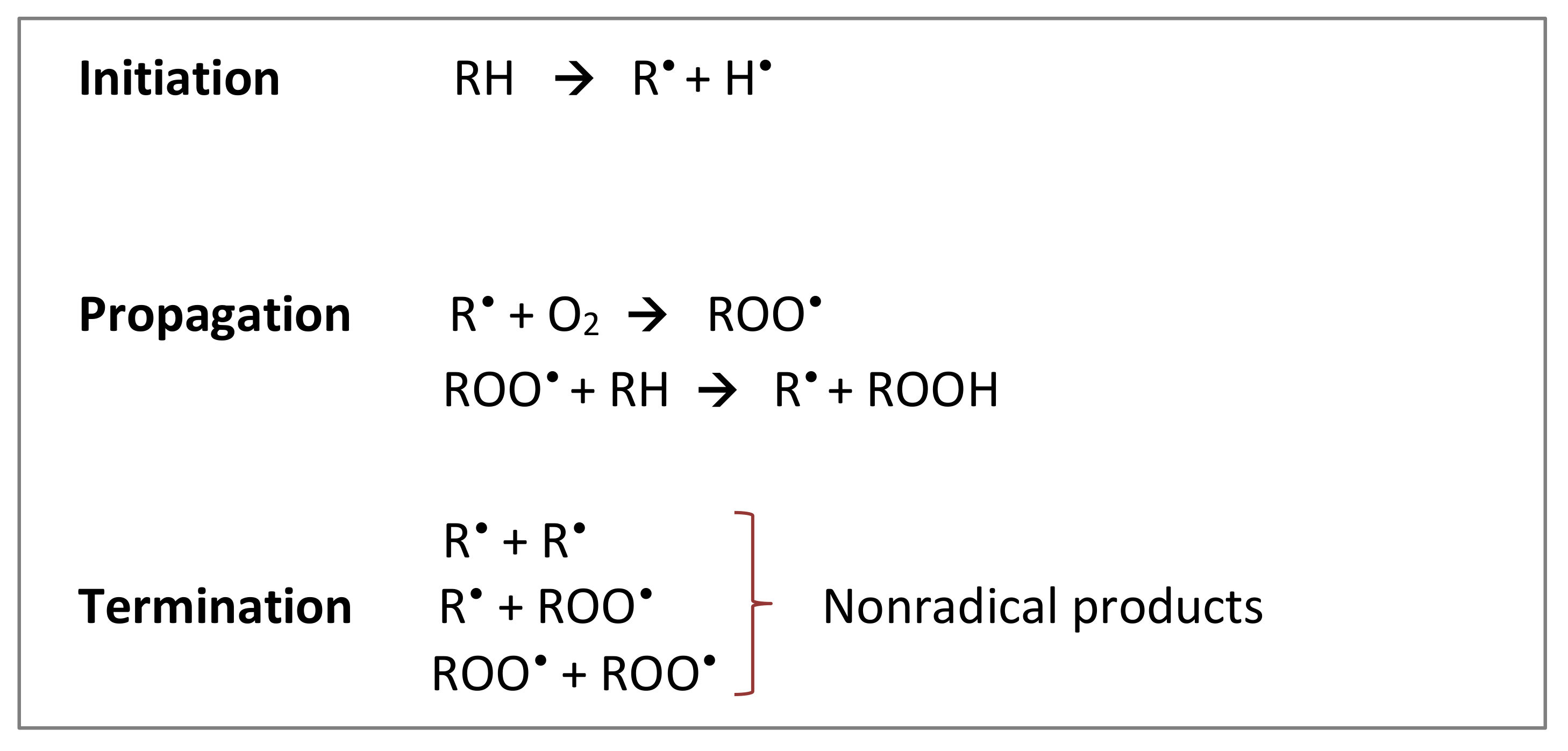
2.1. Common Types of Food Oxidation
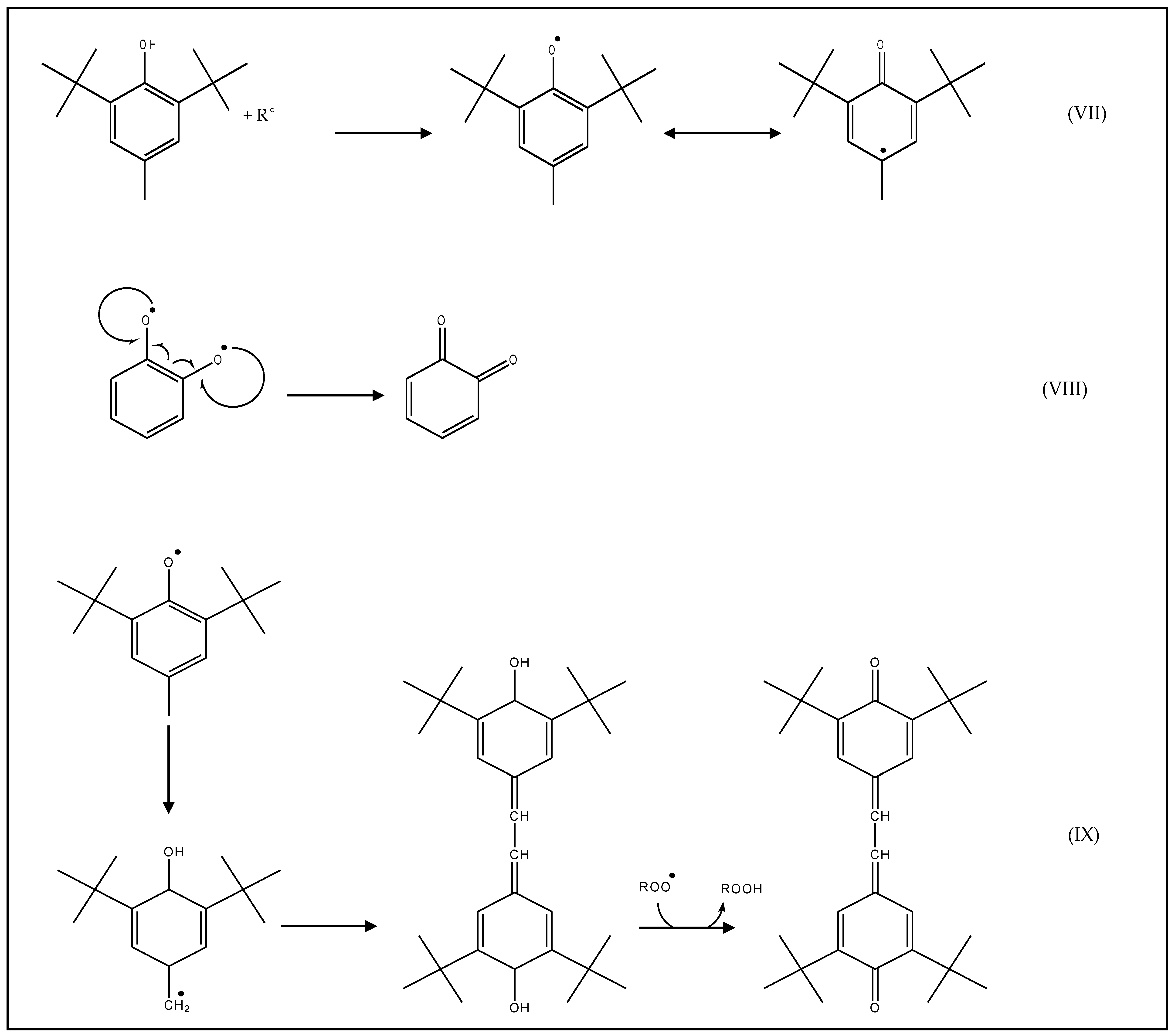
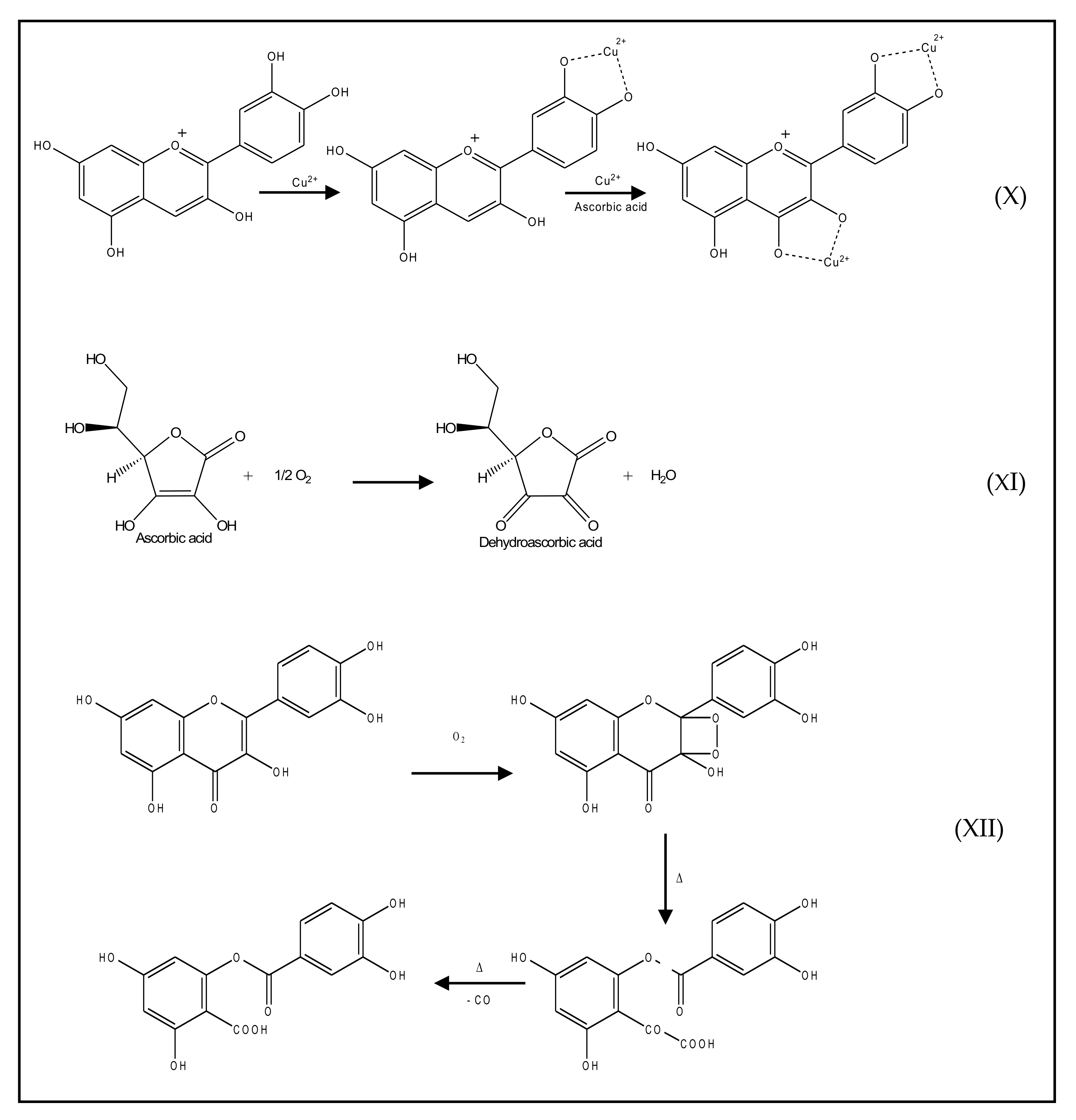
2.2. Mechanism of Action
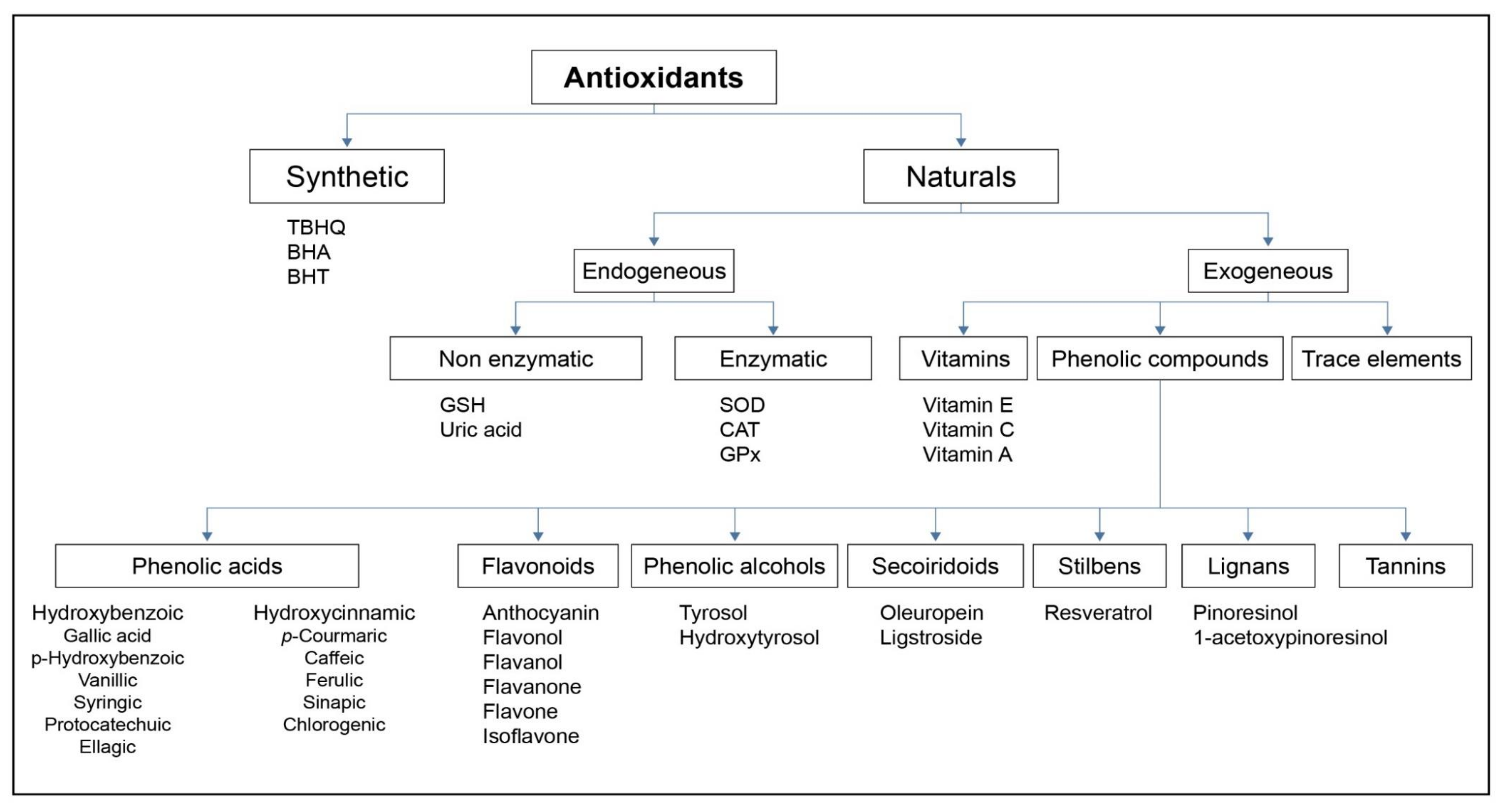
2.3. Main Antioxidant Families
2.4. Total Antioxidant Capacity Assays
2.5. Extraction and Detection of Antioxidants
3. Electrochemistry
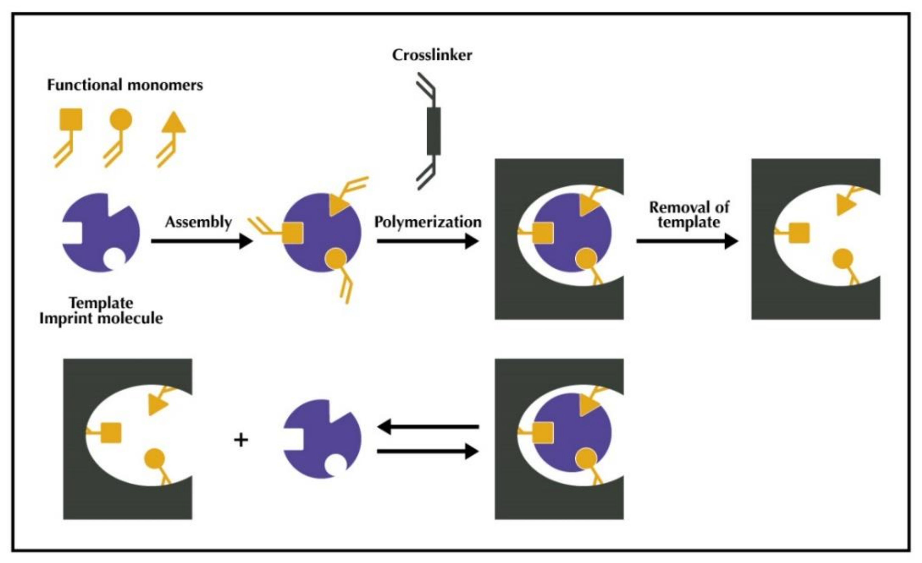
4. Molecular Imprinting
4.1. MIP Synthesis and Applications
4.2. MIPs-Antioxidants
4.3. MIS-Antioxidants
5. Electrochemistry, Molecular Imprinting, and Antioxidants
5.1. Electrochemistry and Antioxidants
5.2. Electrochemistry and Molecular Imprinting
6. Conclusions
Author Contributions
Funding
Conflicts of Interest
References
- Brewer, M.S. Natural Antioxidants: Sources, Compounds. Mech. Action Potential Appl. R. 2011, 10. [Google Scholar] [CrossRef]
- Shahidi, F.; Zhong, Y. Measurement of Antioxidant Activity. J. Funct. Foods 2015, 18, 757–781. [Google Scholar] [CrossRef]
- Apak, R.; Özyürek, M.; Güçlü, K.; Capanoglu, E. Antioxidant Activity/Capacity Measurement. 3. Reactive Oxygen and Nitrogen Species (ROS/RNS) Scavenging Assays, Oxidative Stress Biomarkers, and Chromatographic/Chemometric Assays. J. Agric. Food Chem. 2016, 64, 1046–1070. [Google Scholar] [CrossRef] [PubMed]
- Uslu, B.; Ozkan, S.A. Electroanalytical Methods for the Determination of Pharmaceuticals: A Review of Recent Trends and Developments. Anal. Lett. 2011, 44, 2644–2702. [Google Scholar] [CrossRef]
- Li, M.; Li, D.W.; Xiu, G.; Long, Y.T. Applications of Screen-Printed Electrodes in Current Environmental Analysis. Curr. Opin. Electrochem. 2017, 3, 137–143. [Google Scholar] [CrossRef]
- Giné Bordonaba, J.; Terry, L.A. Electrochemical Behaviour of Polyphenol Rich Fruit Juices Using Disposable Screen-Printed Carbon Electrodes: Towards a Rapid Sensor for Antioxidant Capacity and Individual Antioxidants. Talanta 2012, 90, 38–45. [Google Scholar] [CrossRef] [PubMed]
- Lingxin, S. As Featured in: Molecular Imprinting: Perspectives and Applications. Chem. Soc. Rev. 2016, 45, 2137–2211. [Google Scholar] [CrossRef]
- Garcia-Martinez, O.; Ruiz, C.; Gutierrez-Ibanez, A.; Illescas-Montes, R.; Melguizo-Rodriguez, L. Benefits of Olive Oil Phenolic Compounds in Disease Prevention. Endocr. Metab. Immune Disord. Drug Targets 2018, 18, 333–340. [Google Scholar] [CrossRef]
- Parkinson, L.; Cicerale, S. The Health Benefiting Mechanisms of Virgin Olive Oil Phenolic Compounds. Molecules 2016, 21, 1734. [Google Scholar] [CrossRef] [Green Version]
- Boronat, A.; Mateus, J.; Soldevila-Domenech, N.; Guerra, M.; Rodríguez-Morató, J.; Varon, C.; Muñoz, D.; Barbosa, F.; Morales, J.C.; Gaedigk, A.; et al. Cardiovascular Benefits of Tyrosol and Its Endogenous Conversion into Hydroxytyrosol in Humans. A Randomized, Controlled Trial. Free Radic. Biol. Med. 2019, 143, 471–481. [Google Scholar] [CrossRef]
- Mazué, F.; Delmas, D.; Murillo, G.; Saleiro, D.; Limagne, E.; Latruffe, N. Differential Protective Effects of Red Wine Polyphenol Extracts (RWEs) on Colon Carcinogenesis. Food Funct. 2014, 5, 663–670. [Google Scholar] [CrossRef] [PubMed]
- Kurin, E.; Atanasov, A.G.; Donath, O.; Heiss, E.H.; Dirsch, V.M.; Nagy, M. Synergy Study of the Inhibitory Potential of Red Wine Polyphenols on Vascular Smooth Muscle Cell Proliferation. Planta Med. 2012, 78, 772–778. [Google Scholar] [CrossRef] [PubMed] [Green Version]
- Basli, A.; Soulet, S.; Chaher, N.; Mérillon, J.M.; Chibane, M.; Monti, J.P.; Richard, T. Wine Polyphenols: Potential Agents in Neuroprotection. Oxidative Med. Cell. Longev. 2012, 2012, 805762. [Google Scholar] [CrossRef] [PubMed]
- Shahidi, F.; Ambigaipalan, P. Phenolics and Polyphenolics in Foods, Beverages and Spices: Antioxidant Activity and Health Effects—A Review. J. Funct. Foods 2015, 18, 820–897. [Google Scholar] [CrossRef]
- Lund, M.N.; Baron, C.P. Protein Oxidation in Foods and Food Quality; Woodhead Publishing Limited: Copenhagen, Denmark, 2010. [Google Scholar]
- Baron, C.P.; Berner, L.; Skibsted, L.H.; Refsgaard, H.H.F. Evaluation of Activity of Selected Antioxidants on Proteins in Solution and in Emulsions. Free Radic. Res. 2005, 39, 777–785. [Google Scholar] [CrossRef]
- Jacobsen, C.; Let, M.B.; Nielsen, N.S.; Meyer, A.S. Antioxidant Strategies for Preventing Oxidative Flavour Deterioration of Foods Enriched with N-3 Polyunsaturated Lipids: A Comparative Evaluation. Trends Food Sci. Technol. 2008, 19, 76–93. [Google Scholar] [CrossRef] [Green Version]
- Liu, H.C.; Chen, W.L.; Mao, S.J.T. Antioxidant Nature of Bovine Milk β -Lactoglobulin. J. Dairy Sci. 2007, 90, 547–555. [Google Scholar] [CrossRef]
- Refsgaard, H.H.F.; Tsai, L.; Stadtman, E.R. Modifications of Proteins by Polyunsaturated Fatty Acid Peroxidation Products. Proc. Natl. Acad. Sci. USA 2000, 97, 611–616. [Google Scholar] [CrossRef] [Green Version]
- Díaz, M.; Decker, E.A. Antioxidant Mechanisms of Caseinophosphopeptides and Casein Hydrolysates and Their Application in Ground Beef. J. Agric. Food Chem. 2004, 52, 8208–8213. [Google Scholar] [CrossRef]
- Peña-Ramos, E.A.; Xiong, Y.L. Whey and Soy Protein Hydrolysates Inhibit Lipid Oxidation in Cooked Pork Patties. Meat Sci. 2003, 64, 259–263. [Google Scholar] [CrossRef]
- Sakanaka, S.; Tachibana, Y. Active Oxygen Scavenging Activity of Egg-Yolk Protein Hydrolysates and Their Effects on Lipid Oxidation in Beef and Tuna Homogenates. Food Chem. 2006, 95, 243–249. [Google Scholar] [CrossRef]
- Sakanaka, S.; Tachibana, Y.; Ishihara, N.; Juneja, L.R. Antioxidant Properties of Casein Calcium Peptides and Their Effects on Lipid Oxidation in Beef Homogenates. J. Agric. Food Chem. 2005, 53, 464–468. [Google Scholar] [CrossRef]
- Elias, R.J.; Kellerby, S.S.; Decker, E.A. Antioxidant Activity of Proteins and Peptides. Crit. Rev. Food Sci. Nutr. 2008, 48, 430–441. [Google Scholar] [CrossRef]
- Pietta, P.-G. Flavonoids as Antioxidants. J. Nat. Prod. 2000, 63, 1035–1042. [Google Scholar] [CrossRef]
- Young, I.S.; Woodside, J. V Antioxidants in Health and Disease Antioxidants in Health and Disease. J. Clin. Pathol. 2001, 54, 176–186. [Google Scholar] [CrossRef] [Green Version]
- Tsao, R. Chemistry and Biochemistry of Dietary Polyphenols. Nutrients 2010, 2, 1231–1246. [Google Scholar] [CrossRef] [PubMed]
- Horton, W.; Török, M. Natural and Nature-Inspired Synthetic Small Molecule Antioxidants in the Context of Green Chemistry; Elsevier Inc.: Amsterdam, The Netherlands, 2018; ISBN 9780128095492. [Google Scholar]
- Shahidi, F.; Janitha, P.K.; Wanasundara, P.D. Phenolic Antioxidants. Crit. Rev. Food Sci. Nutr. 1992, 32, 67–103. [Google Scholar] [CrossRef]
- Nimse Balasaheb, S.; Pal, D. Free radicals, natural antioxidants, and their reaction mechanisms. RSC Adv. 2015, 5, 27986–28006. [Google Scholar] [CrossRef] [Green Version]
- Avdeeva, L.V.; Gvozdev, R.I. Oxidation of L-Ascorbic Acid in the Presence of the Copper-Binding Compound from Methanotrophic Bacteria Methylococcus Capsulatus (M). Biomimetics 2020, 5, 48. [Google Scholar] [CrossRef]
- Tournairev, C.; Croux, S.; Maurette, M.T.; Beck, I.; Hocquaux, M.; Braun, A.M.; Oliveros, E. Antioxidant activity of flavonoids: Efficiency of singlet oxygen (1 delta g) quenching. J. Photochem. Photobiol. B 1993, 19, 205–215. [Google Scholar] [CrossRef]
- Herrero, M.; Ibáñez, E.; Cifuentes, A. Analysis of Natural Antioxidants by Capillary Eletromigration Methods. J. Sep. Sci. 2005, 28, 883–897. [Google Scholar] [CrossRef] [Green Version]
- Shahidi, F. Antioxidants in Food and Food Antioxidants. Food Nahr. 2000, 44, 158–163. [Google Scholar] [CrossRef]
- Manach, C.; Scalbert, A.; Morand, C.; Rémésy, C.; Jiménez, L. Polyphenols: Food Sources and Bioavailability. Am. J. Clin. Nutr. 2004, 79, 727–747. [Google Scholar] [CrossRef] [Green Version]
- Robbins, R.J. Phenolic Acids in Foods: An Overview of Analytical Methodology. J. Agric. Food Chem. 2003, 51, 2866–2887. [Google Scholar] [CrossRef] [PubMed]
- King, A.; Young, G. Characteristics and Occurrence of Phenolic Phytochemicals. J. Am. Diet. Assoc. 1999, 99, 213–218. [Google Scholar] [CrossRef]
- Boskou, D.; Blekas, G.; Tsimidou, M. Olive Oil Composition. In Olive Oil: Chemistry and Technology: Second Edition; Elsevier Inc.: Amsterdam, The Netherlands, 2006; pp. 41–72. ISBN 9780128043547. [Google Scholar]
- Rice-Evans, C.A.; Miller, N.J.; Paganga, G. Antioxidant Properties of Phenolic Compounds. Trends Plant Sci. 1997, 2, 152–159. [Google Scholar] [CrossRef]
- Debelo, H.; Li, M.; Ferruzzi, M.G. Processing Influences on Food Polyphenol Profiles and Biological Activity. Curr. Opin. Food Sci. 2020. [Google Scholar] [CrossRef]
- Kliment, C.R.; Oury, T.D. Oxidative stress, extracellular matrix targets, and idiopathic pulmonary fibrosis. Free Radic. Biol. Med. 2010, 49, 707–717. [Google Scholar] [CrossRef]
- Niki, E. Free Radical Biology & Medicine Assessment of Antioxidant Capacity in Vitro and in Vivo. Free Radic. Biol. Med. 2010, 49, 503–515. [Google Scholar] [CrossRef]
- Li, B.; Pratt, D.A. Methods for determining the efficacy of radical-trapping antioxidants. Free Radic. Biol. Med. 2015, 82, 187–202. [Google Scholar] [CrossRef] [PubMed]
- Prieto, M.A.; Vázquez, J.A.; Murado, M.A. Crocin Bleaching Antioxidant Assay Revisited: Application to Microplate to Analyze Antioxidant and pro-Oxidant Activities. Food Chem. 2014. [Google Scholar] [CrossRef] [PubMed] [Green Version]
- Nagatani, N.; Inoue, Y.; Araki, A.; Ushijima, H.; Hattori, G.; Sakurai, Y.; Ogidou, Y.; Saito, M.; Tamiya, E. Rapid Sensing of Antioxidant Capacity Based on Electrochemiluminescence Induced by Electrochemically Generated Reactive Oxygen Species. Electrochim. Acta 2016. [Google Scholar] [CrossRef]
- Apak, R.; Özyürek, M.; Güçlü, K.; Çapanoʇlu, E. Antioxidant Activity/Capacity Measurement. 1. Classification, Physicochemical Principles, Mechanisms, and Electron Transfer (ET)-Based Assays. J. Agric. Food Chem. 2016, 64, 997–1027. [Google Scholar] [CrossRef]
- Apak, R.; Özyürek, M.; Güçlü, K.; Çapanoʇlu, E. Antioxidant Activity/Capacity Measurement. 2. Hydrogen Atom Transfer (HAT)-Based, Mixed-Mode (Electron Transfer (ET)/HAT), and Lipid Peroxidation Assays. J. Agric. Food Chem. 2016, 64, 1028–1045. [Google Scholar] [CrossRef] [PubMed]
- Do, E.; Gökmen, V. Evolution of Food Antioxidants as a Core Topic of Food Science for a Century. Food Res. Int. 2018, 105, 76–93. [Google Scholar] [CrossRef]
- Dai, J.; Mumper, R.J. Plant Phenolics: Extraction, Analysis and Their Antioxidant and Anticancer Properties. Molecules 2010, 15, 7313–7352. [Google Scholar] [CrossRef]
- Faculty, B. Talanta DPPH Assay of Vegetable Oils and Model Antioxidants in Protic and Aprotic Solvents. Talanta 2013, 109, 13–19. [Google Scholar] [CrossRef]
- Marteau, C.; Guitard, R.; Penverne, C.; Favier, D.; Nardello-rataj, V.; Aubry, J. Boosting Effect of Ortho- Propenyl Substituent on the Antioxidant Activity of Natural Phenols. Food Chem. 2016, 196, 418–427. [Google Scholar] [CrossRef]
- Pinto, D.; Vieira, E.F.; Peixoto, A.F.; Freire, C.; Freitas, V.; Costa, P.; Delerue-matos, C.; Rodrigues, F. Optimizing the Extraction of Phenolic Antioxidants from Chestnut Shells by Subcritical Water Extraction Using Response Surface Methodology. Food Chem. 2021, 334, 127521. [Google Scholar] [CrossRef]
- Krylova, E.; Gavrilenko, N.; Saranchina, N.; Gavrilenko, M. Novel Colorimetric Sensor for Cupric Reducing Antioxidant Capacity (CUPRAC) Measurement. Procedia Eng. 2016, 168, 355–358. [Google Scholar] [CrossRef]
- Cicco, N.; Lanorte, M.T.; Paraggio, M.; Viggiano, M.; Lattanzio, V. A Reproducible, Rapid and Inexpensive Folin—Ciocalteu Micro-Method in Determining Phenolics of Plant Methanol Extracts. Microchem. J. 2009, 91. [Google Scholar] [CrossRef]
- Rock, L.; Brunswick, N. Standardized Methods for the Determination of Antioxidant Capacity and Phenolics in Foods and Dietary Supplements. J. Agric. Food Chem. 2005, 53, 4290–4302. [Google Scholar] [CrossRef]
- Pardeshi, S.; Kumar, A.; Dhodapkar, R. Molecular Imprinting: Mimicking Molecular Receptors for Antioxidants. Mater. Sci. Forum 2011, 675–677, 515–520. [Google Scholar] [CrossRef]
- Maroun, R.G.; Rajha, H.N.; El Darra, N.; El Kantar, S.; Chacar, S.; Debs, E.; Vorobiev, E.; Louka, N. 8—Emerging Technologies for the Extraction of Polyphenols from Natural Sources; Woodhead Publishing: Oxford, UK, 2018; ISBN 9780128135723. [Google Scholar]
- Mayachiew, P.; Devahastin, S. Antimicrobial and Antioxidant Activities of Indian Gooseberry and Galangal Extracts. LWT Food Sci. Technol. 2008, 41, 1153–1159. [Google Scholar] [CrossRef]
- Zuo, Y.; Chen, H.; Deng, Y. Simultaneous Determination of Catechins, Caffeine and Gallic Acids in Green, Oolong, Black and Pu-Erh Teas Using HPLC with a Photodiode Array Detector. Talanta 2002, 57, 307–316. [Google Scholar] [CrossRef]
- Chirinos, R.; Rogez, H.; Campos, D.; Pedreschi, R.; Larondelle, Y. Optimization of Extraction Conditions of Antioxidant Phenolic Compounds from Mashua (Tropaeolum Tuberoum RuiZ & Pavon) Tubers. Sep. Purif. Technol. 2007, 55, 217–225. [Google Scholar] [CrossRef]
- Spigno, G.; Tramelli, L.; De Faveri, D.M. Effects of Extraction Time, Temperature and Solvent on Concentration and Antioxidant Activity of Grape Marc Phenolics. J. Food Eng. 2007, 81, 200–208. [Google Scholar] [CrossRef]
- Hidalgo, G.I.; Almajano, M.P. Red Fruits: Extraction of Antioxidants, Phenolic Content, and Radical Scavenging Determination: A Review. Antioxidants 2017, 6, 7. [Google Scholar] [CrossRef] [Green Version]
- Pisoschi, A.M.; Cimpeanu, C.; Predoi, G. Electrochemical Methods for Total Antioxidant Capacity and Its Main Contributors Determination: A Review. Open Chem. 2015, 824–856. [Google Scholar] [CrossRef] [Green Version]
- Blasco, A.J.; Crevillén, A.G.; González, M.C.; Escarpa, A. Direct Electrochemical Sensing and Detection of Natural Antioxidants and Antioxidant Capacity in Vitro Systems. Electroanalysis 2007, 19, 2275–2286. [Google Scholar] [CrossRef]
- Valek, L.; Stipc, T. Electrochemical Determination of Antioxidant Capacity of Fruit Tea Infusions. Food Chem. 2010, 121, 820–825. [Google Scholar] [CrossRef]
- Lyklema, J. Current Opinion in Colloid & Interface Science Principles of Interactions in Non-Aqueous Electrolyte Solutions. Curr. Opin. Colloid Interface Sci. 2013, 18, 116–128. [Google Scholar] [CrossRef]
- Dimé, A.; Cattey, H.; Lucas, D.; Devillers, C. Crystallographic and (Spectro)Electrochemical Characterizations of Cobalt(II) 10-Phenyl-5,15-Di-p-Tolylporphyrin. J. Mol. Struct. 2021, 1226, 129321. [Google Scholar] [CrossRef]
- Rolle, S.D.; Konev, D.V.; Devillers, C.H.; Lizgina, K.V.; Lucas, D.; Stern, C.; Herbst, F.; Heintz, O.; Vorotyntsev, M.A. Efficient Synthesis of a New Electroactive Polymer of Co (II) Porphine by in-situ Replacement of Mg (II) inside Mg (II) Polyporphine film. Electrochim. Acta 2016, 204, 276–286. [Google Scholar] [CrossRef]
- Lima, A.P.; Wallans, T.P.; Nossol, E.; Richter, E.M.; Munoz, R.A.A. Critical Evaluation of Voltammetric Techniques for Antioxidant Capacity and Activity: Presence of Alumina on Glassy-Carbon Electrodes Alters the Results. Electrochim. Acta 2020, 358. [Google Scholar] [CrossRef]
- David, I.G.; Buleandr, M.; Popa, D.E.; Bîzgan, A.C.; Moldovan, Z.; Badea, I.; Iorgulescu, E.E.; Basaga, H. Voltammetric Determination of Polyphenolic Content as Rosmarinic Acid Equivalent in Tea Samples Using Pencil Graphite Electrodes. J. Food Sci. Technol. 2016, 53, 2589–2596. [Google Scholar] [CrossRef] [Green Version]
- David, I.G.; Litescu, C.; Popa, E.; Buleandra, M.; Iordache, L.; Albu, C.; Alecu, A. Analytical Methods Pencil Graphite Electrode—Application to Polyphenol Content Determination in Citrus Juice. Anal. Methods 2018, 10, 5763–5772. [Google Scholar] [CrossRef]
- Kariuki, J.; Ervin, E.; Olafson, C. Development of a Novel, Low-Cost, Disposable Wooden Pencil Graphite Electrode for Use in the Determination of Antioxidants and Other Biological Compounds. Sensors 2015, 15, 18887–18900. [Google Scholar] [CrossRef] [Green Version]
- Rezaei, B.; Boroujeni, M.K.; Ensa, A.A. Biosensors and Bioelectronics Caffeine Electrochemical Sensor Using Imprinted film as Recognition Element Based on Polypyrrole, Sol-Gel, and Gold Nanoparticles Hybrid Nanocomposite Modified Pencil Graphite Electrode. Biosens. Bioelectron. 2014, 60, 77–83. [Google Scholar] [CrossRef]
- Głód, B.K.; Kiersztyn, I.; Piszcz, P. Total Antioxidant Potential Assay with Cyclic Voltammetry and/or Differential Pulse Voltammetry Measurements. J. Electroanal. Chem. 2014, 719, 24–29. [Google Scholar] [CrossRef]
- Maji, P.; Basu, S.; Banik, B.K.; Ganguly, J. Antioxidant Edible Mushrooms: A Green and Rapid Electrochemical Study with the Aqueous Extracts. Mod. Chem. Appl. 2018, 06, 1–7. [Google Scholar] [CrossRef]
- Suliborska, K.; Baranowska, M.; Bartoszek, A.; Chrzanowski, W.; Namieśnik, J. Determination of Antioxidant Activity of Vitamin C by Voltammetric Methods. Proceedings 2019, 11, 23. [Google Scholar] [CrossRef] [Green Version]
- Teixeira, J.; Gaspar, A.; Garrido, E.M.; Garrido, J.; Borges, F. Hydroxycinnamic Acid Antioxidants: An Electrochemical Overview. Biomed Res. Int. 2013, 2013, 251754. [Google Scholar] [CrossRef]
- Vilas-Boas, Â.; Valderrama, P.; Fontes, N.; Geraldo, D.; Bento, F. Evaluation of Total Polyphenol Content of Wines by Means of Voltammetric Techniques: Cyclic Voltammetry vs Differential Pulse Voltammetry. Food Chem. 2019, 276, 719–725. [Google Scholar] [CrossRef]
- José Jara-Palacios, M.; Hernanz, D.; Luisa Escudero-Gilete, M.; Heredia, F.J. Antioxidant Potential of White Grape Pomaces: Phenolic Composition and Antioxidant Capacity Measured by Spectrophotometric and Cyclic Voltammetry Methods. Food Res. Int. 2014, 66, 150–157. [Google Scholar] [CrossRef]
- José Jara-Palacios, M.; Luisa Escudero-Gilete, M.; Miguel Hernández-Hierro, J.; Heredia, F.J.; Hernanz, D. Cyclic Voltammetry to Evaluate the Antioxidant Potential in Winemaking By-Products. Talanta 2017, 165, 211–215. [Google Scholar] [CrossRef]
- Szczepaniak, O.M.; Ligaj, M.; Kobus-Cisowska, J.; Maciejewska, P.; Tichoniuk, M.; Szulc, P. Application for Novel Electrochemical Screening of Antioxidant Potential and Phytochemicals in Cornus Mas Extracts. CyTA J. Food 2019, 17, 781–789. [Google Scholar] [CrossRef] [Green Version]
- Amidi, S.; Mojab, F.; Bayandori, A. A Simple Electrochemical Method for the Rapid Estimation of Antioxidant Potentials of Some Selected Medicinal Plants. Iran. J. Pharm. Res. IJPR 2012, 11, 117–121. [Google Scholar]
- Kissinger, P.T.; Lafayette, W.; Heineman, W.R. Cyclic Voltammetry. J. Chem. Educ. 1983, 60, 702–706. [Google Scholar] [CrossRef]
- Chevion, S.; Roberts, M.A.; Chevion, M. The Use of Cyclic Voltammetry for the Evaluation of Antioxidant Capacity. Free Radic. Biol. Med. 2000, 28, 860–870. [Google Scholar] [CrossRef]
- Hoyos-arbeláez, J.; Vázquez, M.; Contreras-calderón, J. Electrochemical Methods as a Tool for Determining the Antioxidant Capacity of Food and Beverages: A Review. Food Chem. 2017, 221, 1371–1381. [Google Scholar] [CrossRef] [PubMed]
- Sciences, A. Polyphenol Analysis in Black Tea with Carbon Nanotube Electrode. Anal. Sci. 2019, 1–5. [Google Scholar] [CrossRef] [Green Version]
- Zhang, J.-W.; Wang, K.-P.; Zhang, X. Fabrication of SnO2 Decorated Graphene Composite Material and Its Application in Electrochemical Detection of Caffeic Acid in Red Wine. Mater. Res. Bull. 2020, 126, 110820. [Google Scholar] [CrossRef]
- Bharath, G.; Alhseinat, E.; Madhu, R.; Mugo, S.M.; Alwasel, S.; Halim, A. Facile Synthesis of Au@α-Fe2O3@RGO Ternary Nanocomposites for Enhanced Electrochemical Sensing of Caffeic Acid toward Biomedical Applications. J. Alloys Compd. 2018, 750, 819–827. [Google Scholar] [CrossRef]
- Manikandan, V.S.; Sidhureddy, B.; Thiruppathi, A.R.; Chen, A. Sensitive Electrochemical Detection of Caffeic Acid in Wine Based on Fluorine-Doped Graphene Oxide. Sensors 2019, 19, 1604. [Google Scholar] [CrossRef] [Green Version]
- Kokulnathan, T.; Raja, N.; Chen, S.; Liao, W. Nanomolar Electrochemical Detection of Caffeic Acid in Fortified Wine Samples Based on Gold/Palladium Nanoparticles Decorated Graphene Flakes. J. Colloid Interface Sci. 2017, 501, 77–85. [Google Scholar] [CrossRef]
- Palanisamy, S.; Chen, S.-M.; Velusamy, V.; Chen, T.-W.; Ramaraj, S.K. Electrochemical Determination of Caffeic Acid in Wine Samples Using Reduced Graphene Oxide/Polydopamine Composite. J. Electrochem. Soc. 2016, 163, B726–B731. [Google Scholar] [CrossRef]
- Tashkhourian, J.; Nami-ana, S.F. A Sensitive Electrochemical Sensor for Determination of Gallic Acid Based on SiO2 Nanoparticle Modi Fi Ed Carbon Paste Electrode. Mater. Sci. Eng. C 2015, 52, 103–110. [Google Scholar] [CrossRef]
- Gao, F.; Zheng, D.; Tanaka, H.; Zhan, F.; Yuan, X.; Gao, F.; Wang, Q. An Electrochemical Sensor for Gallic Acid Based on Fe2O3/Electro-Reduced Graphene Oxide Composite: Estimation for the Antioxidant Capacity Index of Wines. Mater. Sci. Eng. C. Mater. Biol. Appl. 2015, 57, 279–287. [Google Scholar] [CrossRef] [PubMed]
- Kahl, M.; Golden, T. Electrochemical Determination of Phenolic Acids at a Zn/Al Layered Double Hydroxide Film Modified Glassy Carbon Electrode. Electroanalysis 2014, 26. [Google Scholar] [CrossRef]
- Souza, L.P.; Calegari, F.; Zarbin, A.J.G.; Marcolino-Júnior, L.H.; Bergamini, M.F. Voltammetric Determination of the Antioxidant Capacity in Wine Samples Using a Carbon Nanotube Modified Electrode. J. Agric. Food Chem. 2011, 59, 7620–7625. [Google Scholar] [CrossRef] [PubMed]
- Qi, S.; Zhao, B.; Tang, H.; Jiang, X. Electrochimica Acta Determination of Ascorbic Acid, Dopamine, and Uric Acid by a Novel Electrochemical Sensor Based on Pristine Graphene. Electrochim. Acta 2015, 161, 395–402. [Google Scholar] [CrossRef]
- Raoof, J.B.; Ojani, R.; Beitollahi, H.; Hossienzadeh, R. Electrocatalytic Determination of Ascorbic Acid at the Surface of 2,7-Bis(Ferrocenyl Ethyl)Fluoren-9-one Modified Carbon Paste Electrode. Electroanalysis 2006, 18, 1193–1201. [Google Scholar] [CrossRef]
- Pisoschi, A.M.; Pop, A.; Negulescu, G.P.; Pisoschi, A. Determination of Ascorbic Acid Content of Some Fruit Juices and Wine by Voltammetry Performed at Pt and Carbon Paste Electrodes. Molecules 2011, 16, 1349–1365. [Google Scholar] [CrossRef]
- Barroso, M.F.; de-los-Santos-Álvarez, N.; Lobo-Castañón, M.J.; Miranda-Ordieres, A.J.; Delerue-Matos, C.; Oliveira, M.B.P.P.; Tuñón-Blanco, P. DNA-Based Biosensor for the Electrocatalytic Determination of Antioxidant Capacity in Beverages. Biosens. Bioelectron. 2011, 26, 2396–2401. [Google Scholar] [CrossRef] [Green Version]
- Zokhtareh, R.; Rahimnejad, M. A Novel Sensitive Electrochemical Sensor Based on Nickel Chloride Solution Modified Glassy Carbon Electrode for Curcumin Determination. Electroanalysis 2018, 30, 921–927. [Google Scholar] [CrossRef]
- Ziyatdinova, G.K.; Nizamova, A.M.; Budnikov, H.C. Voltammetric Determination of Curcumin in Spices. J. Anal. Chem. 2012, 67, 591–594. [Google Scholar] [CrossRef]
- Apetrei, C.; Apetrei, I.M.; De Saja, J.A.; Rodriguez-Mendez, M.L. Carbon Paste Electrodes Made from Different Carbonaceous Materials: Application in the Study of Antioxidants. Sensors 2011, 11, 1328–1344. [Google Scholar] [CrossRef]
- Litescu, S.-C.; Radu, G.-L. Estimation of the Antioxidative Properties of Tocopherols—An Electrochemical Approach. Eur. Food Res. Technol. 2000, 211, 218–221. [Google Scholar] [CrossRef]
- Liang, Z.; Zhai, H.; Chen, Z.; Wang, S.; Wang, H.; Wang, S. A Sensitive Electrochemical Sensor for Flavonoids Based on a Multi-Walled Carbon Paste Electrode Modified by Cetyltrimethyl Ammonium Bromide-Carboxylic Multi-Walled Carbon Nanotubes. Sens. Actuators B. Chem. 2017, 244, 897–906. [Google Scholar] [CrossRef]
- Shono, T. 7.1—Oxidation by Electrochemical Methods. In Comprehensive Organic Synthesis; Trost, B.M., Fleming, I., Eds.; Pergamon: Oxford, UK, 1991; pp. 789–813. ISBN 978-0-08-052349-1. [Google Scholar]
- Meites, L.; Zuman, P.; Nurnberg, H. Recommended Terms, Symbols, and Definitions for Electroanalytical Chemistry (Recommendations 1985). Pure Appl. Chem. 1985, 57, 1491–1505. [Google Scholar] [CrossRef]
- Silva, D.H.S.; Pereira, F.C.; Yoshida, M.; Zanoni, M.V.B. Electrochemical Evaluation of Lipophilic Antioxidants from Iryanthera Juruensis Fruits (Myristicaceae). Eclética Química 2005, 30, 15–21. [Google Scholar] [CrossRef]
- Taylor, P.; Bertolino, F.A.; Stege, P.W.; Salinas, E. Electrochemical Study of the Antioxidant Activity and the Synergic Effect of Selenium with Natural and Synthetic Antioxidants. Anal. Lett. 2010, 43, 2078–2090. [Google Scholar] [CrossRef]
- Karikalan, N.; Karthik, R.; Chen, S.M.; Chen, H.A. A Voltammetric Determination of Caffeic Acid in Red Wines Based on the Nitrogen Doped Carbon Modified Glassy Carbon Electrode. Sci. Rep. 2017, 7, 1–10. [Google Scholar] [CrossRef] [Green Version]
- Cormack, P.A.G.; Elorza, A.Z. Molecularly Imprinted Polymers: Synthesis and Characterisation. J. Chromatogr. B 2004, 804, 173–182. [Google Scholar] [CrossRef] [PubMed]
- Shi, Y.; Zhang, J.-H.; Shi, D.; Jiang, M.; Zhu, Y.-X.; Mei, S.-R.; Zhou, Y.-K.; Dai, K.; Lu, B. Selective Solid-Phase Extraction of Cholesterol Using Molecularly Imprinted Polymers and Its Application in Different Biological Samples. J. Pharm. Biomed. Anal. 2006, 42, 549–555. [Google Scholar] [CrossRef]
- Włoch, M.; Datta, J. Synthesis and Polymerisation Techniques of Molecularly Imprinted Polymers. Compr. Anal. Chem. 2019, 86, 17–40. [Google Scholar] [CrossRef]
- Baggiani, C.; Biagioli, F.; Anfossi, L.; Giovannoli, C.; Passini, C.; Giraudi, G. Effect of the Mimic Structure on the Molecular Recognition Properties of Molecularly Imprinted Polymers for Ochratoxin A Prepared by a Fragmental Approach. React. Funct. Polym. 2013, 73, 833–837. [Google Scholar] [CrossRef] [Green Version]
- Gómez-Pineda, L.E.; Pina-Luis, G.E.; Cuán, Á.; García-Calzón, J.A.; Díaz-García, M.E. Physico-Chemical Characterization of Flavonol Molecularly Imprinted Polymers. React. Funct. Polym. 2011, 71, 402–408. [Google Scholar] [CrossRef]
- Sergeyeva, T.A.; Gorbach, L.A.; Piletska, E.V.; Piletsky, S.A.; Brovko, O.O.; Honcharova, L.A.; Lutsyk, O.D.; Sergeeva, L.M.; Zinchenko, O.A.; El’skaya, A. V Colorimetric Test-Systems for Creatinine Detection Based on Composite Molecularly Imprinted Polymer Membranes. Anal. Chim. Acta 2013, 770, 161–168. [Google Scholar] [CrossRef]
- Meier, F.; Schott, B.; Riedel, D.; Mizaikoff, B. Computational and Experimental Study on the Influence of the Porogen on the Selectivity of 4-Nitrophenol Molecularly Imprinted Polymers. Anal. Chim. Acta 2012, 744, 68–74. [Google Scholar] [CrossRef]
- Nomachi, M.; Kubo, T.; Hosoya, K.; Kaya, K. Solvent Effects in the Preparation of Molecularly Imprinted Polymers for Melatonin Using N-Propionyl-5-Methoxytryptamine as the Pseudo Template. Anal. Bioanal. Chem. 2006, 384, 1291–1296. [Google Scholar] [CrossRef]
- Turiel, E.; Martín-Esteban, A. Molecularly Imprinted Polymers for Sample Preparation: A Review. Anal. Chim. Acta 2010, 668, 87–99. [Google Scholar] [CrossRef]
- Pichon, V. Selective Sample Treatment Using Molecularly Imprinted Polymers. J. Chromatogr. A 2007, 1152, 41–53. [Google Scholar] [CrossRef]
- Pichon, V. Aptamer-Based and Immunosorbents; Elsevier Inc.: Amsterdam, The Netherlands, 2019; ISBN 9780128169063. [Google Scholar]
- Acquah, C.; Danquah, M.K.; Yon, J.L.S.; Sidhu, A.; Ongkudon, C.M. A Review on Immobilised Aptamers for High Throughput Biomolecular Detection and Screening. Anal. Chim. Acta 2015, 888, 10–18. [Google Scholar] [CrossRef] [Green Version]
- Du, F.; Guo, L.; Qin, Q.; Zheng, X.; Ruan, G.; Li, J.; Li, G. Recent Advances in Aptamer-Functionalized Materials in Sample Preparation. Trac Trends Anal. Chem. 2015, 67, 134–146. [Google Scholar] [CrossRef]
- Sellergren, B. Direct Drug Determination by Selective Sample Enrichment on an Imprinted Polymer. Anal. Chem. 1994, 66, 1578–1582. [Google Scholar] [CrossRef]
- Puoci, F.; Garreffa, C.; Iemma, F.; Muzzalupo, R.; Spizzirri, U.G.; Picci, N. Molecularly Imprinted Solid Phase Extraction for Detection of Sudan I in Food Matrices. Food Chem. 2005, 93, 349–353. [Google Scholar] [CrossRef]
- Farrington, K.; Magner, E.; Regan, F. Predicting the Performance of Molecularly Imprinted Polymers: Selective Extraction of Caffeine by Molecularly Imprinted Solid Phase Extraction. Anal. Chim. Acta 2006, 566, 60–68. [Google Scholar] [CrossRef]
- Jiang, T.; Zhao, L.; Chu, B.; Feng, Q.; Yan, W.; Lin, J.-M. Molecularly Imprinted Solid-Phase Extraction for the Selective Determination of 17β-Estradiol in Fishery Samples with High Performance Liquid Chromatography. Talanta 2009, 78, 442–447. [Google Scholar] [CrossRef] [PubMed]
- Sun, X.; Wang, M.; Peng, J.; Yang, L.; Wang, X.; Wang, F.; Zhang, X.; Wu, Q.; Chen, R.; Chen, J. Dummy Molecularly Imprinted Solid Phase Extraction of Climbazole from Environmental Water Samples. Talanta 2019, 196, 47–53. [Google Scholar] [CrossRef]
- Sun, X.; Wang, M.; Yang, L.; Wen, H.; Wang, L.; Li, T.; Tang, C.; Yang, J. Preparation and Evaluation of Dummy-Template Molecularly Imprinted Polymer as a Potential Sorbent for Solid Phase Extraction of Imidazole Fungicides from River Water. J. Chromatogr. A 2019, 1586, 1–8. [Google Scholar] [CrossRef]
- Li, Z.; Qian, Z.; Hu, S.; Gong, T.; Xian, Q. Molecularly Imprinted Solid Phase Extraction Coupled with Gas Chromatography-Mass Spectrometry for Determination of N-Nitrosodiphenylamine in Water Samples. Chemosphere 2018, 212, 872–880. [Google Scholar] [CrossRef] [PubMed]
- Wang, C.; Cheng, L.; Zhang, L.; Zuo, Y. Graphene Oxide Based Molecularly Imprinted Polymers Modified with β-Cyclodextrin for Selective Extraction of Di(2-Ethylhexyl) Phthalate in Environmental Waters. J. Sep. Sci. 2019, 42, 1248–1256. [Google Scholar] [CrossRef] [PubMed]
- Zhu, G.; Cheng, G.; Wang, P.; Li, W.; Wang, Y.; Fan, J. Water Compatible Imprinted Polymer Prepared in Water for Selective Solid Phase Extraction and Determination of Ciprofloxacin in Real Samples. Talanta 2019, 200, 307–315. [Google Scholar] [CrossRef] [PubMed]
- Kadhirvel, P.; Combès, A.; Bordron, L.; Pichon, V. Development and Application of Water-Compatible Molecularly Imprinted Polymers for the Selective Extraction of Carbamazepine from Environmental Waters. Anal. Bioanal. Chem. 2019, 411, 1525–1536. [Google Scholar] [CrossRef]
- Wang, M.; Chang, X.; Wu, X.; Yan, H.; Qiao, F. Water-Compatible Dummy Molecularly Imprinted Resin Prepared in Aqueous Solution for Green Miniaturized Solid-Phase Extraction of Plant Growth Regulators. J. Chromatogr. A 2016, 1458, 9–17. [Google Scholar] [CrossRef]
- Wang, M.; Liang, S.; Bai, L.; Qiao, F.; Yan, H. Green Protocol for the Preparation of Hydrophilic Molecularly Imprinted Resin in Water for the Efficient Selective Extraction and Determination of Plant Hormones from Bean Sprouts. Anal. Chim. Acta 2019, 1064, 47–55. [Google Scholar] [CrossRef]
- Wan, Y.; Wang, M.; Fu, Q.; Wang, L.; Wang, D.; Zhang, K.; Xia, Z.; Gao, D. Novel Dual Functional Monomers Based Molecularly Imprinted Polymers for Selective Extraction of Myricetin from Herbal Medicines. J. Chromatogr. B 2018, 1097–1098, 1–9. [Google Scholar] [CrossRef]
- Mansour, M.S.M.; Abdel-Shafy, H.I.; Mehaya, F.M.S. Valorization of Food Solid Waste by Recovery of Polyphenols Using Hybrid Molecular Imprinted Membrane. J. Environ. Chem. Eng. 2018, 6, 4160–4170. [Google Scholar] [CrossRef]
- Rui, C.; He, J.; Li, Y.; Liang, Y.; You, L.; He, L.; Li, K.; Zhang, S. Selective Extraction and Enrichment of Aflatoxins from Food Samples by Mesoporous Silica FDU-12 Supported Aflatoxins Imprinted Polymers Based on Surface Molecularly Imprinting Technique. Talanta 2019, 201, 342–349. [Google Scholar] [CrossRef] [PubMed]
- Yu, H.; He, Y.; She, Y.; Wang, M.; Yan, Z.; Ren, J.H.; Cao, Z.; Shao, Y.; Wang, S.; Abd El-Aty, A.M.; et al. Preparation of Molecularly Imprinted Polymers Coupled with High-Performance Liquid Chromatography for the Selective Extraction of Salidroside from Rhodiola Crenulata. J. Chromatogr. B 2019, 1118–1119, 180–186. [Google Scholar] [CrossRef]
- Balamurugan, K.; Gokulakrishnan, K.; Prakasam, T. Preparation and Evaluation of Molecularly Imprinted Polymer Liquid Chromatography Column for the Separation of Cathine Enantiomers. Saudi Pharm. J. SPJ Off. Publ. Saudi Pharm. Soc. 2012, 20, 53–61. [Google Scholar] [CrossRef] [Green Version]
- Yang, S.; Wang, Y.; Jiang, Y.; Li, S.; Liu, W. Molecularly Imprinted Polymers for the Identification and Separation of Chiral Drugs and Biomolecules. Polymers 2016, 8, 216. [Google Scholar] [CrossRef] [PubMed]
- Remcho, V.T.; Tan, Z.J. MIPs as Chromatographic Stationary. Anal. Chem. 1999, 71. [Google Scholar] [CrossRef]
- Zander, Å.; Findlay, P.; Renner, T.; Sellergren, B.; Swietlow, A. Analysis of Nicotine and Its Oxidation Products in Nicotine Chewing Gum by a Molecularly Imprinted Solid-Phase Extraction. Anal. Chem. 1998, 70, 3304–3314. [Google Scholar] [CrossRef]
- Li, H.; Liu, Y.; Zhang, Z.; Liao, H.; Nie, L.; Yao, S. Separation and Purification of Chlorogenic Acid by Molecularly Imprinted Polymer Monolithic Stationary Phase. J. Chromatogr. A 2005, 1098, 66–74. [Google Scholar] [CrossRef]
- Alexander, C.; Andersson, H.S.; Andersson, L.I.; Ansell, R.J.; Kirsch, N.; Nicholls, I.A.; O’Mahony, J.; Whitcombe, M.J. Molecular Imprinting Science and Technology: A Survey of the Literature for the Years up to and Including 2003. J. Mol. Recognit. 2006, 19, 106–180. [Google Scholar] [CrossRef] [PubMed]
- Huang, X.; Zou, H.; Chen, X.; Luo, Q.; Kong, L. Molecularly Imprinted Monolithic Stationary Phases for Liquid Chromatographic Separation of Enantiomers and Diastereomers. J. Chromatogr. A 2003, 984, 273–282. [Google Scholar] [CrossRef]
- Kriz, D.; Kempe, M.; Mosbach, K. Introduction of Molecularly Imprinted Polymers as Recognition Elements in Conductometric Chemical Sensors. Sens. Actuators B Chem. 1996, 33, 178–181. [Google Scholar] [CrossRef]
- Ansell, R.J. Characterization of the Binding Properties of Molecularly Imprinted Polymers. Adv. Biochem. Eng. Biotechnol. 2015, 150, 51–93. [Google Scholar] [CrossRef] [PubMed]
- Dickert, F.L.; Forth, P.; Lieberzeit, P.; Tortschanoff, M. Molecular Imprinting in Chemical Sensing—Detection of Aromatic and Halogenated Hydrocarbons as Well as Polar Solvent Vapors. Fresenius. J. Anal. Chem. 1998, 360, 759–762. [Google Scholar] [CrossRef]
- Jakusch, M.; Janotta, M.; Mizaikoff, B.; Mosbach, K.; Haupt, K. Molecularly Imprinted Polymers and Infrared Evanescent Wave Spectroscopy. A Chemical Sensors Approach. Anal. Chem. 1999, 71, 4786–4791. [Google Scholar] [CrossRef]
- Gui, R.; Jin, H.; Guo, H.; Wang, Z. Biosensors and Bioelectronics Recent Advances and Future Prospects in Molecularly Imprinted Polymers-Based Electrochemical Biosensors. Biosens. Bioelectron. 2018, 100, 56–70. [Google Scholar] [CrossRef] [PubMed]
- Puoci, F.; Cirillo, G.; Curcio, M.; Iemma, F.; Parisi, O.I.; Castiglione, M.; Picci, N.; Puoci, F.; Cirillo, G.; Curcio, M.; et al. Molecularly Imprinted Polymers for α-Tocopherol Delivery. Drug Deliv. 2008, 15, 253–258. [Google Scholar] [CrossRef] [Green Version]
- Piacham, T.; Nantasenamat, C.; Suksrichavalit, T.; Puttipanyalears, C.; Pissawong, T.; Maneewas, S.; Isarankura-Na-Ayudhya, C.; Prachayasittikul, V. Synthesis and Theoretical Study of Molecularly Imprinted Nanospheres for Recognition of Tocopherols. Molecules 2009, 14, 2985–3002. [Google Scholar] [CrossRef] [PubMed]
- Castro López, M.d.M.; Cela Pérez, M.C.; Dopico García, M.S.; López Vilariño, J.M.; González Rodríguez, M.V.; Barral Losada, L.F. Preparation, Evaluation and Characterization of Quercetin-Molecularly Imprinted Polymer for Preconcentration and Clean-up of Catechins. Anal. Chim. Acta 2012, 721, 68–78. [Google Scholar] [CrossRef]
- Piacham, T.; Isarankura-Na-Ayudhya, C.; Prachayasittikul, V. Quercetin-Imprinted Polymer for Anthocyanin Extraction from Mangosteen Pericarp. Mater. Sci. Eng. C 2015, 51, 127–131. [Google Scholar] [CrossRef]
- Xie, J.; Zhu, L.; Luo, H.; Zhou, L.; Li, C.; Xu, X. Direct Extraction of Specific Pharmacophoric Flavonoids from Gingko Leaves Using a Molecularly Imprinted Polymer for Quercetin. J. Chromatogr. A 2001, 934, 1–11. [Google Scholar] [CrossRef]
- Zhu, L.; Xu, X. Selective Separation of Active Inhibitors of Epidermal Growth Factor Receptor from Caragana Jubata by Molecularly Imprinted Solid-Phase Extraction. J. Chromatogr. A 2003, 991, 151–158. [Google Scholar] [CrossRef]
- Theodoridis, G.; Tegou, A.; Giantsiou, N.; Jandera, P. Molecular Imprinting of Natural Flavonoid Antioxidants: Application in Solid-Phase Extraction for the Sample Pretreatment of Natural Products Prior to HPLC Analysis. J. Sep. Sci. 2006, 29, 2310–2321. [Google Scholar] [CrossRef] [PubMed]
- Blahová, E.; Lehotay, J.; Skačáni, I. The Use of Molecularly Imprinted Polymer for Selective Extraction of (+)-Catechin. J. Liq. Chromatogr. Relat. Technol. 2004, 27, 2715–2731. [Google Scholar] [CrossRef]
- Triadhi, U.; Zulfikar, M.A.; Setiyanto, H.; Amran, M.B. Effects of (Monomer-Crosslinker Initiator) Composition during Non Imprinted Polymers Synthesis for Catechin Retention. J. Phys. Conf. Ser. 2018, 1013. [Google Scholar] [CrossRef] [Green Version]
- Valero-Navarro, Á.; Gómez-Romero, M.; Fernández-Sánchez, J.F.; Cormack, P.A.G.; Segura-Carretero, A.; Fernández-Gutiérrez, A. Synthesis of Caffeic Acid Molecularly Imprinted Polymer Microspheres and High-Performance Liquid Chromatography Evaluation of Their Sorption Properties. J. Chromatogr. A 2011, 1218, 7289–7296. [Google Scholar] [CrossRef]
- Michailof, C.; Manesiotis, P.; Panayiotou, C. Synthesis of Caffeic Acid and P-Hydroxybenzoic Acid Molecularly Imprinted Polymers and Their Application for the Selective Extraction of Polyphenols from Olive Mill Waste Waters. J. Chromatogr. A 2008, 1182, 25–33. [Google Scholar] [CrossRef] [PubMed]
- Fan, D.; Jia, L.; Xiang, H.; Peng, M.; Li, H.; Shi, S. Synthesis and Characterization of Hollow Porous Molecular Imprinted Polymers for the Selective Extraction and Determination of Caffeic Acid in Fruit Samples. Food Chem. 2017, 224, 32–36. [Google Scholar] [CrossRef]
- Li, N.; Bun, T.; Ho, J.; Xuan, J.; Ni, Y.; Zhou, R.; Rong, R. Separation and Purification of the Antioxidant Compounds, Caffeic Acid Phenethyl Ester and Caffeic Acid from Mushrooms by Molecularly Imprinted Polymer. Food Chem. 2013, 139, 1161–1167. [Google Scholar] [CrossRef]
- Schwarz, L.J.; Danylec, B.; Harris, S.J.; Boysen, R.I.; Hearn, M.T.W. Preparation of Molecularly Imprinted Polymers for the Selective Recognition of the Bioactive Polyphenol, (E)-Resveratrol. J. Chromatogr. A 2011, 1218, 2189–2195. [Google Scholar] [CrossRef]
- Pilau, E.J.; Silva, R.G.C.; Jardim, I.C.F.S.; Augusto, F. Molecularly Imprinted Sol-Gel Silica for Solid Phase Extraction of Phenobarbital. J. Braz. Chem. Soc. 2008, 19, 1136–1143. [Google Scholar] [CrossRef]
- Cummins, W.; Duggan, P.; McLoughlin, P. A Comparative Study of the Potential of Acrylic and Sol-Gel Polymers for Molecular Imprinting. Anal. Chim. Acta 2005, 542, 52–60. [Google Scholar] [CrossRef]
- Bitar, M.; Lafarge, C.; Sok, N.; Cayot, P.; Bou-Maroun, E. Molecularly Imprinted Sol-Gel Polymers for the Analysis of Iprodione Fungicide in Wine: Synthesis in Green Solvent. Food Chem. 2019, 293, 226–232. [Google Scholar] [CrossRef] [PubMed]
- Lafarge, C.; Bitar, M.; El Hosry, L.; Cayot, P.; Bou-Maroun, E. Comparison of Molecularly Imprinted Polymers (MIP) and Sol–Gel Molecularly Imprinted Silica (MIS) for Fungicide in a Hydro Alcoholic Solution. Mater. Today Commun. 2020, 24, 101157. [Google Scholar] [CrossRef]
- Díaz-García, M.E.; Laíño, R.B. Molecular Imprinting in Sol-Gel Materials: Recent Developments and Applications. Microchim. Acta 2005, 149, 19–36. [Google Scholar] [CrossRef]
- Moein, M.M.; Abdel-Rehim, A.; Abdel-Rehim, M. Recent Applications of Molecularly Imprinted Sol-Gel Methodology in Sample Preparation. Molecules 2019, 24, 2889. [Google Scholar] [CrossRef] [Green Version]
- Marx, S.; Liron, Z. Molecular Imprinting in Thin Films of Organic−Inorganic Hybrid Sol−Gel and Acrylic Polymers. Chem. Mater. 2001, 13, 3624–3630. [Google Scholar] [CrossRef]
- da Costa Silva, R.G.; Augusto, F. Sol–Gel Molecular Imprinted Ormosil for Solid-Phase Extraction of Methylxanthines. J. Chromatogr. A 2006, 1114, 216–223. [Google Scholar] [CrossRef] [PubMed]
- Wang, L.; Zhi, K.; Zhang, Y.; Liu, Y.; Zhang, L.; Yasin, A.; Lin, Q. Molecularly Imprinted Polymers for Gossypol via Sol-Gel, Bulk, and Surface Layer Imprinting-A Comparative Study. Polymers 2019, 11, 602. [Google Scholar] [CrossRef] [Green Version]
- Junping, W.; Mingfei, P.; Guozhen, F.; Shuo, W. Preparation of a Novel Molecularly Imprinted Polymer by a Sol-Gel Process for on-Line Solid-Phase Extraction Coupled with High Performance Liquid Chromatography to Detect Trace Enrofloxacin in Fish and Chicken Samples. Microchim. Acta 2009, 166, 295–302. [Google Scholar] [CrossRef]
- Duan, Z.-J.; Fan, L.-P.; Fang, G.-Z.; Yi, J.-H.; Wang, S. Novel Surface Molecularly Imprinted Sol-Gel Polymer Applied to the Online Solid Phase Extraction of Methyl-3-Quinoxaline-2-Carboxylic Acid and Quinoxaline-2-Carboxylic Acid from Pork Muscle. Anal. Bioanal. Chem. 2011, 401, 2291–2299. [Google Scholar] [CrossRef]
- Sadeghi, S.; Jahani, M. Solid-Phase Extraction of Florfenicol from Meat Samples by a Newly Synthesized Surface Molecularly Imprinted Sol–Gel Polymer. Food Anal. Methods 2014, 7, 2084–2094. [Google Scholar] [CrossRef]
- Boulanouar, S.; Combès, A.; Mezzache, S.; Pichon, V. Synthesis and Application of Molecularly Imprinted Silica for the Selective Extraction of Some Polar Organophosphorus Pesticides from Almond Oil. Anal. Chim. Acta 2018, 1018, 35–44. [Google Scholar] [CrossRef]
- Rajabi Khorrami, A.; Pasandideh, Y. Preparation of a Novel Sol-Gel Molecularly Imprinted Polymer with Dummy Template for On-Line Solid-Phase Extraction of Patulin from Apple Juice Samples. Int. J. Anal. Tech. 2016, 2, 1–7. [Google Scholar] [CrossRef]
- Kia, S.; Fazilati, M.; Salavati, H.; Bohlooli, S. Preparation of a Novel Molecularly Imprinted Polymer by the Sol–Gel Process for Solid Phase Extraction of Vitamin D3. Rsc Adv. 2016, 6, 31906–31914. [Google Scholar] [CrossRef]
- Bagheri, H.; Piri-Moghadam, H.; Bayat, P.; Balalaie, S. Application of Sol-Gel Based Molecularly Imprinted Xerogel for on-Line Capillary Microextraction of Fentanyl from Urine and Plasma Samples. Anal. Methods 2013, 5. [Google Scholar] [CrossRef]
- Moein, M.M.; Jabbar, D.; Colmsjö, A.; Abdel-Rehim, M. A Needle Extraction Utilizing a Molecularly Imprinted-Sol-Gel Xerogel for on-Line Microextraction of the Lung Cancer Biomarker Bilirubin from Plasma and Urine Samples. J. Chromatogr. A 2014, 1366, 15–23. [Google Scholar] [CrossRef]
- Lin, C.I.; Joseph, A.K.; Chang, C.K.; Wang, Y.C.; Lee, Y. Der Synthesis of Molecular Imprinted Organic–Inorganic Hybrid Polymer Binding Caffeine. Anal. Chim. Acta 2003, 481, 175–180. [Google Scholar] [CrossRef]
- Arabi, M.; Ghaedi, M.; Ostovan, A. Synthesis and Application of In-Situ Molecularly Imprinted Silica Monolithic in Pipette-Tip Solid-Phase Microextraction for the Separation and Determination of Gallic Acid in Orange Juice Samples. J. Chromatogr. B Anal. Technol. Biomed. Life Sci. 2017, 1048, 102–110. [Google Scholar] [CrossRef]
- Yang, P.; Hou, W.D.; Qiu, H.D.; Liu, X.; Jiang, S.X. Preparation of Quercetin Imprinted Core–Shell Organosilicate Microspheres Using Surface Imprinting Technique. Chin. Chem. Lett. 2012, 23, 615–618. [Google Scholar] [CrossRef]
- Braga, L.R.; Rosa, A.A.; Dias, A.C.B. Synthesis and Characterization of Molecularly Imprinted Silica Mediated by Al for Solid Phase Extraction of Quercetin in Ginkgo Biloba L. Anal. Methods 2014, 6, 4029–4037. [Google Scholar] [CrossRef]
- Xu, X.; Xu, G.; Wei, F.; Cen, Y.; Shi, M.; Cheng, X.; Chai, Y.; Sohail, M.; Hu, Q. Carbon Dots Coated with Molecularly Imprinted Polymers: A Facile Bioprobe for Fluorescent Determination of Caffeic Acid. J. Colloid Interface Sci. 2018, 529, 568–574. [Google Scholar] [CrossRef]
- Wei, H.-S.; Tsai, Y.-L.; Wu, J.-Y.; Chen, H. Preparation of Inorganic Molecularly Imprinted Polymers with Higher Adsorption and Selectivity by Sol-Gel Method. J. Chromatogr. B. Anal. Technol. Biomed. Life Sci. 2006, 836, 57–62. [Google Scholar] [CrossRef]
- Shin, M. Inorganic Molecularly Imprinted Polymer by Sol-Gel Process for Recognition of Caffeine. Open J. Org. Polym. Mater. 2013, 3, 1–5. [Google Scholar] [CrossRef] [Green Version]
- Bianchini, C.; Curulli, A.; Pasquali, M.; Zane, D. Determination of Caffeic Acid in Wine Using PEDOT Film Modified Electrode. Food Chem. 2014, 156, 81–86. [Google Scholar] [CrossRef]
- Velmurugan, M.; Balasubramanian, P.; Chen, S.M. Determination of Caffeic Acid in Wine Samples Based on the Electrochemical Reduction of Graphene Oxide Modified Screen Printed Carbon Electrode. Int. J. Electrochem. Sci. 2017, 12, 4173–4182. [Google Scholar] [CrossRef]
- Ziyatdinova, G.; Salikhova, I.; Budnikov, H. Chronoamperometric Estimation of Cognac and Brandy Antioxidant Capacity Using MWNT Modified Glassy Carbon Electrode. Talanta 2014, 125, 378–384. [Google Scholar] [CrossRef]
- Ziyatdinova, G.; Aytuganova, I.; Nizamova, A.; Budnikov, H. Differential Pulse Voltammetric Assay of Coffee Antioxidant Capacity with MWNT-Modified Electrode. Food Anal. Methods 2013, 6, 1629–1638. [Google Scholar] [CrossRef]
- Ziyatdinova, G.; Kozlova, E.; Budnikov, H. Chronocoulometry of Wine on Multi-Walled Carbon Nanotube Modified Electrode: Antioxidant Capacity Assay. Food Chem. 2016, 196, 405–410. [Google Scholar] [CrossRef]
- Raymundo-Pereira, P.A.; Campos, A.M.; Prado, T.M.; Furini, L.N.; Boas, N.V.; Calegaro, M.L.; Machado, S.A.S. Synergy between Printex Nano-Carbons and Silver Nanoparticles for Sensitive Estimation of Antioxidant Activity. Anal. Chim. Acta 2016, 926, 88–98. [Google Scholar] [CrossRef] [Green Version]
- Hui, K.H.; Ambrosi, A.; Pumera, M.; Bonanni, A. Improving the Analytical Performance of Graphene Oxide towards the Assessment of Polyphenols. Chemistry 2016, 22, 3830–3834. [Google Scholar] [CrossRef] [PubMed]
- Chng, C.; Sofer, Z.; Pumera, M.; Bonanni, A. Doped and Undoped Graphene Platforms: The Influence of Structural Properties on the Detection of Polyphenols. Sci. Rep. 2016, 6, 20673. [Google Scholar] [CrossRef] [PubMed]
- Tirawattanakoson, R.; Rattanarat, P.; Ngamrojanavanich, N.; Rodthongkum, N.; Chailapakul, O. Free Radical Scavenger Screening of Total Antioxidant Capacity in Herb and Beverage Using Graphene/PEDOT: PSS-Modified Electrochemical Sensor. J. Electroanal. Chem. 2016, 767, 68–75. [Google Scholar] [CrossRef]
- Arribas, A.S.; Martínez-Fernández, M.; Moreno, M.; Bermejo, E.; Zapardiel, A.; Chicharro, M. Analysis of Total Polyphenols in Wines by FIA with Highly Stable Amperometric Detection Using Carbon Nanotube-Modified Electrodes. Food Chem. 2013, 136, 1183–1192. [Google Scholar] [CrossRef]
- Eguílaz, M.; Gutiérrez, A.; Gutierrez, F.; González-Domínguez, J.M.; Ansón-Casaos, A.; Hernández-Ferrer, J.; Ferreyra, N.F.; Martínez, M.T.; Rivas, G. Covalent Functionalization of Single-Walled Carbon Nanotubes with Polytyrosine: Characterization and Analytical Applications for the Sensitive Quantification of Polyphenols. Anal. Chim. Acta 2016, 909, 51–59. [Google Scholar] [CrossRef]
- Del Carlo, M.; Amine, A.; Haddam, M.; della Pelle, F.; Fusella, G.C.; Compagnone, D. Selective Voltammetric Analysis of O-Diphenols from Olive Oil Using Na2MoO4 as Electrochemical Mediator. Electroanalysis 2012, 24, 889–896. [Google Scholar] [CrossRef]
- Andrei, V.; Sharpe, E.; Vasilescu, A.; Andreescu, S. A Single Use Electrochemical Sensor Based on Biomimetic Nanoceria for the Detection of Wine Antioxidants. Talanta 2016, 156–157, 112–118. [Google Scholar] [CrossRef] [PubMed] [Green Version]
- Cetó, X.; Céspedes, F.; Pividori, M.I.; Gutiérrez, J.M.; del Valle, M. Resolution of Phenolic Antioxidant Mixtures Employing a Voltammetric Bio-Electronic Tongue. Analyst 2012, 137, 349–356. [Google Scholar] [CrossRef]
- Kumar, A.S.; Shanmugam, R.; Nellaiappan, S.; Thangaraj, R. Tea Quality Assessment by Analyzing Key Polyphenolic Functional Groups Using Flow Injection Analysis Coupled with a Dual Electrochemical Detector. Sens. Actuators B Chem. 2016, 227, 352–361. [Google Scholar] [CrossRef]
- Magro, M.; Bonaiuto, E.; Baratella, D.; de Almeida Roger, J.; Jakubec, P.; Corraducci, V.; Tuček, J.; Malina, O.; Zbořil, R.; Vianello, F. Electrocatalytic Nanostructured Ferric Tannates: Characterization and Application of a Polyphenol Nanosensor. Chemphyschem 2016, 17, 3196–3203. [Google Scholar] [CrossRef]
- De Beer, D.; Harbertson, J.F.; Kilmartin, P.A.; Roginsky, V.; Barsukova, T.; Adams, D.O.; Waterhouse, A.L. Phenolics: A Comparison of Diverse Analytical Methods. Am. J. Enol. Vitic. 2004, 55, 389–400. [Google Scholar]
- Florea, A.; Cristea, C.; Vocanson, F.; Səndulescu, R.; Jaffrezic-Renault, N. Electrochemical Sensor for the Detection of Estradiol Based on Electropolymerized Molecularly Imprinted Polythioaniline Film with Signal Amplification Using Gold Nanoparticles. Electrochem. Commun. 2015, 59, 36–39. [Google Scholar] [CrossRef]
- Han, Q.; Shen, X.; Zhu, W.; Zhu, C.; Zhou, X.; Jiang, H. Magnetic Sensing Film Based on Fe3O4@Au-GSH Molecularly Imprinted Polymers for the Electrochemical Detection of Estradiol. Biosens. Bioelectron. 2016, 79, 180–186. [Google Scholar] [CrossRef]
- Liu, W.; Ma, Y.; Sun, G.; Wang, S.; Deng, J.; Wei, H. Molecularly Imprinted Polymers on Graphene Oxide Surface for EIS Sensing of Testosterone. Biosens. Bioelectron. 2017, 92, 305–312. [Google Scholar] [CrossRef] [PubMed]
- Wang, Y.; Cao, Y.; Fang, C.; Gong, Q. Electrochemical Sensor for Parabens Based on Molecular Imprinting Polymers with Dual-Templates. Anal. Chim. Acta 2010, 673, 145–150. [Google Scholar] [CrossRef]
- Jolly, P.; Tamboli, V.; Harniman, R.L.; Estrela, P.; Allender, C.J.; Bowen, J.L. Aptamer-MIP Hybrid Receptor for Highly Sensitive Electrochemical Detection of Prostate Specific Antigen. Biosens. Bioelectron. 2016, 75, 188–195. [Google Scholar] [CrossRef] [Green Version]
- Silva, B.V.M.; Rodríguez, B.A.G.; Sales, G.F.; Sotomayor, M.D.P.T.; Dutra, R.F. An Ultrasensitive Human Cardiac Troponin T Graphene Screen-Printed Electrode Based on Electropolymerized-Molecularly Imprinted Conducting Polymer. Biosens. Bioelectron. 2016, 77, 978–985. [Google Scholar] [CrossRef] [PubMed] [Green Version]
- Wang, Y.; Han, M.; Liu, G.; Hou, X.; Huang, Y.; Wu, K.; Li, C. Molecularly Imprinted Electrochemical Sensing Interface Based on In-Situ-Polymerization of Amino-Functionalized Ionic Liquid for Specific Recognition of Bovine Serum Albumin. Biosens. Bioelectron. 2015, 74, 792–798. [Google Scholar] [CrossRef] [PubMed]
- Chen, H.J.; Zhang, Z.H.; Luo, L.J.; Yao, S.Z. Surface-Imprinted Chitosan-Coated Magnetic Nanoparticles Modified Multi-Walled Carbon Nanotubes Biosensor for Detection of Bovine Serum Albumin. Sens. Actuators B Chem. 2012, 163, 76–83. [Google Scholar] [CrossRef]
- Cieplak, M.; Szwabinska, K.; Sosnowska, M.; Chandra, B.K.C.; Borowicz, P.; Noworyta, K.; D’Souza, F.; Kutner, W. Selective Electrochemical Sensing of Human Serum Albumin by Semi-Covalent Molecular Imprinting. Biosens. Bioelectron. 2015, 74, 960–966. [Google Scholar] [CrossRef] [Green Version]
- Moreira, F.T.C.; Sharma, S.; Dutra, R.A.F.; Noronha, J.P.C.; Cass, A.E.G.; Sales, M.G.F. Smart Plastic Antibody Material (SPAM) Tailored on Disposable Screen Printed Electrodes for Protein Recognition: Application to Myoglobin Detection. Biosens. Bioelectron. 2013, 45, 237–244. [Google Scholar] [CrossRef] [Green Version]
- Karaseva, N.; Ermolaeva, T.; Mizaikoff, B. Piezoelectric Sensors Using Molecularly Imprinted Nanospheres for the Detection of Antibiotics. Sens. Actuators B Chem. 2016, 225, 199–208. [Google Scholar] [CrossRef]
- Turco, A.; Corvaglia, S.; Mazzotta, E. Electrochemical Sensor for Sulfadimethoxine Based on Molecularly Imprinted Polypyrrole: Study of Imprinting Parameters. Biosens. Bioelectron. 2015, 63, 240–247. [Google Scholar] [CrossRef] [PubMed]
- Singh, P.; Kim, Y.-J.; Hoang, V.-A.; Farh, M.E.-A.; Yang, D.-C. Sphingomonas Panacis Sp. Nov., Isolated from Rhizosphere of Rusty Ginseng. Antonie Van Leeuwenhoek 2015, 108, 711–720. [Google Scholar] [CrossRef]
- Lopes, F.; Pacheco, J.G.; Rebelo, P.; Delerue-Matos, C. Molecularly Imprinted Electrochemical Sensor Prepared on a Screen Printed Carbon Electrode for Naloxone Detection. Sens. Actuators B Chem. 2017, 243, 745–752. [Google Scholar] [CrossRef] [Green Version]
- Huang, B.; Xiao, L.; Dong, H.; Zhang, X.; Gan, W.; Mahboob, S.; Al-Ghanim, K.A.; Yuan, Q.; Li, Y. Electrochemical Sensing Platform Based on Molecularly Imprinted Polymer Decorated N,S Co-Doped Activated Graphene for Ultrasensitive and Selective Determination of Cyclophosphamide. Talanta 2017, 164, 601–607. [Google Scholar] [CrossRef]
- Long, F.; Zhang, Z.; Yang, Z.; Zeng, J.; Jiang, Y. Imprinted Electrochemical Sensor Based on Magnetic Multi-Walled Carbon Nanotube for Sensitive Determination of Kanamycin. J. Electroanal. Chem. 2015, 755, 7–14. [Google Scholar] [CrossRef]
- Teng, Y.; Fan, L.; Dai, Y.; Zhong, M.; Lu, X.; Kan, X. Electrochemical Sensor for Paracetamol Recognition and Detection Based on Catalytic and Imprinted Composite Film. Biosens. Bioelectron. 2015, 71, 137–142. [Google Scholar] [CrossRef]
- Prasad, B.B.; Jauhari, D. A Dual-Template Biomimetic Molecularly Imprinted Dendrimer-Based Piezoelectric Sensor for Ultratrace Analysis of Organochlorine Pesticides. Sens. Actuators B Chem. 2015, 207, 542–551. [Google Scholar] [CrossRef]
- Bakas, I.; Hayat, A.; Piletsky, S.; Piletska, E.; Chehimi, M.M.; Noguer, T.; Rouillon, R. Electrochemical Impedimetric Sensor Based on Molecularly Imprinted Polymers/Sol-Gel Chemistry for Methidathion Organophosphorous Insecticide Recognition. Talanta 2014, 130, 294–298. [Google Scholar] [CrossRef]
- Gu, Y.; Yan, X.; Li, C.; Zheng, B.; Li, Y.; Liu, W.; Zhang, Z.; Yang, M. Biomimetic Sensor Based on Molecularly Imprinted Polymer with Nitroreductase-like Activity for Metronidazole Detection. Biosens. Bioelectron. 2016, 77, 393–399. [Google Scholar] [CrossRef]
- Yang, Y.; Cao, Y.; Wang, X.; Fang, G.; Wang, S. Prussian Blue Mediated Amplification Combined with Signal Enhancement of Ordered Mesoporous Carbon for Ultrasensitive and Specific Quantification of Metolcarb by a Three-Dimensional Molecularly Imprinted Electrochemical Sensor. Biosens. Bioelectron. 2015, 64, 247–254. [Google Scholar] [CrossRef] [PubMed]
- Zhao, L.; Zhao, F.; Zeng, B. Synthesis of Water-Compatible Surface-Imprinted Polymer via Click Chemistry and RAFT Precipitation Polymerization for Highly Selective and Sensitive Electrochemical Assay of Fenitrothion. Biosens. Bioelectron. 2014, 62, 19–24. [Google Scholar] [CrossRef]
- Li, Y.; Song, H.; Zhang, L.; Zuo, P.; ce Ye, B.; Yao, J.; Chen, W. Supportless Electrochemical Sensor Based on Molecularly Imprinted Polymer Modified Nanoporous Microrod for Determination of Dopamine at Trace Level. Biosens. Bioelectron. 2016, 78, 308–314. [Google Scholar] [CrossRef]
- Yao, T.; Gu, X.; Li, T.; Li, J.; Li, J.; Zhao, Z.; Wang, J.; Qin, Y.; She, Y. Enhancement of Surface Plasmon Resonance Signals Using a MIP/GNPs/RGO Nano-Hybrid Film for the Rapid Detection of Ractopamine. Biosens. Bioelectron. 2016, 75, 96–100. [Google Scholar] [CrossRef]
- Zhong, M.; Chen, W.; Su, X. A Comparison of One and Two-Dimensional S-Transform in Fringe Pattern Demodulation. Opt. Lasers Eng. 2014, 55, 212–220. [Google Scholar] [CrossRef]
- Li, B.; Zhou, Y.; Wu, W.; Liu, M.; Mei, S.; Zhou, Y.; Jing, T. Highly Selective and Sensitive Determination of Dopamine by the Novel Molecularly Imprinted Poly(Nicotinamide)/CuO Nanoparticles Modified Electrode. Biosens. Bioelectron. 2015, 67, 121–128. [Google Scholar] [CrossRef] [PubMed]
- Afsarimanesh, N.; Mukhopadhyay, S.C.; Kruger, M. MIP-based sensor for CTx-I detection. In Smart Sensors, Measurement and Instrumentation; Springer: Cham, Switzerland, 2019; pp. 59–91. ISBN 978-3-030-03705-5. [Google Scholar]
- Zhao, J.I.E. Solution-Processable Conductive Graphene-Based Materials for Flexible Electronics; Acta Universitatis Upsaliensis: Uppsala, Sweden, 2019; ISBN 9789151306360. [Google Scholar]
- Dechtrirat, D.; Sookcharoenpinyo, B.; Prajongtat, P.; Sriprachuabwong, C.; Sanguankiat, A.; Tuantranont, A.; Hannongbua, S. An Electrochemical MIP Sensor for Selective Detection of Salbutamol Based on a Graphene/PEDOT:PSS Modified Screen Printed Carbon Electrode. Rsc Adv. 2018, 8, 206–212. [Google Scholar] [CrossRef] [Green Version]
- Dechtrirat, D.; Yingyuad, P.; Prajongtat, P.; Chuenchom, L.; Sriprachuabwong, C.; Tuantranont, A.; Tang, I.M. A Screen-Printed Carbon Electrode Modified with Gold Nanoparticles, Poly(3,4-Ethylenedioxythiophene), Poly(Styrene Sulfonate) and a Molecular Imprint for Voltammetric Determination of Nitrofurantoin. Microchim. Acta 2018, 185, 1–9. [Google Scholar] [CrossRef] [PubMed]
- Lahcen, A.A.; Amine, A. Recent Advances in Electrochemical Sensors Based on Molecularly Imprinted Polymers and Nanomaterials. Electroanalysis 2019, 31, 188–201. [Google Scholar] [CrossRef]
- Pernites, R.; Ponnapati, R.; Felipe, M.J.; Advincula, R. Biosensors and Bioelectronics Electropolymerization Molecularly Imprinted Polymer (E-MIP) SPR Sensing of Drug Molecules: Pre-Polymerization Complexed Terthiophene and Carbazole Electroactive Monomers. Biosens. Bioelectron. 2011, 26, 2766–2771. [Google Scholar] [CrossRef] [PubMed]
- Zhang, X.; Peng, Y.; Bai, J.; Ning, B.; Sun, S.; Hong, X.; Liu, Y.; Liu, Y.; Gao, Z. Sensors and Actuators B: Chemical A Novel Electrochemical Sensor Based on Electropolymerized Molecularly Imprinted Polymer and Gold Nanomaterials Amplification for Estradiol Detection. Sens. Actuators B. Chem. 2014, 200, 69–75. [Google Scholar] [CrossRef]
- Zhang, Z.; Li, Y.; Xu, J.; Wen, Y. Electropolymerized Molecularly Imprinted Polypyrrole Decorated with Black Phosphorene Quantum Dots onto Poly(3,4-Ethylenedioxythiophene) Nanorods and Its Voltammetric Sensing of Vitamin C. J. Electroanal. Chem. 2018, 814, 153–160. [Google Scholar] [CrossRef]
- Granado, V.L.V.; Gutiérrez-capitán, M.; Fernández-sánchez, C.; Gomes, M.T.S.R.; Rudnitskaya, A.; Jimenez-jorquera, C. Thin-Film Electrochemical Sensor for Diphenylamine Detection Using Molecularly Imprinted Polymers. Anal. Chim. Acta 2013. [Google Scholar] [CrossRef]
- Mugo, S.M.; Edmunds, B.J.; Berg, D.J.; Gill, N.K. Analytical Methods An Integrated Carbon Entrapped Molecularly Imprinted Polymer (MIP) Electrode for Voltammetric Detection of Resveratrol in Wine. Anal. Methods 2015, 7, 9092–9099. [Google Scholar] [CrossRef] [Green Version]
- Roberto, F.; Leite, F.; De Jesus, W.; Santos, R.; Tatsuo, L. Selective Determination of Caffeic Acid in Wines with Electrochemical Sensor Based on Molecularly Imprinted Siloxanes. Sens. Actuators B Chem. 2014, 193, 238–246. [Google Scholar]
- Santos, W.D.J.R.; Santhiago, M.; Yoshida, I.V.P.; Kubota, L.T. Electrochemical Sensor Based on Imprinted Sol-Gel and Nanomaterial for Determination of Caffeine. Sens. Actuators B Chem. 2012, 166–167, 739–745. [Google Scholar] [CrossRef]
- Yang, L.; Yang, J.; Xu, B.; Zhao, F.; Zeng, B. Facile Preparation of Molecularly Imprinted Polypyrrole-Graphene-Multiwalled Carbon Nanotubes Composite Film Modified Electrode for Rutin Sensing. Talanta 2016, 161, 413–418. [Google Scholar] [CrossRef] [PubMed]
- Sun, S.; Zhang, M.; Li, Y.; He, X. A Molecularly Imprinted Polymer with Incorporated Graphene Oxide for Electrochemical Determination of Quercetin. Sensors 2013, 13, 5493–5506. [Google Scholar] [CrossRef] [PubMed]
- Zhao, P.; Hao, J. Tert-Butylhydroquinone Recognition of Molecular Imprinting Electrochemical Sensor Based on Core–Shell Nanoparticles. Food Chem. 2013, 139, 1001–1007. [Google Scholar] [CrossRef] [PubMed]
- Alipour, S.; Aberoomand, P.; Waqif, S.; Reza, H. Determination of Rosmarinic Acid in Plant Extracts Using a Modified Sensor Based on Magnetic Imprinted Polymeric Nanostructures. Sens. Actuators B Chem. 2020, 323, 128668. [Google Scholar] [CrossRef]


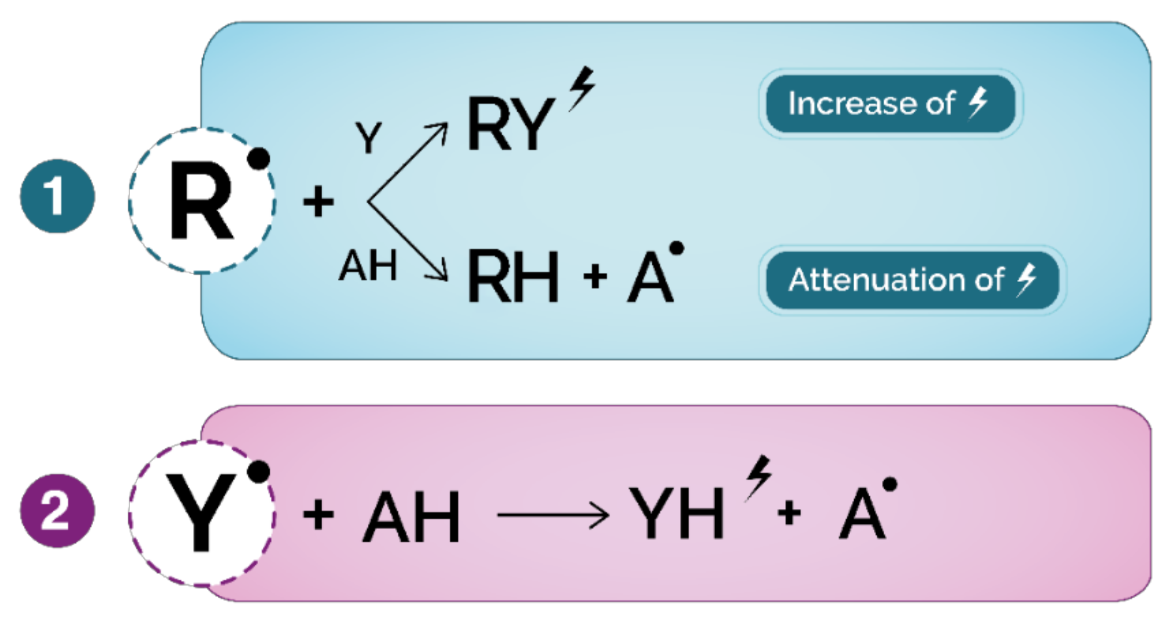
 : fluorescence, absorption, light emission, oxygen consumption; (2) ET-based assays: Y•: oxidized probe, AH: antioxidant; YH: reduced probe; : color modification of reduced probe.
: fluorescence, absorption, light emission, oxygen consumption; (2) ET-based assays: Y•: oxidized probe, AH: antioxidant; YH: reduced probe; : color modification of reduced probe.
: fluorescence, absorption, light emission, oxygen consumption; (2) ET-based assays: Y•: oxidized probe, AH: antioxidant; YH: reduced probe; : color modification of reduced probe.


| Amino Acid | Oxidation Products |
|---|---|
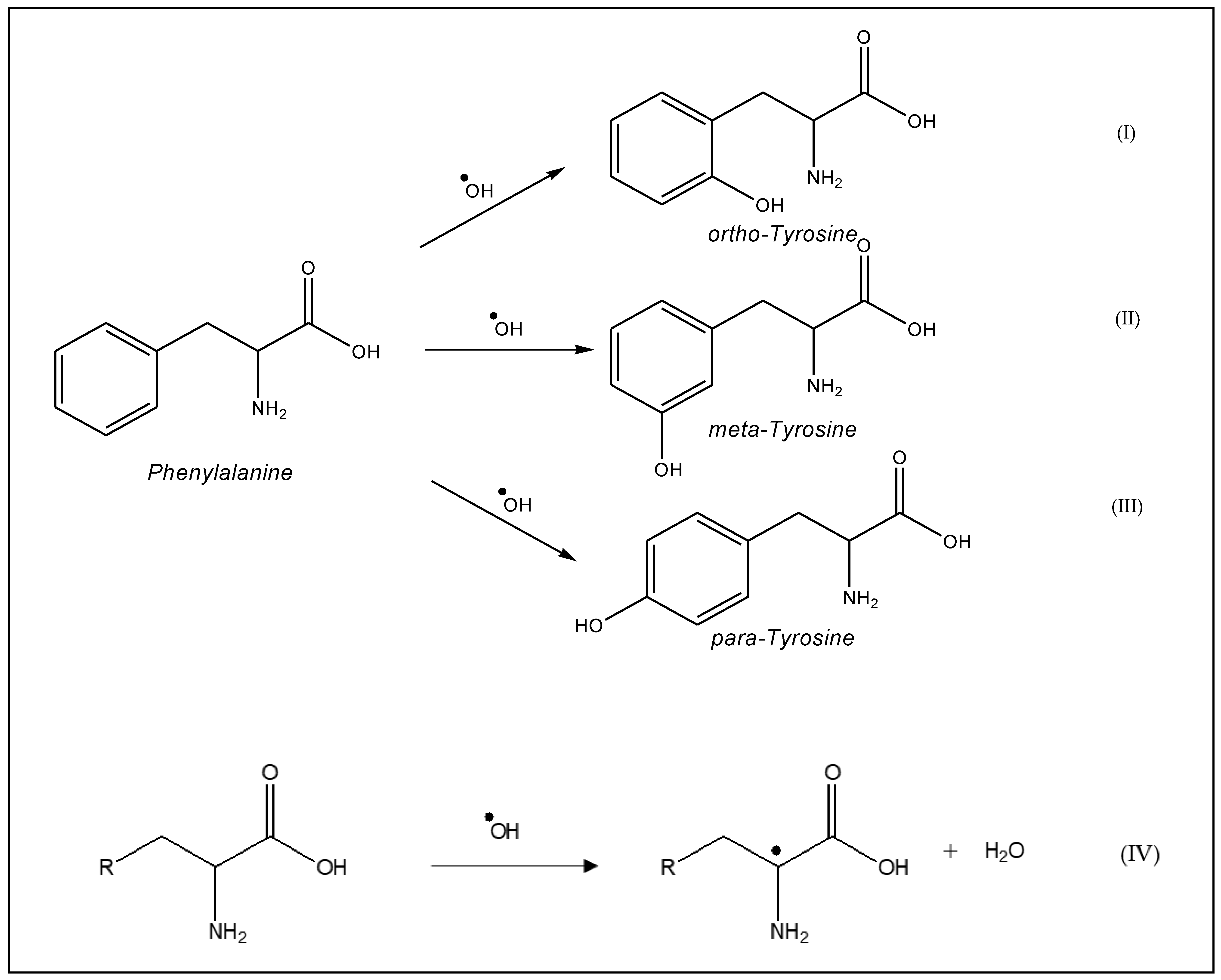
| Cysteine Methionine Tyrosine Tryptophan Phenylalanine Valine, leucine Histidine Proline Threonine Arginine Lysine | Disulfide, cystine Methione sulfoxide/sulphone Dityrosine, 3,4-dihydrophenylalanine (DOPA) Hydroxytryptophan,N-kynurenine, N-formylkynurenine, 3 hydroxylkynurenine Hydroxyphenylalanine, o-tyrosine, m-tyrosine Hydroxyperoxides 2-oxohistidine Hydroxyproline, glutamic semialdehyde pyrrolidinone 2-amino-3-ketobutyric acid Glutamic semialdehyde Hydroxylysine, 2-aminoadipic semialdehyde |
| Antioxidants | Application Media | Working Electrode | Method | Linear Range (µM) | Detection Limit (µM) | References |
|---|---|---|---|---|---|---|
| Polyphenols | Black tea infusion | CNT electrode | CV | 0.23–94 | 0.11 | [86] |
| Caffeic acid | Red wine | SnO2-RGO/GCE | DPV | 0.15–25 | 80.10−3 | [87] |
| Coffee | Au@α-Fe2O3@RGO/GCE | CV | 19–1869 | 0.098 | [88] | |
| Wine | F-GO/GCE | DPV | 0.5–100 | 0.018 | [89] | |
| Wine | Au/PdNPs-GRF | DPV | 0.03–938.97 | 6 × 10⁻³ | [90] | |
| Wine | RGO@PDA/GCE | DPV | 5 × 10⁻³–450.55 | 1.2 × 10⁻³ | [91] | |
| Gallic acid | Tap water, tea and orange juice | SiO2 nanoparticles/CPE | DPV | 8.0 × 10− 1–1.0 × 10⁻² | 2.5 × 10⁻¹ | [92] |
| Wine | CS–fFe2O3–ERGO/GCE | DPV | 1.0–1.0 × 10⁶ | 1.5 × 10⁻¹ | [93] | |
| Phosphate buffer solution | Zn-Al-NO3 layered double hydroxide film/GCE | DPV | 4–600 | 1.6 | [94] | |
| Gallic acid and total polyphenols | Red and white wines | CNT modified carbon paste electrode | DPV | 5.0 × 10⁻¹–15 | 3.0 × 10⁻¹ | [95] |
| Ascorbic acid | Mixture of ascorbic acid, dopamine and uric acid | PG/GCE | CV | 9.00–2314 | 6.45 | [96] |
| Aqueous solution | 2,7-BFEFO/CPE | CV; DPV | 50–2.65×10³; 9–3.5×10³ | 18; 4.2 | [97] | |
| Fruit juices and wines | CPE; Pt strip electrode | DPV | 70–20 × 103; 310–20 × 103 | 20; 87 | [98] | |
| Flavored beverages | DNA/CPE | DPV | 0.05–1.00 | 5 × 10⁻⁴ | [99] | |
| Curcumin | Human blood serum | NiCl2/GCE | DPV | 10–600 | 0.109 | [100] |
| Spices | GCE | CV | 9.9–1.07 × 10² | 41 | [101] | |
| Vanillic acid | Artificial wine solutions | Graphite; carbon microspheres and CNT CPE | CV | 10–400 | 2.85; 3.82; 4.13 | [102] |
| α-tocopherol; γ-tocopherol and δ-tocopherol | Non-aqueous media | Pt electrode | DPV | 2 × 10⁻²–10; 2.2 × 10⁻²–1.4; 2.21 × 10⁻²–31.1 | 1 × 10⁻² | [103] |
| Quercetin | Rhizoma kaempferiae and buds of Styphnolobium japonicum (L.) Schott | CTAB-cMWCNTs/MWCPE | CV | 0.01–20 | 5.3 × 10⁻³ | [104] |
| Template | Application | Reference |
|---|---|---|
| Tocopherols | α-tocopherol delivery in gastrointestinal simulating fluids. | [151] |
| Tocopherol recognition | [152] | |
| Quercetin | Preconcentration and clean-up of catechins | [153] |
| Extraction of anthocyanin from mangosteen pericarp | [154] | |
| Extraction of quercetin and kaempferol from the hydrolyzate of ginkgo leaves | [155] | |
| Separation of active inhibitors of epidermal growth factor receptor (EGRF) from Caragana Jubata | [156] | |
| solid-phase extraction for the sample pretreatment of natural products prior to HPLC analysis | [157] | |
| (+)-Catechin | Extraction of catechins from tea extracts | [158] |
| Retention of catechin | [159] | |
| Caffeic acid | Separation and purification of chlorogenic acid | [143] |
| Extraction of CA in commercial apple juice samples | [160] | |
| Selective extraction of polyphenols from olive mill waste waters | [161] | |
| Extraction of CA from fruits | [162] | |
| Separation and purification of the antioxidant compounds from mushrooms | [163] | |
| p-hydroxybenzoic acid | Selective extraction of polyphenols from olive mill waste waters | [161] |
| Resveratrol | Selective recognition of resveratrol | [164] |
Publisher’s Note: MDPI stays neutral with regard to jurisdictional claims in published maps and institutional affiliations. |
© 2021 by the authors. Licensee MDPI, Basel, Switzerland. This article is an open access article distributed under the terms and conditions of the Creative Commons Attribution (CC BY) license (http://creativecommons.org/licenses/by/4.0/).
Share and Cite
Elhachem, M.; Cayot, P.; Abboud, M.; Louka, N.; Maroun, R.G.; Bou-Maroun, E. The Importance of Developing Electrochemical Sensors Based on Molecularly Imprinted Polymers for a Rapid Detection of Antioxidants. Antioxidants 2021, 10, 382. https://0-doi-org.brum.beds.ac.uk/10.3390/antiox10030382
Elhachem M, Cayot P, Abboud M, Louka N, Maroun RG, Bou-Maroun E. The Importance of Developing Electrochemical Sensors Based on Molecularly Imprinted Polymers for a Rapid Detection of Antioxidants. Antioxidants. 2021; 10(3):382. https://0-doi-org.brum.beds.ac.uk/10.3390/antiox10030382
Chicago/Turabian StyleElhachem, Marie, Philippe Cayot, Maher Abboud, Nicolas Louka, Richard G. Maroun, and Elias Bou-Maroun. 2021. "The Importance of Developing Electrochemical Sensors Based on Molecularly Imprinted Polymers for a Rapid Detection of Antioxidants" Antioxidants 10, no. 3: 382. https://0-doi-org.brum.beds.ac.uk/10.3390/antiox10030382