Article

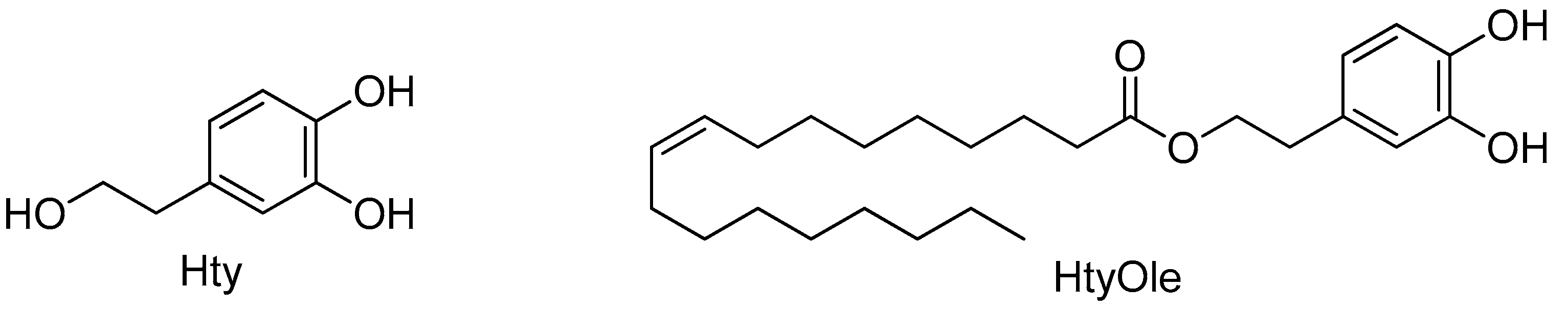
Hydroxytyrosyl Oleate: Improved Extraction Procedure from Olive Oil and By-Products, and In Vitro Antioxidant and Skin Regenerative Properties

1
CREA–Research Centre for Olive, Citrus and Tree Fruit, C.da Li Rocchi, 87036 Rende (CS), Italy
2
Department of Pharmacy, Health and Nutrition Sciences, University of Calabria, Via Pietro Bucci, 87036 Arcavacata di Rende (CS), Italy
3
Clinical Pharmacology and Pharmacovigilance Operative Unit, Department of Health Science, University of Magna Graecia, Mater Domini Hospital Catanzaro, 88100 Catanzaro, Italy
*
Authors to whom correspondence should be addressed.
Submission received: 28 June 2019 / Revised: 15 July 2019 / Accepted: 16 July 2019 / Published: 20 July 2019
(This article belongs to the Special Issue Antioxidants and Skin Protection)

Abstract

:
Recently, we identified hydroxytyrosyl oleate (HtyOle) in the by-products of olive oil, pomace and olive mill waste water (OMWW). Herein, we report that HtyOle is more accurately quantified by extracting the phenolic fraction from both matrices by using aqueous methanol (80%). By applying this method, HtyOle was also detected in extra virgin olive oil (EVOO). Since olive oil is used in the preparation of many cosmetic formulations, we explored the antioxidant capacity of HtyOle in human keratinocytes. Formation of reactive oxygen species (ROS) and malondialdehyde (MDA), as well as activity of Glutathione-S-transferase (GST) and superoxide dismutase (SOD) were decreased by HtyOle. In addition to that, microRNAs (miRs) involved in both redox status balance and skin regeneration potential were also tested. The following miRs, hsa-miR-21 and hsa-miR-29a, were increased while has-miR-34a was not affected by HtyOle.

1. Introduction

Hydroxytyrosol (Hty, Figure 1) is one of the main phenolic compounds in olive fruit. It can occur in its free form, or as a moiety in more complex structures, such as secoiridoids (oleuropein and its aglycone). The distribution among the different forms depends on many factors, such as cultivar, ripening stage, and processing and storage conditions, affecting the hydrolytic activity of endogenous enzymes that release Hty from the secoiridoids [1,2]. As in the case of other polar phenolics, Hty is more abundant in the by-products of olive oil production, namely olive mill waste water (OMWW) and pomace, rather than in olive oil itself [3]. Hydroxytyrosol has been reported to display a number of biological activities, including anticancer, antioxidant, and anti-inflammatory properties [2,4,5]. Despite its potential health benefits, its uses in food and cosmetic industries are limited by its hydrophilic character that eventually leads to low bioavailability. Lipophilization has been suggested as a promising strategy to improve the properties of Hty as well as of other polar phenolics [6]. A number of lipophilic derivatives, including esters bearing fatty acyl chains, have been investigated. Indeed, some hydroxytyrosyl esters were found to display enhanced antioxidant activities, compared to parent Hty, and the activity was found to depend on the chain length [7,8,9,10,11,12,13,14,15,16,17,18,19,20,21,22]. The lipophilic character of hydroxytyrosyl esters suggests their potential use as active compounds in epidermal and dermal formulations for the treatment of the inflammation of the cutaneous stratus [23]. Remarkably, we recently reported on the in vitro anti-inflammatory properties of hydroxytyrosyl oleate (HtyOle, Figure 1) [24].
Although no correlation has been established yet, several antimicrobial agents have antioxidant capabilities [25]. In particular, hydroxytyrosyl esters show both antioxidant and antimicrobial activities at significant levels [26,27]. Reactive Oxygen Species (ROS) are crucial biochemical mediators for senescence and age-related diseases, including disorders of human skin [28,29]. MicroRNAs (miRs) have been well recognized as biomarkers of pathology (especially in cancer) and regulators of gene expression by nutrients and nutrition special regimen [30,31]. Their role in the regulation of antioxidant defense and oxidative stress is emerging [32,33]. Indeed, dysfunction of the antioxidant defense system and the imbalance between formation and removal of ROS can lead to cell damage due to free radical reactions. Herein we studied the effect of HtyOle as a free radical scavenger on the HaCat human keratinocyte cell line, on ROS formation and antioxidant enzymes, as well as its capability to modulate miRs linked to redox cellular state and skin regeneration.

2. Materials and Methods

2.1. Chemicals and Reagents

Hydroxytyrosol was obtained by reducing 3,4-dihydroxyphenylacetic acid with LiAlH4 (Sigma-Aldrich, Milan, Italy), as already reported [34]. n-Hexane and acetone (analytical grade) were supplied from Carlo Erba Reagenti (Milan, Italy). Methyl oleate and LC/MS grade solvents (methanol and formic acid) were acquired from Sigma-Aldrich (Milan, Italy). Ultrapure water was obtained by Milli-Q plus system (Millipore, Bedford, MA, USA). Novozym®435 (immobilized Candida antarctica Lipase B) was from Novozymes (Bagsværd, Denmark). Dulbecco’s modified Eagle’s medium (DMEM), fetal bovine serum (FBS), l-glutamine, penicillin/streptomycin, paraformaldehyde (PFA), and TaqMan microRNA primers were obtained from Thermo Fisher Scientific (Waltham, MA, USA).

2.2. Instrumentation

1H NMR and 13C NMR analyses were performed at 25 °C on a Bruker AC 300 spectrometer at 300 MHz and 75 MHz, respectively, using chloroform-d as the solvent and tetramethylsilane as the internal standard. HPLC analyses were conducted by an Agilent Technologies 1200 series liquid chromatograph provided with a G1379B degasser, a G1312A pump, and a G1329A autosampler. Separations were attained by means of a Discovery C-18 column (3 μm particle size; 150 mm length; 4.6 mm i.d., Merck KGaA, Darmstadt, Germany). MS/MS spectra were recorded by using an API 4000 Q-Trap (AB Sciex, Framingham, MA, USA) mass spectrometer. Detailed instrumental conditions have been previously reported [24].

2.3. Chemistry

Hydroxytyrosyl oleate (HtyOle) was synthesized as previously reported [24]. Briefly, Hty was reacted with methyl oleate, with Novozym®435 as the catalyst, in t-BuOH. Reaction was conducted in an orbital shaker at 50 °C for 24 h. After filtering off the lipase and evaporating the solvent, column chromatography on silica gel (n-hexane-acetone as the eluent) yielded HtyOle. Spectroscopic data were in line with literature [21], and purity was ˃98% as assessed by HPLC.

2.4. Sample Preparation

Pomace and olive mill waste water (OMWW) were obtained by using Oliomio 50 hammer mill (Toscana Enologica Mori, Val di Pesa (FI)) at the Research Center for Olive, Citrus and Fruit Trees (Rende, Italy) starting from 10 kg of olives. Monovarietal extra virgin olive oil (EVOO) was produced from olives of Carolea cultivar in the crop year 2017/2018. All the samples were stored in the refrigerator until analysis.

2.5. Extraction of the Phenolic Fraction from Olive Oil and By-Products

Three different methods for the extraction of the phenolic fraction from matrices were used. Method 1. The first method was employed in our previous work [24]. In particular, the extraction procedure from olive oil and by-products was carried out according to Bianco and coworkers, using 1/1 MeOH-acetone (v/v) solution containing 0.5% sodium metabisulfite [35]. Method 2. The second method previously reported by Sivakumar and coworkers [36], is a modification of the first one. Method 3. The third method for the extraction of phenolic fraction was carried out according to the International Olive Council [37], using 8/2 MeOH-water (v/v) solution.

2.6. Determination of Hydroxytyrosyl Oleate in the Phenolic Fractions Obtained by Different Extraction Methods

The quantitative determination of HtyOle in the phenolic fractions, obtained by means of the three different extraction methods discussed in the previous paragraph, was performed according to our previously published analytical method [24].

2.7. Cell Culture

A human immortalized HaCat keratinocyte cell line was obtained from CLS Cell Lines Service GmbH (Germany) after a material transfer agreement and cultured in 75 cm2 flasks. Keratinocytes were cultured at 37 °C, 5% CO2, in DMEM containing 10% FBS and 1% antibiotics (10,000 μg mL−1 streptomycin and 10,000 units∙mL−1 penicillin). Cell counting was achieved using a Countess Automated Cell Counter (Thermo Fisher Scientific, Waltham, MA, USA) by Trypan Blue staining. Samples were solubilized in dimethyl sulfoxide (DMSO) and diluted in complete medium in order to reach the final concentration. DMSO concentration in each final treatment was maintained below 0.1% and the untreated control was represented by the same amount of DMSO present in the treatments. For each treatment, cells were plated in a 96-well plate for the MTT assay, 24-well plate for fluorescence microscopy and in 100 mm polystyrene dishes for microRNA analyses (Falcon, Becton-Dickinson, Lincoln Park, NJ, USA).

2.8. Cell Viability Assay

Cell viability was estimated by MTT assay, evaluating the decrease of 3-(4,5-dimethylthiasol-2-yl)-2,4-diphenyltetrazolium bromide (MTT) by mitochondrial succinate dehydrogenase [38]. The absorbance (Abs) was read with a microtiter plate reader (Synergy H1 by BioTeck, Winooski, VT, USA) at 570 nm (test wavelength, tw) and at 690 nm (reference wavelength, rw). The optical density (OD) was calculated as Abs rw − Abs tw.

2.9. Fluorescent Staining

Glass cover slips were inserted into 24 well plates and HaCat cells were grown on it. HtyOle pre-treatment at 5 µM with HtyOle for 24 h was performed in order to determine cytosolic ROS formation. After 30 min of UV lamp exposure at 254 nm (UVC spectrum to avoid vitamin D synthesis). CellRox Deep Red (5 µM, λ = ex/em = 640/665) dye was added. Cells were then fixed with PFA (4%) and imaged using the confocal FV-3000 Olympus microscope as previously described [39].

2.10. Antioxidant Enzymes Activity and Lipid Peroxidation

Cell lysate was sonicated and divided into two equal parts. One aliquot was centrifuged at 10,000 rpm for 5 min and the supernatant was used for glutathione-S-transferase (GST) and superoxide dismutase (SOD) enzymatic activity assays. The second aliquot was used for the lipid peroxidation assay. The enzymatic activity of SOD and GST in the cell extracts was evaluated as previously described [40]. The Lowry method was used to estimate protein concentration in the samples [41]. The extent of lipid peroxidation was evaluated by measuring thiobarbituric acid reactive species (TBARS) formation on acid heating reaction, as previously described [42], with slight modifications. Briefly, the samples were mixed with 5% trichloroacetic acid and 0.7% TBA (1 mL each), then heated at 100 °C for 15 min. The amount of TBARS was evaluated by measuring the absorbance at 535 nm and was expressed as malondialdehyde (MDA) equivalents (nmol mg−1 protein−1).

2.11. MicroRNA Extraction and Loop Primer Method

MicroRNA was isolated using PureLink™ miRNA Isolation Kit (K1570-01 Ambion by Life Technologies) as previously described [30]. The amount of miRNA was quantified with a termed looped primer RT-PCR method. Total RNA (10 ng) was reverse transcribed by polymerase chain reaction using the TaqMan MicroRNA Reverse Transcription kit (Thermo Fisher Scientific, Waltham, MA, USA) for both the miR target and endogenous control, according to manufacturer’s instructions. The thermocycling conditions were: 30 min at 16 °C, followed by 30 min at 42 °C, 5 min at 85 °C and 5 min at 4 °C.

2.12. Quantitative Real Time PCR (qRT-PCR)

The quantitative real-time polymerase chain reaction (qRT-PCR) was achieved by TaqMan Universal PCR Master Mix Kit (Thermo Fisher Scientific, Waltham, MA, USA), according to the manufacturer’s instructions and QuantumStudio3TM Real-Time PCR Systems equipment. The thermocycling conditions used have been previously described [43]. After completion of the qRT-PCR experiments, the average values of the cycle threshold (Ct) of the reactions in triplicate were calculated. The relative expression of the miR target was plotted as follows: 40 total qRT-PCR cycles –Ct target miR. This difference (ΔCt) was plotted directly.

2.13. Statistical Analysis

Prism GraphPad Prism version 5.0 for Windows (GraphPad Software, San Diego, CA, USA) was used to build graphs. Differences were evaluated by one-way ANOVA, followed by multi-comparison Dunnett’s test (* p < 0.05, ** p <0.02, *** p < 0.01, compared to controls).

3. Results

3.1. Quantification of Hydroxytyrosyl Oleate in Olive Oil and By-Products Subjected to Different Extraction Procedures

Three methods for the extraction of the phenolic fraction from EVOO and olive oil by-products were compared, with respect to the amount of HtyOle. The results are reported in Table 1. Although similar to the first method, the second method of extraction of the phenolic fraction led to higher amounts of HtyOle in both pomace and OMWW. The highest amount of HtyOle was found in the phenolic fraction obtained by the third extraction method in both by-products. Remarkably, by this approach, it was also possible to detect and quantify HtyOle in extra virgin olive oil (EVOO).

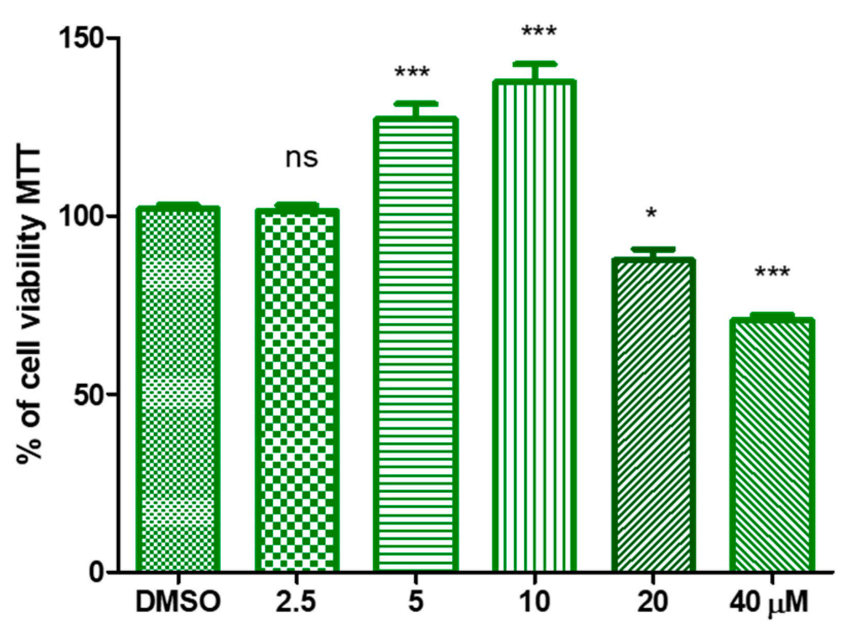
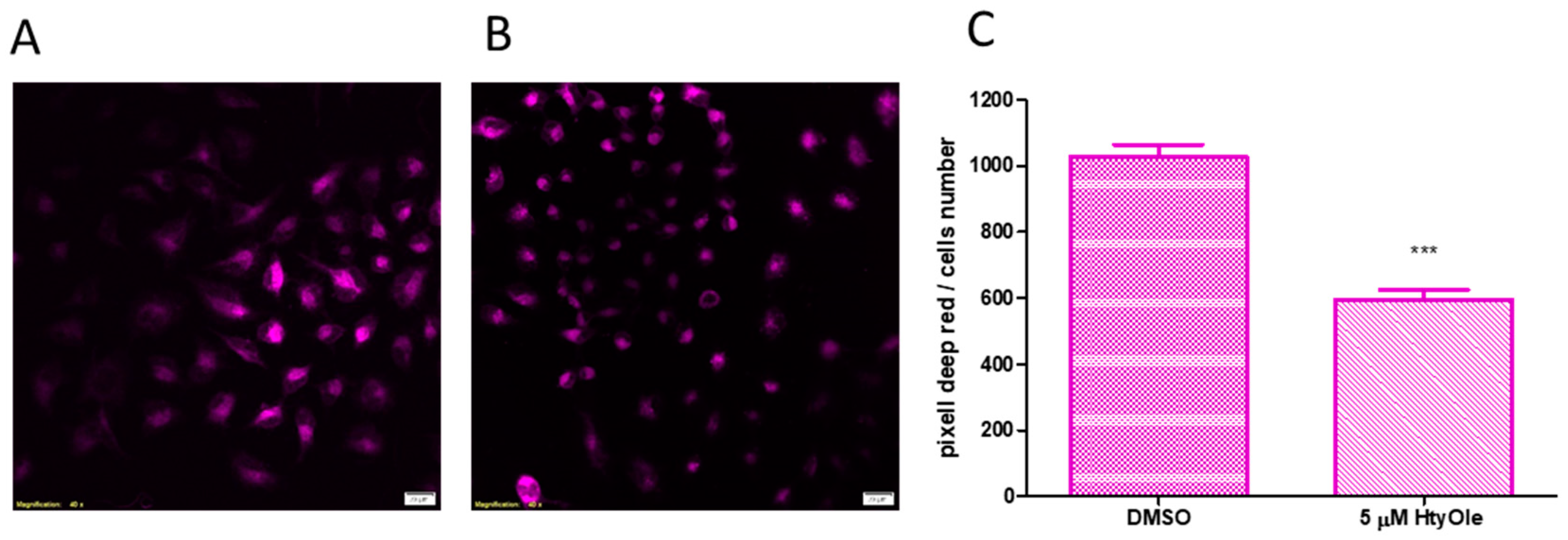
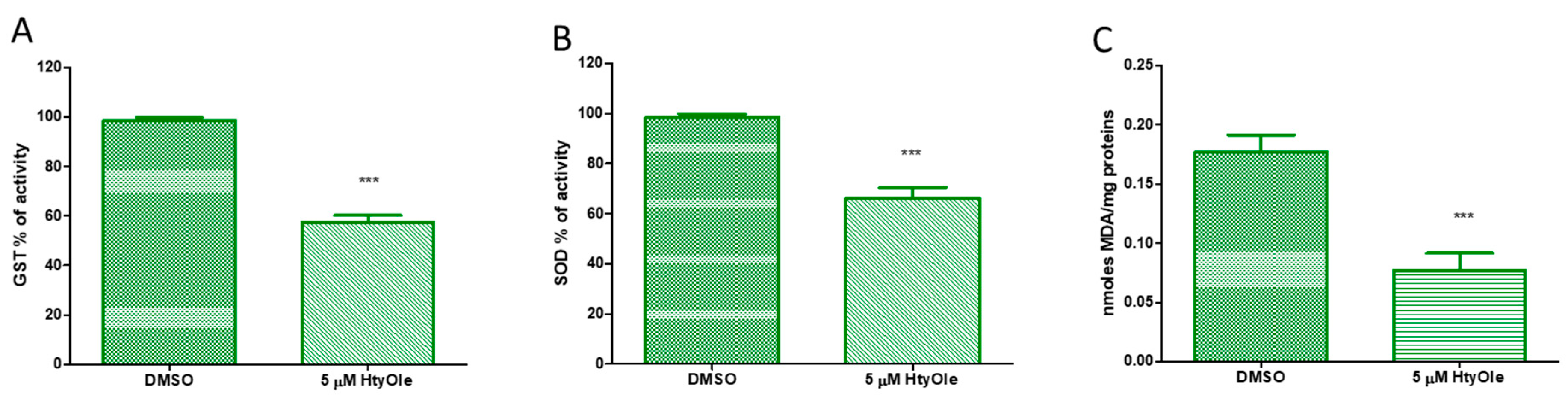
3.2. Hydroxytyrosyl Oleate Affects Cell Viability, ROS Formation, SOD and GST Activities and Lipid Peroxidation in Human Keratinocytes

A significant increase in cell vitality was appreciated when HaCat cells were treated with the HtyOle. In particular, a proliferative concentration-dependent action was observed with up to 10 µM of HtyOle, while at 20 and 40 µM cytotoxicity was recorded, as shown in Figure 2.
Therefore, we chose the lower effective concentration of HtyOle (5 µM) for further experiments. To investigate HtyOle’s potential modulation of ROS formation, CellRox Deep Red staining was performed. As shown in Figure 3, keratinocytes cultured for 24 h with HtyOle and then exposed to UV (Figure 3B) showed a decrease of stain intensity with respect to the control (Figure 3A). Figure 3C showed the average values of cell area in pixels for red stained keratinocytes.
The potential of HtyOle in modulating the antioxidant defenses in human keratinocytes was also investigated. In our experiments, glutathione-S-transferase (GST) and superoxide dismutase (SOD) activities were decreased by HtyOle (Figure 4, panels A and B, respectively). The oxidative degradation of lipids leads to cell damage. Reactive aldehydes, including malondialdehyde (MDA), are the end products of lipid peroxidation [44]. In our experimental set-up, MDA levels decreased upon treatment with 5 µM of HtyOle when compared to control samples (Figure 4C).

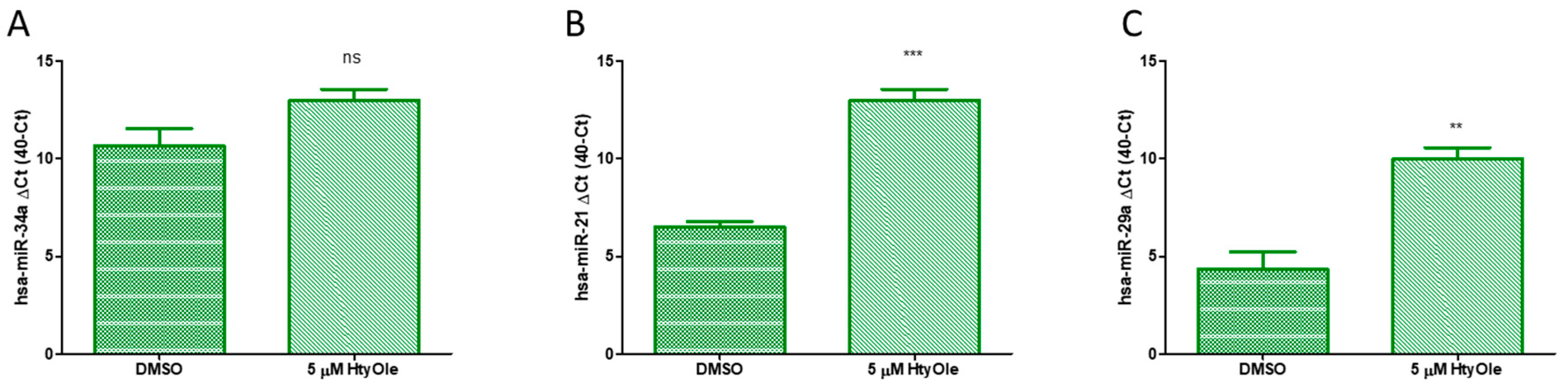
3.3. Hydroxytyrosyl Oleate Modulates hsa-miRs Linked to Redox State Status and Human Keratinocyte Regeneration

The action of HtyOle on miRs was also investigated to verify whether it can have a skin regeneration potential. Here we tested the following miRs: hsa-miR-34a, hsa-miR-21 and hsa-miR-29a. As shown in Figure 5, up-regulation upon HtyOle treatment was found for hsa-miR-21 (Figure 5B) and hsa-miR-29a (Figure 5C). No significant modulation was recorded for hsa-miR-34a (Figure 5A).

4. Discussion

We recently reported the presence of HtyOle in pomace and olive mill waste water (OMWW) [24]. The extraction procedure utilized to recover this hydroxytyrosyl ester from olive oil by-products was the one reported by Bianco and coworkers that allowed the isolation and identification of the analogue tyrosyl oleate (TyOle) from olive drupes of Cassanese cultivar [35]. Herein we show that using a simpler and more economical extraction procedure leads to a higher quantity of HtyOle, both in pomace and in OMWW. Moreover, by means of this approach, it was also possible to identify and quantify HtyOle in EVOO and the results obtained are in line with literature data [45]. This compound was found to be the most active among a series of Hty fatty esters in decreasing nitric oxide (NO) production by lipopolysaccharide (LPS)-stimulated murine macrophages. Moreover, HtyOle suppressed prostaglandin E2 (PGE2) production and the expression of inducible NO synthase, cyclooxygenase-2 (COX-2) and interleukin-1β (IL-1β) at a transcriptional level [24]. Even though there is not any correlation yet, several anti-inflammatory and antimicrobial agents have antioxidant capabilities [25]. ROS balance is fundamental in all organs and tissues, including skin. In this concern, increased levels of keratinocyte oxidative stress and decreased antioxidant defenses have been correlated with skin-dermal dysfunction [29,46]. Antioxidant defenses of human keratinocytes depend on SOD and GST activities. Both enzymes are involved in the detoxification of the cells from ROS [47]. SOD catalyzes the dismutation of superoxide into oxygen and hydrogen peroxide, and alterations in its activity can lead to unbalanced free radical production. SOD is mainly linked to the mitochondrial redox state [48], while GST is essential to maintain the homeostasis of ROS production and clearance [49]. Our experiments showed that SOD and GST activities are decreased by HtyOle treatment, pointing out its action as free radical scavenger per se. During oxidative stress the cells use their protection apparatus to minimize the process of lipid peroxidation by using the antioxidant enzymes [50]. Therefore, the enzyme activity is shut down because free radicals and lipid peroxidation are dropped [51]. In fact, when cellular redox state is lower also, detoxifications mediated by enzyme activity are resultantly lower [52]. This assumes an important meaning also from a nutritional point of view in skin healing care [53]. The oxidative degradation of lipids results in cell damage. The end products of lipid peroxidation are reactive aldehydes, such as malondialdehyde [44]. In our experimental model, the MDA levels also decreased upon HtyOle treatment when compared to control samples. Recently, the role of nutrition in the regulation of microRNAs (miRs) as well as their capability to regulate cellular redox status and keratinocytes’ regeneration has emerged [31,54]. In fact, it was established that Dicer activity is important for the maintenance of the redox status balance [55]. Moreover, Dicer activation declines with age and Dicer knockout mice might have an impaired antioxidant defense system. We found that hsa-miR-21 and hsa-miR-29a were increased upon HtyOle while hsa-miR-34a was not affected. The physiological action of those miRs is less studied compared to what we know about them in pathophysiology, especially cancer. Of note is that has-miR-29a and hsa-miR-21 are able to regulate skin healing through multiple aspects of this regenerative process. Particularly hsa-miR-21 promoting keratinocytes cell migration [56]. Although increasing levels of this latter miR seem worst in the pathophysiology of many types of cancer and it is considered an oncomiR [57], physiologically, in the context of skin injury and regeneration, it assumes a different and fundamental role. By contrast, hsa-miR34a inhibits proliferation and stimulates apoptosis in human keratinocyte, and it was proposed as a potential therapeutic target for psoriasis [58]. In addition, this miR is suppressed in skin and oral squamous cell carcinomas and in keratinocytes with a compromised differentiation program [59].

5. Conclusions

The identification of hydroxytyrosyl oleate in extra virgin olive oil opens a new scenario for the rational use of olive oil in topical formulations. The capability of hydroxytyrosyl oleate to control cellular redox status per se as well as microRNA expression linked to skin regenerative processes, outlines its role as an epigenetic regulator in a plethora of skin related diseases, from healing to aging.

Author Contributions

C.B., P.P. and E.C. designed the study and the experiments; C.B. and C.L.T. carried out the experiments; C.B., A.F. and M.C.C analyzed the data and revised the statistical analyses; E.P., L.G. and R.C. facilitated the experiments; P.P. and E.C. prepared and edited the manuscript.

Funding

This research received no external funding.

Conflicts of Interest

The authors declare no conflict of interest.

References

  1. Gambacorta, A.; Tofani, D.; Bernini, R.; Migliorini, A. High-yielding preparation of a stable precursor of hydroxytyrosol by total synthesis and from the natural glycoside oleuropein. J. Agric. Food Chem. 2007, 55, 3386–3391. [Google Scholar] [CrossRef] [PubMed]
  2. Robles-Almazan, M.; Pulido-Moran, M.; Moreno-Fernandez, J.; Ramirez-Tortosa, C.; Rodriguez-Garcia, C.; Quiles, J.L.; Ramirez-Tortosa, M.C. Hydroxytyrosol: Bioavailability, toxicity, and clinical applications. Food Res. Int. 2018, 105, 654–667. [Google Scholar] [CrossRef] [PubMed]
  3. Ciriminna, R.; Meneguzzo, F.; Fidalgo, A.; Ilharco, L.M.; Pagliaro, M. Extraction, benefits and valorization of olive polyphenols. Eur. J. Lipid Sci. Technol. 2016, 118, 503–511. [Google Scholar] [CrossRef]
  4. Bernini, R.; Merendino, N.; Romani, A.; Velotti, F. Naturally occurring hydroxytyrosol: Synthesis and anticancer potential. Curr. Med. Chem. 2013, 20, 655–670. [Google Scholar] [CrossRef] [PubMed]
  5. Zhang, X.; Cao, J.; Zhong, L. Hydroxytyrosol inhibits pro-inflammatory cytokines, iNOS, and COX-2 expression in human monocytic cells. Naunyn-Schmiedeberg's Arch. Pharmacol. 2009, 379, 581–586. [Google Scholar] [CrossRef] [PubMed]
  6. Bayrasy, C.; Chabi, B.; Laguerre, M.; Lecomte, J.; Jublanc, E.; Villeneuve, P.; Wrutniak-Cabello, C.; Cabello, G. Boosting antioxidants by lipophilization: A strategy to increase cell uptake and target mitochondria. Pharm. Res. 2013, 30, 1979–1989. [Google Scholar] [CrossRef] [PubMed]
  7. Akanbi, T.O.; Barrow, C.J. Lipase-Produced Hydroxytyrosyl Eicosapentaenoate is an Excellent Antioxidant for the Stabilization of Omega-3 Bulk Oils, Emulsions and Microcapsules. Molecules 2018, 23, 275. [Google Scholar] [CrossRef]
  8. Almeida, J.; Losada-Barreiro, S.; Costa, M.; Paiva-Martins, F.; Bravo-Díaz, C.; Romsted, L.S. Interfacial Concentrations of Hydroxytyrosol and Its Lipophilic Esters in Intact Olive Oil-in-Water Emulsions: Effects of Antioxidant Hydrophobicity, Surfactant Concentration, and the Oil-to-Water Ratio on the Oxidative Stability of the Emulsions. J. Agric. Food Chem. 2016, 64, 5274–5283. [Google Scholar] [CrossRef]
  9. Bernini, R.; Crisante, F.; Barontini, M.; Tofani, D.; Balducci, V.; Gambacorta, A. Synthesis and Structure/Antioxidant Activity Relationship of Novel Catecholic Antioxidant Structural Analogues to Hydroxytyrosol and Its Lipophilic Esters. J. Agric. Food Chem. 2012, 60, 7408–7416. [Google Scholar] [CrossRef]
  10. Bernini, R.; Gilardini Montani, M.S.; Merendino, N.; Romani, A.; Velotti, F. Hydroxytyrosol-derived compounds: A basis for the creation of new pharmacological agents for cancer prevention and therapy. J. Med. Chem. 2015, 58, 9089–9107. [Google Scholar] [CrossRef]
  11. Bernini, R.; Carastro, I.; Palmini, G.; Tanini, A.; Zonefrati, R.; Pinelli, P.; Brandi, M.L.; Romani, A. Lipophilization of Hydroxytyrosol-Enriched Fractions from Olea europaea L. Byproducts and Evaluation of the in Vitro Effects on a Model of Colorectal Cancer Cells. J. Agric. Food Chem. 2017, 65, 6506–6512. [Google Scholar] [CrossRef]
  12. Bouallagui, Z.; Bouaziz, M.; Lassoued, S.; Engasser, J.M.; Ghoul, M.; Sayadi, S. Hydroxytyrosol acyl esters: Biosynthesis and activities. Appl. Biochem. Biotechnol. 2011, 163, 592–599. [Google Scholar] [CrossRef]
  13. Candiracci, M.; Madrona, A.; Espartero, J.L.; Zappia, G.; Piatti, E. Lipophilic hydroxytyrosol esters significantly improve the oxidative state of human red blood cells. J. Funct. Foods 2016, 23, 339–347. [Google Scholar] [CrossRef]
  14. Grasso, S.; Siracusa, L.; Spatafora, C.; Renis, M.; Tringali, C. Hydroxytyrosol lipophilic analogues: Enzymatic synthesis, radical scavenging activity and DNA oxidative damage protection. Bioorg. Chem. 2007, 35, 137–152. [Google Scholar] [CrossRef]
  15. Lucas, R.; Comelles, F.; Alcántara, D.; Maldonado, O.S.; Curcuroze, M.; Parra, J.L.; Moreales, J.C. Surface-active properties of lipophilic antioxidants tyrosol and hydroxytyrosol fatty acid esters: A potential explanation for the nonlinear hypothesis of the antioxidant activity in oil-in-water emulsions. J. Agric. Food Chem. 2010, 58, 8021–8026. [Google Scholar] [CrossRef]
  16. Mateos, R.; Trujillo, M.; Pereira-Caro, G.; Madrona, A.; Cert, A.; Espartero, J.L. New lipophilic tyrosyl esters. Comparative antioxidant evaluation with hydroxytyrosyl esters. J. Agric. Food Chem. 2008, 56, 10960–10966. [Google Scholar] [CrossRef]
  17. Medina, I.; Lois, S.; Alcántara, D.; Lucas, R.; Morales, J.C. Effect of lipophilization of hydroxytyrosol on its antioxidant activity in fish oils and fish oil-in-water emulsions. J. Agric. Food Chem. 2009, 57, 9773–9779. [Google Scholar] [CrossRef]
  18. Sun, Y.; Zhou, D.; Shahidi, F. Antioxidant properties of tyrosol and hydroxytyrosol saturated fatty acid esters. Food Chem. 2018, 245, 1262–1268. [Google Scholar] [CrossRef]
  19. Tofani, D.; Balducci, V.; Gasperi, T.; Incerpi, S.; Gambacorta, A. Fatty Acid Hydroxytyrosyl Esters: Structure/Antioxidant Activity Relationship by ABTS and in Cell-Culture DCF Assays. J. Agric. Food Chem. 2010, 58, 5292–5299. [Google Scholar] [CrossRef]
  20. Torres de Pinedo, A.; Peñalver, P.; Pérez-Victoria, I.; Rondón, D.; Morales, J.C. Synthesis of new phenolic fatty acid esters and their evaluation as lipophilic antioxidants in an oil matrix. Food Chem. 2007, 105, 657–665. [Google Scholar] [CrossRef]
  21. Trujillo, M.; Mateos, R.; de Teran, L.C.; Espartero, J.L.; Cert, R.; Jover, M.; Alcudia, F.; Bautista, J.; Cert, A.; Parrado, J. Lipophilic hydroxytyrosyl esters. Antioxidant activity in lipid matrices and biological systems. J. Agric. Food Chem. 2006, 54, 3779–3785. [Google Scholar] [CrossRef]
  22. Zhou, D.-Y.; Sun, Y.-X.; Shahidi, F. Preparation and antioxidant activity of tyrosol and hydroxytyrosol ester. J. Funct. Foods 2017, 37, 66–73. [Google Scholar] [CrossRef]
  23. Procopio, A.; Celia, C.; Nardi, M.; Oliverio, M.; Paolino, D.; Sindona, G. Lipophilic Hydroxytyrosol Esters: Fatty Acid Conjugates for Potential Topical Administration. J. Nat. Prod. 2011, 74, 2377–2381. [Google Scholar] [CrossRef]
  24. Plastina, P.; Benincasa, C.; Perri, E.; Fazio, A.; Augimeri, G.; Poland, M.; Witkamp, R.; Meijerink, J. Identification of hydroxytyrosyl oleate, a derivative of hydroxytyrosol with anti-inflammatory properties, in olive oil by-products. Food Chem. 2019, 279, 105–113. [Google Scholar] [CrossRef]
  25. Angiolella, L.; Sacchetti, G.; Efferth, T. Antimicrobial and Antioxidant Activities of Natural Compounds. Evid.-Based Complement. Altern. Med. 2018, 2018, 1945179. [Google Scholar] [CrossRef]
  26. Bernini, R.; Carastro, I.; Santoni, F.; Clemente, M. Synthesis of Lipophilic Esters of Tyrosol, Homovanillyl Alcohol and Hydroxytyrosol. Antioxidants 2019, 8, 174. [Google Scholar] [CrossRef]
  27. Ghalandari, M.; Naghmachi, M.; Oliverio, M.; Nardi, M.; Shirazi, H.R.G.; Eilami, O. Antimicrobial effect of Hydroxytyrosol, Hydroxytyrosol Acetate and Hydroxytyrosol Oleate on Staphylococcus Aureus and Staphylococcus Epidermidis. Electr. J. Gen. Med. 2018, 15, em46. [Google Scholar] [CrossRef]
  28. Davalli, P.; Mitic, T.; Caporali, A.; Lauriola, A.; D’Arca, D. ROS, Cell Senescence, and Novel Molecular Mechanisms in Aging and Age-Related Diseases. Oxid. Med. Cell. Longev. 2016, 2016, 3565127. [Google Scholar] [CrossRef]
  29. Rinnerthaler, M.; Bischof, J.; Streubel, M.K.; Trost, A.; Richter, K. Oxidative stress in aging human skin. Biomolecules 2015, 5, 545–589. [Google Scholar] [CrossRef]
  30. Perri, M.; Caroleo, M.C.; Liu, N.; Gallelli, L.; De Sarro, G.; Kagechika, H.; Cione, E. 9-cis Retinoic acid modulates myotrophin expression and its miR in physiological and pathophysiological cell models. Exp. Cell Res. 2017, 354, 25–30. [Google Scholar] [CrossRef]
  31. Cannataro, R.; Perri, M.; Caroleo, M.C.; Gallelli, L.; De Sarro, G.; Cione, E. Modulation of MicroRNAs Linked to Pain-migraine by Ketogenic Diet (P14-007-19). Curr. Dev. Nutr. 2019, 3 (Suppl. 1). [Google Scholar] [CrossRef] [Green Version]
  32. Catalanotto, C.; Cogoni, C.; Zardo, G. MicroRNA in Control of Gene Expression: An Overview of Nuclear Functions. Int. J. Mol. Sci. 2016, 17, 1712. [Google Scholar] [CrossRef]
  33. Yaribeygi, H.; Atkin, S.L.; Sahebkar, A. Potential roles of microRNAs in redox state: An update. J. Cell. Biochem. 2018, 120, 1679–1684. [Google Scholar] [CrossRef]
  34. Fazio, A.; Caroleo, M.C.; Cione, E.; Plastina, P. Novel acrylic polymers for food packaging: Synthesis and Antioxidant Properties. Food Pack. Shelf Life 2017, 11, 84–90. [Google Scholar] [CrossRef]
  35. Bianco, A.; Melchioni, C.; Ramunno, A.; Romeo, G.; Uccella, N. Phenolic components of Olea Europaea isolation of tyrosol derivatives. Nat. Prod. Res. 2004, 18, 29–32. [Google Scholar] [CrossRef]
  36. Sivakumar, G.; Briccoli Bati, C.; Uccella, N. HPLC-MS Screening of the antioxidant profile of Italian olive cultivars. Chem. Nat. Compd. 2005, 41, 588–591. [Google Scholar] [CrossRef]
  37. International Olive Council. Determination of Biophenols in Olive Oils By HPLC; COI/T.20/Doc No 29; COI: Madrid, Spain, 2009; pp. 1–8. [Google Scholar]
  38. Perri, M.; Yap, J.L.; Fletcher, S.; Cione, E.; Kane, M.A. Therapeutic potential of Bcl-xL/Mcl-1 synthetic inhibitor JY-1-106 and retinoids for human triple-negative breast cancer treatment. Oncol. Lett. 2018, 15, 7231–7236. [Google Scholar] [CrossRef]
  39. Ferri, F.; Olivieri, F.; Cannataro, R.; Caroleo, M.C.; Cione, E. Phytomelatonin Regulates Keratinocytes Homeostasis Counteracting Aging Process. Cosmetics 2019, 6, 27. [Google Scholar] [CrossRef]
  40. Perri, M.; Pingitore, A.; Cione, E.; Vilardi, E.; Perrone, V.; Genchi, G. Proliferative and anti-proliferative effects of retinoic acid at doses similar to endogenous levels in Leydig MLTC-1/R2C/TM-3 cells. Biochim. Biophys. Acta 2010, 1800, 993–1001. [Google Scholar] [CrossRef]
  41. Cione, E.; Pingitore, A.; Perri, M.; Genchi, G. Influence of all-trans-retinoic acid on oxoglutarate carrier via retinoylation reaction. Biochim. Biophys. Acta 2009, 1791, 3–7. [Google Scholar] [CrossRef]
  42. Cione, E.; Tucci, P.; Senatore, V.; Perri, M.; Trombino, S.; Iemma, F.; Picci, N.; Genchi, G. Synthesized esters of ferulic acid induce release of cytochrome c from rat testes mitochondria. J. Bioenerg. Biomembr. 2008, 40, 19–26. [Google Scholar] [CrossRef]
  43. Gallelli, L.; Cione, E.; Peltrone, F.; Siviglia, S.; Verano, A.; Chirchiglia, D.; Zampogna, S.; Guidetti, V.; Sammartino, L.; Montana, A.; et al. Hsa-miR-34a-5p and hsa-miR-375 as Biomarkers for Monitoring the Effects of Drug Treatment for Migraine Pain in Children and Adolescents: A Pilot Study. J. Clin. Med. 2019, 8, 928. [Google Scholar] [CrossRef]
  44. Ayala, A.; Muñoz, M.F.; Argüelles, S. Lipid peroxidation: Production, metabolism, and signaling mechanisms of malondialdehyde and 4-hydroxy-2-nonenal. Oxid. Med. Cell. Longev. 2014, 2014, 360438. [Google Scholar] [CrossRef]
  45. Lee, Y.Y.; Crauste, C.; Wang, H.; Leung, H.H.; Vercauteren, J.; Galano, J.-M.; Oger, C.; Durand, T.; Wan, J.M.-F.; Lee, J.C.-Y. Extra Virgin Olive Oil Reduced Polyunsaturated Fatty Acid and Cholesterol Oxidation in Rodent Liver: Is This Accounted for Hydroxytyrosol-Fatty Acid Conjugation? Chem. Res. Toxicol. 2016, 29, 1689–1698. [Google Scholar] [CrossRef] [Green Version]
  46. Cano Sanchez, M.; Lancel, S.; Boulanger, E.; Neviere, R. Targeting Oxidative Stress and Mitochondrial Dysfunction in the Treatment of Impaired Wound Healing: A Systematic Review. Antioxidants 2018, 7, 98. [Google Scholar] [CrossRef]
  47. Lim, S.; Kwon, M.; Joung, E.-J.; Shin, T.; Oh, C.-W.; Choi, J.S.; Kim, H.-R. Meroterpenoid-Rich Fraction of the Ethanolic Extract from Sargassum serratifolium Suppressed Oxidative Stress Induced by Tert-Butyl Hydroperoxide in HepG2 Cells. Mar. Drugs 2018, 16, 374. [Google Scholar] [CrossRef]
  48. Younus, H. Therapeutic potentials of superoxide dismutase. Int. J. Health Sci. 2018, 12, 88–93. [Google Scholar]
  49. Birben, E.; Sahiner, U.M.; Sackesen, C.; Erzurum, S.; Kalayci, O. Oxidative stress and antioxidant defense. World Allergy Organ. J. 2012, 5, 9–19. [Google Scholar] [CrossRef]
  50. Gomes, E.C.; Silva, A.N.; de Oliveira, M.R. Oxidants, Antioxidants, and the Beneficial Roles of Exercise-Induced Production of Reactive Species. Oxid. Med. Cell. Longev. 2012, 2012, 756132. [Google Scholar] [CrossRef]
  51. He, L.; He, T.; Farrar, S.; Ji, L.; Liu, T.; Ma, X. Antioxidants Maintain Cellular Redox Homeostasis by Elimination of Reactive Oxygen Species. Cell. Physiol. Biochem. 2017, 44, 532–553. [Google Scholar] [CrossRef]
  52. Kurutas, E.B. The importance of antioxidants which play the role in cellular response against oxidative/nitrosative stress: Current state. Nutr. J. 2016, 15, 71. [Google Scholar] [CrossRef]
  53. Molnar, J.A.; Underdown, M.J.; Clark, W.A. Nutrition and Chronic Wounds. Adv. Wound Care 2014, 3, 663–681. [Google Scholar] [CrossRef]
  54. Liu, Y.; Zhong, L.; Liu, D.; Ye, H.; Mao, Y.; Hu, Y. Differential miRNA expression profiles in human keratinocytes in response to protein kinase C inhibitor. Mol. Med. Rep. 2017, 16, 6608–6619. [Google Scholar] [CrossRef] [Green Version]
  55. Kitscha, P.; Mann, G.E.; Siow, R.C. Mechanosensitive microRNAs in endothelial responses to shear stress and Nrf2-mediated redox signaling. Free Radic. Biol. Med. 2017, 108, S37. [Google Scholar] [CrossRef]
  56. Yang, X.; Wang, J.; Guo, S.L.; Fan, K.J.; Li, J.; Wang, Y.L.; Yang, X. miR-21 promotes keratinocyte migration and re-epithelialization during wound healing. Int. J. Biol. Sci. 2017, 7, 685–690. [Google Scholar] [CrossRef]
  57. Medina, P.P.; Nolde, M.; Slack, F.J. OncomiR addiction in an in vivo model of microRNA-21-induced pre-B-cell lymphoma. Nature 2010, 467, 86–90. [Google Scholar] [CrossRef]
  58. Chen, H.; Xiong, Y.; Liu, L.; Lu, L.; Tian, F.; Wang, Z.; Zhao, Y. MicroRNA-34a inhibits proliferation and stimulates apoptosis in human keratinocyte through activation of Smac-mediated mitochondrial apoptotic pathway: A potential therapeutic target for psoriasis. Int. J. Clin. Exp. Pathol. 2017, 10, 266–273. [Google Scholar]
  59. Lefort, K.; Brooks, Y.; Ostano, P.; Cario-André, M.; Calpini, V.; Guinea-Viniegra, J.; Albinger-Hegyi, A.; Hoetzenecker, W.; Kolfschoten, I.; Dotto, G.P.; et al. A miR-34a-SIRT6 axis in the squamous cell differentiation network. EMBO J. 2013, 32, 2248–2263. [Google Scholar] [CrossRef] [Green Version]
Figure 1. Structure of hydroxytyrosol (Hty) and hydroxytyrosyl oleate (HtyOle).
Antioxidants 08 00233 g001
Figure 2. Concentration response of hydroxytyrosyl oleate (HtyOle) in human keratinocyte cell viability. HtyOle significantly affected cell vitality in the range 5–40 µM (* p < 0.05; *** p < 0.01).
Antioxidants 08 00233 g002
Figure 3. Reactive oxygen species (ROS) formation upon hydroxytyrosyl oleate (HtyOle) treatment in cytosolic compartment. Representative images of ROS stained in keratinocytes cultured without (A) and with 5 µM of HtyOle (B) for 24 h. Scale bars: 20 µm. Average values of cell area in pixels for red stained keratinocytes (C). Data in (C) are expressed as the means ± sd of n = 3 independent experiments (*** p < 0.01).
Antioxidants 08 00233 g003
Figure 4. Activity of antioxidant enzymes and lipid peroxidation in human keratinocytes treated with 5 µM of hydroxytyrosyl oleate (HtyOle). Enzymatic activity of Glutathione-S-transferase (GST, (A)) and superoxide dismutase (SOD, (B)); malondialdehyde (MDA) formation (panel (C)). Tests were performed in three independent experiments each done in duplicate (*** p < 0.01 versus control).
Antioxidants 08 00233 g004
Figure 5. Hydroxytyrosyl (HtyOle) modulates hsa-miR expression linked to redox state and keratinocyte’s regenerative biochemical pathway. Expression of hsa-miR-34a (A); hsa-miR-21 (B) and hsa-miR-29a (C) in the presence or absence of HtyOle. Keratinocytes were cultured with and without HtyOle for 24 h. Tests were performed in three independent experiments each done in duplicate (** p < 0.02, *** p < 0.01 versus control).
Antioxidants 08 00233 g005
Table 1. Amount of hydroxytyrosyl oleate (HtyOle) depending on extraction procedure.
Amount of HtyOle (mg kg−1)
PomaceOMWW 1EVOO 2
Method 14.3 ± 0.3 a2.6 ± 0.2 a< LOQ a
Method 29.3 ± 0.3 b3.4 ± 0.1 b< LOQ a
Method 316.0 ± 0.2 c7.1 ± 0.1 c4.9 ± 0.3 b
1 Olive mill waste water (OMWW). 2 Extra virgin olive oil (EVOO). Data are expressed as means ± S.D. (n= 3). a–c Different lower case letter superscripts in the same column indicate a significant difference (p < 0.05).

