Article

Use of a Novel Peptide Welding Technology Platform for the Development of B- and T-Cell Epitope-Based Vaccines

1
Department of Chemical, Pharmaceutical and Agricultural Sciences, University of Ferrara, 44121 Ferrara, Italy
2
Division of Infection and Immunity, Cardiff University School of Medicine, Cardiff CF14 4XN, UK
3
Systems Immunity Research Institute, Cardiff University School of Medicine, Cardiff CF14 4XN, UK
4
CNRS UMR 5164, ImmunoConcEpT, Université de Bordeaux, 33000 Bordeaux, France
*
Author to whom correspondence should be addressed.
Shared last authorship.
Submission received: 6 April 2021 / Revised: 29 April 2021 / Accepted: 14 May 2021 / Published: 19 May 2021
(This article belongs to the Special Issue Development of Epitope-Based Peptide Vaccine)

Abstract

:
Peptide vaccines incorporating B- and T-cell epitopes have shown promise in the context of various cancers and infections. These vaccines are relatively simple to manufacture, but more immunogenic formulations are considered a priority. We developed tetrabranched derivatives for this purpose based on a novel peptide welding technology (PWT). PWTs provide molecular scaffolds for the efficient synthesis of ultrapure peptide dendrimers, which allow the delivery of multiple ligands within a single macromolecular structure. Peptide vaccines incorporating T-cell epitopes derived from melanoma and B-cell epitopes derived from human immunodeficiency virus, synthesized using this approach, elicited primary immune responses in vitro and in vivo. Subcutaneous administration of the B-cell epitope-based vaccines also elicited more potent humoral responses than subcutaneous administration of the corresponding peptides alone. Highly immunogenic peptide epitope-based vaccines can therefore be generated quickly and easily using a novel PWT.

1. Introduction

Immunization is the most effective available strategy for preventing the spread of infectious diseases. However, the eradication of vaccine-preventable infections has been impeded by common misconceptions and uncertainties [1], highlighted by current efforts to immunize the global population against SARS-CoV-2 [2]. The most prevalent concern is safety. These reservations are further compounded in terms of overall coverage by logistical issues related to vaccine storage and transportation. Peptide-based vaccines are attractive in this context, because they are generally safe and have low handling requirements [3,4]. Immunization with linear peptides can be used to elicit specific B- and/or T-cell responses, although the latter approach requires epitope matching to individual human leukocyte antigens (HLAs) [5]. In the setting of personalized medicines for the treatment of cancer, new formulations may also be required periodically to counteract the ongoing emergence of neoantigen specificities, necessitating fast and efficient synthesis methods based on sequence information [6].
The biocompatibility and stability of peptide vaccines render them flexible tools for the local or systemic administration of multiple antigens [3]. However, peptide-based antigens suffer from unfavorable pharmacokinetic properties, including short half-lives in vivo. In addition, the low molecular weight of a peptide antigen may negatively affect its immunogenicity [5,7]. These drawbacks can be overcome using a chemical multimerization strategy to generate immunogenic, peptidase-resistant molecules termed multiple antigen peptides (MAPs) [8]. We recently described a novel approach based on convergent chemistry that allows the rapid synthesis of a new form of MAPs, namely peptide welding technology (PWT) [9]. The tetramerization of different G protein-coupled receptor-derived peptides to PWTs prolonged their action in vivo, potentially as a consequence of reduced susceptibility to the activity of various proteolytic enzymes, including trypsin and chymotrypsin [10,11].
In this study, we present the synthesis of tetrabranched derivatives of three peptide antigens using two cores, PWT1 and PWT2. The PWT1 core is structured on the classical Lys-Lys branched moiety originally developed by Chang et al. [12], whereas the PWT2 core is a cyclam-based structure. B- and T-cell peptide epitopes conjugated to PWT1 or PWT2 were assessed for their immunogenicity in vitro and in vivo. Our results show that the PWT approach allows the efficient delivery of peptides epitopes in a form that elicits primary cellular and humoral immune responses.

2. Materials and Methods

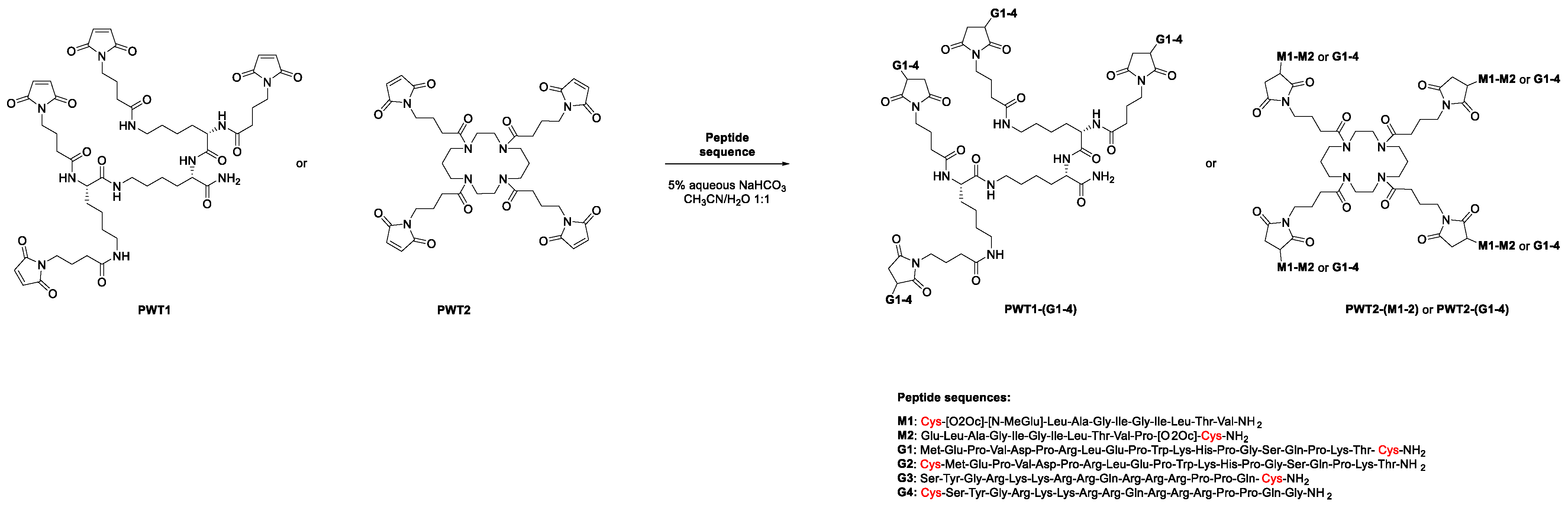
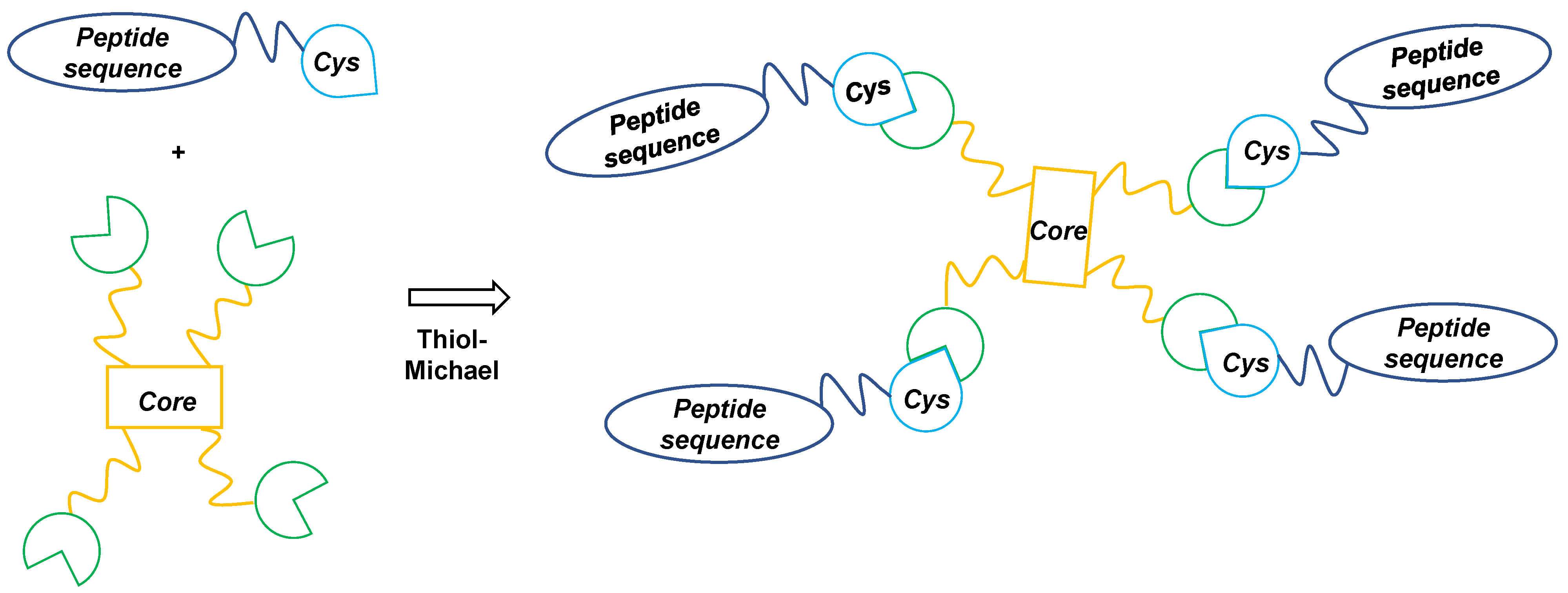
2.1. Peptides and PWT-Derivatives

Peptides were synthesized using solid-phase Fmoc/tBu chemistry as reported previously [13]. The commercially available Rink amide resin (100 mg, 0.55 mmol/g) was used as a functionalized support for peptides M1, M2, and G1–G4 (Appendix A). The threonine and glutamine Wang resins were used similarly for peptides Tat1–20 and Tat46–60, respectively. Coupling reactions were conducted for 1 h at room temperature using a 4-fold excess of N,N′-diisopropylcarbodiimide/1-hydroxybenzotriazole (DIC/HOBt). Fmoc removal and further coupling reactions were performed repeatedly to obtain the desired peptide-bound resins. Peptides were removed from the resins via incubation with a TFA/H2O/Et3SiH mixture (95:2.5:2.5) for 5 h at room temperature. The resulting crude products were triturated with cold diethyl ether, purified using a Waters Prep 600 HPLC System equipped with a Jupiter C18 column (250 × 30 mm, 300 Å, 15 µm particle size), and eluted in a binary mobile phase consisting of solution A (100% H2O, 0. 1% v/v TFA) and solution B (40% H2O, 60% CH3CN, 0.1% v/v TFA) at a flow rate of 20 mL/min. Gradients were established individually considering the analytical HPLC profile of the crude product. The molecular weights of the reaction intermediates and final compounds were determined using an electrospray mass spectrometer (ESI MICROMASS ZMD 2000). Purity was assessed using a Beckman Coulter Gold HPLC System 168 equipped with an XBridge C18 column (4.6 × 150 mm, 5 μm particle size) at a flow rate of 0.7 mL/min across a linear gradient from 100% solvent A (H2O, 0.1% TFA) to 100% solvent B (CH3CN, 0.1% TFA) over 25 min. The analytical profiles of the final compounds (monitored at 220 nm) showed purities > 98%. Resins for solid-phase peptide synthesis and enantiopure Fmoc-protected amino acids were purchased from Bachem (Bubendorf, Switzerland), BLDpharm (Kaiserslautern, Germany), and Fluka (Milan, Italy).

2.2. In Vitro Priming of Human Antigen-Specific CD8+ T Cells

Buffy coats from healthy blood donors were obtained from the University Hospital of Ferrara. Peripheral blood mononuclear cells (PBMCs) were isolated using Ficoll-Hypaque (Lonza, Basel, Switzerland) and frozen in liquid nitrogen [14,15]. Thawed PBMCs were resuspended at 107 cells/mL in 48-well tissue-culture plates (2.5 × 10⁶ cells/well) containing AIM medium (Thermo Fisher Scientific, Monza, Italy) supplemented with Flt3 ligand (50 ng/mL; Miltenyi Biotec, Bologna, Italy) to mobilize resident dendritic cells. After 24 h (day 1), the Melan-A26–35 peptide (ELA) was added to the cultures in its native form or conjugated to PWTs (Appendix A). Negative control wells lacked peptide (PWT scaffold alone). PWT-derivatives were used at a 1:4 molar ratio compared with peptide alone to compensate for stoichiometry (1 µM for ELA and 0.25 µM for PWT2-M1 and PWT2-M2). Dendritic cell maturation was induced with TNF (1000 U/mL; Miltenyi Biotec), IL-1β (10 ng/mL; Miltenyi Biotec), IL-7 (0.5 ng/mL; R&D Systems, Minneapolis, Minnesota, USA), and prostaglandin E2 (1 μM; Calbiochem, Milan, Italy). On day 2, fetal bovine serum (FBS; Euroclone, Milan, Italy) was added at a final v/v ratio of 10%. Medium was replaced on days 4 and 7 with fresh RPMI 1640 (Euroclone) enriched with 10% FBS, nonessential amino acids (Euroclone), and sodium pyruvate (Sigma-Aldrich, Milan, Italy). Antigen-specific CD8+ T cells were characterized on day 10 as described previously [16,17]. Briefly, cells were labeled with PE-conjugated ELA/HLA-A2 tetramers for 15 min at 37 °C and then surface-stained for 15 min at room temperature with anti-CD3–FITC and anti-CD8–APC-Cy7 (BD Biosciences, Milan, Italy). Intracellular staining for T-bet was performed using a Transcription Factor Buffer Set (BD Pharmingen, Milan, Italy) and anti-T-bet–eFluor 660 (Thermo Fisher Scientific). Non-viable cells were eliminated from the analysis using LIVE/DEAD Fixable Aqua (Thermo Fisher Scientific). Samples were acquired using a FACSCanto II cytometer (BD Biosciences). Data were analyzed using FACSDiva version 7.0 and FlowJo version 10 (BD Biosciences).

2.3. Mouse Immunization

Animals (n = 5 or 6 per group) were handled according to European and Institutional guidelines. Six-week-old female BALB/c mice (Charles River Laboratories, Lecco, Italy) were immunized subcutaneously (SC) or via the oral mucosa (OM) with the Tat1–20 or Tat46–60 peptides (3–30 µg), either alone or conjugated to PWT scaffolds (Appendix A). SC injections were performed at two sites on the back with the relevant immunogen diluted in a total of 100 µL of PBS. OM immunization was performed using a pipette tip to deliver the relevant immunogen directly into the mouth in a total of 10 µL of PBS. Mice were deprived of water for 6 h before immunization via this route. Immunogens were administered on days 1, 14, and 28. Serum samples were collected from the retro-orbital plexus 2 or 10 weeks later and processed as described previously [18,19].

2.4. Serology

IgG titers specific for the Tat protein from human immunodeficiency virus (HIV) type 1 (isolate IIIB, clone BH10) [15,20] or the Tat1–20 or Tat46–60 peptides were determined via enzyme-linked immunoassays (ELISAs) as described previously [21,22]. Briefly, 96-well plates were coated with Tat or Tat-derived peptides (100 ng/200 µL/well) in 0.05 M carbonate buffer (pH 9.6) for 18 h at 4 °C. Plates were then washed with PBS containing 0.05% Tween 20 (Sigma-Aldrich) and incubated for 90 min at 37 °C with blocking buffer (PBS containing 0.05% Tween 20 and 1% BSA). After extensive further washes, serial dilutions of each serum sample were dispensed in duplicate wells (100 µL/well) and incubated for 90 min at 37 °C. Plates were washed again before the addition of 100 µL/well of HRP-conjugated goat anti-mouse IgG (Sigma-Aldrich) and then incubated for 90 min at 37 °C. After incubation, plates were washed another five times and developed using a solution (150 µL/well) of 2,2′-azinobis [3-ethylbenzothiazoline-6-sulfonic acid]-diammonium salt (ABTS) substrate (Roche, Monza, Italy). Absorbance values were measured at 405 nm using a Sunrise Absorbance Microplate Reader (Tecan, Salzburg, Austria). Cut-off values were estimated as the mean OD of three negative control sera plus 0.05. Each OD value was subtracted from the blank and cut-off values to obtain a net OD value, and IgG titers were calculated using the intercept function [18,23].

2.5. Statistical Analysis

Independent groups were compared using the two-tailed Mann–Whitney U test with Bonferroni correction for multiple comparisons. Time-dependent variations in the number of mice with detectable immune responses were compared using Fisher’s exact test. Significance was assigned at p < 0.05.

3. Results

3.1. Chemistry

The PWT1 and PWT2 scaffolds were synthesized as described previously [9,24,25,26]. The maleimide derivatives PWT1 and PWT2 were tetrafunctionalized via a thiol-Michael reaction (Figure 1) in the presence of catalytic amounts of a base (NaHCO3 5% aqueous solution) with peptide sequences derived from the melanoma-associated ELA T-cell epitope (M1 and M2) or the HIV-specific Tat1–20 (G1 and G2) or Tat46–60 (G3 and G4) B-cell epitopes, opportunely modified with a cysteine residue (Appendix A).
Two derivatives of the ELA epitope were synthesized by inserting an N- (M1) or C-terminal (M2) cysteine into the native sequence, and a polyoxyethylene chain was inserted between the peptide sequence and the cysteine residue to increase the solubility of these compounds. Indeed, one of the main practical issues associated with these peptide sequences is that they are highly hydrophobic and insoluble (both in water and organic solvents), with a strong tendency to form gels. Similarly, the N-terminal glutamate residue of M1 was modified to N-methyl glutamate. M1 and M2 were conjugated to PWT2, generating two different constructs [PWT2-(M1–M2)] (Appendix A). This scaffold was used for proof-of-concept immunogenicity studies, because most of the peptide dendrimers generated previously were synthesized using the PWT2 core [9].
The HIV-derived peptides were modified by inserting the cysteine anchoring site at the N- or C-terminal position of Tat1–20 (G1 and G2) or Tat46–60 (G3 and G4). All four peptides were conjugated separately to PWT1 and PWT2, generating eight different constructs [PWT1-(G1–G4) and PWT2-(G1–G4)] (Appendix A).
Tetrafunctionalization via a thiol-Michael reaction offers the advantage of having an excellent nucleophile under mild reaction conditions (in terms of solvent, temperature, and pH), making it a very efficient and fast procedure (the tetrafunctionalization reaction occurs within 5 min). These features make this chemical strategy applicable to a plethora of peptide sequences and, potentially, also to non-peptide products [27].

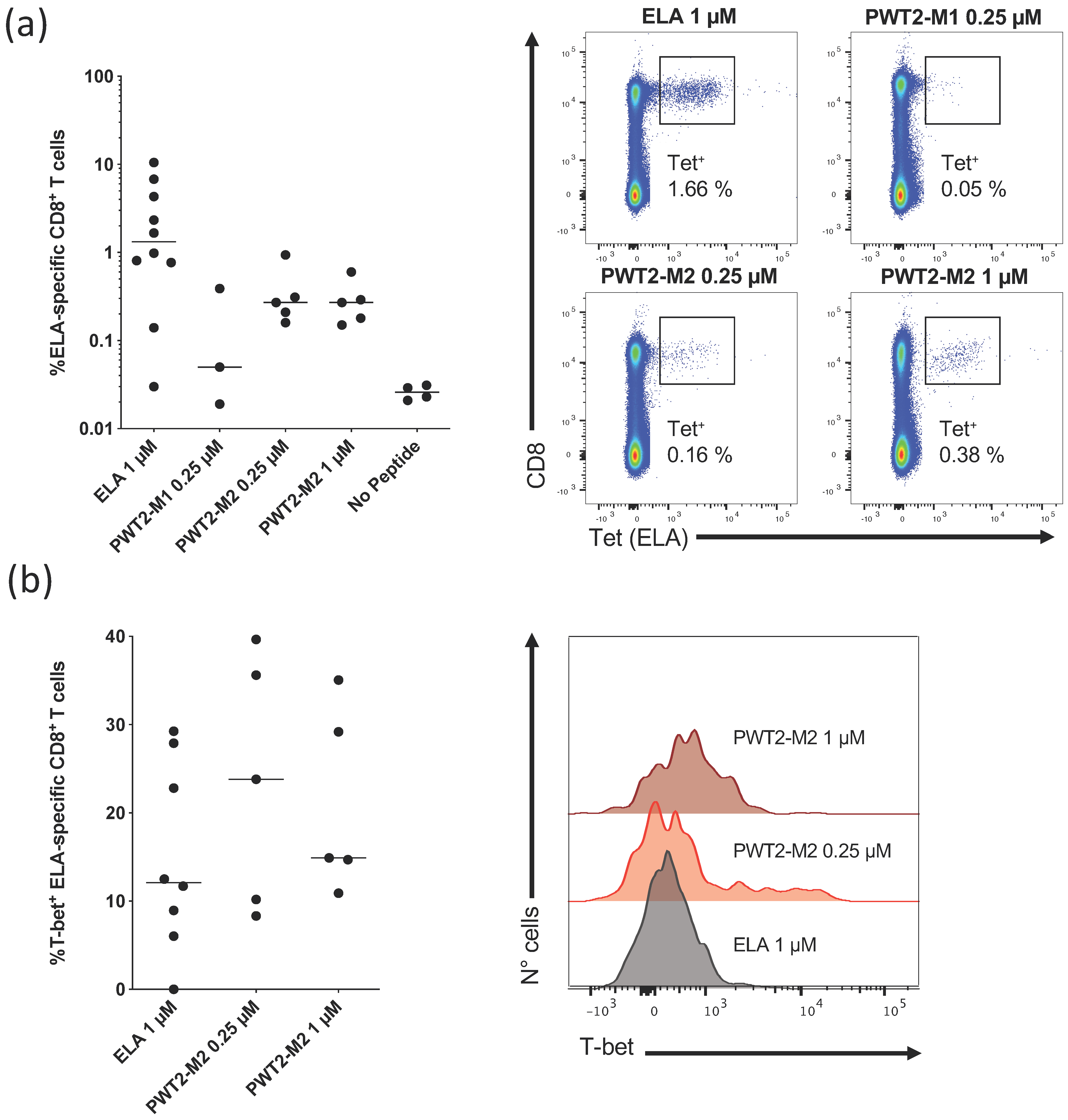
3.2. PWT Scaffolds Can Present T-Cell Epitopes

In preliminary experiments, we used an in vitro priming system to determine whether HLA class I-restricted peptide epitopes linked to PWT scaffolds could activate naive CD8+ T cells [16,28,29]. As a model antigen, we used the HLA-A2-restricted Melan-A26–35 peptide epitope (ELA), which is recognized by a relatively large fraction of naive precursors in the human CD8+ T-cell pool, facilitating the generation of reproducible data from low numbers of cells [29]. PBMCs from healthy, HLA-A2+, melanoma-naive donors were cultured with Flt3 ligand, a standard cocktail of cytokines, and the ELA peptide, either alone or conjugated via its N- (M1) or C-terminus (M2) to PWT2 (Figure 1 and Appendix A). Epitope-specific CD8+ T cells were quantified via tetramer staining on day 10.
PWT2-M1 induced very few ELA-specific CD8+ T cells (Figure 2a). Higher response frequencies were observed after priming with PWT2-M2, albeit at levels below those elicited by the cognate peptide alone (Figure 2a). This pattern could not be reversed by increasing the concentration of PWT2-M2 to 1 µM (Figure 2a).
We then evaluated the functionality of ELA-specific CD8+ T cells by measuring the expression of T-bet, a master regulator of effector differentiation associated with enhanced cytokine production and cytolytic activity [16,17]. Irrespective of the priming condition, ELA-specific CD8+ T cells expressed T-bet at similar frequencies, indicating a consistent pattern of differentiation in vitro (Figure 2b).
Collectively, these results show that the PWT system can present T-cell epitopes in immunogenic form, enabling the differentiation of at least some naive precursors, and that tetrafunctionalization of a cognate peptide does not completely abolish antigen recognition via the TCR.

3.3. PWT Scaffolds Can Present B-Cell Epitopes

In further experiments, we used in vivo models to determine whether B-cell epitopes could also elicit immune responses after tetrafunctionalization. Two different PWT scaffolds were used for this purpose: PWT1, which is structurally related to classical MAPs, and PWT2, which is chemically more diverse (Figure 1). As model antigens, we chose two linear B-cell peptides, one immunodominant (Tat1–20) and one subdominant (Tat46–60), derived from HIV [30]. Each peptide was conjugated separately via the N- and C-terminal domains to PWT1 and PWT2 (Figure 1 and Appendix A). BALB/c mice were injected SC with 3 µg of each construct on days 1, 14, and 28. Control mice were inoculated with the Tat1–20 or Tat46–60 peptides alone. IgG titers specific for each peptide or the whole Tat protein were measured 2 weeks after the last immunization via ELISA.
As shown in Table 1, the Tat1–20 peptide was not immunogenic when administered alone. In contrast, responses were observed in some mice immunized with Tat1–20 conjugated to PWT1 via its C-terminal domain (PWT1-G1) or to PWT2 via its N-terminal domain (PWT2-G2). Responses directed against the Tat1–20 peptide and the whole Tat protein were observed in both cases. Similarly, the Tat46–60 peptide was not very immunogenic when administered alone, inducing a response in only one mouse. Higher levels of immunogenicity were observed with PWT1-G4, PWT2-G3, and PWT2-G4, but not with PWT1-G3. Responses were again specific for both the cognate peptide (Tat46–60) and the whole Tat protein.
Collectively, these results show that the PWT system can present B-cell epitopes in immunogenic form, enabling the induction of primary humoral immune responses directed against the cognate peptide and the whole protein antigen.

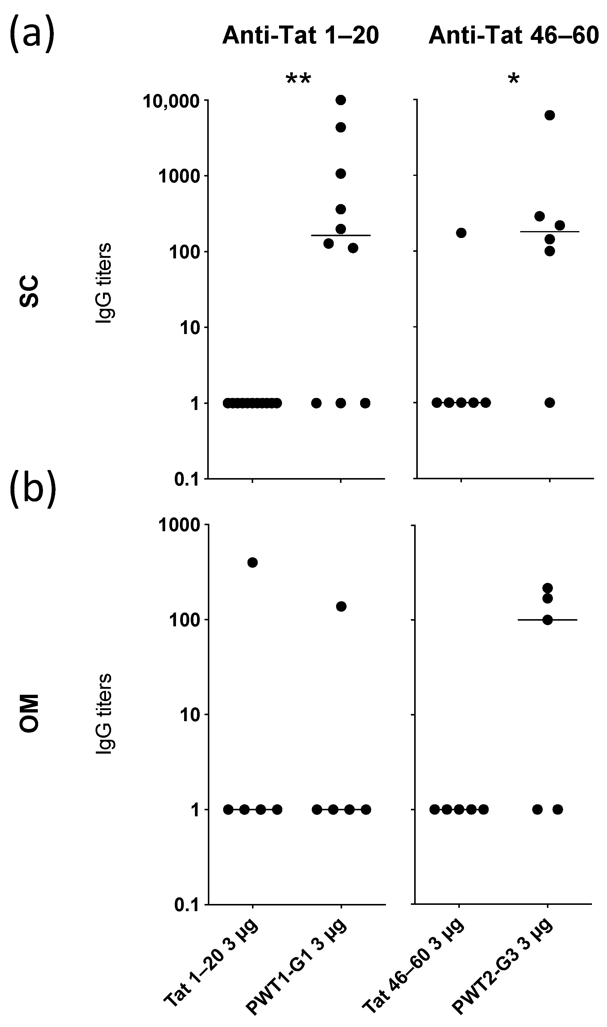
3.4. Conjugation of Peptide Epitopes to PWT Scaffolds Enhances Their Immunogenicity

To extend these findings, we focused on the most promising constructs for each specificity, namely PWT1-G1 (Tat1–20 conjugated to PWT1 via its C-terminal domain) and PWT2-G3 (Tat46–60 conjugated to PWT2 via its N-terminal domain). PWT1-G1 and PWT2-G3 induced significantly higher levels of peptide-specific IgG than the corresponding Tat1–20 and Tat46–60 peptides alone in mice immunized SC (Figure 3a).
As peptides typically display short half-lives in vivo, with negative consequences for immunogenicity, we immunized mice with the same constructs delivered via the OM. This route of administration exposes antigens to several proteolytic enzymes. Peptides alone (Tat1–20 or Tat46–60) were poorly immunogenic, and likewise, PWT1-G1 induced Tat1–20-specific IgG in only one mouse (Figure 3b). In contrast, OM administration of PWT2-G3 induced Tat46–60-specific IgG in 60% of mice (3/5), although this difference was not significant compared with the peptide alone (Figure 3b).
Collectively, these results suggest that humoral immunogenicity can be enhanced by conjugating linear B-cell epitopes to PWT scaffolds, delivered either SC or via the OM.

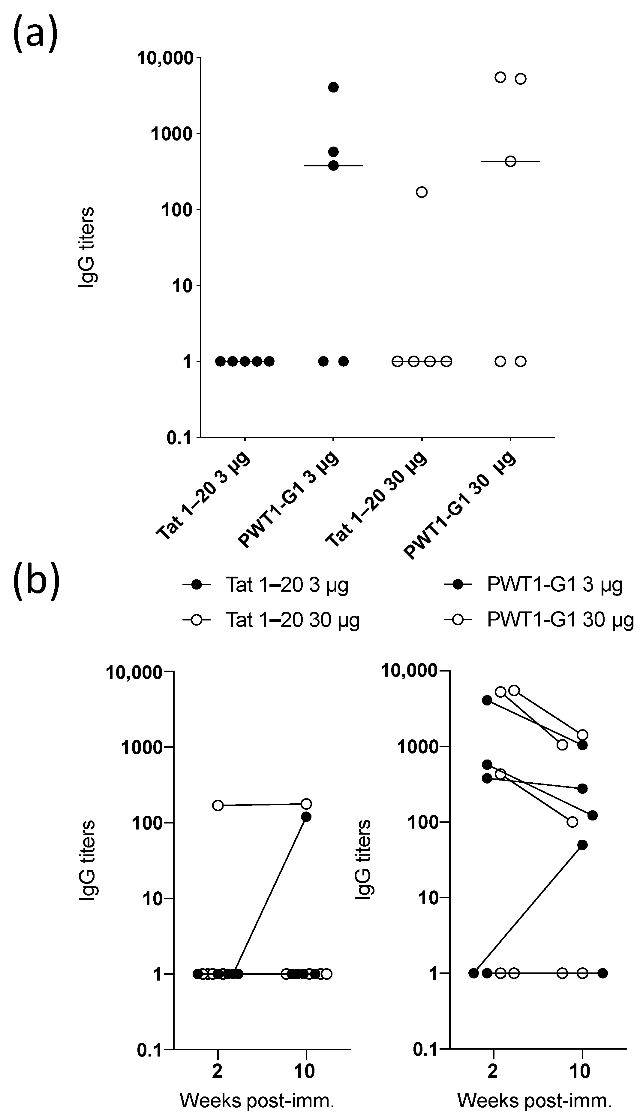
3.5. IgG Titers Induced by Peptides Conjugated to PWT Scaffolds Are Maintained Over Time

Data collected via the in vitro T-cell priming system suggested that increasing the dose of the PWT construct did not affect the magnitude of the subsequent immune response (Figure 2). To explore this phenomenon in the context of B-cell responses, we immunized mice SC with Tat1–20 alone or PWT1-G1 at doses of 3 or 30 µg. No significant differences in the resulting IgG titers were observed between the dosing schedules, either for Tat1–20 alone or for PWT1-G1 (Figure 4a). Irrespective of dose, however, all mice with detectable IgG responses after 2 weeks also had detectable IgG responses after 10 weeks, indicating that PWT1-G1 induced durable humoral immunity (Figure 4b).

4. Discussion

It has been estimated that peptide vaccines are currently being tested for different indications in more than 600 clinical studies, 15 of which have reached phase III [3]. The production of peptide epitopes does not represent a major limitation in this context, because chemical synthesis is rapid and allows the inclusion of modifications at the level of single amino acids, as well as the introduction of linkers and/or stabilizers [3,31]. However, native peptides are susceptible to enzymatic degradation, and more refined approaches are required to enhance the proteolytic stability of clinical-grade products without compromising immunogenicity or purity [3].
In this study, we describe a fast, reproducible, and high-yield technology that allows the generation of ultrapure tetrabranched peptides. The classical MAP strategy relies on a solid-phase platform to synthesize branched tetrameric or octameric peptide antigens using an appropriate polylysine-based scaffold [32]. Purification of target compounds prepared via this chemical approach is complex, reflecting contamination by tens of deletion compounds (lacking one or more amino acids) with physicochemical properties (molecular weight, charge, polarity, hydrophilicity, etc.) very similar to those of the desired product. Consequently, overall yields are often very low, with no guarantee of purity [33]. We have developed a convergent chemical strategy for the synthesis of multimeric peptide sequences based on a two-step procedure, in which the peptides and the core are first synthesized and purified separately and then stapled together via the thiol-Michael reaction (Figure 5).
The chemoselectivity of this reaction perfectly matches the need for a highly selective reaction between the core and the peptide binding site. Indeed, the reaction occurs only between the thiol group (characterizing the side-chain of the cysteine residue included in the peptide sequence as Michael’s donor group) and the maleimide function (a typical Michael’s acceptor group) to generate a thioether bond. Using this strategy, both B- and T-cell epitopes could be linked effectively to PWT structures, allowing the induction of primary cellular and humoral immune responses. Moreover, in vivo experiments showed that the levels of epitope-specific IgG induced by peptides conjugated to PWT scaffolds were higher than those elicited by the corresponding native peptides. Of note, not all derivatives were able to prime naive B or T cells. For example, the Tat1–20 peptide was not immunogenic when bound to PWT1 via its N-terminus, whereas the Tat46–60 peptide was immunogenic in this format. Further studies are necessary to uncover a rational explanation for such discrepancies, which likely relate to the conformation of the final product.
Peptide vaccines have been advocated in the context of various tumors, including melanoma, glioblastoma, and breast cancer [34,35], showing better toxicity profiles than most classical treatments [34]. The first clinical trial of a peptide vaccine was performed in 1995 [36]. More recently, the use of multiple peptides and personalized approaches have shown very promising results in the treatment of melanoma [37,38]. The HLA-A2-restricted, melanoma-associated ELA epitope is among the peptides that have been used in this context [39]. Conjugation of this peptide to a PWT scaffold in the present study allowed the in vitro priming of epitope-specific CD8+ T cells. Although the corresponding native peptide was more immunogenic in our system, T-bet expression was induced at similar frequencies among ELA-specific CD8+ T cells. In vivo experiments are therefore warranted to pursue these observations and determine whether the greater stability of tetrabranched derivatives could be exploited for clinical applications.
Peptide vaccines have also been advocated for the prevention and treatment of several infectious diseases, including HIV [40,41,42]. For example, the Tat protein has been used in two phase II clinical trials as a therapeutic vaccine against HIV, showing promising results in terms of immunological normalization and viral reservoir reduction [43,44,45]. High levels of antibodies against Tat1–20 or Tat46–60 have also been associated with undetectable plasma viral loads [46]. In line with this observation, antibodies directed against the N-terminus of Tat, which is the most immunogenic region in terms of humoral responses [47,48], protect monkeys from infection [49]. Moreover, the Tat46–60 peptide contains a basic domain that is critical for several Tat-related activities, including transactivation and transportation into the nucleus [50]. In the present study, both peptides were highly immunogenic when conjugated to PWT scaffolds, suggesting that this approach could facilitate the development of vaccines against HIV. In addition, the Tat46–60 peptide conjugated to PWT2 elicited more potent humoral immune responses than the corresponding native peptide after mucosal administration. Further studies are required to determine if this effect is a consequence of resistance to proteolysis and in vivo stability. This route of administration nonetheless mimics the anatomy of natural infection and may enhance vaccine compliance as a less invasive mode of immunization [51].

5. Conclusions

Our collective data show that PWT scaffolds can present B- and T-cell epitopes. Moreover, humoral immune responses elicited in vivo by peptides conjugated to PWT scaffolds were superior to those elicited in vivo by native peptides and were maintained over time. In conjunction with the advantages of this synthetic approach in terms of rapidity, yield, and purity, these observations suggest that the delivery of peptide vaccines on a global or personalized basis could be enhanced by the widespread adoption of PWT.

6. Patents

Remo Guerrini is the inventor of a patent application claiming the PWT technology (EP13162532.9).

Author Contributions

Conceptualization, F.N., R.G. (Remo Guerrini), and R.G. (Riccardo Gavioli); methodology, F.N., S.P., E.G., E.M., V.A. (Valentina Albanese), V.F., and V.A. (Victor Appay); formal analysis, F.N. and V.F.; resources, S.P., E.M., V.A. (Valentina Albanese), S.L.-L., and D.A.P.; data curation, F.N.; writing—original draft preparation, F.N., S.P., and A.C.; writing—review and editing, E.G., E.M., V.A. (Valentina Albanese), V.F., S.L.-L., D.A.P., V.A. (Victor Appay), P.M., R.G. (Remo Guerrini), and R.G. (Riccardo Gavioli); supervision, D.A.P., P.M., R.G. (Remo Guerrini), A.C., and R.G. (Riccardo Gavioli); funding acquisition, D.A.P., R.G. (Remo Guerrini), A.C., and R.G. (Riccardo Gavioli). All authors have read and agreed to the published version of the manuscript.

Funding

This work was supported by grants from the Wellcome Trust (100326/Z/12/Z) to D.A.P. and by grants from the University of Ferrara (Fondo di Ateneo per la Ricerca Scientifica) to R.G. (Remo Guerrini), A.C., and R.G. (Riccardo Gavioli). Core provision of the FACSCanto II was funded by the University of Ferrara (Bando per l’Acquisizione di Strumenti per la Ricerca di Ateneo 2015).

Institutional Review Board Statement

The study was conducted according to the guidelines of the Declaration of Helsinki and was approved by the University of Ferrara (protocol number 10246).

Data Availability Statement

All data pertaining to this study are included in the article.

Conflicts of Interest

R.G. (Remo Guerrini) is the inventor of a patent application claiming the PWT technology (EP13162532.9). R.G. (Remo Guerrini) is also a cofounder of the spin-off company UFPeptides s.r.l., the assignee of this patent application, located at the University of Ferrara.

Appendix A

Chemical structures of the compounds used in this study (native peptides and peptides conjugated to PWTs).
ELA peptide sequence:
Vaccines 09 00526 i001
H-Glu-Leu-Ala-Gly-Ile-Gly-Ile-Leu-Thr-Val-NH2.
ELA derivative M1:
Vaccines 09 00526 i002
H-Cys-[O2Oc]-[N-Me]Glu-Leu-Ala-Gly-Ile-Gly-Ile-Leu-Thr-Val-NH2
ELA derivative M2:
Vaccines 09 00526 i003
H-Glu-Leu-Ala-Gly-Ile-Gly-Ile-Leu-Thr-Val-Pro-[O2Oc]-Cys-NH2
Tetrafunctionalized PWT2 with ELA derivative M1 (cysteine at N-terminus):
Vaccines 09 00526 i004
Tetrafunctionalized PWT2 with ELA derivative M2 (cysteine at C-terminus):
Vaccines 09 00526 i005
Tat1–20 peptide sequence:
Vaccines 09 00526 i006
H-Met-Glu-Pro-Val-Asp-Pro-Arg-Leu-Glu-Pro-Trp-Lys-His-Pro-Gly-Ser-Gln-Pro-Lys-Thr-OH
Tat1–20 derivative G1:
Vaccines 09 00526 i007
H-Met-Glu-Pro-Val-Asp-Pro-Arg-Leu-Glu-Pro-Trp-Lys-His-Pro-Gly-Ser-Gln-Pro-Lys-Thr-Cys-NH2
Tetrafunctionalized PWT1 with Tat1–20 derivative G1 (cysteine at C-terminus):
Vaccines 09 00526 i008
Tetrafunctionalized PWT2 with Tat1–20 derivative G1 (cysteine at C-terminus):
Vaccines 09 00526 i009
Tat1–20 derivative G2:
Vaccines 09 00526 i010
H-Cys-Met-Glu-Pro-Val-Asp-Pro-Arg-Leu-Glu-Pro-Trp-Lys-His-Pro-Gly-Ser-Gln-Pro-Lys-Thr-NH2
Tetrafunctionalized PWT1 with Tat1–20 derivative G2 (cysteine at N-terminus):
Vaccines 09 00526 i011
Tetrafunctionalized PWT2 with Tat1–20 derivative G2 (cysteine at N-terminus):
Vaccines 09 00526 i012
Tat46–60 peptide sequence:
Vaccines 09 00526 i013
H-Ser-Tyr-Gly-Arg-Lys-Lys-Arg-Arg-Gln-Arg-Arg-Arg-Pro-Pro-Gln-OH
Tat46–60 derivative G3:
Vaccines 09 00526 i014
H-Ser-Tyr-Gly-Arg-Lys-Lys-Arg-Arg-Gln-Arg-Arg-Arg-Pro-Pro-Gln-Cys-NH2
Tetrafunctionalized PWT1 with Tat46–60 derivative G3 (cysteine at C-terminus):
Vaccines 09 00526 i015
Tetrafunctionalized PWT2 with Tat46–60 derivative G3 (cysteine at C-terminus):
Vaccines 09 00526 i016
Tat46–60 derivative G4:
Vaccines 09 00526 i017
H-Cys-Ser-Tyr-Gly-Arg-Lys-Lys-Arg-Arg-Gln-Arg-Arg-Arg-Pro-Pro-Gln-Gly-NH2
Tetrafunctionalized PWT1 with Tat46–60 derivative G4 (cysteine at N-terminus):
Vaccines 09 00526 i018
Tetrafunctionalized PWT2 with Tat46–60 derivative G4 (cysteine at N-terminus):
Vaccines 09 00526 i019

References

  1. Nicoli, F.; Appay, V. Immunological considerations regarding parental concerns on pediatric immunizations. Vaccine 2017, 35, 3012–3019. [Google Scholar] [CrossRef] [Green Version]
  2. Sallam, M. COVID-19 Vaccine Hesitancy Worldwide: A Concise Systematic Review of Vaccine Acceptance Rates. Vaccines 2021, 9, 160. [Google Scholar] [CrossRef] [PubMed]
  3. Sabatino, D. Medicinal Chemistry and Methodological Advances in the Development of Peptide-Based Vaccines. J. Med. Chem. 2020, 63, 14184–14196. [Google Scholar] [CrossRef]
  4. Yoshida, K.; Noguchi, M.; Mine, T.; Komatsu, N.; Yutani, S.; Ueno, T.; Yanagimoto, H.; Kawano, K.; Itoh, K.; Yamada, A. Characteristics of severe adverse events after peptide vaccination for advanced cancer patients: Analysis of 500 cases. Oncol. Rep. 2011, 25, 57–62. [Google Scholar]
  5. Nelde, A.; Rammensee, H.G.; Walz, J.S. The Peptide Vaccine of the Future. Mol. Cell. Proteom. 2021, 20, 100022. [Google Scholar] [CrossRef] [PubMed]
  6. Hilf, N.; Kuttruff-Coqui, S.; Frenzel, K.; Bukur, V.; Stevanovic, S.; Gouttefangeas, C.; Platten, M.; Tabatabai, G.; Dutoit, V.; van der Burg, S.H.; et al. Actively personalized vaccination trial for newly diagnosed glioblastoma. Nature 2019, 565, 240–245. [Google Scholar] [CrossRef] [PubMed]
  7. Finessi, V.; Nicoli, F.; Gallerani, E.; Sforza, F.; Sicurella, M.; Cafaro, A.; Caputo, A.; Ensoli, B.; Gavioli, R. Effects of different routes of administration on the immunogenicity of the Tat protein and a Tat-derived peptide. Hum. Vaccin. Immunother. 2015, 11, 1489–1493. [Google Scholar] [CrossRef] [PubMed] [Green Version]
  8. Tam, J.P. Synthetic peptide vaccine design: Synthesis and properties of a high-density multiple antigenic peptide system. Proc. Natl. Acad. Sci. USA 1988, 85, 5409–5413. [Google Scholar] [CrossRef] [Green Version]
  9. Calo, G.; Rizzi, A.; Ruzza, C.; Ferrari, F.; Pacifico, S.; Gavioli, E.C.; Salvadori, S.; Guerrini, R. Peptide welding technology—A simple strategy for generating innovative ligands for G protein coupled receptors. Peptides 2018, 99, 195–204. [Google Scholar] [CrossRef] [PubMed]
  10. Cerlesi, M.C.; Ding, H.; Bird, M.F.; Kiguchi, N.; Ferrari, F.; Malfacini, D.; Rizzi, A.; Ruzza, C.; Lambert, D.G.; Ko, M.C.; et al. Pharmacological studies on the NOP and opioid receptor agonist PWT2-[Dmt(1)]N/OFQ(1-13). Eur. J. Pharmacol. 2017, 794, 115–126. [Google Scholar] [CrossRef] [PubMed] [Green Version]
  11. Bracci, L.; Falciani, C.; Lelli, B.; Lozzi, L.; Runci, Y.; Pini, A.; De Montis, M.G.; Tagliamonte, A.; Neri, P. Synthetic peptides in the form of dendrimers become resistant to protease activity. J. Biol. Chem. 2003, 278, 46590–46595. [Google Scholar] [CrossRef] [Green Version]
  12. Chang, K.J.; Pugh, W.; Blanchard, S.G.; McDermed, J.; Tam, J.P. Antibody specific to the alpha subunit of the guanine nucleotide-binding regulatory protein Go: Developmental appearance and immunocytochemical localization in brain. Proc. Natl. Acad. Sci. USA 1988, 85, 4929–4933. [Google Scholar] [CrossRef] [PubMed] [Green Version]
  13. Behrendt, R.; White, P.; Offer, J. Advances in Fmoc solid-phase peptide synthesis. J. Pept. Sci. 2016, 22, 4–27. [Google Scholar] [CrossRef] [Green Version]
  14. Telatin, V.; Nicoli, F.; Frasson, C.; Menegotto, N.; Barbaro, F.; Castelli, E.; Erne, E.; Palu, G.; Caputo, A. In Chronic Hepatitis C Infection, Myeloid-Derived Suppressor Cell Accumulation and T Cell Dysfunctions Revert Partially and Late After Successful Direct-Acting Antiviral Treatment. Front. Cell. Infect. Microbiol. 2019, 9, 190. [Google Scholar] [CrossRef] [PubMed] [Green Version]
  15. Nicoli, F.; Gallerani, E.; Sforza, F.; Finessi, V.; Chachage, M.; Geldmacher, C.; Cafaro, A.; Ensoli, B.; Caputo, A.; Gavioli, R. The HIV-1 Tat protein affects human CD4+ T-cell programing and activation, and favors the differentiation of naive CD4+ T cells. AIDS 2018, 32, 575–581. [Google Scholar] [CrossRef]
  16. Papagno, L.; Kuse, N.; Lissina, A.; Gostick, E.; Price, D.A.; Appay, V.; Nicoli, F. The TLR9 ligand CpG ODN 2006 is a poor adjuvant for the induction of de novo CD8+ T-cell responses in vitro. Sci. Rep. 2020, 10, 11620. [Google Scholar] [CrossRef] [PubMed]
  17. Nicoli, F.; Papagno, L.; Frere, J.J.; Cabral-Piccin, M.P.; Clave, E.; Gostick, E.; Toubert, A.; Price, D.A.; Caputo, A.; Appay, V. Naive CD8+ T-Cells Engage a Versatile Metabolic Program Upon Activation in Humans and Differ Energetically From Memory CD8+ T-Cells. Front. Immunol. 2018, 9, 2736. [Google Scholar] [CrossRef] [Green Version]
  18. Caputo, A.; Brocca-Cofano, E.; Castaldello, A.; Voltan, R.; Gavioli, R.; Srivastava, I.K.; Barnett, S.W.; Cafaro, A.; Ensoli, B. Characterization of immune responses elicited in mice by intranasal co-immunization with HIV-1 Tat, gp140 DeltaV2Env and/or SIV Gag proteins and the nontoxicogenic heat-labile Escherichia coli enterotoxin. Vaccine 2008, 26, 1214–1227. [Google Scholar] [CrossRef] [PubMed]
  19. Nicoli, F.; Finessi, V.; Sicurella, M.; Rizzotto, L.; Gallerani, E.; Destro, F.; Cafaro, A.; Marconi, P.; Caputo, A.; Ensoli, B.; et al. The HIV-1 Tat protein induces the activation of CD8+ T cells and affects in vivo the magnitude and kinetics of antiviral responses. PLoS ONE 2013, 8, e77746. [Google Scholar] [CrossRef] [PubMed] [Green Version]
  20. Nicoli, F.; Chachage, M.; Clowes, P.; Bauer, A.; Kowour, D.; Ensoli, B.; Cafaro, A.; Maboko, L.; Hoelscher, M.; Gavioli, R.; et al. Association between different anti-Tat antibody isotypes and HIV disease progression: Data from an African cohort. BMC Infect. Dis. 2016, 16, 344. [Google Scholar] [CrossRef] [PubMed] [Green Version]
  21. Nicoli, F.; Gallerani, E.; Sicurella, M.; Pacifico, S.; Cafaro, A.; Ensoli, B.; Marconi, P.; Caputo, A.; Gavioli, R. The Tat Protein of HIV-1 Prevents the Loss of HSV-Specific Memory Adaptive Responses and Favors the Control of Viral Reactivation. Vaccines 2020, 8, 274. [Google Scholar] [CrossRef]
  22. Sicurella, M.; Nicoli, F.; Gallerani, E.; Volpi, I.; Berto, E.; Finessi, V.; Destro, F.; Manservigi, R.; Cafaro, A.; Ensoli, B.; et al. An attenuated herpes simplex virus type 1 (HSV1) encoding the HIV-1 Tat protein protects mice from a deadly mucosal HSV1 challenge. PLoS ONE 2014, 9, e100844. [Google Scholar] [CrossRef] [PubMed] [Green Version]
  23. Nicoli, F.; Gallerani, E.; Skarlis, C.; Sicurella, M.; Cafaro, A.; Ensoli, B.; Caputo, A.; Marconi, P.C.; Gavioli, R. Systemic immunodominant CD8 responses with an effector-like phenotype are induced by intravaginal immunization with attenuated HSV vectors expressing HIV Tat and mediate protection against HSV infection. Vaccine 2016, 34, 2216–2224. [Google Scholar] [CrossRef] [PubMed]
  24. Guerrini, R.; Marzola, E.; Trapella, C.; Pela, M.; Molinari, S.; Cerlesi, M.C.; Malfacini, D.; Rizzi, A.; Salvadori, S.; Calo, G. A novel and facile synthesis of tetra branched derivatives of nociceptin/orphanin FQ. Bioorg. Med. Chem. 2014, 22, 3703–3712. [Google Scholar] [CrossRef] [PubMed]
  25. Lambert, T.H.; Danishefsky, S.J. Total synthesis of UCS1025A. J. Am. Chem. Soc. 2006, 128, 426–427. [Google Scholar] [CrossRef] [PubMed]
  26. Song, H.Y.; Ngai, M.H.; Song, Z.Y.; MacAry, P.A.; Hobley, J.; Lear, M.J. Practical synthesis of maleimides and coumarin-linked probes for protein and antibody labelling via reduction of native disulfides. Org. Biomol. Chem. 2009, 7, 3400–3406. [Google Scholar] [CrossRef]
  27. Nair, D.P.; Podgorski, M.; Chatani, S.; Gong, T.; Xi, W.X.; Fenoli, C.R.; Bowman, C.N. The Thiol-Michael Addition Click Reaction: A Powerful and Widely Used Tool in Materials Chemistry. Chem. Mater. 2014, 26, 724–744. [Google Scholar] [CrossRef]
  28. Gayet, R.; Michaud, E.; Nicoli, F.; Chanut, B.; Paul, M.; Rochereau, N.; Guillon, C.; He, Z.; Papagno, L.; Bioley, G.; et al. Impact of IgA isoforms on their ability to activate dendritic cells and to prime T cells. Eur. J. Immunol. 2020, 50, 1295–1306. [Google Scholar] [CrossRef]
  29. Lissina, A.; Briceno, O.; Afonso, G.; Larsen, M.; Gostick, E.; Price, D.A.; Mallone, R.; Appay, V. Priming of Qualitatively Superior Human Effector CD8+ T Cells Using TLR8 Ligand Combined with FLT3 Ligand. J. Immunol. 2016, 196, 256–263. [Google Scholar] [CrossRef] [PubMed] [Green Version]
  30. Longo, O.; Tripiciano, A.; Fiorelli, V.; Bellino, S.; Scoglio, A.; Collacchi, B.; Alvarez, M.J.; Francavilla, V.; Arancio, A.; Paniccia, G.; et al. Phase I therapeutic trial of the HIV-1 Tat protein and long term follow-up. Vaccine 2009, 27, 3306–3312. [Google Scholar] [CrossRef]
  31. Skwarczynski, M.; Toth, I. Peptide-based synthetic vaccines. Chem. Sci. 2016, 7, 842–854. [Google Scholar] [CrossRef] [Green Version]
  32. Tam, J.P. Recent advances in multiple antigen peptides. J. Immunol. Methods 1996, 196, 17–32. [Google Scholar] [CrossRef]
  33. Niederhafner, P.; Sebestik, J.; Jezek, J. Peptide dendrimers. J. Pept. Sci. 2005, 11, 757–788. [Google Scholar] [CrossRef] [PubMed]
  34. Schneble, E.; Clifton, G.T.; Hale, D.F.; Peoples, G.E. Peptide-Based Cancer Vaccine Strategies and Clinical Results. Methods Mol. Biol. 2016, 1403, 797–817. [Google Scholar] [CrossRef] [PubMed]
  35. Ma, M.; Liu, J.; Jin, S.; Wang, L. Development of tumour peptide vaccines: From universalization to personalization. Scand. J. Immunol. 2020, 91, e12875. [Google Scholar] [CrossRef] [PubMed]
  36. Mukherji, B.; Chakraborty, N.G.; Yamasaki, S.; Okino, T.; Yamase, H.; Sporn, J.R.; Kurtzman, S.K.; Ergin, M.T.; Ozols, J.; Meehan, J.; et al. Induction of antigen-specific cytolytic T cells in situ in human melanoma by immunization with synthetic peptide-pulsed autologous antigen presenting cells. Proc. Natl. Acad. Sci. USA 1995, 92, 8078–8082. [Google Scholar] [CrossRef] [Green Version]
  37. Ott, P.A.; Hu, Z.; Keskin, D.B.; Shukla, S.A.; Sun, J.; Bozym, D.J.; Zhang, W.; Luoma, A.; Giobbie-Hurder, A.; Peter, L.; et al. An immunogenic personal neoantigen vaccine for patients with melanoma. Nature 2017, 547, 217–221. [Google Scholar] [CrossRef]
  38. Hu, Z.; Leet, D.E.; Allesoe, R.L.; Oliveira, G.; Li, S.; Luoma, A.M.; Liu, J.; Forman, J.; Huang, T.; Iorgulescu, J.B.; et al. Personal neoantigen vaccines induce persistent memory T cell responses and epitope spreading in patients with melanoma. Nat. Med. 2021, 27, 515–525. [Google Scholar] [CrossRef]
  39. Carretero-Iglesia, L.; Couturaud, B.; Baumgaertner, P.; Schmidt, J.; Maby-El Hajjami, H.; Speiser, D.E.; Hebeisen, M.; Rufer, N. High Peptide Dose Vaccination Promotes the Early Selection of Tumor Antigen-Specific CD8 T-Cells of Enhanced Functional Competence. Front. Immunol. 2019, 10, 3016. [Google Scholar] [CrossRef]
  40. Malonis, R.J.; Lai, J.R.; Vergnolle, O. Peptide-Based Vaccines: Current Progress and Future Challenges. Chem. Rev. 2020, 120, 3210–3229. [Google Scholar] [CrossRef] [PubMed] [Green Version]
  41. Azuar, A.; Jin, W.; Mukaida, S.; Hussein, W.M.; Toth, I.; Skwarczynski, M. Recent Advances in the Development of Peptide Vaccines and Their Delivery Systems Against Group A Streptococcus. Vaccines 2019, 7, 58. [Google Scholar] [CrossRef] [Green Version]
  42. Bellini, C.; Horvati, K. Recent Advances in the Development of Protein- and Peptide-Based Subunit Vaccines against Tuberculosis. Cells 2020, 9, 2673. [Google Scholar] [CrossRef]
  43. Ensoli, B.; Bellino, S.; Tripiciano, A.; Longo, O.; Francavilla, V.; Marcotullio, S.; Cafaro, A.; Picconi, O.; Paniccia, G.; Scoglio, A.; et al. Therapeutic immunization with HIV-1 Tat reduces immune activation and loss of regulatory T-cells and improves immune function in subjects on HAART. PLoS ONE 2010, 5, e13540. [Google Scholar] [CrossRef] [PubMed] [Green Version]
  44. Ensoli, B.; Nchabeleng, M.; Ensoli, F.; Tripiciano, A.; Bellino, S.; Picconi, O.; Sgadari, C.; Longo, O.; Tavoschi, L.; Joffe, D.; et al. HIV-Tat immunization induces cross-clade neutralizing antibodies and CD4+ T cell increases in antiretroviral-treated South African volunteers: A randomized phase II clinical trial. Retrovirology 2016, 13, 34. [Google Scholar] [CrossRef] [PubMed] [Green Version]
  45. Ensoli, F.; Cafaro, A.; Casabianca, A.; Tripiciano, A.; Bellino, S.; Longo, O.; Francavilla, V.; Picconi, O.; Sgadari, C.; Moretti, S.; et al. HIV-1 Tat immunization restores immune homeostasis and attacks the HAART-resistant blood HIV DNA: Results of a randomized phase II exploratory clinical trial. Retrovirology 2015, 12, 33. [Google Scholar] [CrossRef] [Green Version]
  46. Re, M.C.; Vignoli, M.; Furlini, G.; Gibellini, D.; Colangeli, V.; Vitone, F.; La Placa, M. Antibodies against full-length Tat protein and some low-molecular-weight Tat-peptides correlate with low or undetectable viral load in HIV-1 seropositive patients. J. Clin. Virol. 2001, 21, 81–89. [Google Scholar] [CrossRef]
  47. Butto, S.; Fiorelli, V.; Tripiciano, A.; Ruiz-Alvarez, M.J.; Scoglio, A.; Ensoli, F.; Ciccozzi, M.; Collacchi, B.; Sabbatucci, M.; Cafaro, A.; et al. Sequence conservation and antibody cross-recognition of clade B human immunodeficiency virus (HIV) type 1 Tat protein in HIV-1-infected Italians, Ugandans, and South Africans. J. Infect. Dis. 2003, 188, 1171–1180. [Google Scholar] [CrossRef]
  48. Moreau, E.; Belliard, G.; Partidos, C.D.; Pradezinsky, F.; Le Buanec, H.; Muller, S.; Desgranges, C. Important B-cell epitopes for neutralization of human immunodeficiency virus type 1 Tat in serum samples of humans and different animal species immunized with Tat protein or peptides. J. Gen. Virol. 2004, 85, 2893–2901. [Google Scholar] [CrossRef] [PubMed]
  49. Bachler, B.C.; Humbert, M.; Palikuqi, B.; Siddappa, N.B.; Lakhashe, S.K.; Rasmussen, R.A.; Ruprecht, R.M. Novel biopanning strategy to identify epitopes associated with vaccine protection. J. Virol. 2013, 87, 4403–4416. [Google Scholar] [CrossRef] [Green Version]
  50. Kurnaeva, M.A.; Sheval, E.V.; Musinova, Y.R.; Vassetzky, Y.S. Tat basic domain: A “Swiss army knife” of HIV-1 Tat? Rev. Med. Virol. 2019, 29, e2031. [Google Scholar] [CrossRef]
  51. Skwarczynski, M.; Toth, I. Non-invasive mucosal vaccine delivery: Advantages, challenges and the future. Expert Opin. Drug Deliv. 2020, 17, 435–437. [Google Scholar] [CrossRef] [PubMed] [Green Version]
Figure 1. Tetrafunctionalization of PWT1 and PWT2 with the peptide sequences M1–2 and G1–4.
Vaccines 09 00526 g001
Figure 2. In vitro priming of human naive CD8+ T cells specific for a melanoma-associated epitope conjugated to PWT2 (PWT2-M1 and PWT2-M2). (a) Data summary (left panel) and representative flow cytometry plots (right panel) showing tetramer+ ELA-specific CD8+ T cells expanded in the presence of Flt3 ligand, a cocktail of cytokines (TNF, IL-1β, IL-7, and PGE2), and the ELA peptide, either alone or conjugated to PWT2. PBMCs primed in the absence of peptide (PWT2 alone) were used as a negative control. (b) Data summary (left panel) and representative flow cytometry histogram plots (right panel) showing intracellular expression of T-bet among the corresponding tetramer+ ELA-specific CD8+ T cells. Left panels: each dot represents one HLA-A2+ donor per condition, and horizontal bars indicate median values (a,b). Right panels: plots are gated on viable CD3+ events (a) or on tetramer+ ELA-specific CD8+ T cells (b). Significance was determined using the two-tailed Mann–Whitney U test with Bonferroni correction for multiple comparisons.
Vaccines 09 00526 g002
Figure 3. Analysis of IgG responses induced by Tat peptides alone or Tat peptides conjugated to PWT scaffolds. Mice were immunized on three separate occasions with 3 µg of Tat1–20, PWT1-G1, Tat46–60, or PWT2-G3 delivered either SC (a) or via the OM (b). Serum samples were collected 2 weeks after the final immunization. IgG titers specific for Tat1–20 or Tat46–60 were measured via ELISA. Each dot represents one mouse, and horizontal bars indicate median values. Significance was determined using the two-tailed Mann–Whitney U test. * p < 0.05, ** p < 0.01.
Vaccines 09 00526 g003
Figure 4. Analysis of IgG responses induced by Tat1–20 alone or Tat1–20 conjugated to a PWT scaffold over time. (a,b) Mice were immunized SC on three separate occasions with 3 or 30 µg of Tat1–20 or PWT1-G1. Serum samples were collected 2 (a,b) and 10 weeks (b) after the third immunization. IgG titers specific for Tat1–20 were measured via ELISA. Each dot represents one mouse, and horizontal bars indicate median values. Significance was determined using the two-tailed Mann–Whitney U test with Bonferroni correction for multiple comparisons (a) or Fisher’s exact test (b).
Vaccines 09 00526 g004
Figure 5. Representation of the convergent chemical strategy.
Vaccines 09 00526 g005
Table 1. Numbers of mice with detectable humoral responses after immunization.
ConstructPWTConjugation DomainIgG Responses vs. Tat1–20IgG Responses vs. Tat Protein
Tat1–20Peptide aloneNA 10/50/5
PWT1-G1PWT1C-terminus3/52/5
PWT1-G2PWT1N-terminus0/60/6
PWT2-G1PWT2C-terminus0/60/6
PWT2-G2PWT2N-terminus2/63/6
ConstructPWTConjugation DomainIgG Responses vs. Tat46–60IgG Responses vs. Tat Protein
Tat46–60Peptide aloneNA 11/61/6
PWT1-G3PWT1C-terminus0/60/6
PWT1-G4PWT1N-terminus3/63/6
PWT2-G3PWT2C-terminus5/65/6
PWT2-G4PWT2N-terminus2/66/6
1 NA, not applicable.
Publisher’s Note: MDPI stays neutral with regard to jurisdictional claims in published maps and institutional affiliations.

