Review

The COVID-19 Pandemic: A Comprehensive Review of Taxonomy, Genetics, Epidemiology, Diagnosis, Treatment, and Control

1
Department of Veterinary Preventive Medicine, Ohio Agricultural Research and Development Center, The Ohio State University, Wooster, OH 44691, USA
2
Department of Animal Hygiene, Zoonoses and Animal Ethology, Faculty of Veterinary Medicine, Suez Canal University, Ismailia 41522, Egypt
3
Department of Virology, Faculty of Veterinary Medicine, Suez Canal University, Ismailia 41522, Egypt
4
Department of Animal Wealth Development, Faculty of Veterinary Medicine, Suez Canal University, Ismailia 41522, Egypt
5
Department of Radiology, University of Massachusetts Medical School, Worcester, MA 01655, USA
6
Avian and Rabbit Diseases Department, Faculty of Veterinary Medicine, Sadat City University, Sadat 32897, Egypt
7
Research and Development Section, PerNaturam GmbH, 56290 Gödenroth, Germany
*
Authors to whom correspondence should be addressed.
Submission received: 18 March 2020 / Revised: 19 April 2020 / Accepted: 21 April 2020 / Published: 24 April 2020

Abstract

:
A pneumonia outbreak with unknown etiology was reported in Wuhan, Hubei province, China, in December 2019, associated with the Huanan Seafood Wholesale Market. The causative agent of the outbreak was identified by the WHO as the severe acute respiratory syndrome coronavirus-2 (SARS-CoV-2), producing the disease named coronavirus disease-2019 (COVID-19). The virus is closely related (96.3%) to bat coronavirus RaTG13, based on phylogenetic analysis. Human-to-human transmission has been confirmed even from asymptomatic carriers. The virus has spread to at least 200 countries, and more than 1,700,000 confirmed cases and 111,600 deaths have been recorded, with massive global increases in the number of cases daily. Therefore, the WHO has declared COVID-19 a pandemic. The disease is characterized by fever, dry cough, and chest pain with pneumonia in severe cases. In the beginning, the world public health authorities tried to eradicate the disease in China through quarantine but are now transitioning to prevention strategies worldwide to delay its spread. To date, there are no available vaccines or specific therapeutic drugs to treat the virus. There are many knowledge gaps about the newly emerged SARS-CoV-2, leading to misinformation. Therefore, in this review, we provide recent information about the COVID-19 pandemic. This review also provides insights for the control of pathogenic infections in humans such as SARS-CoV-2 infection and future spillovers.

1. Introduction

Coronaviruses are enveloped, single-strand RNA viruses that can infect a wide range of hosts including avian, wild, domestic mammalian species, and humans. Coronaviruses are well known for their ability to mutate rapidly, alter tissue tropism, cross the species barrier, and adapt to different epidemiological situations [1]. Six human coronaviruses have been reported since the 1960s; four of them (OC43, 229E, NL63, and HKU1) cause mild illness similar to the common cold and gastrointestinal tract infection. The other two, severe acute respiratory syndrome coronavirus (SARS-CoV) and Middle East respiratory syndrome coronavirus (MERS-CoV), have raised significant public health concerns due to their zoonotic emergence and crossing of the species barrier, causing high pathogenicity and mortality in humans [2]. SARS- and MERS-CoVs were reported to be transmitted from the main host (bats) to palm civets or dromedary camels, respectively, then finally to humans [3,4,5]. Both SARS- and MERS-CoVs were and are highly pathogenic, resulting in 8096 and 2519 human cases, with 9.6% and 34.3% fatality rate in 2003–2004 and 2012–present, respectively [6,7].
Several clusters of pneumonia cases of unknown causes were reported in Wuhan city, Hubei province, China, in December 2019. By epidemiological investigations, most of these patients were related to the Huanan Seafood Wholesale Market. The causative agent of this pneumonia was confirmed as the severe acute respiratory syndrome coronavirus-2 (SARS-CoV-2), previously named 2019 novel coronavirus (2019-nCoV), and the diseases was termed coronavirus disease-2019 (COVID-19) [8,9]. Based on phylogenetic analysis, SARS-CoV-2 forms a distinct lineage with Bat-SARS-like coronaviruses that belong to the order Nidovirales, family Coronaviridae, genus Betacoronavirus, and subgenus Sarbecovirus [9]. SARS-CoV-2 shares 96.3%, 89%, and 82% nucleotide similarity with bat CoV RaTG13, SARS-like CoV ZXC21, and SARS-CoV, respectively, which confirms its zoonotic origin [10,11].
At the beginning of the outbreak, scientists thought that the disease was initially only transmitted from animals to humans, then only between people who are symptomatic, until the first human-to-human transmission case from an asymptomatic carrier was documented in Germany [10,12,13]. This is also now evidenced by cases of community spread in which no direct links between current patients and suspected COVID-19 carriers can be made. As of 13 April 2020, the disease has caused a worldwide pandemic in more than 200 countries, with more than 1,700,000 confirmed human cases and 111,600 deaths. As a result of the rapid spread of the virus, authorities worldwide have begun limiting international and domestic travel and large gatherings at educational institutions, restaurants, bars, etc.
SARS-CoV-2 is reported to be transmitted between humans through direct contact, aerosol droplets, fecal–oral route, and intermediate fomites from both symptomatic and asymptomatic patients during the incubation period [9,14]. The disease is characterized by fever, dry cough, dyspnea, and diarrhea in 20–25% of patients who do not exhibit upper respiratory signs such as sneezing or sore throat [8,13]. In severe cases, the disease is characterized by pneumonia, metabolic acidosis, septic shock, and bleeding [14,15].
Several control measures are being instituted by nations around the world to extinguish the SARS-CoV-2 pandemic, including the issuance of travel advisories or even flight bans to and from infected countries, strict quarantine measures and traveler screenings, implementation of mitigation measures by healthcare specialists, application of social distancing measures for schools and popular gatherings, strict personal hygiene such as frequent handwashing, and wearing face masks [16]. Currently, world public health authorities such as the Centers for Disease Control and Prevention (CDC), World Health Organization (WHO), and other global partners are trying to control and prevent the spread of SARS-CoV-2. Additionally, WHO issued a guide to managing the recent pandemic including instructions for the rapid detection of the disease, emergency treatment, application of prevention and control strategies, supportive therapy, and prevention of disease complications [15].
Due to misleading information that is circulating and knowledge gaps about the newly emerged SARS-CoV-2, this review provides the most recent information about SARS-CoV-2, with emphasis on (1) the current status of SARS-CoV-2 outbreaks, (2) evidence regarding its origin by phylogenetic analysis, (3) epidemiological characteristics required for efficient control strategies, (4) diagnosis of the disease, (5) current control strategies and effective therapies, and (6) challenges to control future epidemics like those caused by coronaviruses. This review also provides insights for the control of pathogenic infections in humans with novel coronaviruses and spillovers in the future.

2. History of Coronavirus Outbreaks

Coronaviruses have repeatedly evolved during the past 1000 years [17]. The first recovery of coronaviruses involved the identification of illnesses in animals followed by the isolation of infectious bronchitis virus (IBV) from chickens in 1937 [18] and murine hepatitis viruses (MHV) from mice in 1949 [19]. Pigs were found to carry a transmissible gastroenteritis virus (TGEV) in the United States in 1946 [19]. Human coronaviruses were first characterized in the 1960s from respiratory tract infections [20]. The two first isolated viruses were B814 and 229E [21,22]. Since then, several other coronavirus strains have been isolated from humans using tissue culture (OC16 and OC43) [23,24]. The number of identified coronaviruses has continued to increase significantly to include viruses of several additional animal species such as calves, dogs, cats, bats, sparrows, rabbits, and turkeys [25].
In 2002–2003, SARS-CoV caused a disease outbreak with deaths in 29 countries, most cases being in China and Hong Kong. The total number of reported cases was 8096, of which 774 died, corresponding to a 9.6% fatality rate [6], before the disease died out in part due to strict quarantine protocols. Based on the genome sequence, SARS-CoV appeared to be very closely related to another virus from Himalayan palm civets, from which it may have emerged [3]. Later, civets were considered an intermediate host for SARS-CoV, with bats as the natural host [5].
Hu et al. [26] conducted a five-year surveillance study of SARS-related coronaviruses isolated from horseshoe bats in Yunnan province, China, where 11 SARS-like CoVs were identified. Genome comparisons revealed high genetic diversity among these viruses in several genes, including S, ORF3, and ORF8. Despite the differences in S protein sequences, all 11 SARS-like CoVs are still able to use the same human angiotensin-converting enzyme-2 (hACE2) receptor, demonstrating a close relationship with SARS-CoV. Therefore, SARS-CoV likely emerged through recombination of bat SARS-like CoVs before infecting civets, from which the recombinant virus spread to humans, causing the SARS epidemic [5,26].
Ten years later, MERS-CoV emerged in Middle Eastern countries where the virus was transmitted to humans from dromedary camels [27]. As of January 2020, MERS-CoV has resulted in 2519 laboratory-confirmed cases and 866 deaths (34.3% fatality rate), with more than 80% of the cases reported from Saudi Arabia [7]. The human and camel MERS-CoV strains share more than 99% identity with variations (substitutions) located in the S, ORF3, and ORF4b genes [28]. Phylogenetically, MERS-CoV is very close to bat coronaviruses HKU4 and HKU5 [29]. A comprehensive analysis of the evolutionary relationships indicated that MERS-CoV may have originated from bats as a result of recombination events within ORF1ab and S genes [30,31]. To gain access into the cell, MERS-CoV uses the human dipeptidyl peptidase 4 (DPP4) receptor [32]. This is also the case for MERS-related CoVs isolated from bats in China, whose spike proteins are able to bind to the same receptor as MERS-CoV, confirming the possibility of a bat origin for MERS-CoV [33].
In December 2019, SARS-CoV-2 emerged in Wuhan City, China, causing severe respiratory illness and mortality (see the epidemiology section). Early studies reported that it may have evolved from bats, as revealed by phylogenetic analysis [9] and its high identity (96.3%) with the bat coronavirus RaTG13.

3. Coronavirus Taxonomy

Coronaviruses are enveloped, icosahedral symmetric particles, approximately 80–220 nm in diameter containing a non-segmented, single-strand, positive-sense RNA genome of about 26–32 kb in size [34]. Coronaviruses (CoVs) are one of the largest groups of viruses that belong to the order Nidovirales, suborder Cornidovirineae, and family Coronaviridae. Coronaviridae is classified into two subfamilies, namely, Letovirinae and Orthocoronavirinae. Letovirinae includes the Alphaletovirus genus, while Orthocoronaviridae is further classified on the basis of phylogenetic analysis and genome structure into four genera: Alphacoronavirus (αCoV), Betacoronavirus (βCoV), Gammacoronavirus (γCoV), and Deltacoronavirus (δCoV), which contain 17, 12, 2, and 7 unique species, respectively (ICTV 2018). The most recent classification of the Coronaviridae is shown in Table 1. Corona in Latin means crown, and this name was attributed to the virus due to the presence of spike projections from the virus envelope that give it the shape of a crown under the electron microscope; Nido means nest and refers to the ability of the viruses of this order to make a nested set of subgenomic mRNA [25,35].
Coronaviruses infect a wide range of wild and domestic animals; α- and βCoVs infect mammals, while γ- and δCoVs primarily infect birds (Table 1). A human coronavirus (HCoV) was first isolated in 1960 from hospitalized patients who suffered from common cold symptoms and was named B814 [36]. So far, the seven different HCoVs that infect humans are 229E, NL63, which belong to α CoVs, and HKU1, OC43, SARS, MERS, SARS-CoV-2, which belong to βCoVs. In 2002–2003, a pandemic caused by SARS-CoV (lineage B βCoV) originated in China [37]. In the Middle East, MERS-CoV (lineage C βCoV) emerged in 2012 [27]. In 2019, a newly emerged SARS-CoV-2, closely related to bat SARS-related CoVs, was clustered with lineage B βCoV. Chan et al. [13] demonstrated that SARS-CoV-2 represents a distinct lineage in the subgenus Sarbecovirus (previously, lineage 2b of βCoV) [38].
Additionally, other coronaviruses have caused pandemic diseases in domestic and wild mammals and birds, leading to high mortality rates and severe economic losses. These viruses include IBV in chickens [40], Beluga whale coronavirus SW1 (BWCoV-SW1) [41], bat coronaviruses CDPHE15 and HKU10 (ICTV 2018), porcine epidemic diarrhea virus (PEDV), TGEV, and sudden acute diarrhea syndrome (SADS-CoV) [42].

4. SARS-CoV-2 Genome Organization

Since the emergence of SARS-CoV-2 in Wuhan City, China, in December 2019, many laboratories have been working on sequencing the genome of the causative agent. As of 14 April 2020, there are a total of 7655 complete genomes from 67 countries in the Global Initiative on Sharing All Influenza Data (GISAID) database [43] (Table 2). A reference genome is now available in the NCBI genome database (29,903 nucleotide, Reference Sequence: NC_045512.3) [44]. To date, there are a total of 875 sequences including one RefSeq sequence and 768 complete genomes at NCBI.
SARS-CoV-2 is a monopartite, single-stranded, and positive-sense RNA virus with a genome size of 29,903 nucleotides, making it the second-largest known RNA genome. The virus genome consists of two untranslated regions (UTRs) at the 5′ and 3′ ends and 11 open reading frames (ORFs) that encode 27 proteins (Table 3).
The first ORF (ORF1/ab) constitutes about two-thirds of the virus genome, encoding 16 non-structural proteins (NSPS), while the remaining third of the genome encodes 4 structural proteins and at least 6 accessory proteins. The structural proteins are spike glycoprotein (S), matrix protein (M), envelope protein (E), and nucleocapsid protein (N), while the accessory proteins are orf3a, orf6, orf7a, orf7b, orf8, and orf10, as shown in Figure 1 [2,13,38,45].
The 5′UTR and 3′UTR of SARS CoV-2 are comprised of 265 and 229 nucleotides, respectively. Orf1ab is 21,290 nucleotides and encodes either replicase proteins pp1a of 4405 amino acids (aa) (nsp1–nsp11) or pp1ab of 7096 aa (nsp1–nsp16), according to ribosomal frameshift. Of these proteins, (1) nsp1 suppresses the antiviral host response, (2) nsp3 is a papain-like protease, (3) nsp5 is a 3CLpro (3C-like protease domain), (4) nsp7 makes a complex with nsp8 to form a primase, (5) nsp9 is responsible for RNA/DNA binding activity, (6) nsp12 is an RNA-dependent RNA polymerase (RdRp), (7) nsp13 is confirmed as a helicase, (8) nsp14 is a 3′–5′ exonuclease (ExoN), 9) nsp15 is a poly(U)-specific endoribonuclease (XendoU). The remaining nsps are involved in transcription and replication of the viral genome [13,46,47].

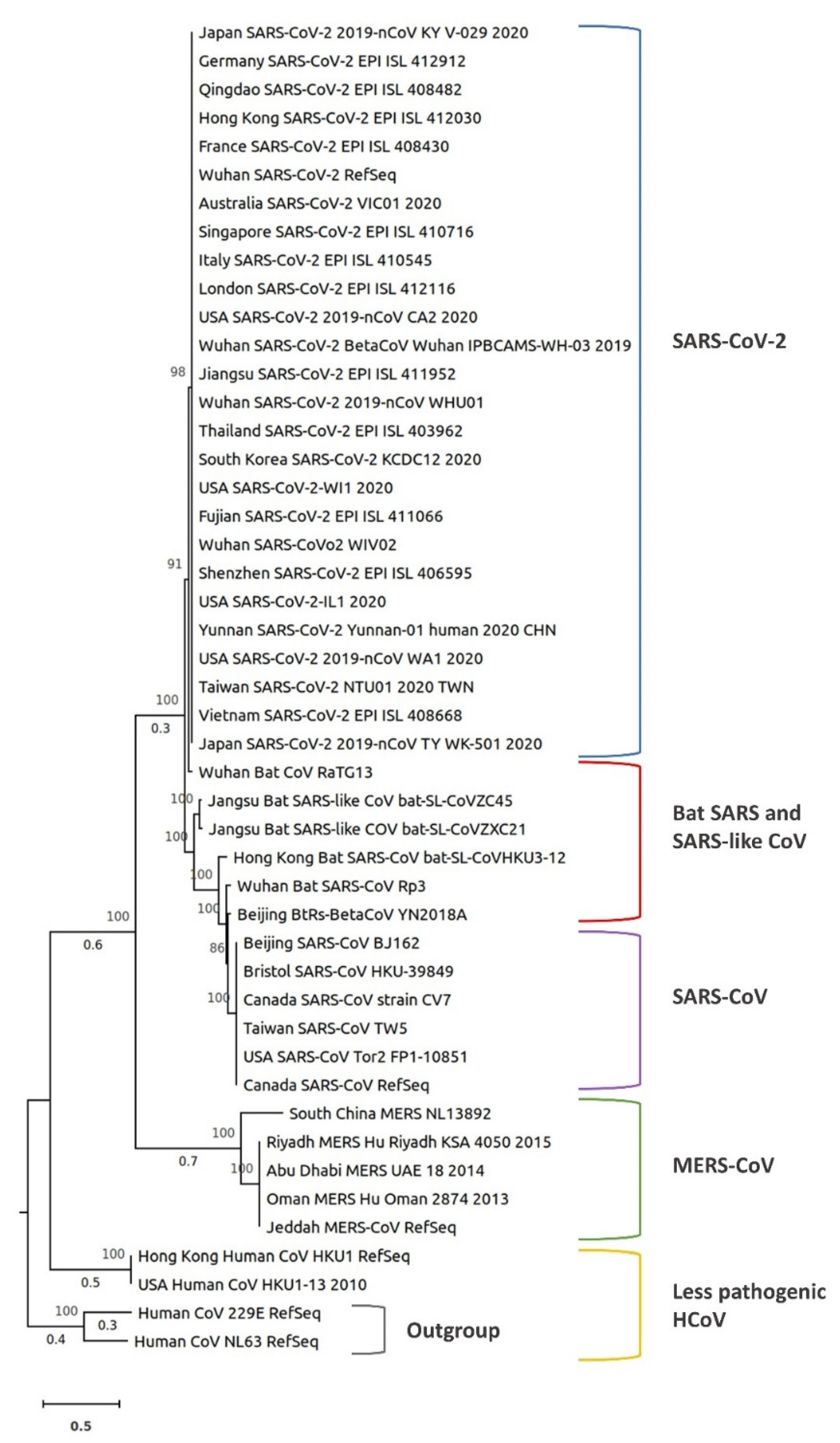
5. Comparative Phylogenetic Analysis of SARS-CoV-2

Single-stranded RNA viruses exhibit a faster biological mutation rate due to the lack of proofreading activity of viral RNA polymerases [48]; however, unlike other mutation-prone RNA viruses, with the exception of the Arenaviridae family, CoVs do have limited proofreading capabilities, with the nsp14 protein allowing for the enhanced genome size of CoV family members [49].
Recombination is another mechanism of evolution in coronaviruses [50]. A high recombination frequency was demonstrated in murine hepatitis virus during mixed infection, where the majority of viruses recovered after three passages were recombinants [51]. Recombination was also reported for MERS-CoV and SARS-CoV. Seven putative recombination regions were detected in ORF1ab and S protein between SARS-CoV and six other coronaviruses by in silico analysis of their genomes [52]. Similarly, bioinformatic analysis of MERS-CoV genomic data revealed 28 recombinant sequences from humans and camels [53]. Recombination in SARS-CoV-2 is not yet clearly understood. Initial studies suggested that it may have occurred in the course of SARS-CoV-2 evolution [9], while other researchers excluded the possibility of recombination based on a full genome evolutionary analysis investigating putative recombination events [11].
To better understand the evolution of SARS-CoV-2, we performed a phylogenetic analysis of 45 representative coronaviruses from 18 countries including SARS-CoV, SARS-CoV-2, HCoV, bat SARS CoV, bat SARS-like CoV, and MERS-CoV. The viral genomes were obtained from the GISAID and NCBI databases. Multiple sequence alignment was performed using kalign 3 [54]. A phylogenetic tree was constructed based on whole-genome sequences (coding sequences of all genes) in IQ-TREE, using the maximum likelihood method, ultrafast bootstrap approximation, and ModelFinder [55,56]. The tree was drawn to scale, with branch lengths measured in the number of substitutions per site. The bootstrap values were determined by 1,000 replicates. The tree was visualized in MEGA X [57] (Figure 2).
In the analysis we performed, all SARS-CoV-2 samples from the 18 countries clustered together and were close to bat SARS or SARS-like coronaviruses, with Wuhan bat CoV RaTG13 being the closest virus. In addition, MERS-CoV and human CoV HKU1 were very distant from SARS-CoV-2 (Figure 2). Within MERS-CoV samples, the South China MERS-NL13892 clustered separately from other MERS-CoVs. The two bat SARS-like CoVs (bat-SL-CoVZC45 and bat-SL-CoVZXC21) were the second closest viruses from bats to SARS-CoV-2 (Figure 2). All SARS-CoVs from China, Canada, England, and the US were in a single cluster.
Zhou et al. [9] conducted a phylogenetic analysis of SARS-CoV-2 against previously identified coronaviruses based on their whole-genome sequences, main structural protein genes, and non-structural protein genes. SARS-CoV-2 clustering was different depending on whether the whole genome or specific genes were used in the analysis. For example, SARS-CoV-2 clustered with the members of the subgenus Sarbecovirus including the SARS-CoV (79.5% identical) that caused the global pandemic in 2003 and other bat SARS-like viruses (96% identical at the whole-genome level), but the topological position within the Sarbecoviruses changed when individual genes (ORF1ab, S, E, M, and N) were used for clustering [2,9].

6. SARS-CoV-2 Mutations and Their Effects

Based on the whole-genome sequence alignment, SARS-CoV-2 shares 89% identity with bat SARS-like CoVZXC21, 82% with SARS-CoV, and 96.3% with bat CoV RaTG13 [11,13]. Alignment of the predicted protein sequences of SARS-CoV-2 to those of SARS-CoV or SARS-like coronaviruses revealed a total of 380 amino acid substitutions between these viruses [2]. These amino acid substitutions were distributed as follows: 348 mutations in nonstructural proteins (ORF1ab, 3a, 3b, 7a, 7b, 9b, and ORF14), 27 in S protein, and 5 in N protein. No amino acid substitutions were detected in E or M proteins, indicating that E and M proteins are highly conserved among these viruses.
It has been reported that SARS-CoV-2 uses the same cellular receptor, hACE2, as SARS-CoV to gain entry into the cell [9,58,59]. The analysis of the receptor-binding domains (RBD) of SARS-CoV and SARS-CoV-2 S protein revealed similar binding affinities [60]. Wu et al. [2] found a total of 27 amino acid substitutions in the S protein but not in the receptor-binding motif (RBM) that directly interacts with hACE2, which may affect host tropism. These 27 substituted residues were distributed as follows: 17 in the S1 subunit [6 in the RBD and 6 in the subdomain (SD)] and 10 in the S2 subunit. Wan et al. [58] reported similarity in the spike protein RBD, including RBM, of both SARS-CoV and SARS-CoV-2, in addition to the presence of several residues in SARS-CoV-2 RBM that favor the interaction with human ACE2. These results agree with the genomic analysis of SARS-CoV-2, according to which the S2 subunit of the spike protein shares 99% identity with those of two bat SARS-like CoVs (SL-CoVZXC21 and ZC45) and of human SARS-CoV [13]. While the SARS-CoV-2 S2 subunit was conserved, the S1 subunit shares an overall 70% identity with those of bat and human SARS-CoV. The RBD core domain of S1 is highly conserved, with most of the amino acid differences located in the external subdomain that is responsible for the direct interaction with host receptors [13]. Investigators have also reported the presence of a polybasic cleavage site and predicted O-linked glycans that are unique to SARS-CoV-2 S protein. Differences in SARS-CoV-2 S protein and the high contagious nature of this virus suggest that SARS-CoV-2 has evolved via natural selection for binding to human ACE2 receptor [61].
ORF3b also differs in SARS-CoV-2. ORF3b deletion mutations in SARS-CoV do not affect viral replication in vitro [62]. ORF3b may play a role in viral pathogenicity in addition to its inhibitory effects on interferon (IFN) expression and signaling [63,64]. Recently, a novel short putative protein was identified in ORF3b of SARS-CoV-2 [13]; however, the function of this novel protein is still not known. SARS-CoV-2 ORF8 is closer to those of bat SARS CoV ZXC21 and ZC45 and distant from that of human SARS-CoV [13].

7. Genetic Diversity of SARS-CoV-2

The assessment of genetic diversity among 86 complete or semi-complete genomes of SARS-CoV-2 viruses revealed three deletions in the genome of isolates from Japan, USA, and Australia in addition to many other substitution mutations. The deletion mutations were in the ORF1ab gene (3-nucleotide and 24-nucleotide deletion) and at the 3′ end of the genome (10-nucleotide deletion). Of the 93 substitution mutations, 42 changed the amino acid sequence of structural and non-structural proteins [65]. The 3- and 24-nucleotide deletions in ORF1ab are expected to reduce the protein sequence by 1 and 8 amino acid residues, respectively, without changing the reading frame, but the functional effects have yet to be investigated.
The alignment of SARS-CoV-2 reference S protein gene against all SARS-CoV-2 sequenced genomes from China, USA, Japan, Australia, and Taiwan revealed 99.97–100% identity, with 100% query coverage (also confirmed by our phylogenetic analysis, Figure 2), while the identity and coverage for SARS-CoV S protein gene were 74.5% and 91%, respectively. Also, the S protein gene from bat SARS and SARS-like coronavirus isolates shared 76.5–83% identity with that of SARS-CoV-2. This agrees with previous conclusions regarding the evolutionary analysis of SARS-CoV-2 [11,44]. In the phylogenetic analysis we performed, SARS-CoV-2 viruses were in the same cluster regardless of the geographic region (Figure 2). These results strongly suggest the possibility of a recent common ancestor for all SARS-CoV-2 or the transmission of the same virus strain across countries.

8. Epidemiology of COVID-19

The outbreak of COVID-19 originated from Wuhan City, Hubei province, in China. Fifty-five percent of the infected cases before 1 January 2020 were linked to the Huanan Seafood Wholesale Market. However, the first human-to-human case of SARS-CoV-2 infection reported on 1 December 2019 did not have any exposure to this market [66,67]. In mid-January 2020, SARS-CoV-2 spread to other provinces of China due to the Spring Festival travel season. SARS-CoV-2 was transmitted from China to other countries via international travelers. On 13 January 2020, the first case of SARS-CoV-2 infection was confirmed outside China in Thailand, and on 16 January 2020 the first infected case was confirmed in Japan. These cases were also linked to the Huanan Seafood Wholesale Market. By 25 January 2020, the number of confirmed cases had risen to 2062, including 2,016 in China, Thailand, Hong Kong, Macau, Australia, Malaysia, Singapore, France, Japan, South Korea, Taiwan, the US, Vietnam, Nepal, and Sweden. On 30 January 2020, China reported a sharp rise in the number of infected cases, with the presence of infection in more than 18 countries. Therefore, WHO declared the SARS-CoV-2 outbreak to be a Public Health Emergency of International Concern [68].
As of 16 March 2020, more than 150 countries and territories have been affected, with major outbreaks in central China, South Korea, Italy, Iran, France, and Germany [69]. There were 167,511 confirmed cases of SARS-CoV-2 infections, with 6606 deaths and about 8% estimated mortality rate. More than 73% of these cases have been reported in mainland China [69]. At this time, the number of global cases has shown a drastic increase within a short time, confirmed cases and deaths in China have not increased too much, while confirmed cases and deaths in other countries have drastically increased (Table 4). The number of confirmed cases increased from 2798 to 17,391 in one week (between 27 January and 3 February), and the number of infected countries doubled (from 12 to 24). Due to the rapid increase of the number of infected cases and infected countries, the WHO declared SARS-CoV-2 a pandemic on 11 March 2020 and on 13 March 2020, the WHO declared Europe to be the new center of the pandemic due to the massive increase of confirmed cases there [70]. On 23 March 2020, Italy reported the highest number of deaths (5560) followed by China (3276), Spain (1720), and Iran (1685). One week later (30 March 2020), the global map of COVID-19 had changed. For example, the highest number of cases was reported in the USA (122,653 cases; 2112 deaths) followed by Italy (97,689 cases; 10,781 deaths), China (82,447 cases; 3310 deaths), Spain (78,797 cases; 6528 deaths), Germany (57,298 cases; 455 deaths), France (39,642 cases; 2602 deaths), and Iran (38,309 cases; 2640 deaths). As of 6 April 2020, there were 1,210,956 confirmed cases of SARS-CoV-2 infection (most of the cases, 307,318, were in the USA) and 67,594 deaths (most of the deaths, 15,889, were in Italy). One week later (13 April 2020), the number of confirmed cases of SARS-CoV-2 increased 1.7 times (up to 524,514 confirmed cases), and the number of deaths increased 2.5 times (up to 20,444 deaths) in the USA alone. The number of confirmed cases, deaths, and infected countries are shown in Table 4.

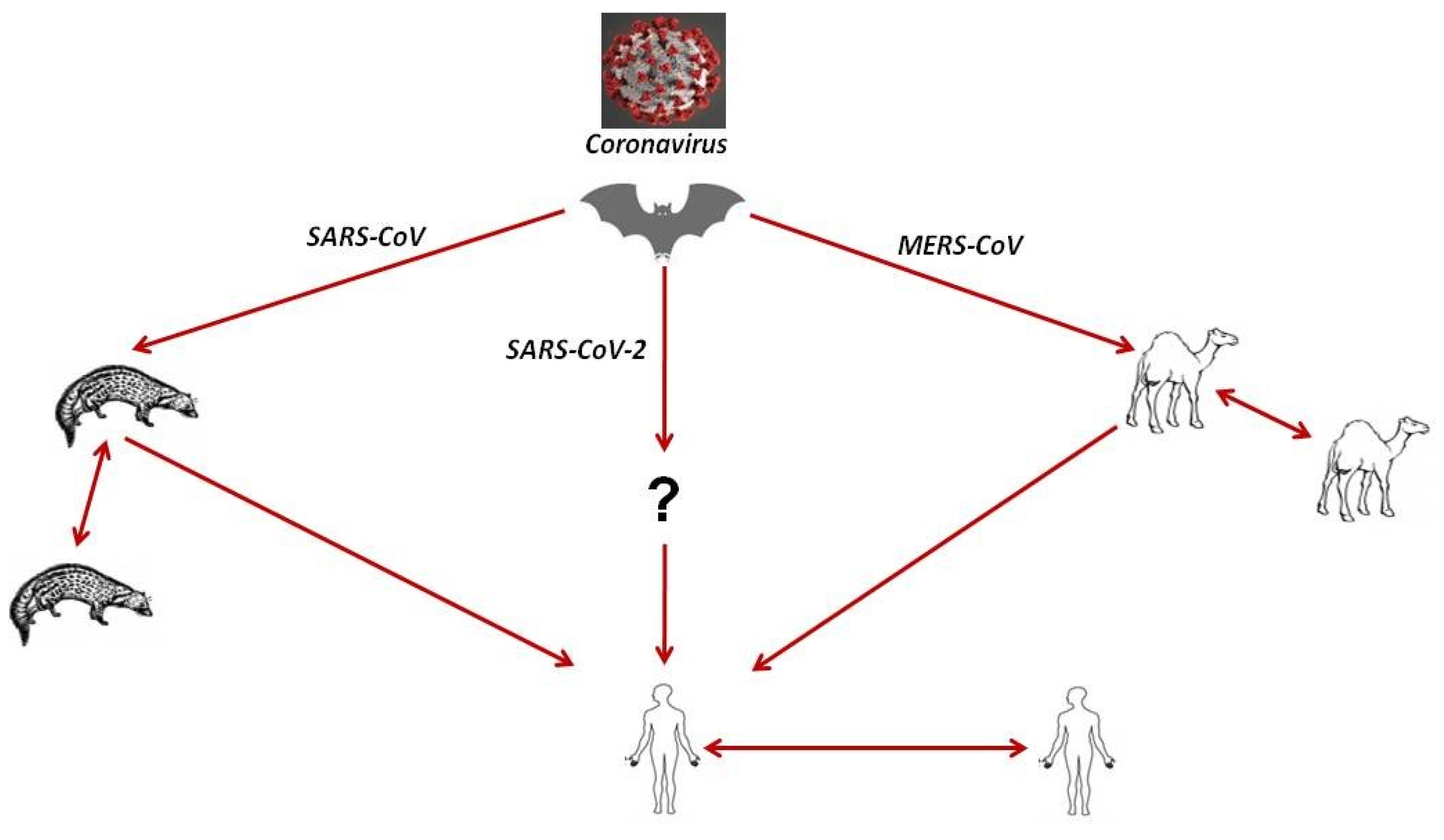
8.1. Source of Infection and Evolution of SARS-CoV-2

The origins of more than 75% of coronavirus infections are considered zoonotic, i.e., animals are the main source of the outbreaks. For example, SARS-CoV was transmitted from palm civets to humans, and MERS-CoV from dromedary camels to humans. Bats are currently considered a reservoir for all human coronaviruses, as mentioned above [5,71]. Many coronaviruses are circulating in animals but have not yet infected humans. The type of animal that SARS-CoV-2 originated from is still unclear. At the beginning of the outbreak in Wuhan, China, many patients were linked to the Huanan Seafood Wholesale Market, suggesting animal-to-person spread. After retrospectively studying case reports, the number of patients that did not have exposure to animal markets has risen, indicating person-to-person spread was also occurring at that time [66]. SARS-CoV-2 is closely related to bat coronaviruses and SARS-CoV [8]. A group of researchers reported early in the outbreak that the novel SARS-CoV-2 has the highest similarity of codon usage bias with snakes [72,73]; however, this method to determine initial host origins is dubious. Interestingly, researchers also reported one amino acid difference in the receptor-binding domain of the S protein of Pangolin-CoV compared to that of SARS-CoV-2, suggesting that pangolins might play a role as an intermediate host (Xiao et al., data currently under review). Another group of researchers reported that the virus originated from bats based on the genome sequence of SARS-CoV-2, which is 96% identical to bat coronavirus RaTG13. There were speculations that SARS-CoV-2 is a laboratory-engineered CoV and leaked directly from a laboratory in Wuhan where a bat CoV (RaTG13) was recently reported. However, there is no evidence to support this allegation [74]. Recently, a group of researchers found that SARS-CoV-2 replicates poorly in dogs, pigs, chickens, and ducks but efficiently in ferrets and cats [75]. Scientists are still trying to find the main source of the disease outbreak and identify the definitive intermediate hosts.

8.2. Mode of SARS-CoV-2 Transmission to Humans: Transmission Dynamics and Virus Persistence

Both established (SARS-CoV, MERS-CoV) and novel (SARS-CoV-2) coronaviruses were reported to spread from an infected person to a non-infected person through direct or indirect contact. SARS-CoV-2 infection was reported to be transmitted directly from person to person like most respiratory viruses via close contact with an infected person or through respiratory droplets (aerosol) produced when an infected person coughs or sneezes. These droplets can be inhaled to reach the lung. The virus can be indirectly transmitted via touching a surface or an object that was previously contaminated with the virus and then touching the face, eyes, or mouth [76] and possibly via the fecal–oral route [77,78]. Asymptomatic carriers (during the incubation period of the virus) and patients after recovery from the acute form of the disease are also considered a potential source of virus transmission to healthy persons [10,12]. Interestingly, human coronaviruses are able to survive on steel, metal, wood, aluminum, paper, glass, plastic, ceramic, disposable gowns, and surgical gloves for 2–9 days. High temperature (≥30 °C) can reduce the persistence period, while low temperature (4 °C) increases the persistence time up to 28 days [79]. Transmission of the virus vertically from mother to fetus or via breast milk has not been confirmed yet [80]. The transmission cycle of coronavirus among animals and humans is shown in Figure 3.

8.3. Risk Factors for SARS-CoV-2 Infection and Their Assessment

There are many factors that affect SARS-CoV-2 transmission and spread. These factors include, but are not limited to: (1) travel to or contact with individuals who have recently visited Wuhan, China, or other places experiencing an outbreak; (2) close contact with persons who are diagnosed positive for the disease, such as healthcare workers caring for patients with SARS-CoV-2; (3) contact with droplets and secretions (produced by sneezing or coughing) from an infected person and eating or handling wild animals native to China such as bats. Additionally, the risk of infection is higher for the elderly and for patients suffering from pre-existing illnesses such as cardiovascular disease, hypertension, diabetes, and chronic respiratory disease [66]. The reported fatality rate based on age is 14.8% for people ˃80 years of age, 8% for people between 70 and 79 years, 3.6% for people between 60 and 69 years, 1.3% for people between 50 and 59 years, 0.4% for people between 40 and 49 years, 0.2% for people between 10 and 39 years; no fatalities have been reported for children under 10 years of age. Notably, the fatality rate is higher in males (2.8%) than in females (1.7%) [81,82].

8.4. Clinical Characteristics and Susceptibility of SARS-CoV-2 Infection in Humans

The estimated incubation period of the novel coronavirus ranges from 2 to 14 days. However, some cases had an incubation period of 21, 24, or 27 days [83]. The complete clinical picture of SARS-CoV-2 is still unclear. The disease begins with flu-like symptoms that include fever, fatigue, dry cough, sore throat, shortness of breath, headache, chest tightness, chest pain, and muscle pain. Some of SARS-CoV-2 patients have runny nose, nausea, vomiting, and diarrhea [84]. People can be infected without showing symptoms, which allows the virus to spread more effectively from person to person. Complications can occur due to COVID-19 leading to severe infections, such as pneumonia (infection of the lungs), kidney failure, and death [8]. The mild phase of the disease can last up to 2 weeks, while severe or critical disease lasts approximately 3 to 6 weeks (this analysis was conducted on 55,924 confirmed cases). Additionally, the time from the disease onset to the development of severe disease is one week, while the time from the onset of symptoms to death ranges from 2 to 8 weeks [82].
Based on the data analysis of 72,314 confirmed cases of SARS-CoV-2 in Wuhan City, China, by 11 February, 80.9% of the cases were mild with flu-like symptoms, and patients recovered at home, 13.8% were severe with pneumonia and shortness of breath, 4.7% were critical with respiratory failure and septic shock resulting in organs failure, and approximately 2% of the cases were fatal [85]. Another study was conducted on 99 hospitalized patients, and symptoms were classified as follow: fever (83%), cough (82%), shortness of breath (31%), muscle ache (11%), confusion (9%), headache (8%), sore throat (5%), runny nose (4%), chest pain (2%), diarrhea (2%), and nausea and vomiting (1%) [8].

9. Diagnosis of SARS-CoV-2 Infection

The rapid diagnosis of SARS-CoV-2 infection is the cornerstone of disease control. It depends on several criteria including case history, clinical symptoms, serology, molecular diagnosis, and computed tomography (CT) imaging. On 2 March 2020, WHO published interim guidance for laboratory testing of suspected human cases, with precautions for specimen collection, packing, shipment, and amplification of nucleic acid to detect viral genes (N, E, S, and RdRp) [82]. SARS-CoV-2 uses the same cell entry receptor, hACE2, as SARS-CoV. Therefore, oral swabs, bronchoalveolar lavage fluid (BALF), blood, as well as anal swabs are the best samples used for virus diagnosis [86]. A proper diagnosis depends primarily on the factors described below.

9.1. Epidemiological History

The strict monitoring of case history in clinically suspicious patients is considered the first step in the early diagnosis of SARS-CoV-2 infection. Clinically suspicious patients are those who suffer from fever and lower respiratory tract infection symptoms (for details, see the clinical characteristics section) and reside within or have traveled to endemic regions or had close contact with a confirmed or suspected case. Additionally, SARS-CoV-2 can be transmitted by symptomatic and asymptomatic patients especially to the high-risk group mentioned above (for details, see the risk assessment section) [13].

9.2. Laboratory Diagnosis

The blood profiles of patients suffering from SARS-CoV-2 infection revealed the following: (1) increased C-reactive protein and erythrocytes, (2) increased myohemoglobin, liver enzymes, and muscle enzymes, with a high level of D-dimer in severe cases, and (3) normal or decreased white blood cell counts and lymphocytes in the early stage of the disease, with advanced lymphocytopenia in severe cases [13]. In ICU patients, high levels of plasma granulocyte colony-stimulating factor (GCSF), IP10, IL2, IL7, IL10, TNF-α, and MIP1a were reported [38].

9.3. Virus Detection

Electron microscope examination of SARS-CoV-2 revealed the typical coronavirus morphology. Further, SARS-CoV-2 was successfully isolated from human respiratory epithelial cells or BALF samples of infected patients using Huh7 cells and Vero E6 cells. The isolated strain was confirmed by immunofluorescent antibody techniques using the cross-reactive nucleoprotein (NP) antibody. Serum neutralization tests (SNT) using Vero E6 cells were conducted to confirm the neutralization activity in IgG-positive viral samples [87].

9.4. Serological and Molecular Diagnosis

IgM and IgG ELISA detection kits using bat SARSr-CoV Rp3 NP were developed with no cross-reaction against human coronaviruses except SARSr-CoV [80]. Using these serological tools, viral antibody titers were increased in SARS-CoV-2-infected patients [38]. The procedures of ELISA for the determination of SARS-CoV-2 IgG were described before [86]. Nucleic acid detection is the main, fastest, and most sensitive test for the diagnosis of SARS-CoV-2 infection. Recently, two nested RT-PCR and two real-time RT-PCR assays have been developed with successful detection of the first 25 positive cases of infection in Japan [88]. Three real-time RT-PCR techniques have been designed based on the E, RdRp, and N genes [89]. Also, scientists established molecular detection tools for SARS-CoV-2 based on the S gene [86].

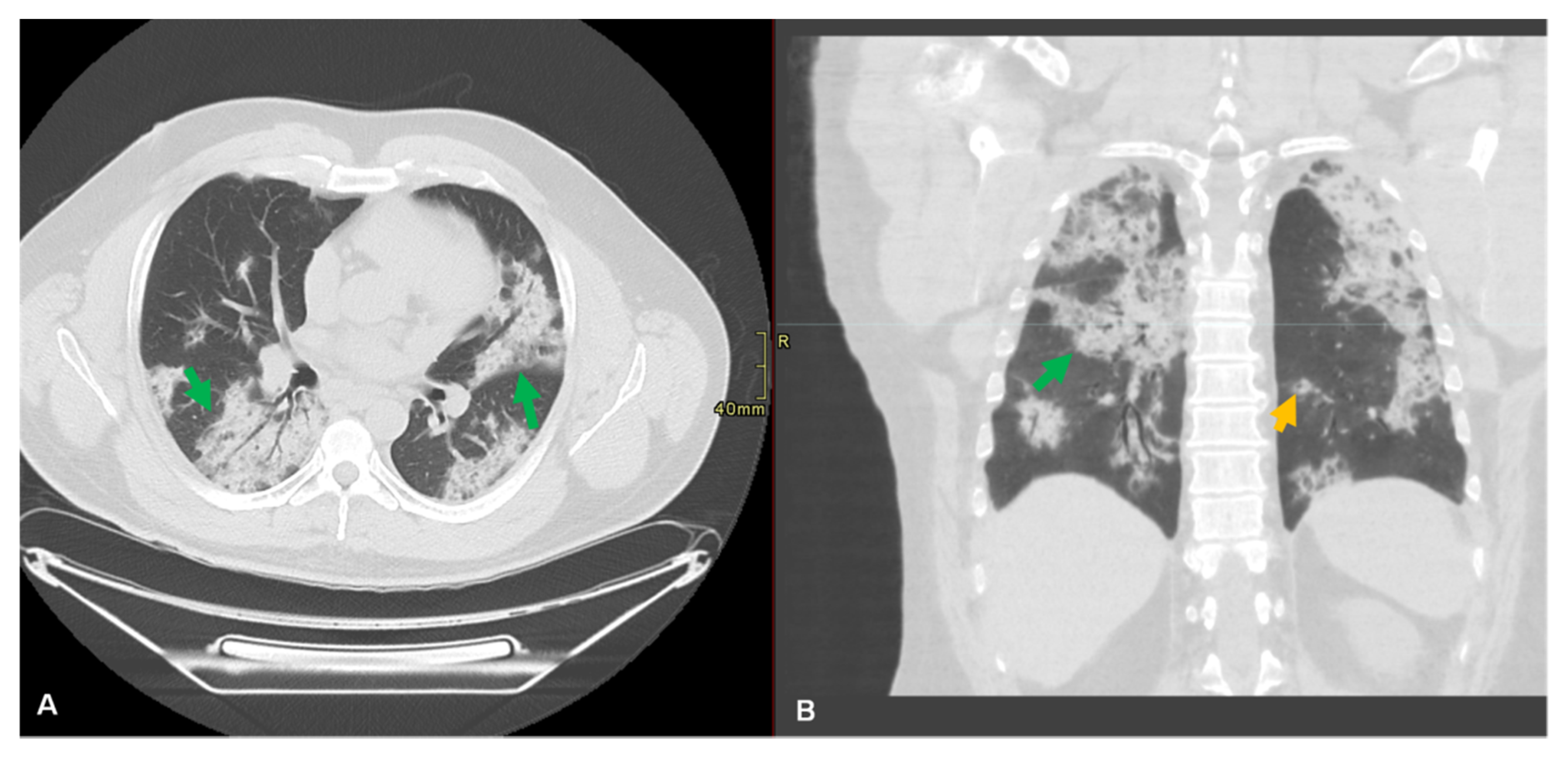
9.5. Radiological Diagnosis

Chest X-ray examination in the early stage of the disease shows interstitial changes and multiple small plaque shadows. Chest CT scans play an important role in the diagnosis of acute respiratory disease syndrome (ARDS) and pneumonia as well as in the early detection of lung parenchymal abnormalities in patients at risk and provide an impression of secondary infection (Figure 4).
Assessing these lungs parenchymal abnormalities conveys disease severity to clinicians. Using artificial intelligence models in the future may be useful in mass screening, to allow risk prioritization and help to minimize turnaround time [90]. Pan et al. [91] conducted a retrospective study to elaborate the time course of lung changes during recovery from infection. They described findings using international standard nomenclatures such as ground-glass opacity (GGO), consolidation, and crazy paving patterns. They established a semi-quantitative scoring system of 5 grades to quantify the degree of involvement based on an area ranging from 0% to >75%. The total score ranged from 0 to 25 (max), and involvement was subpleural, random, or diffuse. They found that in early stages (0–4 days after the onset of symptoms), GGO was the main finding in lower lung lobes; in progressive stages (5–8 days), the progression of lung disease involved three patterns of ground-glass, consolidation, and crazy paving, while in peak stages (9–13 days), dense consolidation became the prevalent feature; in absorption stages (>14 days), GGO was detected with no crazy paving and resolution of consolidations [91]. More than 75% of SARS-CoV-2-affected patients suffered from bilateral lung involvement, and 71% have multilobe involvement. CT examinations of 21 patients showed 29% consolidation and 86% GGO in the chest [8,9,92,93]. Another study examined 51 cases by CT and reported that 77% showed pure GGO, 75% exhibited GGO with reticular and/or interlobular septal thickness, 59% had GGO with consolidation, while 55% revealed pure consolidation. Bilateral lung involvement was reported in 86% of cases; in 80% of the cases the posterior part of the lung was involved, while in 86%, the periphery was involved [94].

10. Control and Treatment of COVID-19 Infection

In January 2020, the WHO issued guidance for the clinical management of SARS when SARS-CoV-2 infection was suspected. In this guidance, the start of emergency treatments, immediate implementation of prevention and control strategies, early supportive therapy and prevention of SARS-CoV-2 complications were described in detail [15]. So far, there are no approved specific antiviral drugs for SARS-CoV-2 infection. Therefore, preventive measures and inactivation of the virus are essential to stop and control the spread of the disease. Human coronaviruses can be inactivated using 0.5% hydrogen peroxide, 62–71% ethanol, 0.1% sodium hypochlorite, 0.7–1% formaldehyde, 2% glutaraldehyde, or 0.23% povidone iodine within 1 minute. Other disinfectants such as 0.02% chlorhexidine digluconate, 0.55% orthophtalaldehyde, or 0.05–0.2% benzalkonium chloride are less effective [79].
In light of the urgent clinical demand, many drugs are approved to be used for clinical trials against SARS-CoV-2 infection, such as lopinavir/ritonavir, arbidol, interferon-alpha, favipiravir, chloroquine phosphate, darunavir/cobicistat, oseltamivir, and methylprednisolone. The most used antiviral drugs [95] are summarized in Table 5.
Generally, coronaviruses are not sensitive to current antiviral drugs, and high concentrations of drugs effective on these viruses cannot be used in vivo. Therefore, combinations of different therapies have been used for the treatment of coronavirus infections [96]. Some drug combinations that could be successful for the treatment of SARS-CoV-2 patients are lopinavir and ritonavir [97,98], lopinavir/ritonavir plus arbidol [99], and ribavirin and interferon [100,101]. The use of anti-inflammatory drugs such as glucocorticoids, IL-6 antagonist, janus kinase inhibitors (JAK), and choloroquine/hydrocholoroquine in SARS-CoV-2 patients is a dilemma, especially in patients suffering from an impaired immune system. Balancing the risk–benefit ratio is a critical issue. Corticosteroids may delay the elimination of the virus and increase the risk of secondary infection. In addition, drugs targeting pro-inflammatory cytokines can only inhibit specific inflammatory factors and thus may not be very effective in curbing the cytokine storm (excessive and uncontrolled release of pro-inflammatory cytokines). Moreover, some anti-inflammatory drugs such as JAK block INF-α production, which is important in fighting the virus [102].
Additionally, fecal transplantation was approved for clinical trials as a therapeutic option for SARS-CoV-2-related pneumonia based on the promising results obtained from fecal microbiota transplantation in patients suffering from antibiotic-associated diarrhea, active ulcerative colitis, and other viral infections [103,104,105,106]. Recently, it was found that intestinal microbiota-derived IFN in lung stroma confers protection against viral diseases such as avian influenza and respiratory syncytial virus [103]. Moreover, based on historical records of the effect of antiviral herbs on SARS and influenza H1N1, Chinese herbal formulas could be an alternative approach for the prevention of SARS-CoV-2 in a high-risk population [107], if no scientifically based therapeutics are available. It was found that Sambucus formasana Nakai exhibited a strong antiviral effect against human coronavirus NL63 [108].

11. Vaccination

To date, there is no vaccine to prevent SARS-CoV-2 infection, and trials for vaccine development are in the preliminary stages of research. Several vaccine candidates such as live attenuated, adenovirus-vectored, recombinant protein, and nucleic acid (DNA and mRNA) vaccines are in the pipeline [123].

12. Preventive Measures to Control the SARS-CoV-2 Spread

The epidemiology of SARS-CoV-2 is still unclear, and data availability is limited. Therefore, it is imperative to follow preventive measures and safety precautions issued by health authorities to limit exposure to the virus and to reduce further spread. General hygienic measures should be implemented, such as (1) washing hands often with soap and water or an alcohol-based hand sanitizer, (2) cough or sneeze etiquette, recommending covering of the mouth, (3) avoiding touching eyes, nose, and mouth if the hands are not clean, (4) avoiding close contact with sick persons, (5) avoiding sharing dishes, glasses, bedding, and other household items with sick people, (6) cleaning and disinfection of surfaces that are often touched, and (7) staying home from work, school, and public areas when feeling sick.
The transmission route of SARS-CoV-2 is probably not only through cough, respiratory droplets, and/or contaminated surfaces [13,124], but also through fecal–oral transmission [78]. Therefore, strict hygienic measures should be followed, especially in dense cities or agricultural spaces [125].
Since the SARS-CoV-2 spread is primarily driven by travel, screening of travelers who arrive at airports from pandemic areas for possible SARS-CoV-2 infection and entry-screening procedures are necessary. Also, general hygienic precautions during travel are highly recommended. Travelers who suffered from acute respiratory infection should be tested and reported to the respective public health authorities [73]. In addition, people should be motivated to notify and report about travel history and close contacts in case of SARS-CoV-2 infection.
Asymptomatic carriers (during the incubation period) and patients after recovery from the acute form are also considered potential sources of the virus [12,13]. Strict hygienic measures should be implemented to avoid virus transmission to healthcare workers and other contacts, i.e., placement of SARS-CoV-2 suspected or confirmed patients in single-person rooms and wearing personal protection equipment (PPE) such as masks, goggles, and protective gowns. Because early diagnosis and detection of asymptomatic carriers of SARS-CoV-2 are successful factors for the treatment and prevention of transmission, health authorities should designate laboratories to implement tests for a rapid and accurate diagnosis [126]. The control of coronaviruses is based on biosecurity regarding animals as well as on shifts in food habits, including discouraging the consumption of bushmeat and of animal products without appropriate cooking [127]. Ban of wet marketplaces where live or dead animals are handled should be implemented. Surveillance among people who have contact with wildlife and improvement of biosecurity regarding wildlife trade are urgently needed to prevent the next pandemic outbreak [128].

13. Challenges to Control SARS-CoV-2 and the Role of the One-Health Approach in Disease Control

The epidemiology of SARS-CoV-2 is still unclear. Many unresolved questions related to SARS-CoV-2 epidemiology and pathogenicity pose great challenges for researchers. These unresolved questions include: What is the origin of SARS-CoV-2? What is the intermediate host that transmitted the virus from bats? Why does the virus cause severe disease and mortality in the elderly or those with co-morbidities, while it is milder in children? Are aerosol, saliva, feces, urine, and foodborne the only routes of transmission? What are the other unknown routes of transmission?
Control of the SARS-CoV-2 outbreak and future epidemics requires global efforts among medical and veterinary clinicians, diagnosticians, epidemiologists, public health experts, vaccinologists, pharmaceutical industries, economists, and governments to implement a One-Health approach [128,129]. These measures must include: (1) writing policies and supporting funds required for the implementation of One Health, prevention, and control measures, (2) hiring well-trained and professional personnel, (3) performing rapid and accurate diagnosis and treatment of infected persons, (4) developing and providing vaccines for virus control in humans, (5) conducting surveillance among wildlife for the identification and characterization of possible reservoirs and surveillance among people who are in contact with wildlife to identify risk factors in human behaviors and living environment, (6) improving hygienic measures, (7) assessing the social and economic impacts of COVID-19 on the population, (8) utilizing veterinary experience in the disinfection of premises and gatherings under the supervision of health authorities to decrease outbreaks in humans, (9) providing antiviral drugs for the treatment of the disease in humans, and (10) increasing public health awareness about the virus and its transmission.

14. Conclusions and Future Perspective

The SARS-CoV-2 outbreak started in Wuhan City, China, in December 2019. It is now a global pandemic, with 1,773,084 confirmed cases, 111,652 deaths, and 467,074 recoveries (as of 13 April 2020). The virus has the potential for rapid and extensive spread between people and countries. There are a lot of misleading information and knowledge gaps on the newly emerged SARS-CoV-2. Therefore, we reviewed the latest updates about different aspects including epidemiology, source of infection, transmission dynamics, zoonotic potential, virus characteristics, and discovery of novel strategies for disease control to avoid spillover of infection in the future. Bats play an important role in the transmission of the infection to humans. Coronaviruses are genetically diverse and have a high tendency towards frequent genetic mutations and gene recombination, which increases the risk of interspecies transmission. Information about the incubation period can help in establishing an effective quarantine for asymptomatic carriers, thus preventing the virus spread. From our perspectives and based on the currently available information about the virus and its epidemiology, the control of the SARS-CoV-2 requires an effective and global disease coordination effort including multidisciplinary research efforts (One-Health approach) through collaboration between governments, epidemiologists, virologists, health authorities, veterinarians, and physicians. At this stage of the disease outbreak, developing vaccines is crucial to limit the spread of the infection.

Author Contributions

Conceptualization, Y.A.H. and M.F.; methodology Y.A.H. and M.F.; software Y.A.H., M.F., A.E. and A.S.; validation, Y.A.H., M.F. and A.E.; formal analysis, Y.A.H., M.F. and A.E..; data curation, Y.A.H., M.F., and A.E.; writing—original draft preparation, Y.A.H, M.F., A.E., A.S., S.P.K. and A.A.S..; writing—review and editing, Y.A.H., M.F., A.E., A.S., S.P.K., and A.A.S.; visualization, investigation, and supervision, Y.A.H. All authors have read and agreed to the published version of the manuscript.

Funding

This research received no external funding.

Acknowledgments

We gratefully acknowledge the authors, originating, and submitting laboratories of the sequences from the GISAID’s EpiCoV™ Database, on which part of the phylogenetic tree construction is based.

Conflicts of Interest

The authors declare no conflict of interest.

References

  1. Decaro, N.; Mari, V.; Elia, G.; Addie, D.D.; Camero, M.; Lucente, M.S.; Martella, V.; Buonavoglia, C. Recombinant canine coronaviruses in dogs, Europe. Emerg. Infect. Dis. 2010, 16, 41–47. [Google Scholar] [CrossRef] [PubMed]
  2. Wu, A.; Peng, Y.; Huang, B.; Ding, X.; Wang, X.; Niu, P.; Meng, J.; Zhu, Z.; Zhang, Z.; Wang, J. Genome composition and divergence of the novel coronavirus (2019-nCoV) originating in China. Cell Host Microbe 2020, 27, 325–328. [Google Scholar] [CrossRef] [PubMed] [Green Version]
  3. Guan, Y.; Zheng, B.; He, Y.; Liu, X.; Zhuang, Z.; Cheung, C.; Luo, S.; Li, P.; Zhang, L.; Guan, Y. Isolation and characterization of viruses related to the SARS coronavirus from animals in southern China. Science 2003, 302, 276–278. [Google Scholar] [CrossRef] [PubMed] [Green Version]
  4. Drosten, C.; Meyer, B.; Müller, M. Supplement to: Transmission of MERS-coronavirus in household contacts. N. Engl. J. Med. 2014, 371, 1–10. [Google Scholar] [CrossRef]
  5. Cui, J.; Li, F.; Shi, Z.-L. Origin and evolution of pathogenic coronaviruses. Nat. Rev. Microbiol. 2019, 17, 181–192. [Google Scholar] [CrossRef] [Green Version]
  6. WHO. Summary of Probable SARS Cases with Onset of Illness from 1 November 2002 to 31 July 2003. Available online: https://www.who.int/csr/sars/country/table2004_04_21/en/ (accessed on 10 March 2020).
  7. WHO. MERS Situation Update, January 2020. Available online: http://www.emro.who.int/health-topics/mers-cov/mers-outbreaks.html (accessed on 11 March 2020).
  8. Huang, C.; Wang, Y.; Li, X.; Ren, L.; Zhao, J.; Hu, Y.; Zhang, L.; Fan, G.; Xu, J.; Gu, X.; et al. Clinical features of patients infected with 2019 novel coronavirus in Wuhan, China. Lancet 2020, 395, 497–506. [Google Scholar] [CrossRef] [Green Version]
  9. Zhou, P.; Yang, X.-L.; Wang, X.-G.; Hu, B.; Zhang, L.; Zhang, W.; Si, H.-R.; Zhu, Y.; Li, B.; Huang, C.-L. A pneumonia outbreak associated with a new coronavirus of probable bat origin. Nature 2020, 579, 270–273. [Google Scholar] [CrossRef] [Green Version]
  10. Chan, J.F.; Yuan, S.; Kok, K.H.; To, K.K.; Chu, H.; Yang, J.; Xing, F.; Liu, J.; Yip, C.C.; Poon, R.W.; et al. A familial cluster of pneumonia associated with the 2019 novel coronavirus indicating person-to-person transmission: A study of a family cluster. Lancet 2020, 395, 514–523. [Google Scholar] [CrossRef] [Green Version]
  11. Paraskevis, D.; Kostaki, E.G.; Magiorkinis, G.; Panayiotakopoulos, G.; Sourvinos, G.; Tsiodras, S. Full-genome evolutionary analysis of the novel corona virus (2019-nCoV) rejects the hypothesis of emergence as a result of a recent recombination event. Infect. Genet. Evol. 2020, 79, 104212. [Google Scholar] [CrossRef]
  12. Rothe, C.; Schunk, M.; Sothmann, P.; Bretzel, G.; Froeschl, G.; Wallrauch, C.; Zimmer, T.; Thiel, V.; Janke, C.; Guggemos, W. Transmission of 2019-nCoV infection from an asymptomatic contact in Germany. N. Engl. J. Med. 2020, 382, 970–971. [Google Scholar] [CrossRef] [Green Version]
  13. Chan, J.F.; Kok, K.H.; Zhu, Z.; Chu, H.; To, K.K.; Yuan, S.; Yuen, K.Y. Genomic characterization of the 2019 novel human-pathogenic coronavirus isolated from a patient with atypical pneumonia after visiting Wuhan. Emerg. Microbes Infect. 2020, 9, 221–236. [Google Scholar] [CrossRef] [PubMed] [Green Version]
  14. Li, J.-Y.; You, Z.; Wang, Q.; Zhou, Z.-J.; Qiu, Y.; Luo, R.; Ge, X.-Y. The epidemic of 2019-novel-coronavirus (2019-nCoV) pneumonia and insights for emerging infectious diseases in the future. Microb. Infect. 2020, 22, 80–85. [Google Scholar] [CrossRef] [PubMed]
  15. WHO. Clinical Management of Severe Acute Respiratory Infection When Novel Coronavirus (nCoV) Infection Is Suspected. Available online: https://www.who.int/publications-detail/clinical-management-of-severe-acute-respiratory-infection-when-novel-coronavirus-(ncov)-infection-is-suspected (accessed on 17 March 2020).
  16. Cowling, B.J.; Leung, G.M. Epidemiological research priorities for public health control of the ongoing global novel coronavirus (2019-nCoV) outbreak. Euro Surveill. 2020, 25, 2000110. [Google Scholar] [CrossRef] [PubMed]
  17. Forni, D.; Cagliani, R.; Clerici, M.; Sironi, M. Molecular evolution of human coronavirus genomes. Trends Microbiol. 2017, 25, 35–48. [Google Scholar] [CrossRef] [Green Version]
  18. Beaudette, F.R.; Hudson, C.B. Cultivation of the virus of infectious bronchitis. J. Am. Vet. Med. A. 1937, 90, 51–56. [Google Scholar]
  19. Cheever, F.S.; Daniels, J.B.; Pappenheimer, A.M.; Bailey, O.T. A murine virus (JHM) causing disseminated encephalomyelitis with extensive destruction of myelin: I. Isolation and biological properties of the virus. J. Exp. Med. 1949, 90, 181–194. [Google Scholar] [CrossRef] [Green Version]
  20. Kahn, J.S.; McIntosh, K. History and recent advances in coronavirus discovery. Pediatr. Infect. Dis. J. 2005, 24, S223–S227. [Google Scholar] [CrossRef]
  21. Tyrrell, D.; Bynoe, M. Cultivation of viruses from a high proportion of patients with colds. Lancet 1966, 1, 76–77. [Google Scholar] [CrossRef]
  22. Hamre, D.; Procknow, J.; Medicine. A new virus isolated from the human respiratory tract. Proc. Soc. Exp. Biol. Med. 1966, 121, 190–193. [Google Scholar] [CrossRef]
  23. Tyrrell, D.; Almeida, J.; Cunningham, C.; Dowdle, W.; Hofstad, M.; McIntosh, K.; Tajima, M.; Zakstelskaya, L.Y.; Easterday, B.; Kapikian, A. Coronaviridae. Intervirology 1975, 5, 76–82. [Google Scholar] [CrossRef]
  24. McIntosh, K.; Becker, W.B.; Chanock, R.M. Growth in suckling-mouse brain of “IBV-like” viruses from patients with upper respiratory tract disease. Proc. Natl. Acad. Sci. USA 1967, 58, 2268–2273. [Google Scholar] [CrossRef] [PubMed] [Green Version]
  25. Lai, M.M.; Perlman, S.; Anderson, L. Coronaviridae. In Fields Virology; Knipe, D.M., Howley, P.M., Eds.; Lippincott Williams & Wilkins: Philadelphia, PA, USA, 2007; Volume 1, pp. 1305–1318. [Google Scholar]
  26. Hu, B.; Zeng, L.-P.; Yang, X.-L.; Ge, X.-Y.; Zhang, W.; Li, B.; Xie, J.-Z.; Shen, X.-R.; Zhang, Y.-Z.; Wang, N. Discovery of a rich gene pool of bat SARS-related coronaviruses provides new insights into the origin of SARS coronavirus. PLoS Pathog. 2017, 13, e1006698. [Google Scholar] [CrossRef] [PubMed]
  27. Zaki, A.M.; Van Boheemen, S.; Bestebroer, T.M.; Osterhaus, A.D.; Fouchier, R.A. Isolation of a novel coronavirus from a man with pneumonia in Saudi Arabia. N. Engl. J. Med. 2012, 367, 1814–1820. [Google Scholar] [CrossRef] [PubMed]
  28. Chu, D.K.; Hui, K.P.; Perera, R.A.; Miguel, E.; Niemeyer, D.; Zhao, J.; Channappanavar, R.; Dudas, G.; Oladipo, J.O.; Traoré, A. MERS coronaviruses from camels in Africa exhibit region-dependent genetic diversity. Proc. Natl. Acad. Sci. USA 2018, 115, 3144–3149. [Google Scholar] [CrossRef] [Green Version]
  29. Lau, S.K.; Li, K.S.; Tsang, A.K.; Lam, C.S.; Ahmed, S.; Chen, H.; Chan, K.-H.; Woo, P.C.; Yuen, K.-Y. Genetic characterization of Betacoronavirus lineage C viruses in bats reveals marked sequence divergence in the spike protein of pipistrellus bat coronavirus HKU5 in Japanese pipistrelle: Implications for the origin of the novel Middle East respiratory syndrome coronavirus. J. Virol. 2013, 87, 8638–8650. [Google Scholar]
  30. Dudas, G.; Rambaut, A. MERS-CoV recombination: Implications about the reservoir and potential for adaptation. Virus Evol. 2016, 2. [Google Scholar] [CrossRef] [Green Version]
  31. Wang, Y.; Liu, D.; Shi, W.; Lu, R.; Wang, W.; Zhao, Y.; Deng, Y.; Zhou, W.; Ren, H.; Wu, J. Origin and possible genetic recombination of the Middle East respiratory syndrome coronavirus from the first imported case in China: Phylogenetics and coalescence analysis. mBio 2015, 6, e01280-15. [Google Scholar] [CrossRef] [Green Version]
  32. Raj, V.S.; Mou, H.; Smits, S.L.; Dekkers, D.H.; Müller, M.A.; Dijkman, R.; Muth, D.; Demmers, J.A.; Zaki, A.; Fouchier, R.A. Dipeptidyl peptidase 4 is a functional receptor for the emerging human coronavirus-EMC. Nature 2013, 495, 251–254. [Google Scholar] [CrossRef] [Green Version]
  33. Luo, C.-M.; Wang, N.; Yang, X.-L.; Liu, H.-Z.; Zhang, W.; Li, B.; Hu, B.; Peng, C.; Geng, Q.-B.; Zhu, G.-J. Discovery of novel bat coronaviruses in south China that use the same receptor as Middle East respiratory syndrome coronavirus. J. Virol. 2018, 92, e00116-18. [Google Scholar] [CrossRef] [Green Version]
  34. Weiss, S.R.; Navas-Martin, S. Coronavirus pathogenesis and the emerging pathogen severe acute respiratory syndrome coronavirus. Microbiol. Mol. Biol. Rev. 2005, 69, 635–664. [Google Scholar] [CrossRef] [Green Version]
  35. Lai, M.M.; Cavanagh, D. The molecular biology of coronaviruses. In Advances in Virus Research; Elsevier: Amsterdam, The Netherlands, 1997; Volume 48, pp. 1–100. [Google Scholar]
  36. Tyrrell, D.; Bynoe, M. Cultivation of a novel type of common-cold virus in organ cultures. Br. Med. J. 1965, 1, 1467–1470. [Google Scholar] [CrossRef] [PubMed] [Green Version]
  37. Drosten, C.; Günther, S.; Preiser, W.; Van Der Werf, S.; Brodt, H.-R.; Becker, S.; Rabenau, H.; Panning, M.; Kolesnikova, L.; Fouchier, R.A. Identification of a novel coronavirus in patients with severe acute respiratory syndrome. N. Engl. J. Med. 2003, 348, 1967–1976. [Google Scholar] [CrossRef] [PubMed]
  38. Chen, N.; Zhou, M.; Dong, X.; Qu, J.; Gong, F.; Han, Y.; Qiu, Y.; Wang, J.; Liu, Y.; Wei, Y.; et al. Epidemiological and clinical characteristics of 99 cases of 2019 novel coronavirus pneumonia in Wuhan, China: A descriptive study. Lancet 2020, 395, 507–513. [Google Scholar] [CrossRef] [Green Version]
  39. Fan, Y.; Zhao, K.; Shi, Z.-L.; Zhou, P. Bat Coronaviruses in China. Viruses 2019, 11, 210. [Google Scholar] [CrossRef] [Green Version]
  40. Zhao, Y.; Zhang, H.; Zhao, J.; Zhong, Q.; Jin, J.-H.; Zhang, G.-Z. Evolution of infectious bronchitis virus in China over the past two decades. J. Gen. Virol. 2016, 97, 1566–1574. [Google Scholar] [CrossRef]
  41. Vlasova, A.N.; Halpin, R.; Wang, S.; Ghedin, E.; Spiro, D.J.; Saif, L.J. Molecular characterization of a new species in the genus Alphacoronavirus associated with mink epizootic catarrhal gastroenteritis. J. Gen. Virol. 2011, 92, 1369–1379. [Google Scholar] [CrossRef]
  42. Zhou, P.; Fan, H.; Lan, T.; Yang, X.L.; Shi, W.F.; Zhang, W.; Zhu, Y.; Zhang, Y.W.; Xie, Q.M.; Mani, S.; et al. Fatal swine acute diarrhoea syndrome caused by an HKU2-related coronavirus of bat origin. Nature 2018, 556, 255–258. [Google Scholar] [CrossRef]
  43. GISAID. Global Initiative on Sharing All Influenza Data. Available online: https://www.gisaid.org/ (accessed on 14 April 2020).
  44. Wu, F.; Zhao, S.; Yu, B.; Chen, Y.-M.; Wang, W.; Song, Z.-G.; Hu, Y.; Tao, Z.-W.; Tian, J.-H.; Pei, Y.-Y. A new coronavirus associated with human respiratory disease in China. Nature 2020, 579, 265–269. [Google Scholar] [CrossRef] [Green Version]
  45. Ceraolo, C.; Giorgi, F.M. Genomic variance of the 2019-nCoV coronavirus. J. Med. Virol. 2020, 92, 522–528. [Google Scholar] [CrossRef] [Green Version]
  46. Angeletti, S.; Benvenuto, D.; Bianchi, M.; Giovanetti, M.; Pascarella, S.; Ciccozzi, M. COVID-2019: The role of the nsp2 and nsp3 in its pathogenesis. J. Med. Virol. 2020, in press. [Google Scholar] [CrossRef] [Green Version]
  47. Krichel, B.; Falke, S.; Hilgenfeld, R.; Redecke, L.; Uetrecht, C. Processing of the SARS-CoV pp1a/ab nsp7-10 region. Biochem. J. 2019, 477, 1009–1019. [Google Scholar] [CrossRef] [PubMed] [Green Version]
  48. Elena, S.F.; Sanjuán, R. Adaptive value of high mutation rates of RNA viruses: Separating causes from consequences. J. Virol. 2005, 79, 11555–11558. [Google Scholar] [CrossRef] [PubMed] [Green Version]
  49. Ferron, F.; Subissi, L.; De Morais, A.T.S.; Le, N.T.T.; Sevajol, M.; Gluais, L.; Decroly, E.; Vonrhein, C.; Bricogne, G.; Canard, B. Structural and molecular basis of mismatch correction and ribavirin excision from coronavirus RNA. Proc. Natl. Acad. Sci. USA 2018, 115, E162–E171. [Google Scholar] [CrossRef] [PubMed] [Green Version]
  50. Lau, S.K.; Lee, P.; Tsang, A.K.; Yip, C.C.; Tse, H.; Lee, R.A.; So, L.-Y.; Lau, Y.-L.; Chan, K.-H.; Woo, P.C. Molecular epidemiology of human coronavirus OC43 reveals evolution of different genotypes over time and recent emergence of a novel genotype due to natural recombination. J. Virol. 2011, 85, 11325–11337. [Google Scholar] [CrossRef] [Green Version]
  51. Makino, S.; Keck, J.G.; Stohlman, S.A.; Lai, M. High-frequency RNA recombination of murine coronaviruses. J. Virol. 1986, 57, 729–737. [Google Scholar] [CrossRef] [Green Version]
  52. Zhang, X.; Yap, Y.; Danchin, A. Testing the hypothesis of a recombinant origin of the SARS-associated coronavirus. Arch. Virol. 2005, 150, 1–20. [Google Scholar] [CrossRef] [Green Version]
  53. Zhang, Z.; Shen, L.; Gu, X. Evolutionary dynamics of MERS-CoV: Potential recombination, positive selection and transmission. Sci. Rep. 2016, 6, 25049. [Google Scholar] [CrossRef] [Green Version]
  54. Lassmann, T. Kalign 3: Multiple sequence alignment of large datasets. Bioinformatics 2019, 36, 1928–1929. [Google Scholar] [CrossRef]
  55. Kalyaanamoorthy, S.; Minh, B.Q.; Wong, T.K.; von Haeseler, A.; Jermiin, L.S. ModelFinder: Fast model selection for accurate phylogenetic estimates. Nat. Methods 2017, 14, 587–589. [Google Scholar] [CrossRef] [Green Version]
  56. Nguyen, L.-T.; Schmidt, H.A.; Von Haeseler, A.; Minh, B.Q. IQ-TREE: A fast and effective stochastic algorithm for estimating maximum-likelihood phylogenies. Mol. Biol. Evol. 2015, 32, 268–274. [Google Scholar] [CrossRef]
  57. Kumar, S.; Stecher, G.; Li, M.; Knyaz, C.; Tamura, K. MEGA X: Molecular evolutionary genetics analysis across computing platforms. Mol. Biol. Evol. 2018, 35, 1547–1549. [Google Scholar] [CrossRef] [PubMed]
  58. Wan, Y.; Shang, J.; Graham, R.; Baric, R.S.; Li, F. Receptor recognition by novel coronavirus from Wuhan: An analysis based on decade-long structural studies of SARS. J. Virol. 2020, 94, e00127-20. [Google Scholar] [CrossRef] [PubMed] [Green Version]
  59. Park, Y.-J.; Walls, A.C.; Wang, Z.; Sauer, M.M.; Li, W.; Tortorici, M.A.; Bosch, B.-J.; DiMaio, F.; Veesler, D. Structures of MERS-CoV spike glycoprotein in complex with sialoside attachment receptors. Nat. Struct. Mol. Biol. 2019, 26, 1151–1157. [Google Scholar] [CrossRef] [PubMed] [Green Version]
  60. Walls, A.C.; Park, Y.-J.; Tortorici, M.A.; Wall, A.; McGuire, A.T.; Veesler, D. Structure, function and antigenicity of the SARS-CoV-2 spike glycoprotein. Cell 2020, in press. [Google Scholar] [CrossRef]
  61. Andersen, K.; Rambaut, A.; Lipkin, W.; Holmes, E.C.; Garry, R. The proximal origin of SARS-CoV-2. Nat. Med. 2020, 26, 450–452. [Google Scholar] [CrossRef] [Green Version]
  62. Yount, B.; Roberts, R.S.; Sims, A.C.; Deming, D.; Frieman, M.B.; Sparks, J.; Denison, M.R.; Davis, N.; Baric, R.S. Severe acute respiratory syndrome coronavirus group-specific open reading frames encode nonessential functions for replication in cell cultures and mice. J. Virol. 2005, 79, 14909–14922. [Google Scholar] [CrossRef] [Green Version]
  63. Khan, S.; Fielding, B.C.; Tan, T.H.; Chou, C.-F.; Shen, S.; Lim, S.G.; Hong, W.; Tan, Y.-J. Over-expression of severe acute respiratory syndrome coronavirus 3b protein induces both apoptosis and necrosis in Vero E6 cells. Virus Res. 2006, 122, 20–27. [Google Scholar] [CrossRef] [Green Version]
  64. Kopecky-Bromberg, S.A.; Martínez-Sobrido, L.; Frieman, M.; Baric, R.A.; Palese, P. Severe acute respiratory syndrome coronavirus open reading frame (ORF) 3b, ORF 6, and nucleocapsid proteins function as interferon antagonists. J. Virol. 2007, 81, 548–557. [Google Scholar] [CrossRef] [Green Version]
  65. Phan, T. Genetic diversity and evolution of SARS-CoV-2. Infect. Genet. Evol. 2020, 81, 104260. [Google Scholar] [CrossRef]
  66. CDC. Coronavirus Disease 2019 (COVID-19) Situation Summary. Available online: https://www.cdc.gov/coronavirus/2019-nCoV/summary.html#risk-assessment (accessed on 3 April 2020).
  67. WHO. Coronavirus Disease (COVID-2019) Situation Reports-48. Available online: https://www.who.int/docs/default-source/coronaviruse/situation-reports/20200308-sitrep-48-covid-19.pdf?sfvrsn=16f7ccef_4 (accessed on 10 March 2020).
  68. WHO. Rolling Updates on Coronavirus Disease (COVID-19). Available online: https://www.who.int/emergencies/diseases/novel-coronavirus-2019/events-as-they-happen (accessed on 14 April 2020).
  69. WHO. Coronavirus Disease (COVID-2019) Situation Reports. Available online: https://www.who.int/docs/default-source/coronaviruse/situation-%E2%80%8Ereports/20200304-sitrep-44-covid-19.pdf?sfvrsn=783b4c9d_2%E2%80%8E (accessed on 8 March 2020).
  70. WHO. WHO Director-General’s Opening Remarks at the Media Briefing on COVID-19-11 March 2020. Available online: https://www.who.int/dg/speeches/detail/who-director-general-s-opening-remarks-at-the-media-briefing-on-covid-19---11-march-2020 (accessed on 12 March 2020).
  71. Helmy, Y.A.; El-Adawy, H.; Abdelwhab, E.M. A comprehensive review of common bacterial, parasitic and viral zoonoses at the human-animal interface in Egypt. Pathogens 2017, 6, 33. [Google Scholar] [CrossRef]
  72. Ji, W.; Wang, W.; Zhao, X.; Zai, J.; Li, X. Cross-species transmission of the newly identified coronavirus 2019-nCoV. J. Med. Virol. 2020, 92, 433–440. [Google Scholar] [CrossRef] [PubMed]
  73. ECDC. Outbreak of Severe Acute Respiratory Syndrome Coronavirus 2 (SARS-CoV-2): Increased Transmission Beyond China—Fourth Update. Available online: https://www.ecdc.europa.eu/sites/default/files/documents/SARS-CoV-2-risk-assessment-14-feb-2020.pdf (accessed on 26 February 2020).
  74. Liu, S.L.; Saif, L.J.; Weiss, S.R.; Su, L. No credible evidence supporting claims of the laboratory engineering of SARS-CoV-2. Emerg. Microbes Infect. 2020, 9, 505–507. [Google Scholar] [CrossRef] [PubMed] [Green Version]
  75. Shi, J.; Wen, Z.; Zhong, G.; Yang, H.; Wang, C.; Huang, B.; Liu, R.; He, X.; Shuai, L.; Sun, Z. Susceptibility of ferrets, cats, dogs, and other domesticated animals to SARS–coronavirus 2. Science 2020. [Google Scholar] [CrossRef] [PubMed] [Green Version]
  76. CDC. What You Need to Know about Coronavirus Disease 2019 (COVID-19). Available online: https://www.cdc.gov/coronavirus/2019-ncov/downloads/2019-ncov-factsheet.pdf (accessed on 6 April 2020).
  77. Lai, C.C.; Shih, T.P.; Ko, W.C.; Tang, H.J.; Hsueh, P.R. Severe acute respiratory syndrome coronavirus 2 (SARS-CoV-2) and corona virus disease-2019 (COVID-19): The epidemic and the challenges. Int. J. Antimicrob. Agents 2020, 55, 105924. [Google Scholar] [CrossRef]
  78. Holshue, M.L.; DeBolt, C.; Lindquist, S.; Lofy, K.H.; Wiesman, J.; Bruce, H.; Spitters, C.; Ericson, K.; Wilkerson, S.; Tural, A.; et al. First case of 2019 novel coronavirus in the United States. N. Engl. J. Med. 2020, 382, 929–936. [Google Scholar] [CrossRef]
  79. Kampf, G.; Todt, D.; Pfaender, S.; Steinmann, E. Persistence of coronaviruses on inanimate surfaces and its inactivation with biocidal agents. J. Hosp. Infect. 2020, 104, 246–251. [Google Scholar] [CrossRef] [Green Version]
  80. Chen, Z.M.; Fu, J.F.; Shu, Q.; Chen, Y.H.; Hua, C.Z.; Li, F.B.; Lin, R.; Tang, L.F.; Wang, T.L.; Wang, W.; et al. Diagnosis and treatment recommendations for pediatric respiratory infection caused by the 2019 novel coronavirus. World J. Pediatr. 2020. [Google Scholar] [CrossRef] [Green Version]
  81. Wang, D.; Hu, B.; Hu, C.; Zhu, F.; Liu, X.; Zhang, J.; Wang, B.; Xiang, H.; Cheng, Z.; Xiong, Y.; et al. Clinical characteristics of 138 hospitalized patients with 2019 novel coronavirus-infected pneumonia in Wuhan, China. JAMA 2020, 323, 1061–1069. [Google Scholar] [CrossRef]
  82. WHO. Coronavirus Disease (COVID-19) Technical Guidance: Laboratory Testing for 2019-nCoV in Humans. Available online: https://www.who.int/emergencies/diseases/novel-coronavirus-2019/technical-guidance/laboratory-guidance (accessed on 12 March 2020).
  83. Bai, Y.; Yao, L.; Wei, T.; Tian, F.; Jin, D.Y.; Chen, L.; Wang, M. Presumed asymptomatic carrier transmission of COVID-19. JAMA 2020, 323, 1406–1407. [Google Scholar] [CrossRef] [Green Version]
  84. WHO. Coronavirus. Available online: https://www.who.int/health-topics/coronavirus#tab=tab_3 (accessed on 14 April 2020).
  85. NCPERE. The epidemiological characteristics of an outbreak of 2019 novel coronavirus diseases (COVID-19) in China. Zhonghua Liu Xing Bing Xue Za Zhi 2020, 41, 145–151. [Google Scholar] [CrossRef]
  86. Zhang, W.; Du, R.-H.; Li, B.; Zheng, X.-S.; Yang, X.-L.; Hu, B.; Wang, Y.-Y.; Xiao, G.-F.; Yan, B.; Shi, Z.-L. Molecular and serological investigation of 2019-nCoV infected patients: Implication of multiple shedding routes. Emerg. Microbes Infect. 2020, 9, 386–389. [Google Scholar] [CrossRef] [PubMed] [Green Version]
  87. Perlman, S. Another decade, another coronavirus. N. Engl. J. Med. 2020, 382, 760–762. [Google Scholar] [CrossRef] [PubMed]
  88. Shirato, K.; Nao, N.; Katano, H.; Takayama, I.; Saito, S.; Kato, F.; Katoh, H.; Sakata, M.; Nakatsu, Y.; Mori, Y.; et al. Development of genetic diagnostic methods for novel coronavirus 2019 (nCoV-2019) in Japan. Jpn. J. Infect. Dis. 2020, in press. [Google Scholar] [CrossRef] [PubMed] [Green Version]
  89. Corman, V.M.; Landt, O.; Kaiser, M.; Molenkamp, R.; Meijer, A.; Chu, D.K.W.; Bleicker, T.; Brunink, S.; Schneider, J.; Schmidt, M.L.; et al. Detection of 2019 novel coronavirus (2019-nCoV) by real-time RT-PCR. Euro. Surveill. 2020, 25, 2000045. [Google Scholar] [CrossRef] [PubMed] [Green Version]
  90. Kim, H. Outbreak of novel coronavirus (COVID-19): What is the role of radiologists? Eur. Radiol. 2020, in press. [Google Scholar] [CrossRef] [PubMed] [Green Version]
  91. Pan, F.; Ye, T.; Sun, P.; Gui, S.; Liang, B.; Li, L.; Zheng, D.; Wang, J.; Hesketh, R.L.; Yang, L.; et al. Time course of lung changes on chest CT during recovery from 2019 novel coronavirus (COVID-19) pneumonia. Radiology 2020, in press. [Google Scholar] [CrossRef] [Green Version]
  92. Ryu, S.; Chun, B. An interim review of the epidemiological characteristics of 2019 novel coronavirus. Epidemiol. Health 2020, 42, e2020006. [Google Scholar] [CrossRef] [Green Version]
  93. Chung, M.; Bernheim, A.; Mei, X.; Zhang, N.; Huang, M.; Zeng, X.; Cui, J.; Xu, W.; Yang, Y.; Fayad, Z.A. CT imaging features of 2019 novel coronavirus (2019-nCoV). Radiology 2020, 295, 202–207. [Google Scholar] [CrossRef] [Green Version]
  94. Song, F.; Shi, N.; Shan, F.; Zhang, Z.; Shen, J.; Lu, H.; Ling, Y.; Jiang, Y.; Shi, Y. Emerging 2019 novel coronavirus (2019-nCoV) pneumonia. Radiology 2020, 295, 210–217. [Google Scholar] [CrossRef] [Green Version]
  95. Li, H.; Wang, Y.; Xu, J.; Cao, B. Potential antiviral therapeutics for 2019 novel coronavirus. Zhonghua Jie He He Hu Xi Za Zhi 2020, 43, E002. [Google Scholar]
  96. Zylka-Menhorn, V.; Grunert, G. Coronavirus 2019-nCoV: Der Steckbrief des Virus ist im Fluss. Deutsches Ärzteblatt Jg. 117, Heft 6. Dtsch. Arztebl. 2020, 117, A-250/B-219/C-215. [Google Scholar]
  97. Han, W.; Quan, B.; Guo, Y.; Zhang, J.; Lu, Y.; Feng, G.; Wu, Q.; Fang, F.; Cheng, L.; Jiao, N.; et al. The course of clinical diagnosis and treatment of a case infected with coronavirus disease 2019. J. Med. Virol. 2020, 92, 461–463. [Google Scholar] [CrossRef] [PubMed]
  98. Lim, J.; Jeon, S.; Shin, H.Y.; Kim, M.J.; Seong, Y.M.; Lee, W.J.; Choe, K.W.; Kang, Y.M.; Lee, B.; Park, S.J. Case of the index patient who caused tertiary transmission of COVID-19 infection in Korea: The application of lopinavir/ritonavir for the treatment of COVID-19 infected pneumonia monitored by quantitative RT-PCR. J. Korean Med. Sci. 2020, 35, e79. [Google Scholar] [CrossRef] [PubMed]
  99. Huang, X.; Xu, Y.; Yang, Q.; Chen, J.; Zhang, T.; Li, Z.; Guo, C.; Chen, H.; Wu, H.; Li, N. Efficacy and biological safety of lopinavir/ritonavir based anti-retroviral therapy in HIV-1-infected patients: A meta-analysis of randomized controlled trials. Sci. Rep. 2015, 5, 8528. [Google Scholar] [CrossRef] [Green Version]
  100. Al-Tawfiq, J.A.; Momattin, H.; Dib, J.; Memish, Z.A. Ribavirin and interferon therapy in patients infected with the Middle East respiratory syndrome coronavirus: An observational study. Int. J. Infect. Dis. 2014, 20, 42–46. [Google Scholar] [CrossRef] [Green Version]
  101. Arabi, Y.M.; Shalhoub, S.; Mandourah, Y.; Al-Hameed, F.; Al-Omari, A.; Al Qasim, E.; Jose, J.; Alraddadi, B.; Almotairi, A.; Al Khatib, K.; et al. Ribavirin and interferon therapy for critically ill patients with Middle East respiratory syndrome: A multicenter observational study. Clin. Infect. Dis. 2019, in press. [Google Scholar] [CrossRef] [Green Version]
  102. Zhang, W.; Zhao, Y.; Zhang, F.; Wang, Q.; Li, T.; Liu, Z.; Wang, J.; Qin, Y.; Zhang, X.; Yan, X.; et al. The use of anti-inflammatory drugs in the treatment of people with severe coronavirus disease 2019 (COVID-19): The experience of clinical immunologists from China. Clin. Immunol. 2020, 214, 108393. [Google Scholar] [CrossRef]
  103. Bradley, K.C.; Finsterbusch, K.; Schnepf, D.; Crotta, S.; Llorian, M.; Davidson, S.; Fuchs, S.Y.; Staeheli, P.; Wack, A. Microbiota-driven tonic interferon signals in lung stromal cells protect from influenza virus infection. Cell Rep. 2019, 28, 245–256.e244. [Google Scholar] [CrossRef] [Green Version]
  104. Dai, M.; Liu, Y.; Chen, W.; Buch, H.; Shan, Y.; Chang, L.; Bai, Y.; Shen, C.; Zhang, X.; Huo, Y. Rescue fecal microbiota transplantation for antibiotic-associated diarrhea in critically ill patients. Crit. Care 2019, 23, 324. [Google Scholar] [CrossRef] [Green Version]
  105. Ding, X.; Li, Q.; Li, P.; Zhang, T.; Cui, B.; Ji, G.; Lu, X.; Zhang, F. Long-Term safety and efficacy of fecal microbiota transplant in active ulcerative colitis. Drug Saf. 2019, 42, 869–880. [Google Scholar] [CrossRef]
  106. Domínguez-Díaz, C.; García-Orozco, A.; Riera-Leal, A.; Padilla-Arellano, J.R.; Fafutis-Morris, M. Microbiota and its role on viral evasion: Is it with us or against us? Front. Cell. Infect. Microbiol. 2019, 9, 256. [Google Scholar] [CrossRef] [PubMed] [Green Version]
  107. Luo, H.; Tang, Q.-L.; Shang, Y.-X.; Liang, S.-B.; Yang, M.; Robinson, N.; Liu, J.-P. Can Chinese medicine be used for prevention of corona virus disease 2019 (COVID-19)? A review of historical classics, research evidence and current prevention programs. Chin. J. Integr. Med. 2020, 26, 243–250. [Google Scholar] [CrossRef] [PubMed] [Green Version]
  108. Weng, J.-R.; Lin, C.-S.; Lai, H.-C.; Lin, Y.-P.; Wang, C.-Y.; Tsai, Y.-C.; Wu, K.-C.; Huang, S.-H.; Lin, C.-W. Antiviral activity of Sambucus FormosanaNakai ethanol extract and related phenolic acid constituents against human coronavirus NL63. Virus Res. 2019, 273, 197767. [Google Scholar] [CrossRef] [PubMed]
  109. Chu, C.M.; Cheng, V.C.; Hung, I.F.; Wong, M.M.; Chan, K.H.; Chan, K.S.; Kao, R.Y.; Poon, L.L.; Wong, C.L.; Guan, Y.; et al. Role of lopinavir/ritonavir in the treatment of SARS: Initial virological and clinical findings. Thorax 2004, 59, 252–256. [Google Scholar] [CrossRef] [PubMed] [Green Version]
  110. Wang, Y.; Ding, Y.; Yang, C.; Li, R.; Du, Q.; Hao, Y.; Li, Z.; Jiang, H.; Zhao, J.; Chen, Q.; et al. Inhibition of the infectivity and inflammatory response of influenza virus by Arbidol hydrochloride in vitro and in vivo (mice and ferret). Biomed. Pharmacother. 2017, 91, 393–401. [Google Scholar] [CrossRef]
  111. Furuta, Y.; Gowen, B.B.; Takahashi, K.; Shiraki, K.; Smee, D.F.; Barnard, D.L. Favipiravir (T-705), a novel viral RNA polymerase inhibitor. Antiviral Res. 2013, 100, 446–454. [Google Scholar] [CrossRef] [Green Version]
  112. Furuta, Y.; Komeno, T.; Nakamura, T. Favipiravir (T-705), a broad spectrum inhibitor of viral RNA polymerase. Proc. Jpn. Acad. Ser. B Phys. Biol. Sci. 2017, 93, 449–463. [Google Scholar] [CrossRef] [Green Version]
  113. Janowski, A.B.; Dudley, H.; Wang, D. Antiviral activity of ribavirin and favipiravir against human astroviruses. J. Clin. Virol. 2020, 123, 104247. [Google Scholar] [CrossRef]
  114. Gao, J.; Li, J.; Shao, X.; Jin, Y.; Lü, X.W.; Ge, J.F.; Huang, Y.; Zhang, L.; Chen, L. Antiinflammatory and immunoregulatory effects of total glucosides of Yupingfeng powder. Chin. Med. J. 2009, 122, 1636–1641. [Google Scholar]
  115. Sheahan, T.P.; Sims, A.C.; Leist, S.R.; Schäfer, A.; Won, J.; Brown, A.J.; Montgomery, S.A.; Hogg, A.; Babusis, D.; Clarke, M.O.; et al. Comparative therapeutic efficacy of remdesivir and combination lopinavir, ritonavir, and interferon beta against MERS-CoV. Nat. Commun. 2020, 11, 222. [Google Scholar] [CrossRef] [Green Version]
  116. Lathouwers, E.; Wong, E.Y.; Luo, D.; Seyedkazemi, S.; De Meyer, S.; Brown, K. HIV-1 resistance rarely observed in patients using darunavir once-daily regimens across clinical studies. HIV Clin. Trials 2017, 18, 196–204. [Google Scholar] [CrossRef] [PubMed] [Green Version]
  117. Mifsud, E.J.; Tilmanis, D.; Oh, D.Y.; Ming-Kay Tai, C.; Rossignol, J.F.; Hurt, A.C. Prophylaxis of ferrets with nitazoxanide and oseltamivir combinations is more effective at reducing the impact of influenza a virus infection compared to oseltamivir monotherapy. Antiviral Res. 2020, 176, 104751. [Google Scholar] [CrossRef] [PubMed]
  118. Lee, N.; Allen Chan, K.C.; Hui, D.S.; Ng, E.K.; Wu, A.; Chiu, R.W.; Wong, V.W.; Chan, P.K.; Wong, K.T.; Wong, E.; et al. Effects of early corticosteroid treatment on plasma SARS-associated Coronavirus RNA concentrations in adult patients. J. Clin. Virol. 2004, 31, 304–309. [Google Scholar] [CrossRef] [PubMed]
  119. Shen, C.; Wang, Z.; Zhao, F.; Yang, Y.; Li, J.; Yuan, J.; Wang, F.; Li, D.; Yang, M.; Xing, L.; et al. Treatment of 5 critically ill patients with COVID-19 with convalescent plasma. JAMA 2020, in press. [Google Scholar] [CrossRef]
  120. Jawhara, S. Could Intravenous Immunoglobulin Collected from Recovered Coronavirus Patients Protect against COVID-19 and Strengthen the Immune System of New Patients? Int. J. Mol. Sci. 2020, 21, 2272. [Google Scholar] [CrossRef] [Green Version]
  121. Atluri, S.; Manchikanti, L.; Hirsch, J. Expanded umbilical cord mesenchymal stem cells (UC-MSCs) as a therapeutic strategy in managing critically ill COVID-19 patients: The case for compassionate use. Pain Phys. 2020, 23, E71–E83. [Google Scholar]
  122. Clay, L.; Druce, J.; Catton, M.; Jans, D.; Wagstaff, K. The FDA-approved drug ivermectin inhibits the replication of SARS-CoV-2 in vitro. Antiviral Res. 2020, in press. [Google Scholar] [CrossRef]
  123. Chen, W.-H.; Strych, U.; Hotez, P.; Bottazzi, M.E. The SARS-CoV-2 vaccine pipeline: An overview. Curr. Trop. Med. Rep. 2020, in press. [Google Scholar] [CrossRef] [Green Version]
  124. Phan, L.T.; Nguyen, T.V.; Luong, Q.C.; Nguyen, T.V.; Nguyen, H.T.; Le, H.Q.; Nguyen, T.T.; Cao, T.M.; Pham, Q.D. Importation and human-to-human transmission of a novel coronavirus in Vietnam. N. Engl. J. Med. 2020, 382, 872–874. [Google Scholar] [CrossRef] [Green Version]
  125. Neiderud, C.-J. How urbanization affects the epidemiology of emerging infectious diseases. Infect. Ecol. Epidemiol. 2015, 5, 27060. [Google Scholar] [CrossRef]
  126. WHO. Infection Prevention and Control during Health Care When Novel Coronavirus (nCoV) Infection Is Suspected Interim Guidance. Available online: https://www.who.int/publications-detail/infection-prevention-and-control-during-health-care-when-novel-coronavirus-(ncov)-infection-is-suspected-20200125 (accessed on 17 March 2020).
  127. Kock, R.A.; Karesh, W.B.; Veas, F.; Velavan, T.P.; Simons, D.; Mboera, L.E.; Dar, O.; Arruda, L.B.; Zumla, A. 2019-nCoV in context: Lessons learned? Lancet Planet. Health 2020, 4, E87–E88. [Google Scholar] [CrossRef] [Green Version]
  128. Daszak, P.; Olival, K.J.; Li, H. A strategy to prevent future pandemics similar to the 2019-nCoV outbreak. Biosaf. Health 2020, 2, 6–8. [Google Scholar] [CrossRef]
  129. Fawzy, M.; Helmy, Y.A. The one health approach is necessary for the control of Rift Valley fever infections in Egypt: A comprehensive review. Viruses 2019, 11, 139. [Google Scholar] [CrossRef] [PubMed] [Green Version]
Figure 1. Genome organization of SARS CoV-2 and its encoded proteins. The orf1ab gene constitutes two-thirds of the genome, encodes a total of 16 non-structural proteins (NSPs) within the pp1ab gene, as shown in yellow, which are nsp1 (180 aa), nsp2 (638 aa), nsp3 (1945 aa), nsp4 (500 aa), nsp5 (306 aa), nsp6 (290 aa), nsp7 (83 aa), nsp8 (198 aa), nsp9 (113 aa), nsp10 (139 aa), nsp11 (13 aa), nsp12 (932 aa), nsp13 (601 aa), nsp14 (527 aa), nsp15 (346 aa), and nsp16 (298 aa). The other third of SARS CoV-2 includes four genes (in green) that encode four structural proteins (S, M, E, N), and six accessory genes (in blue) that encode six accessory proteins (orf3a, orf6, orf7a, orf7b, orf8, and orf10).
Jcm 09 01225 g001
Figure 2. Phylogenetic tree based on the complete genome sequences of 45 selected coronaviruses from 18 countries including the SARS-CoV-2, SARS-CoV, HCoV, bat SARS, SARS-like CoV, and MERS-CoV. The tree was constructed in IQ-TREE using the maximum likelihood method, ModelFinder, and ultrafast bootstrap approximation (1000 replicates). The tree is drawn to scale, with branch lengths (numbers below the branches) measured in the number of substitutions per site. Branch lengths less than 0.3 are not shown. Numbers above the branches represent the percentage of replicate trees in which the associated viruses clustered together in the bootstrap test. The tree is rooted with two human coronavirus species from the genus Alphacoronavirus as an outgroup (HCoV-229E and HCoV-NL63).
Jcm 09 01225 g002
Figure 3. The transmission cycle of coronaviruses including MERS-CoV, SARS-CoV, and SARSCoV-2. The transmission of the virus to humans occurs by direct contact with infected animals. The continuous line represents direct transmission.
Jcm 09 01225 g003
Figure 4. Lung of a 51-year-old male patient with a history of hepatitis C and symptoms of dry cough and shortness of breathing for three weeks. No recent travel or known contacts with infected subjects. Axial (A) and coronal computed tomography (CT) (B) of chest without contrast revealed bilateral peribronchial and subpleural consolidative opacities noted throughout both lungs (green arrow). There were scattered nodular consolidative opacities in a peribronchial distribution (orange arrow). The patient tested positive for SARS-CoV-2 RNA.
Jcm 09 01225 g004
Table 1. Classification of Coronaviridae according to the International Committee of Taxonomy of Viruses (ICTV), with special emphasis on reservoir host, zoonotic importance, and major epidemics.
FamilySubfamilyGenusSubgenusSpeciesAbbreviationsAccession NumbersReservoir HostZoonotic [39]
CoronaviridaeLetovirinae AlphaletovirusMilecovirusMicrohyla letovirus 1 No
OrthocoronavirinaeAlphacoronavirusColacovirusBat coronavirus CDPHE15BtCoV-CDPHE15NC_022103.1BatsNo
DecacovirusBat coronavirus HKU10BtCoV-HKU10NC_018871.1BatsNo
Rhinolophus ferrumeguinum alphacoronavirus HuB-2013BtRfCoV-HuB13KJ473807.1BatsNo
DuvinacovirusHuman coronavirus 229EHCoV-229ENC_002645.1HumanNo
LuchacovirusLucheng Rn rat coronavirusLRNVNC_032730.1RatsNo
MinacovirusFerret coronavirusFRCoVNC_030292.1FerretsNo
Mink coronavirus 1MCoVNC_023760.1MinksNo
MinunacovirusMiniopetrus batcoronavirus 1BtMiCoV-1EU420138.1BatsNo
Miniopetrus batcoronavirus HKU8BtMiCoV-HKU8EU420139.1BatsNo
MytacovirusMyotis ricketti alphacoronavirus Sax-2011BtMy-Sax11NC_028811.1BatsNo
NyctacovirusNyctalus velutinus alphacoronavirus SC-2013BtNy-Sc13NC_028833.1BatsNo
PedacovirusPorcine epidemic diarrhea virus PEDVNC_003436.1PigsNo
Scotophilus bat coronavirus 512BtScCoV-512NC_009657.1Bats
RhinacovirusRhinolophus bat coronavirus HKU2BtRhCoV-HKU2 (SADS)NC_009988.1Bats and pigsYes
SetracovirusHuman coronavirus NL63HCoV-NL63NC_005831.2HumanNo
NL63-related bat coronavirus strain BtKYNL63-9bBtKYNL63NC_032107.1BatsNo
TegacovirusAlphacoronavirus 1TGEV
CCoV
FeCoV
NC_038861.1
KP_849472.1
JQ_408980.1
Porcines,
canines,
felines
No
No
No
BetacoronavirusEmbecovirusBetacoronavirus 1HCoV-OC43
BCoV
ECoV
NC_006213.1
NC_003045.1
EF_446615.1
Human
Bovines
Equines
No
No
No
China Rattus coronavirus HKU24RtCoV-HKU24NC_026011.1RatsNo
Human coronavirus HKU1HCoV-HKU1NC_006577.2HumanNo
Murine coronavirusMHVNC_001846.1MouseNo
Rabbit coronavirus HKU14RbCoV HKU14JN_874559RabbitsNo
HibecovirusBat Hp-betacoronavirus Zhejiang2013BtHpCoV-ZJ13NC_025217.1BatsNo
MerbecovirusHedgehog coronavirus 1EriCoV-1NC_039207.1HedgehogNo
Middle East respiratory syndrome-related coronavirusMERSr-CoVNC_019843.3Human, camels, and batsYes
Pipistrellus bat coronavirus HKU5BtPiCoV-HKU5NC_009020.1BatsNo
Tylonycteris bat coronavirus HKU4BtTyCoV-HKU4NC_009019.1BatsNo
NobecovirusRoussetus bat coronavirus GCCDC1BtEoCoV-GCCDC1NC_030886.1BatsNo
Roussetus bat coronavirus HKU9BtRoCoV-HKU9MG762674.1BatsNo
SarbecovirusSevere acute respiratory syndrome-related coronavirusSARSr-CoVNC_004718.3Human, palm civets, and batsYes
Unclassified BetacoronavirusPangolin coronavirusPangolin-CoVNA_606875.1PangolinsNo
GammacoronavirusCegacovirusBeluga whale coronavirus SW1BWCoV-SW1NC_010646.1WhaleNo
IgacovirusAvian coronavirusIBVNC_001451.1BirdsNo
DeltacoronavirusAndecovirusWigeon coronavirus HKU20WiCoV-HKU20NC_016995.1BirdsNo
BuldecovirusBulbul coronavirus HKU11BuCoV-HKU11NC_011547.1BirdsNo
Coronavirus HKU15PoCoV-HKU15NC_039208.1PigsNo
Munia coronavirus HKU13MuCoV-HKU13NC_011550.1BirdsNo
White-eye coronavirus HKU16WECoV-HKU13NC_016991.1BirdsNo
HerdecovirusNight heron coronavirus HKU19NHCoV-HKU19NC_016994.1BirdsNo
MoordecovirusCommon moorhen coronavirus HKU21CMCoV-HKU21NC_016996.1BirdsNo
Human coronaviruses (HCoVs) are in bold, while major epidemic-causing mammalian and avian viruses are in red.
Table 2. Number of completed genomes, partial sequences, or incomplete genomes of severe acute respiratory syndrome coronavirus-2 (SARS-CoV-2) from different countries submitted to the Global Initiative on Sharing All Influenza Data (GISAID) as of 14 April 2020.
CountryNumber of Complete GenomesPartial Sequences/Incomplete GenomesTotal
Algeria, Argentina, Czech Republic, Greece, Hungary, Saudi Arabia, Slovenia3 *03 *
Australia3910391
Austria21021
Belarus, Columbia, Pakistan, Thailand, Turkey2 *02 *
Belgium3220322
Brazil36036
Cambodia, Ecuador, Lithuania, Mexico, Nepal, Nigeria, Panama, Poland, South Africa, Sweden1 *01 *
Canada1290129
Chile707
China34647393
Congo42042
Denmark, Mexico9 *09 *
Finland40040
France2040204
Georgia13013
Germany64064
Ghana15015
Hong Kong642690
Iceland6010601
India32133
Indonesia, Philippines04 *4 *
Iran12324
Ireland, South Korea13 *013 *
Italy39544
Japan1021103
Kuwait, New Zealand, Vietnam8 *08 *
Latvia505
Russia, Slovakia, Estonia4 *04 *
Luxembourg86086
Malaysia7310
Netherlands5850585
Norway29029
Peru112
Portugal1000100
Senegal23023
Singapore37037
South Africa606
Spain1050105
Switzerland52052
Taiwan22022
United Kingdom254012541
USA146721469
Total76551187773
Available at: https://www.gisaid.org/, * Numbers are for each country.
Table 3. SARS CoV-2 genes and encoded polyproteins.
GeneFromToGene Length (Nucleotide)ProteinProtein Length (Amino Acid)
5′ UTR1265265Untranslated
orf1ab26621,55521,290pp1ab7096
pp1a4405
S21,56325,3843822S1273
orf3a25,39326,220828orf3a275
E26,24526,472228E75
M26,52327,191669M222
orf627,20227,387186orf661
orf7a27,39427,759366orf7a121
orf7b27,75627,887132orf7b43
orf827,89428,259366orf8121
N28,27429,5331260N419
orf1029,55829,674117orf1038
3′UTR29,67529,903229Untranslated
Table 4. Number of confirmed cases, deaths, and infected countries inside and outside China weekly [69].
DateNumber of Infected Countries and Territories Cumulative Confirmed CasesCumulative Number of Deaths
GloballyChinaOutside ChinaGloballyChinaOutside China
13 December–6 January144440000
13 January245441110
20 January42822793660
27 January12279827413780800
3 February2417,39117,2381533623611
10 February2540,55440,2353199109091
17 February2671,42970,635794177517723
24 February3079,33177,26220692618259523
2 March6588,94880,174877430432915128
9 March105109,57780,90428,67338093123686
16 March152167,51181,07786,434660632183388
23 March195332,93081,601251,32914,509326711,242
30 March204693,28282,447610,83533,106331029,796
6 April2101,210,95683,0051,127,95167,594334064,254
13 April2131,773,08483,5971,689,487111,6523351108,301
Table 5. Summary of drugs/treatments registered for clinical trials against SARS-CoV-2.
Drug/TreatmentMode of ActionAntiviral Activity against COVID-19-Related VirusesReferences
Lopinavir/RitonavirProtease inhibitor- Provided good results against SARS-CoV decreased the viral load significantly and provided good results in COVID-19 patients[97,98,109]
ArbidolInhibits membrane fusion- Used for the treatment of influenza viruses in Russia and China
- Lopinavir/ritonavir plus arbidol combination improved significantly the conditions of patients suffering from COVID-19 pneumonia
[102,110]
Interferon therapyInhibits many stages of virus replication: viral entry, transcription, replication, translation, assembly- Opinavir/ritonavir plus interferon combination was used for the treatment of HIV infection
- Ribavirin and interferon combination was used for the treatment of patients infected with MERS-CoV
[99,100,101]
FavipiravirInhibits viral RNA polymerase and mRNA capping- Demonstrated an inhibitory effect on all influenza subtypes including neuraminidase- and M2 inhibitor-resistant strains
- Showed inhibitory effects against Arenaviruses, Bunyaviruses, and Filoviruses
[111,112,113]
ChloroquineIncreases pH in host cell lysosomes and negatively influences virus–receptor binding, as well as interferes with the glycosylation of cellular receptors of SARS-CoV- Exhibited a promising antiviral effect against SARS-CoV-2 in vitro
- Improved COVID-19-pneumonia patients and shortened the course of the disease
[114]
RemdesivirA monophosphoramidate of adenosine prodrug that incorporates into nascent viral RNA chains causing pre-mature termination- Used against a wide range of RNA viruses such as Filoviridae, Paramyxoviridae, Pneumoviridae, and Coronaviridae; used successfully in COVID-19 treatment in the United States and showed no adverse events[115]
Darunavir and CobicistatInhibit 3 C-like protease (3CLpro).- Used for the treatment of MERS-CoV in experimental animals
- Used for the treatment of HIV-1 patients
[116]
OseltamivirNeuraminidase inhibitor- Anti-influenza drug
- Combination of nitazoxanide and oseltamivir is more effective in the treatment of ferrets infected with influenza virus compared to oseltamivir monotherapy
[97,117]
Steroid treatment (Methylprednisolon)Anti-inflammatory- Commonly used for the treatment of SARS patients suffering from severe pneumonia.
- Because these drugs are immunosuppressive, they may delay viral clearance if given before viral replication is controlled
[102,118]
Convalescent plasmaSARS-CoV-2-neutralizing antibodies- Immunotherapy combined with antiviral drugs is efficient against COVID-19[119,120]
Mesenchymal Stem CellsAnti-inflammatory and immunomodulatory- Enhances recovery in COVID-19 patients.[121]
IvermectinAnti-parasitic and antiviral- Inhibits the in vitro replication of SARS-CoV-2 on Vero-hSLAM cells with 5000-fold reduction in viral RNA in 48 hours[122]

