Article

Bacillus velezensis T149-19 and Bacillus safensis T052-76 as Potential Biocontrol Agents against Foot Rot Disease in Sweet Potato

by
Jackeline Rossetti Mateus
1,
Isabella Dal’Rio
1,
Diogo Jurelevicius
1,
Fabio Faria da Mota
2,
Joana Montezano Marques
1,3,
Rommel Thiago Juca Ramos
3,
Artur Luiz da Costa da Silva
3,
Paulo Roberto Gagliardi
4 and
Lucy Seldin
1,*
1
Instituto de Microbiologia Paulo de Góes, Universidade Federal do Rio de Janeiro, Rio de Janeiro CEP 21941-902, Brazil
2
Laboratório de Biologia Computacional e Sistemas, Instituto Oswaldo Cruz, Rio de Janeiro CEP 21040-360, Brazil
3
Instituto de Ciências Biológicas, Universidade Federal do Pará, Belém CEP 66075-110, Brazil
4
Departamento de Engenharia Agronômica do Campus de São Cristóvão, Universidade Federal do Sergipe, Sergipe CEP 49100-000, Brazil
*
Author to whom correspondence should be addressed.
Submission received: 31 August 2021 / Revised: 12 October 2021 / Accepted: 20 October 2021 / Published: 25 October 2021

Abstract

:
Sweet potato (Ipomoea batatas) tuberous roots are used for human consumption, animal feed, and many industrial products. However, the crop is susceptible to various pests and diseases, including foot rot disease caused by the phytopathogenic fungus Plenodomus destruens. Biological control of plant pathogens by Bacillus species is widely disseminated in agrosystems, but specific biological control agents against the foot rot disease-causing fungus are not yet available. Our previous studies showed that two Bacillus strains isolated from sweet potato roots—B. safensis T052-76 and B. velezensis T149-19—were able to inhibit P. destruens in vitro, but data from in vivo experiments using simultaneously the fungus and the bacteria were missing. In this study, both strains were shown to protect the plant from the disease and to mitigate the symptoms of foot rot disease in pot experiments. Total fungal community quantification using real-time PCR showed a significant decrease in the number of copies of the ITS gene when the bacteria were inoculated, compared to the control (with the fungus only). To determine the genes encoding antimicrobial substances likely to inhibit the fungus, their genomes were sequenced and annotated. Genes coding for mycosubtilin, bacillaene, macrolactin, bacillibactin, bacilysin, plantazolicin, plipastatin, dificidine, fengycin and surfactin were found in B. velezensis T149-19, while those coding for bacylisin, lichenysin, bacillibactin, fengycin and surfactin were found in B. safensis T052-76. Altogether, the data presented here contribute to advancing the knowledge for the use of these Bacillus strains as biocontrol products in sweet potato.

1. Introduction

Sweet potato (Ipomoea batatas (L.) Lam.) belongs to the family Convolvulaceae and is among the most widely cultivated subsistence crops worldwide, with more than 100 million tons per year [1]. Sweet potato is quite tolerant to abiotic stresses such as high temperature, water deficit and low soil fertility, one of the strong reasons why sweet potato is grown in more developing countries than any other root crop [2,3,4]. Sweet potato has two types of roots: absorbing roots and storage roots, which are also called tuberous roots. Tuberous roots are used in human and animal food, and in the industrial production of flour, starch and bioethanol. The tuberous root is a valuable source of carbohydrates, fiber, carotenes, thiamin, riboflavin, niacin, potassium, zinc, calcium, iron, vitamins A and C, and high-quality protein [5,6,7].
Although sweet potato can be produced under unfavorable growing conditions and with low production cost, some diseases and pests severely affect its cultivation. Most pathogens cause damage to the roots, causing a strong reduction or even total loss of production [8,9]. The bacterial pathogens Erwinia chrysanthemi and E. carotovora are the most important phytopathogens of sweet potatoes worldwide [10]. However, pathogenic fungi are responsible for the greatest economic losses in sweet potato cultivation [9]. Ceratocystis fimbriata, Fusarium oxysporum, Rhizopus oryzae, and Monilochaetes infuscans are some examples of fungi that cause different diseases in sweet potato. Jiang et al. [11] have demonstrated the potential of Pantoea dispersa as an effective biocontrol agent for black rot in sweet potato caused by C. fimbriata. However, the most feared by farmers, mainly in tropical countries, is the fungus Plenodomus destruens, which causes foot rot disease [9,12].
Foot rot disease begins with fungal infection at the stem collar, implying interruption of water and nutrient absorption due to the formation of wet necrosis that surrounds the stem [5]. These necrotic lesions are covered by pycnidia of the fungus and, as the disease progresses, infected branches become shriveled and dry, stems die and turn black, and dry, dark decay also occurs at the tuberous root tips of sweet potato [5,13,14]. Although field use of fungicides is not allowed for sweet potato cultivation in many countries, farmers usually use systemic fungicides (such as thiabendazole and difenoconazole) against P. destruens [15], contrary to the use of alternative ecologically safe control methods.
The best strategy to counter the threats caused by pathogens starts with host plant resistance achieved in breeding programs [16]. Despite the efforts to select resistant clones, management programs sometimes fail to find a cultivar that is fully resistant [16], and other strategies are necessary to combat the disease. The application of plant growth-promoting bacteria (PGPB)—in this case, the production of antimicrobial substances by these PGPB—is frequently used for the biological control of different plant diseases (for review, see O’Brien [17], Tariq et al. [18], and others). In agriculture, an increasing number of farmers are recognizing the need for other alternatives for the control of pests and/or diseases that are not so harmful to the environment, land and health of farmers themselves [18,19,20].
In previous studies, Marques et al. [21,22] isolated 93 endophytic bacterial strains from the tuberous roots of different sweet potato genotypes. Among them, two Bacillus strains were able to inhibit P. destruens in in vitro experiments, suggesting their biological control potential. These strains were identified as Bacillus safensis (T052-76) and B. velezensis (T149-19), and they were shown to (i) also inhibit other phytopathogenic fungi; (ii) adapt themselves in the sweet potato rhizosphere and remain stable in the soil for a long period of time (up to 180 days); and (iii) not significantly alter the rhizosphere and soil indigenous microbial communities in pot experiments [23]. However, data obtained from in vivo experiments using simultaneously the fungus and one or two bacteria (B. safensis T052-76 and B. velezensis T149-19) were still missing.
Based on these promising results for biological control in sweet potato, this study aims to determine whether these Bacillus strains are able to inhibit P. destruens and prevent the appearance of foot rot symptoms in pot experiments. Furthermore, the sequencing and annotation of their genomes are presented, contributing to the knowledge about the genes encoding antimicrobial substances able to inhibit the fungus, and for the future development of formulations containing these bacteria.

2. Materials and Methods

2.1. Bacterial and Fungal Strains

The two Bacillus strains (B. safensis T052-76 and B. velezensis T149-19) were previously isolated from the interior of tuberous roots of different sweet potato genotypes [22]. They were stored in trypticase soy broth (TSB) containing 20% glycerol at −80 °C. The same medium was used for growth at 32 °C for 24 h. When necessary, 1.5% agar was added to TSB.
The fungus Plenodomus destruens was isolated from symptomatic sweet potato plantlets collected at the University of Sergipe and identified based on morphological characteristics (including the observation of abundant irregularly shaped and black pycnidia) by Dr. Viviane Talamini of the Embrapa Coastal Tablelands, Sergipe, Brazil. The fungus was maintained in potato-dextrose-agar (PDA), at room temperature.

2.2. Bacterial DNA Extraction

DNA from B. safensis T052-76 and from B. velezensis T149-19 was isolated according to the method described in Seldin et al. [24]. Cells from 60 mL cultures grown in TSB at 32 °C for 24 h were centrifuged (10,000× g, 10 min), resuspended in 4 mL of Tris–EDTA–NaCl buffer [25], and treated with 400 µL lysozyme (10 mg mL−1 for 30 min at 37 °C), and 400 µL of sodium dodecyl sulfate 10% (10 min, 37 °C). Further purification steps were those described in Seldin and Dubnau [25] with the addition of a final step using the ZR Fungal/Bacterial DNA MiniPrep™ system (Zymo Research—D6005). The DNA was quantified spectrophotometrically using a NanoDrop™ and a Qubit™ fluorimeter (both from Thermo Fisher Scientific, Waltham, MA, USA).

2.3. Whole-Genome Sequencing (WGS), De Novo Genome Assembly and Sequence Analyses

Libraries for sequencing were obtained using the ION Xpress Plus gDNA Fragment Library Preparation (Life Technologies, Carlsbad, CA, USA) protocol and using the ION Xpress™ Plus Fragment Library kit (Thermo Fisher Scientific). DNA was fragmented using the enzyme ION Shear Enzyme Mix II, which generated fragments of approximately 400 bp. After purification using the Agencourt AMPure XP reagent kit (Beckman Coulter Life Sciences, IN, USA), the fragments were deposited on a 318v2 chip using the ION PGM protocol ™ Sequencing 400 kit (Thermo Fisher Scientific). Sequencing was performed using the Ion Torrent PGM™ platform (Thermo Fisher Scientific) at the Federal University of Pará, Brazil.
The reads were de novo-assembled with SPAdes (version 3.10; [26]) and the contigs obtained were annotated by the Prokaryotic Genome Annotation Pipeline PGAP tool (NCBI, Bethesda, MD, USA).
The WGS sequences have been deposited in DDBJ/EMBL/Genbank/SRA under the accession numbers: Strain T149-19—No NGUJ00000000, BioProject ID PRJNA386667, BioSample ID SAMN06761372, SUB2674729/SRA: SRR5568584; Strain T052-76—No NEHC00000000, BioProject ID PRJNA383335, BioSample ID SAMN06761400, SUB2590555/SRA: SRR5484649.

2.4. Search for Antimicrobial Substances Coding Genes

Searches for antimicrobial biosynthesis gene clusters and the functional annotation of these ORFs were performed using antiSMASH software (Antibiotics and Secondary Metabolite Analysis Shell) Version 5.0 [27]. Additionally, manual curation was further performed on the files generated by antiSMASH using the UniProt database and the classic RAST web system [28,29].

2.5. Pot Experiments in Greenhouse

The pot experiments were carried out in a greenhouse at the Federal University of Sergipe (UFS). The sweet potato genotype used in the experiments was IPB-149—“Ourinho”, the same genotype used in previous experiments [22,23]. The experiments were conducted in a completely randomized design, with four replications for each treatment. The treatments were designed to determine whether the bacteria would be able to (i) protect the plant from the disease—inoculating the bacteria before (60 days after planting = DAP) and the fungus afterwards (120 DAP); and/or (ii) to mitigate the symptoms of foot rot disease—inoculating the fungus before (60 DAP) and then the bacteria (120 DAP). Seven groups of 1 L-pots (14 cm wide and 12 cm high) containing a mixture of 85% sifted cerrado subsoil, 5% dry rice husk and 10% charred rice husk, enriched with 100 g of dolomine limestone, 200 g of simple superphosphate and 60 g of ammonium sulfate (for 100 L of substrate) were planted with sweet potato plantlets. These plantlets were kept in a greenhouse (25 °C ± 2 °C) throughout the experimental period (180 days). Irrigation was performed with a microsprinkler attached to the greenhouse ceiling at 48 h intervals.
The seven pot groups correspond to: T1—disease control: sweet potato inoculated with the fungus P. destruens (F); T2—sweet potato inoculated with strain T149-19 and then with P. destruens (19 + F); T3—sweet potato inoculated with strain T052-76 and then with P. destruens (76 + F); T4—sweet potato inoculated with strains T052-76 + T149-19 simultaneously and then with P. destruens (19 + 76 + F); T5—sweet potato inoculated with P. destruens and then with strain T149-19 (F + 19); T6—sweet potato inoculated with P. destruens and then with strain T052-76 (F + 76); T7—sweet potato inoculated with P. destruens and then with strains T149-19 + T052-76 (F + 19 + 76).
For inoculation, the fungus was previously cultivated in potato-dextrose-agar for 40 days. Then, the fungus was washed with 15 mL of distilled water, and the suspension was adjusted to 4.0 × 10⁶ conidia mL−1. This suspension was inoculated in the stem of each plant using a sterile scalpel (adapted from Pereira et al. [12]). For the inoculation of the bacterial suspensions, both strains were previously grown in TSB for 24 h at 32 °C, and washed twice with sterile saline (NaCl 0.85%). A groove approximately 2 cm deep was made around the stem of each plant, and 106 CFU g−1 soil in 2 mL saline were inoculated in each pot. In the control pots, the same volume of sterile saline used for bacterial inoculation was added. The development of foot rot disease and plant health were evaluated throughout the experiment (180 DAP). Samplings of rhizosphere soil were obtained from each of the treatments (T1 to T7) at the end of the experiment. They were kept refrigerated before DNA extraction.

2.6. Plant Health Analyses

Physiological and plant development analyses were performed after 180 DAP (corresponding to 60 days after the last inoculum—whether fungal or bacterial). The parameters used in the analyses were the presence of disease symptoms (such as darkening of the stem, with consequent yellowing and/or falling leaves and branch death), plant survival, plant height, emergence of lateral shoots in the stem of plants (regrowth) and number of leaves. The statistical analysis of the quantitative data (plant height and number of leaves) was performed with PAST 3.26 software [30]. As using the Shapiro–Wilk test for normality data a normal distribution was not observed (p < 0.05), the Kruskal–Wallis test was used and it was observed a significant difference between treatments (p < 0.05). A post hoc non-parametric Mann–Whitney U test was then performed to determine between which treatments there was a significant difference (p < 0.05).

2.7. DNA Extraction for Molecular Analysis

Root samples obtained from each pot of the seven treatments were mixed with 10 mL of sterile saline (0.85%) in 250 mL-Erlenmeyers, and incubated under agitation (100 rpm), at 28 °C for 1 h. The roots were then separated from the rhizosphere soil using sterile tweezers, and discarded. Aliquots of 500 mg of rhizosphere samples (n = 28) were used for the extraction of total DNA using the DNeasy PowerSoil kit (Qiagen®, Hilden, Mettmann, Germany).

2.8. Polymerase Chain Reaction (PCR) Amplification and Genetic Fingerprinting Analyses

To determine the influence of the inoculated bacteria on the fungal community of the rhizosphere soil in the different treatments (T1 to T7), the soil samples were analyzed using PCR-denaturing gradient gel electrophoresis (DGGE). For the amplification of the fungal ITS region, an initial PCR using the primers EF4F [31] and ITS4R [32] was conducted. Amplicons obtained from this first PCR were then used as templates for a second amplification procedure using the primers ITS1F-GC [33] and ITS2R [32]. PCR conditions were as previously described in the respective references.
DGGE analysis was performed using an INGENYphorU (INGENY, Leiden, NL) using a denaturant gradient of urea and formamide varying from 23–48%. The electrophoresis conditions were 140 V for 17 h at 60 °C, as described by Heuer et al. [34]. After electrophoresis, the DGGE was stained with SYBR Green I and visualized using a STORM apparatus (Amersham Pharmacia Biotech, Munich, Germany). Bionumerics software® 5.0 was used for the analysis of the band profile generated in the DGGE. The construction of the dendrogram was based on the coefficient of similarity of Dice and the unweighted pair group method with arithmetic mean (UPGMA) method. The binary matrix generated from the DGGE lanes were exported to PAST 3.26 software [30] to perform non-metric multidimensional scaling (NMDS) and PERMANOVA analyses. To correlate the DGGE profiles obtained from the different treatments with plant health (plant height, number of leaves and fungal abundance), these three parameters were added to NMDS as vectors.

2.9. Quantitative Real-Time PCR

Quantification of the total fungal population in the different treatments after 180 DAP was performed by quantitative real-time PCR (qPCR) using ABI Prism 7300 Cycler (Applied Biosystems, Germany) equipment with the SYBR Green I system. Primers ITS 1f (5′-TCCGTAGGTGAACCTGCGG-3′) and 5.8s (5′-CGCTGCGTTCTCTCATCG-3′) were used [35]. Amplification conditions were 2 min at 50 °C; 5 min at 95 °C; followed by 40 cycles of 95 °C for 1 min, 55 °C for 30 s and 72 °C for 1 min. The quantification of the total fungal population in the control pots (inoculated only with the fungus) was compared with those inoculated with the bacteria, before or after the fungus. The data were statistically analyzed with Past 3.26 software [30]. The data were log-transformed and showed a normal distribution (Shapiro-Wilk test, p > 0.05). Therefore, one-way ANOVA test was performed to evaluate if there was a difference between all treatments (p < 0.05). The Tukey’s HSD test was performed to determine between which treatments there was a significant difference (p < 0.05).

3. Results

3.1. Genomes and Functional Annotation

The genome of B. velezensis T149-19 was estimated at 3,894,256 bp in length, totaling 75 contigs, and a GC content of 46.5%. Several genes related to the production of a variety of antimicrobial substances through the ribosomal synthesis pathway and non-ribosomal synthesis pathway were observed. Regions predicted as coding for mycosubtilin, bacillaene, macrolactin, iron-siderophore bacillibactin, and bacilysin showed 100% similarity with gene clusters already known in the literature, while for plantazolicin we observed 91% similarity. With lower similarities (66% and 30%, respectively), regions predicted as coding for dificidine and plipastatin were observed. Three regions encoding surfactin (with 43%, 39% and 8% similarity with already known gene clusters) and two regions predicted as coding for fengycin (with only 13% and 20% similarity) were also found in the T149-19 genome. Table 1 summarizes the gene regions of varying sizes distributed throughout the B. velezensis T149-19 genome encoding different antimicrobial substances.
The gene cluster related to the synthesis of mycosubtilin in B. velezensis T149-19 includes the fenF, mycA, mycB and mycC genes that are part of the biosynthetic core of this compound. The gene cluster related to the synthesis of bacillaene in the genome of strain T149-19 includes the bae genes (I, J, L, M, N) that are part of the polyketide bacillaene biosynthetic core. The gene cluster related to macrolactin synthesis includes the genes pks and pdhA, and that related to plipastatin includes the ppsA gene and other accessory genes. The T149-19 genome analysis also showed clusters encoding the secondary metabolites, bacillibactin and plantazolicin. These clusters include the genes dhb and yui, among others, and the ptn genes that are part of the biosynthetic core of bacillibactin and plantazolicin, respectively. The gene cluster related to bacilysin synthesis in B. velezensis T149-19 includes the genes ywfA and bac A, B, C, D, E and F, while the gene cluster related to dificidine includes the dif genes A, B, C, D, E, F, G, I, J and K (Table 1).
The srf genes, which encode the antimicrobial substance surfactin, are distributed in three different regions of the T149-19 genome, localized in three different contigs (7, 8 and 26). In contig 7, a gene cluster includes the srfAB, srfAC and srfAD genes, but not the srfAA gene. In contig 8, a gene cluster contains only one of the genes of the biosynthetic core of surfactin, the srfAA gene, and other accessory genes. Contig 26 contains the predicted biosynthetic cluster that encodes surfactin. This gene cluster contains the srfAA and srfAB genes, which are part of the biosynthetic core of this substance (Table 1). Finally, the fen genes—encoding the antimicrobial substance fengycin—are distributed in two regions of the T149-19 genome. One gene cluster contains fenA, fenB and fenC and the second gene cluster contains fenC and fenD genes (Table 1).
The genome of B. safensis T052-76 was estimated at 3,662,344 bp, totaling 72 contigs, and a GC content of 41.7%. The coding regions for antimicrobial substances such as bacilysin, lichenysin, bacillibactin and fengycin presented 50% or more similarity with gene clusters already known in the literature. One region encoding surfactin (with 39% similarity with already known gene clusters) was also found in the T052-76 genome. Table 2 summarizes the gene regions of varying sizes distributed throughout the B. safensis T052-76 genome encoding the synthesis of different antimicrobial substances.
The gene cluster related to the synthesis of bacylisin in B. safensis T052-76 includes the bac and ywf genes, while the gene cluster related to the synthesis of the substance lichenysin includes the hyuA and B genes, the putA, B and C genes and the lchAA genes. Moreover, another cluster was found presenting the dhb genes, which direct the synthesis of the nonribosomal peptide bacillibactin. Finally, a biosynthetic cluster was observed carrying the yng genes encoding the secondary metabolite fengycin (Table 2). As in the genome of B. velezensis T149-19, we found in the B. safensis T052-76 genome the srfA and ycx genes, among others, involved in the synthesis of surfactin (Table 2).

3.2. Pot Experiments in Greenhouse
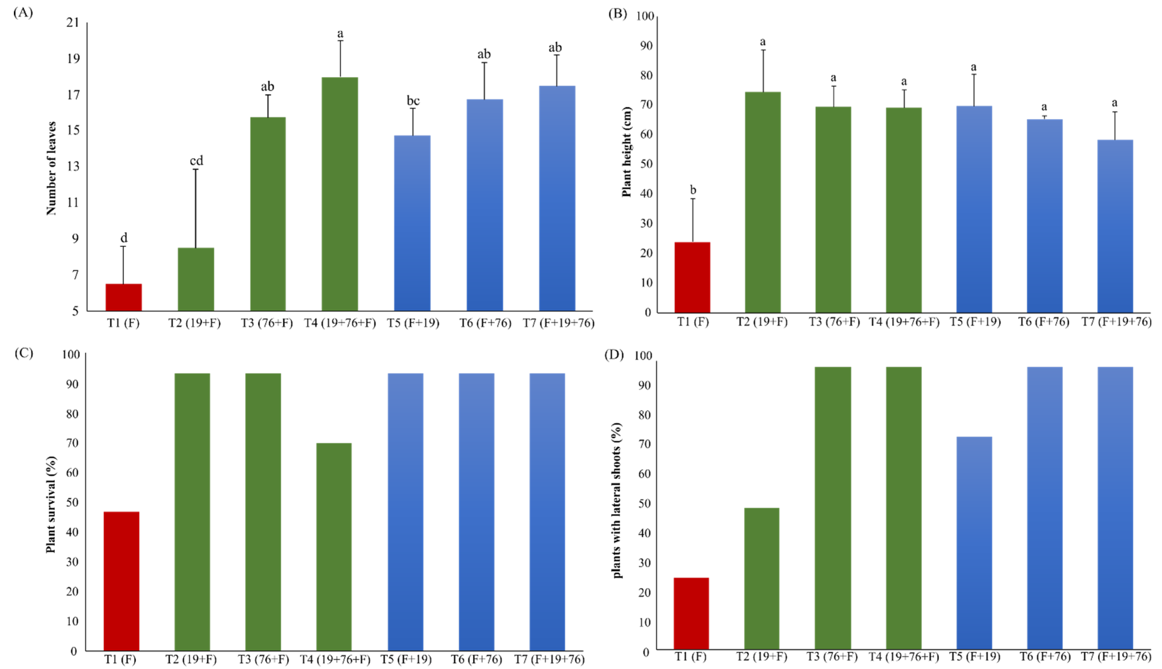
3.2.1. Plant Health

The plants were evaluated at the end of the experiment (180 DAP) in the seven pot groups, observing the number of leaves in each pot, measuring the plant height, determining the percentage of symptomatic plants, and the formation of lateral shoots.
The number of leaves varied among the seven treatments of the experiment, and the pots inoculated only with the fungus P. destruens (T1) showed the lowest number of leaves among the treatments. All treatments differed significantly (Mann–Whitney U test, p < 0.05) from the control treatment, except that one where the bacterium B. velezensis T149-19 (T2) was initially inoculated (Figure 1A).
All plants inoculated with bacteria, before or after fungal inoculum, showed a higher growth performance, reaching the end of the experiment approximately 70 cm long. The mean size of the control treatment plants (approximately 25 cm), inoculated only with the fungus, differed statistically (Mann–Whitney U test, p < 0.05) from the other treatments (Figure 1B).
Regarding the survival of the plants, it was observed that the plants of all treatments inoculated with the bacteria, before or after the fungus, had a higher survival capacity. In the control treatment (T1), 50% of the plants died while only one plant died in the T4 treatment (Figure 1C). During the daily analyses of the plants in the greenhouse, it was observed that sweet potato plants began to emit lateral shoots from the stems closest to the soil. This phenomenon was predominant in the pots where the plants were inoculated with bacteria, either before or after the fungal inoculum (Figure 1D).

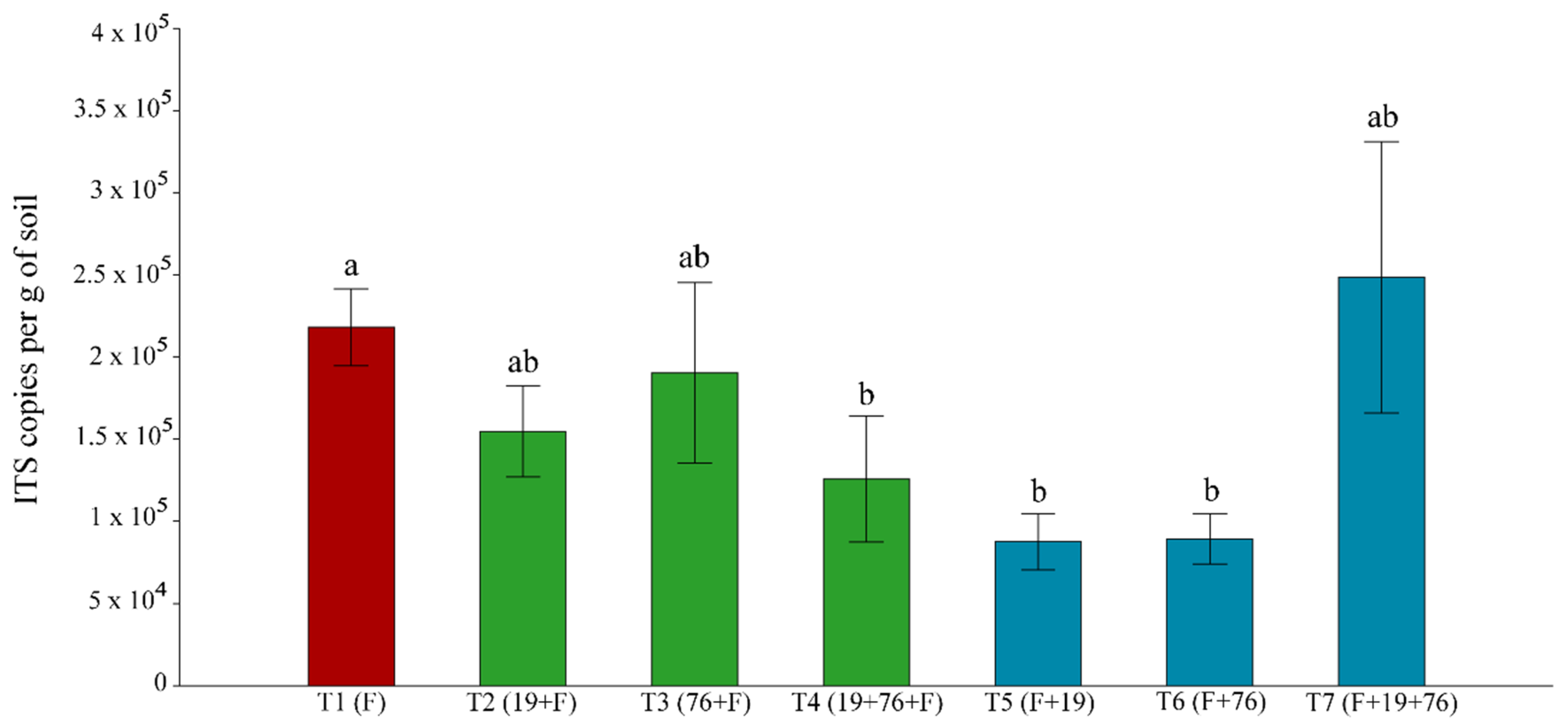
3.2.2. Total Fungal Community Quantification Using Real-Time PCR (qPCR)

The number of copies of the ITS gene for the disease control treatment (T1) samples (inoculated only with the fungus) was determined by qPCR, and this number was compared with the number of ITS genes present in the samples of the pots inoculated with the bacteria before or after the fungus (Figure 2). A significant decrease (Tukey’s HSD test, p < 0.05) in the number of copies of the ITS gene was observed in treatments T4, T5 and T6. No significant reduction was observed in the other treatments (T2, T3 and T7) when compared to T1 (Figure 2).

3.2.3. Response of the Fungal Communities to the Inoculation of Strains T052-76 and T149-19

The effect of the inoculation of the fungus P. destruens and the bacteria B. velezensis T149-19 and B. safensis T052-76 in the different treatments (T1-T7) on the fungal community in the rhizosphere of the plants was analyzed through PCR-DGGE. The dendrogram generated from the DGGE (Figure 3) revealed that the samples grouped together within each treatment. Most samples of the control treatment, where the plantlets were inoculated only with the fungus P. destruens, formed a separate group. The structure of the fungal community was clearly influenced by bacterial inoculation. In addition, the samples that were inoculated first with the fungus showed a slight tendency to form a group (upper part of the dendrogram, Figure 3). The samples inoculated with the bacteria B. velezensis and B. safensis separately (T2 and T3) formed their own subgroups, with approximately 45% similarity.

3.2.4. Non-Metric Multidimensional Scaling (NMDS) Ordination of PCR-Denaturing Gradient Gel Electrophoresis (DGGE) Profiles

The numerical matrix generated by the DGGE bands was submitted to NMDS using the PAST 3.26 statistical software to correlate the DGGE profiles obtained from the different treatments with physiological and plant development parameters (plant height, number of leaves) and fungal abundance (Figure 4). Samples T1, inoculated only with the fungus, were separated from all other treatments (PERMANOVA, p < 0.05). The remaining treatments showed a strong tendency to group among themselves, demonstrating once again the influence of bacterial inoculation in the grouping of samples. PERMANOVA statistically confirmed this observation (p < 0.05). Moreover, a clear correlation between the fungal community structure in the T2 to T7 treatments and the plant parameters (height and number of leaves) was observed, while T1 was correlated to the fungal abundance.

4. Discussion

Bacteria belonging to the genus Bacillus are usually described as producers of a wide array of antimicrobials, many of which have great importance in antibiosis against plant pathogens [36,37]. The main bioactive molecules produced by Bacillus species are non-ribosomally synthesized peptides and lipopeptides, polyketide compounds, bacteriocins and siderophores. Lipopeptides from the surfactin (lichenysin and pumilacidin), iturin (iturin, mycosubtilin, bacillomycin, and mojavensin) and fengycin (fengycin and plipastatin) families seem to be the most prevalent antimicrobials produced by Bacillus strains against different phytopathogens [36,37,38]. Beric et al. [39] demonstrated the presence of multiple biosynthetic operons for the synthesis of non-ribosomal lipopeptides in a considerable number of natural isolates of Bacillus. Several B. subtilis, B. licheniformis, B. natto, B. pumilus, B. thuringiensis, B. velezensis and B. amyloliquefaciens strains have already been described to produce lipopeptides with the potential to be used as biocontrol agents [36,37,40].
In this study, two Bacillus strains belonging to the species B. velezensis and B. safensis are being considered for the biocontrol of sweet potato, more specifically against the foot rot disease agent—the fungus P. destruens. We found in the literature many papers where B. velezensis is used in the biocontrol of different genera of phytopathogenic fungi in crops of high agricultural importance such as potato, mango, avocado, papaya, citrus, tomato, blueberry, blackberry, raspberry, zucchini, melon, cucumber, watermelon and others [41,42,43]. However, to the best of our knowledge, no studies are available considering either sweet potato as the target crop and B. safensis as the antimicrobial substances-producing species, except our previous studies [22,23]. The potential application of B. safensis strain B21 as a biopesticide has previously been described only for the control of rice blast [44]. Therefore, this study contributes to the knowledge about the capacity of these two Bacillus strains to inhibit P. destruens and to prevent the appearance of foot rot symptoms in pot experiments and about the genes encoding antimicrobial substances able to inhibit the fungus.
Genes coding for mycosubtilin, bacillaene, macrolactin, bacillibactin, bacilysin, plantazolicin, fengycin, plipastatin, dificidine and surfactin were found in B. velezensis T149-19, while those coding for bacilysin, lichenysin, bacillibactin, fengycin and surfactin were found in B. safensis T052-76. Likewise, the genomes of Bacillus velezensis CMRP 4490 and B-4 strains showed biosynthetic gene clusters related to the synthesis of molecules with antifungal activity [45,46]. Chen et al. [47] have demonstrated the presence of up to 13 clusters responsible for the synthesis of lipopeptides such as fengycin, iturin, surfactin, butirosine, plantazolicin and its hydrolyzed isomer, kijanimycin, bacilysin, dificidine, bacillaene, and bacillaene B in the genome of the B. velezensis strain LM2303.
All these antifungal substances possibly produced by B. velezensis T149-19 and B. safensis T052-76 have the potential to inhibit P. destruens. Moreover, the simultaneous use of the two strains in vivo experiments may have the potential to increase the spectrum of action against different pathogens. For example, surfactin is mostly known for its powerful biosurfactant properties and its limited fungitoxicity [36,48]. However, the synergistic activity of surfactin and different lipopeptides has already been demonstrated [36]. Nonetheless, we are aware that the presence of different antimicrobial substance coding genes in both Bacillus genomes does not guarantee that they are being expressed and the substances produced. Further experiments are still necessary to prove their presence after being introduced in the sweet potato root environment. However, as other authors have reported that more than one substance can be produced and be active in the same Bacillus strain [49,50], from the broad spectrum of antimicrobial substances produced by B. velezensis T149-19 and B. safensis T052-76 it is very likely that they can act in vivo.
Pot experiments were conducted to determine whether the bacteria would be able to protect the sweet potato plant from the disease—inoculating the bacteria before (60 DAP) and the fungus afterwards (120 DAP) and/or to mitigate the symptoms of foot rot disease—inoculating the fungus first (60 DAP) and then the bacteria (120 DAP). In both situations, all plants inoculated with bacteria (each separately or the two bacteria together) showed a higher growth performance and a higher survival capacity in the presence of the fungus. Lateral shoots during plant growth also appeared in sweet potato treated with bacteria. These observations speak in favor of both activities (plant protection and mitigation of symptoms) carried out by bacteria. Moreover, a significant effect of growth promotion was also observed (greater number of leaves, higher plants, and presence of lateral shoots) in plants treated with one or both bacteria together before or after fungal inoculation. Therefore, B. velezensis T149-19 and B. safensis T052-76 could be potential biocontrol agents with plant growth-promoting ability. Antifungal activity and plant growth-promoting ability were also demonstrated in B. velezensis CMRP 4490 [46].
The response of the sweet potato rhizospheric fungal communities to the inoculation of strains T052-76 and T149-19 was also evaluated using molecular methods. Although we are aware that it would have been beneficial to have performed the experiments more than once, we observed that the structure of the fungal community was influenced by bacterial inoculation in DGGE, and using qPCR, a significant decrease in the number of copies of the ITS gene was observed in the majority of the treatments where bacteria were inoculated. Therefore, our results may suggest that the antimicrobial substances produced by the inoculated strains are controlling fungal abundance, including P. destruens.

5. Conclusions

Based on genome sequencing and data analysis by comparative genomics, B. velezensis T149-19 and B. safensis T052-76 were shown to harbor a broad spectrum of antimicrobial substance-coding genes in their genomes. Data from in vivo experiments using simultaneously the fungus and the bacteria (each separately or the two bacteria together) showed a higher plant growth performance and a higher survival capacity of sweet potato plants than those inoculated only with the fungus. Therefore, both Bacillus strains can be considered promising biocontrol agents for biological control in crop production. The results presented here provide useful information that may affect the development of bioinoculants in sweet potato, protecting this crop against foot rot disease.

Author Contributions

L.S., J.R.M., D.J. conceived the study. J.R.M., I.D., P.R.G. carried out the experiments. J.R.M., J.M.M., R.T.J.R., A.L.d.C.d.S. planned and carried out the sequencing and assembly. J.R.M. planned and carried out the annotation and analyses of both genomes. J.M.M., D.J., F.F.d.M. contributed to the interpretation of the results. L.S. took the lead in writing the manuscript. All authors have read and agreed to the published version of the manuscript.

Funding

This study was supported by grants from Conselho Nacional de Desenvolvimento Científico e Tecnológico (CNPq), Coordenação de Aperfeiçoamento de Pessoal de Nível Superior (CAPES, financial code 001) and Fundação de Amparo à Pesquisa do Estado do Rio de Janeiro (FAPERJ).

Institutional Review Board Statement

Not applicable.

Informed Consent Statement

Not applicable.

Acknowledgments

This study was supported by grants from Conselho Nacional de Desenvolvimento Científico e Tecnológico (CNPq), Coordenação de Aperfeiçoamento de Pessoal de Nível Superior (CAPES, financial code 001) and Fundação de Amparo à Pesquisa do Estado do Rio de Janeiro (FAPERJ).

Conflicts of Interest

The authors declare no conflict of interest.

References

  1. CIP. 2021. Available online: https://cipotato.org/sweetpotato/ (accessed on 15 March 2021).
  2. Kays, S.J. Sweetpotato production worldwide: Assessment, trends and the future. Acta Hortic. 2005, 670, 19–25. [Google Scholar] [CrossRef]
  3. Zengzhi, S.; Bing, D.; Jinxi, H.; Shaozhen, H.; Qingchang, L.; Hong, Z. A genome-wide BAC-end sequence survey provides first insights into sweet potato (Ipomoea batatas (L.) Lam.) genome composition. BMC Genom. 2016, 17, 945. [Google Scholar]
  4. FAOSTAT—Statistics Database. Available online: http://www.faostat.fao.org (accessed on 15 March 2021).
  5. Silva, J.B.C.; Lopes, C.A.; Magalhães, J.S. Batata-Doce (Ipomoea Batatas), In Sistemas De Produção Nº 6, Versão Eletrônica; Embrapa Hortaliças: Brasília, Brazil, 2008. [Google Scholar]
  6. Mussoline, W.A.; Wilkie, A.C. Feed and fuel: The dual-purpose advantage of an industrial sweetpotato. J. Sci. Food Agric. 2016, 97, 1567–1575. [Google Scholar] [CrossRef]
  7. El Sheikha, A.F.; Ray, R.C. Potential impacts of bio-processing of sweet potato: Review. Crit. Rev. Food Sci. Nutr. 2017, 57, 455–471. [Google Scholar] [CrossRef]
  8. Williams, R.; Soares, F.; Pereira, L.; Belo, B.; Soares, A.; Setiawan, A.; Browne, M.; Nesbitt, H.; Erskine, W. Sweet potato can contribute to both nutritional and food security in Timor-Leste. Field Crop. Res. 2013, 146, 38–43. [Google Scholar] [CrossRef]
  9. Lopes, U.P.; Michereff, S.J. Desafios Do Manejo De Doenças Radiculares Causadas Por Fungos; Editora Universitária da UFRPE: Pernambuco, Brazil, 2018. [Google Scholar]
  10. Mikhail, M.S.; Abdel-Alim, A.I.; Abd-El-Khair, H.; Abdel-Gaied, T.G.; Shereen, A.M. Host range and total cellular protein fingerprint of soft rot Erwinia isolated from some vegetables in Egypt. Plant Arch. 2019, 19, 295–306. [Google Scholar]
  11. Jiang, L.; Jeong, J.C.; Lee, J.-S.; Park, J.M.; Yang, J.W.; Lee, M.H.; Choi, S.H.; Kim, C.Y.; Kim, D.-H.; Kim, S.W.; et al. Potential of Pantoea dispersa as an effective biocontrol agent for black rot in sweet potato. Sci. Rep. 2019, 9, 16354. [Google Scholar] [CrossRef] [PubMed] [Green Version]
  12. Pereira, R.B.; Amaro, G.B.; Ribeiro, M.C.V.; Pinheiro, J.B. Reação de genótipos de batata-doce ao mal-do-pé (Plenodomus destruens). In Boletim de Pesquisa e Desenvolvimento, Nº 122; Embrapa Hortaliças: Brasília, Brazil, 2014. [Google Scholar]
  13. Pereira, R.B.; Fernandes, F.R.; Pinheiro, J.B. Recomendações Para Manejo Da Podridão-Do-Pé Em Batata-Doce. In Comunicado Técnico Nº 79; Embrapa Hortaliças: Brasília, Brazil, 2011. [Google Scholar]
  14. Gai, Y.; Ma, H.; Chen, X.; Zheng, J.; Chen, H.; Li, H. Stem blight, foot rot and storage tuber rot of sweet potato caused by Plenodomus destruens in China. J. Gen. Plant Pathol. 2016, 82, 181–185. [Google Scholar] [CrossRef]
  15. Lopes, C.A.; Silva, J.B.C. Management measures to control foot rot of sweet potato caused by Plenodomus destruens. Int. J. Pest Manag. 1993, 39, 72–74. [Google Scholar] [CrossRef]
  16. Okada, Y.; Kobayashi, A.; Tabuchi, H.; Kuranouchi, T. Review of major sweetpotato pests in Japan, with information on resistance breeding programs. Breed. Sci. 2017, 67, 73–82. [Google Scholar] [CrossRef] [Green Version]
  17. O’Brien, P.A. Biological control of plant diseases. Australas. Plant Pathol. 2017, 46, 293–304. [Google Scholar] [CrossRef] [Green Version]
  18. Tariq, M.; Khan, A.; Asif, M.; Khan, F.; Ansari, T.; Shariq, M.; Siddiqui, M.A. Biological control: A sustainable and practical approach for plant disease management. Acta Agric. Scand. B Soil Plant Sci. 2020, 70, 507–524. [Google Scholar] [CrossRef]
  19. Qiao, J.Q.; Wu, H.J.; Huo, R.; Gao, X.W.; Borriss, R. Stimulation of plant growth and biocontrol by Bacillus amyloliquefaciens subsp. plantarum FZB42 engineered for improved action. Chem. Biol. Technol. Agric. 2014, 1, 12. [Google Scholar] [CrossRef] [Green Version]
  20. Wu, L.; Wu, H.-J.; Qiao, J.; Gao, X.; Borriss, R. Novel routes for improving biocontrol activity of Bacillus based bioinoculants. Front. Microbiol. 2015, 6, 1395. [Google Scholar] [CrossRef] [Green Version]
  21. Marques, J.M.; da Silva, T.F.; Vollú, R.E.; Blank, A.F.; Ding, G.-C.; Seldin, L.; Smalla, K. Plant age and genotype affect the bacterial community composition in the tuber rhizosphere of field-grown sweet potato plants. FEMS Microbiol. Ecol. 2014, 88, 424–435. [Google Scholar] [CrossRef]
  22. Marques, J.M.; da Silva, T.F.; Vollú, R.E.; de Lacerda, J.R.M.; Blank, A.F.; Smalla, K.; Seldin, L. Bacterial endophytes of sweet potato tuberous roots affected by the plant genotype and growth stage. Appl. Soil Ecol. 2015, 96, 273–281. [Google Scholar] [CrossRef]
  23. Mateus, J.R.; Marques, J.M.; Dal’Rio, I.; Vollú, R.E.; Coelho, M.R.R.; Seldin, L. Response of the microbial community associated with sweet potato (Ipomoea batatas) to Bacillus safensis and Bacillus velezensis strains. Antonie Van Leeuwenhoek 2019, 112, 501–512. [Google Scholar] [CrossRef]
  24. Seldin, L.; Rosado, A.S.; Cruz, D.W.; Nobrega, A.; van Elsas, J.D.; Paiva, E. Comparison of Paenibacillus azotofixans strains isolated from rhizoplane, rhizosphere and non-rhizosphere soil from maize planted in two different Brazilian soils. Appl. Environ. Microbiol. 1998, 64, 3860–3868. [Google Scholar] [CrossRef] [PubMed] [Green Version]
  25. Seldin, L.; Dubnau, D. DNA homology among Bacillus polymyxa, Bacillus macerans, Bacillus azotofixans and other nitrogen fixing Bacillus strains. Int. J. Syst. Bacteriol. 1985, 35, 151–154. [Google Scholar] [CrossRef] [Green Version]
  26. Bankevich, A.; Nurk, S.; Antipov, D.; Gurevich, A.A.; Dvorkin, M.; Kulikov, A.S.; Lesin, V.M.; Nikolenko, S.I.; Pham, S.; Prjibelski, A.D.; et al. SPAdes: A new genome assembly algorithm and its applications to single-cell sequencing. J. Comp. Biol. 2012, 19, 455–477. [Google Scholar] [CrossRef] [Green Version]
  27. Blin, K.; Shaw, S.; Steinke, K.; Villebro, R.; Ziemert, N.; Lee, S.Y.; Medema, M.H.; Weber, T. antiSMASH 5.0: Updates to the secondary metabolite genome mining pipeline. Nucleic Acids Res. 2019, 47, W81–W87. [Google Scholar] [CrossRef] [Green Version]
  28. Aziz, R.K.; Bartels, D.; Best, A.A.; Dejongh, M.; Disz, T.; Edwards, R.; Formsma, K.; Gerdes, S.; Glass, E.M.; Kubal, M.; et al. The RAST Server: Rapid annotations using subsystems technology. BMC Genom. 2008, 9, 75. [Google Scholar] [CrossRef] [Green Version]
  29. Overbeek, R.; Olson, R.; Pusch, G.D.; Olsen, G.J.; Davis, J.J.; Disz, T.; Edwards, R.; Gerdes, S.; Parrello, B.D.; Shukla, M.; et al. The SEED and the Rapid Annotation of microbial genomes using Subsystems Technology (RAST). Nucleic Acids Res. 2014, 42, D206–D214. [Google Scholar] [CrossRef]
  30. Hammer, O.; Harper, D.A.T.; Ryan, P.D. Past: Paleontological statistics software package for education and data analysis. Palaeontol. Electron. 2001, 1, 9. [Google Scholar]
  31. Smit, E.; Leeflang, P.; Glandorf, B.; Van Elsas, J.D.; Wernars, K. Analysis of fungal diversity in the wheat rhizosphere by sequencing of cloned PCR-amplified genes encoding 18S rRNA and temperature gradient gel electrophoresis. Appl. Environ. Microbiol. 1999, 65, 2614–2621. [Google Scholar] [CrossRef] [Green Version]
  32. White, T.J.; Bruns, T.D.; Lee, S.B.; Taylor, J.W. Analysis of phylogenetic relationships by amplification and direct sequencing of ribosomal RNA genes. In PCR Protocols: A Guide to Methods and Applications; Innis, M.A., Gelfand, D.H., Sninsky, J.J., White, T.J., Eds.; Academic Press: New York, NY, USA, 1990; pp. 315–322. [Google Scholar]
  33. Gardes, M.; Bruns, T.D. ITS primers with enhanced specificity for basidiomycetes: Application to the identification of mycorrhiza and rusts. Mol. Ecol. 1993, 2, 113–118. [Google Scholar] [CrossRef]
  34. Heuer, H.; Wieland, J.; Schonfeld, J.; Schonwalder, A.; Gomes, N.C.M.; Smalla, K. Bacterial community profiling using DGGE or TGGE analysis. In Environmental Molecular Microbiology: Protocols and Applications Horizon; Rouchelle, P., Ed.; Horizon Scientific Press: Wymondham, Norfolk, UK, 2001; pp. 177–190. [Google Scholar]
  35. Fierer, N.; Jackson, J.A.; Vilgalys, R.; Jackson, R.B. Assessment of soil microbial community structure by use of taxon-specific quantitative PCR assays. Appl. Environ. Microbiol. 2005, 71, 4117–4120. [Google Scholar] [CrossRef] [PubMed] [Green Version]
  36. Ongena, M.; Jacques, P. Bacillus lipopeptides: Versatile weapons for plant disease biocontrol. Trends Microbiol. 2008, 16, 115–125. [Google Scholar] [CrossRef] [PubMed]
  37. Fira, D.; Dimkic, I.; Beric, T.; Lozo, J.; Stankovic, S. Biological control of plant pathogens by Bacillus species. J. Biotechnol. 2018, 285, 44–55. [Google Scholar] [CrossRef] [PubMed]
  38. Jacques, P. Surfactin and other lipopeptides from Bacillus spp. In Microbiology; Soberón-Chávez, G., Ed.; Springer: Berlin, Germany, 2011; Volume 20. [Google Scholar]
  39. Berić, T.; Kojić, M.; Stanković, S.; Topisirović, L.; Degrassi, G.; Myers, M.; Venturi, V.; Fira, D. Antimicrobial activity of Bacillus sp. natural isolates and their potential use in the biocontrol of phytopathogenic bacteria. Food Technol. Biotechnol. 2012, 50, 25–31. [Google Scholar]
  40. Grady, E.N.; MacDonald, J.; Ho, M.T.; Weselowski, B.; McDowell, T.; Solomon, O.; Renaud, J.; Yuan, Z.C. Characterization and complete genome analysis of the surfactin-producing, plant-protecting bacterium Bacillus velezensis 9D. BMC Microbiol. 2019, 19, 5. [Google Scholar] [CrossRef]
  41. Galindo, E.; Serrano-Carreón, L.; Gutiérrez, C.; Allende, R.; Balderas, K.; Patiño, M.; Trejo, M.; Wong, M.; Rayo, E.; Isauro, D.; et al. The challenges of introducing a new biofungicide to the market: A case study. Elect. J. Biotechnol. 2013, 16, 5. [Google Scholar] [CrossRef]
  42. Balderas-Ruíz, K.A.; Bustos, P.; Santamaria, R.I.; González, V.; Cristiano-Fajardo, S.A.; Barrera-Ortíz, S.; Mezo-Villalobos, M.; Aranda-Ocampo, S.; Guevara-García, A.; Galindo, E.; et al. Bacillus velezensis 83 a bacterial strain from mango phyllosphere, useful for biological control and plant growth promotion. AMB Express 2020, 10, 163. [Google Scholar] [CrossRef]
  43. Cui, L.; Yang, C.; Wei, L.; Li, T.; Chen, X. Isolation and identification of an endophytic bacteria Bacillus velezensis 8-4 exhibiting biocontrol activity against potato scab. Biol. Control 2020, 141, 104156. [Google Scholar] [CrossRef]
  44. Rong, S.; Xu, H.; Li, L.; Chen, R.; Gao, X.; Xu, Z. Antifungal activity of endophytic Bacillus safensis B21 and its potential application as a biopesticide to control rice blast. Pestic. Biochem. Physiol. 2020, 162, 69–77. [Google Scholar] [CrossRef]
  45. Zhu, Z.; Peng, Q.; Man, Y.; Li, Z.; Zhou, X.; Bai, L.; Peng, D. Analysis of the antifungal properties of Bacillus velezensis B-4 through a bioassay and complete-genome sequencing. Front. Genet. 2020, 11, 703. [Google Scholar] [CrossRef]
  46. Teixeira, G.M.; Mosela, M.; Nicoletto, M.L.A.; Ribeiro, R.A.; Hungria, M.; Youssef, K.; Higashi, A.Y.; Mian, S.; Ferreira, A.S.; Gonçalves, L.S.A.; et al. Genomic insights into the antifungal activity and plant growth-promoting ability in Bacillus velezensis CMRP. Front. Microbiol. 2021, 11, 618415. [Google Scholar] [CrossRef]
  47. Chen, L.; Heng, J.; Qin, S.; Bian, K. A comprehensive understanding of the biocontrol potential of Bacillus velezensis LM2303 against Fusarium head blight. PLoS ONE 2018, 13, e0198560. [Google Scholar] [CrossRef] [Green Version]
  48. Pérez-Garcia, A.; Romero, D.; De Vicente, A. Plant protection and growth stimulation by microorganisms: Biotechnological applications of Bacilli in agriculture. Curr. Opin. Biotechnol. 2011, 22, 187–193. [Google Scholar] [CrossRef] [PubMed]
  49. Yuan, J.; Li, B.; Zhang, N.; Waseem, R.; Shen, Q.; Huang, Q. Production of bacillomycin- and macrolactin-type antibiotics by Bacillus amyloliquefaciens NJN-6 for suppressing soilborne plant pathogens. Agric. Food Chem. 2012, 60, 2976–2981. [Google Scholar] [CrossRef] [PubMed]
  50. Li, Y.G.; Geng, X.B.; Ji, P.S.; Pan, C.Q.; Wei, S. Isolation and evaluation of a Bacillus methylotrophicus strain for control of corn stalk rot. Biocontrol Sci. Technol. 2016, 26, 727–773. [Google Scholar] [CrossRef]
Figure 1. (A) Number of leaves in each plant (180 DAP); (B) plant height; (C) plant survival; (D) lateral shoot formation. F represents the inoculum with the fungus P. destruens, 19 represents the inoculum with the bacterium B. velezensis T149-19 and 76 represents the inoculum with the bacterium B. safensis T052-76. The position of letter F (before or after the numbers) indicates the moment of inoculation of the fungus (before or after bacteria). In (A,B), treatments that do not share a letter have a mean difference that is statistically significant based on the Mann–Whitney U test (p < 0.05).
Agriculture 11 01046 g001
Figure 2. Total fungal population quantified by quantitative polymerase chain reaction (qPCR). The bar values indicate the average of the four replicates (n) of each treatment. T represents each of the treatments from 1 to 7, F represents the inoculum with the fungus P. destruens, 19 represents the inoculum with the bacterium B. velezensis T149-19 and 76 represents the inoculum with the bacterium B. safensis T052-76. The position of letter F (before or after the numbers) indicates the moment of inoculation of the fungus (before or after bacteria). Treatments that do not share a letter have a mean difference that is statistically significant based on Tukey’s HSD test (p < 0.05).
Agriculture 11 01046 g002
Figure 3. Dendrogram generated from the denaturing gradient gel electrophoresis (DGGE) using the unweighted pair group method with arithmetic mean (UPGMA) method and the Dice coefficient based on ITS fragments of the fungal community of the rhizosphere of treatments T1 to T7. F: inoculated with P. destruens; 19: B. velezensis T149-19; 76: B. safensis T052-76.
Agriculture 11 01046 g003
Figure 4. Nonmetric multidimensional scaling (NMDS) plot based on Bray–Curtis dissimilarities of fungal ITS region-based PCR-DGGE bands. Plant health parameters (plant height, number of leaves) and fungal abundance in qPCR were used here as vectors for NMDS analyses. T1 to T7 treatments: T1 = F; T2 = 19 + F; T3 = 76 + F; T4 = 19 + 76 + F; T5 = F + 19; T6 = F + 76; T7 = F + 19 + 76. PH = plant height, LN = number of leaves and FA = fungal abundance.
Agriculture 11 01046 g004
Table 1. Biosynthetic gene clusters related to the production of a variety of antimicrobial substances in the Bacillus velezensis T149-19 genome.
Gene Cluster
(Size in nt)
Similarity
(%) a
Access Number
MIBiG b
Coding forRelated Genes
88,273100BGC0001103MycosubtilinfenF, mycA, mycB and mycC
97,565100BGC0001089Bacillaenebae genes (I, J, L, M, N)
88,300100BGC0000181Macrolactinpks and pdhA
51,787100BGC0000309Bacillibactindhb, yui, among others
41,419100BGC0001184BacilysinywfA and bacA, B, C, D, E and F
22,18291BGC0000569Plantazolicinptn genes
25,25639BGC0000433SurfactinsrfAB, srfAC and srfAD
160,18843BGC0000433SurfactinsrfAA and other accessory genes
91088BGC0000433SurfactinsrfAA and srfAB
13,00820BGC0001095FengycinfenA, fenB and fenC
920513BGC0001095FengycinfenC and fenD
49,12330BGC0000407PlipastatinppsA and other accessory genes
33,89266BGC0000176Dificidinedif genes (A, B, C, D, E, F, G, I, J, K)
a similarity with gene clusters found in bacterial strains determined using antiSMASH software. b access number to the database MIBiG (Minimum Information about a Biosynthetic Gene cluster) for each predicted gene cluster by antiSMASH.
Table 2. Biosynthetic gene clusters related to the production of a variety of antimicrobial substances in the Bacillus safensis T052-76 genome.
Gene Cluster (Size in nt)Similarity
(%) a
Access Number
MIBiG b
Coding forRelated Genes
41,42285BGC0001184Bacylisinbac and ywf genes
27,83050BGC0000381LichenysinhyuA and B, putA, B and C and lchAA genes
49,71053BGC0000309Bacillibactindhb genes
28,41353BGC0001103Fengycinyng genes
44,18639BGC0000433SurfactinsrfA and ycx genes, and others
a similarity with gene clusters found in bacterial strains determined using antiSMASH software. b access number to the database MIBiG (Minimum Information about a Biosynthetic Gene cluster) for each predicted gene cluster by antiSMASH.
Publisher’s Note: MDPI stays neutral with regard to jurisdictional claims in published maps and institutional affiliations.

