Article

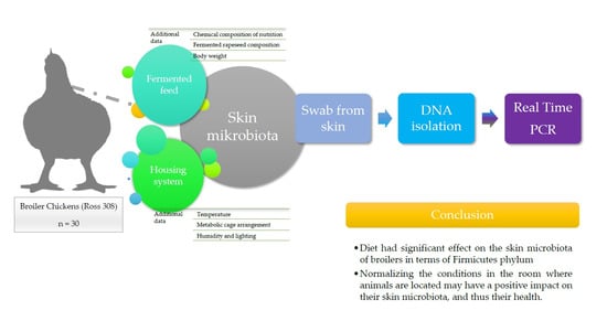
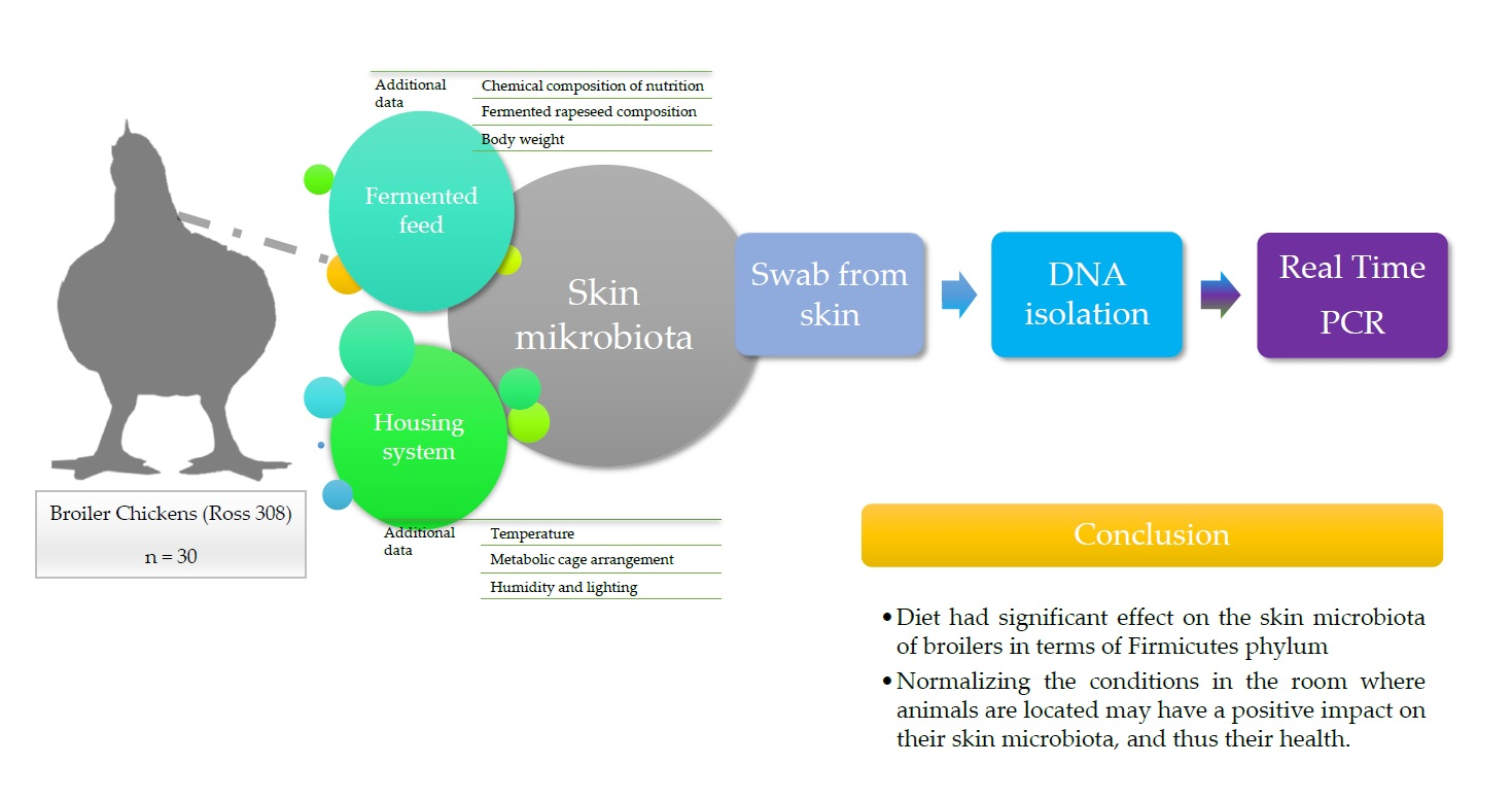
Levels of Firmicutes, Actinobacteria Phyla and Lactobacillaceae Family on the Skin Surface of Broiler Chickens (Ross 308) Depending on the Nutritional Supplement and the Housing Conditions

1
Institute of Animal Breeding, Wroclaw University of Environmental and Life Sciences, 51-630 Wroclaw, Poland
2
Department of Animal Nutrition and Feed Management, Wroclaw University of Environmental and Life Sciences, 51-630 Wroclaw, Poland
*
Author to whom correspondence should be addressed.
Submission received: 22 February 2021 / Revised: 23 March 2021 / Accepted: 23 March 2021 / Published: 26 March 2021
(This article belongs to the Special Issue Poultry: Breeding, Health, Nutrition, and Management)

Abstract

:
The microbiome of animals, both in the digestive tract and in the skin, plays an important role in protecting the host. The skin is one of the largest surface organs for animals; therefore, the destabilization of the microbiota on its surface can increase the risk of diseases that may adversely affect animals’ health and production rates, including poultry. The aim of this study was to evaluate the effect of nutritional supplementation in the form of fermented rapeseed meal and housing conditions on the level of selected bacteria phyla (Firmicutes, Actinobacteria, and family Lactobacillaceae). The study was performed on 30 specimens of broiler chickens (Ross 308), individually kept in metabolic cages for 36 days. They were divided into 5 groups depending on the feed received. On day 36, skin swabs were individually collected. Temperature and humidity were measured in the room. The temperature was measured every 2 days (18 measurements × 6 points). The results of Real-Time PCR analysis have shown a significant effect of the feed additive on the level of Firmicutes phylum on the skin. On the other hand, a variable level of the tested bacteria was shown depending on the location of the cages. The Firmicutes phylum and Lactobacillaceae family achieved the highest level in the top-window zone. However, in the case of the Actinobacteria phylum, the highest level was found at the top-door and middle-door zones. The obtained results suggest that the conditions in which animals live may affect the microbiota of their skin.

Graphical Abstract

1. Introduction

Poultry, and in particular domestic chickens (Gallus gallus domesticus), is one of the main sources of animal protein due to one of the best feed/meat conversion ratios [1]. Due to humanitarian reasons and the production volume itself, it is important to maintain adequate welfare when keeping animals [2]. One of the welfare aspects is avoiding animal exposure to mechanical injuries possibility [3], which often result in disruption of the skin continuity, which may lead to many bacterial diseases [4]. It is not, however, the only circumstance related to the skin that we should pay attention to when keeping animals.
The skin of vertebrates is one of the largest organs in terms of surface, and for decades it has been attributed mainly to the role of an insulator separating the internal system of animals from the external environment. Over time, however, our knowledge about the skin and the roles and mechanisms occurring within or on its surface has grown significantly [5]. We already know that the protective barrier function is maintained not only by ectoderm cells but also by bacteria living on the surface of the skin, creating a specific ecosystem—a microbiota that, in order to fulfill its proper function, requires a balance both in terms of the species composition itself and the prevailing physicochemical conditions [6,7]. Factors such as nutrition, temperature, humidity and sunlight may affect the structure of the microbiota, which may directly affect animal health, and therefore, generate higher breeding costs [6]. The microbiota consists of bacteria, protozoa, fungi and archaea. However, bacteria are the most abundant both inside the body and on the skin. In mammals, the most numerous groups of bacteria belong to the Firmicutes and Bacteroidetes phyla. Yet, on the skin itself, bacteria from the Firmicutes, Bacteroidetes, Actinobacteria and Proteobacteria phyla are largely present; however, it depends on the taxon and even the species of the animal or the place where it lives [8]. Unfortunately, in the case of broiler chickens, no tests were carried out to systematize the correct microbiological composition, which in the future may provide additional information about their health status and thus their welfare.
The use of feed additives, including fermented feed, may affect the microbiological composition of the digestive system, increasing the amount of, e.g., bacteria from the Lactobacillaceae family. Changes in the gut can have an impact on overall health status [9] and thus also on the skin [10]. However, this relationship has not yet been thoroughly investigated for broiler chickens and should be undertaken in the future, and was also the reason for conducting the research presented in this paper.
This research focuses on the effects of nutrition and the housing environment on the levels of selected bacteria phyla on the skin of caged broiler chickens.

2. Materials and Methods

2.1. Animals

The research was conducted on 30 broiler chickens (Ross 308, male) kept in metabolic cages (1 broiler chicken per cage). The animals were kept in cages from the first day of life. They were divided into 5 food groups, 6 chickens per group (Table 1):
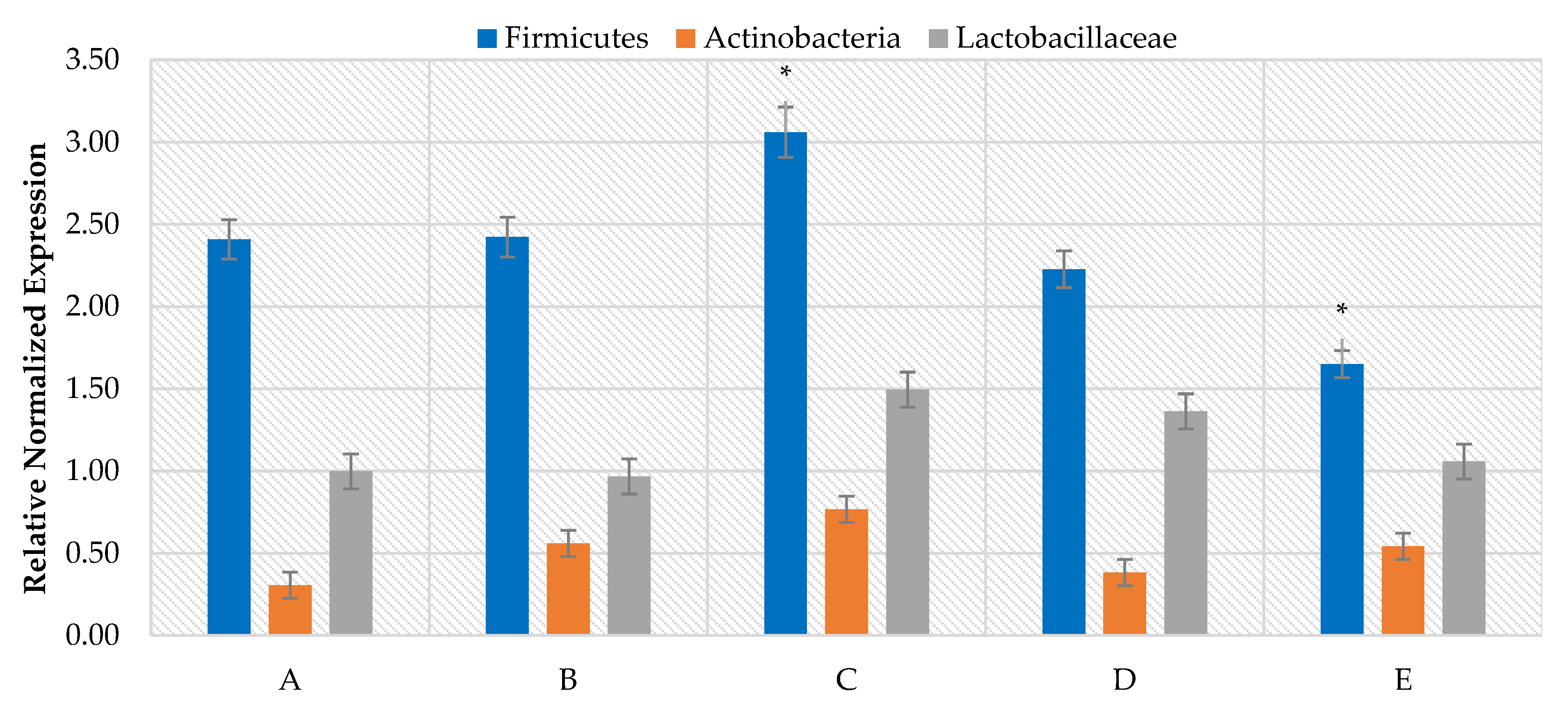
  • A—control (no rapeseed meal);
  • B—addition of 5% of standard rapeseed meal;
  • C—addition of 10% of standard rapeseed meal;
  • D—addition of 5% standard rapeseed meal and 5% fermented rapeseed meal;
  • E—addition of 10% standard rapeseed meal and 10% fermented rapeseed meal.
They received feed and water ad libitum.
The composition of the additive fermented rapeseed meal: protein 29%, fiber 10%, lactic acid 6%, LAB(lactic acid bacteria) log 6–7, glucosinolate reduction.
The room temperature from day 1 was about 32 °C; then it was gradually lowered by about 0.5 to 1 °C until the 20th day to obtain the temperature of 21 °C, at which it was kept until the sampling and weighing day. Humidity in the rooms during their rearing ranged from 60–70%. During the day, they had no access to external light. The only source of light was automatic lighting from 6.00 to 23.00 throughout the entire period of their maintenance. The lighting on both sides of the cages ranged between 24 to 20 lux from top to bottom; LED lighting was used.
The condition of the tested animals was good, and they did not show any symptoms, such as diarrhea, skin problems, increased body temperature, lack of appetite, and apathy, etc.
The authors confirm that the ethical policies of the journal were adhered to. All animals that qualified for the study were subjected to standard procedures without any harm or discomfort. The study did not require the consent of the Local Ethical Committee for Animal Experiments (Act of 15 January 2015 on protection animals used for scientific or educational purposes), OJ 2015, 266, implementing the Directive 2010/63/EU of the European Parliament and the Council of 22 September 2010 on the protection of animals used for scientific purposes.

2.2. Sampling

Samples were collected on the 36th day of age individually from each animal in the area around the neck. Feathers were trimmed on a small area so the swabs would not have contact with them. Skin swabs were collected with sterile brushes, placed in test tubes with sterile water, and then frozen at −26 °C until analysis (5 days).

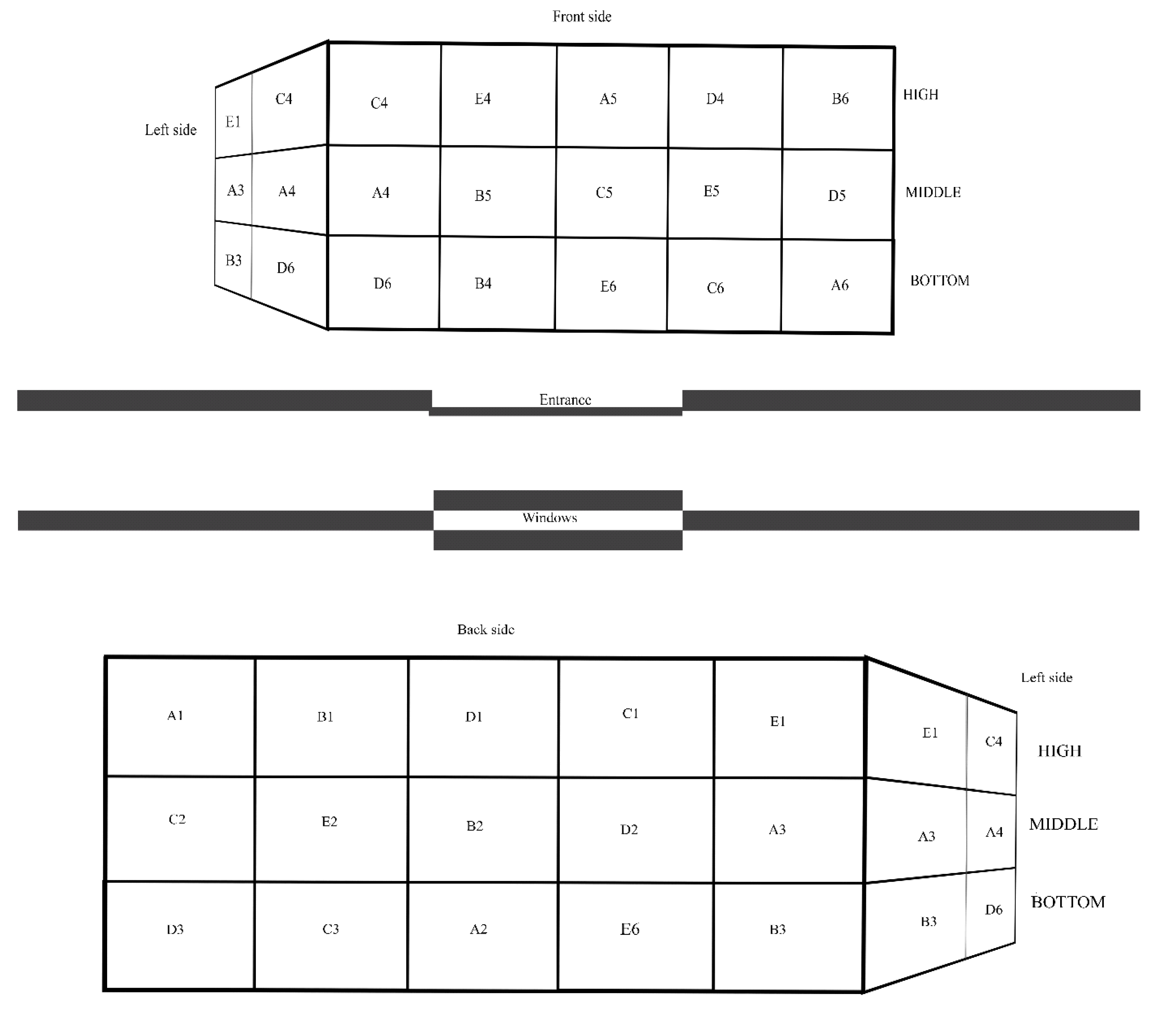
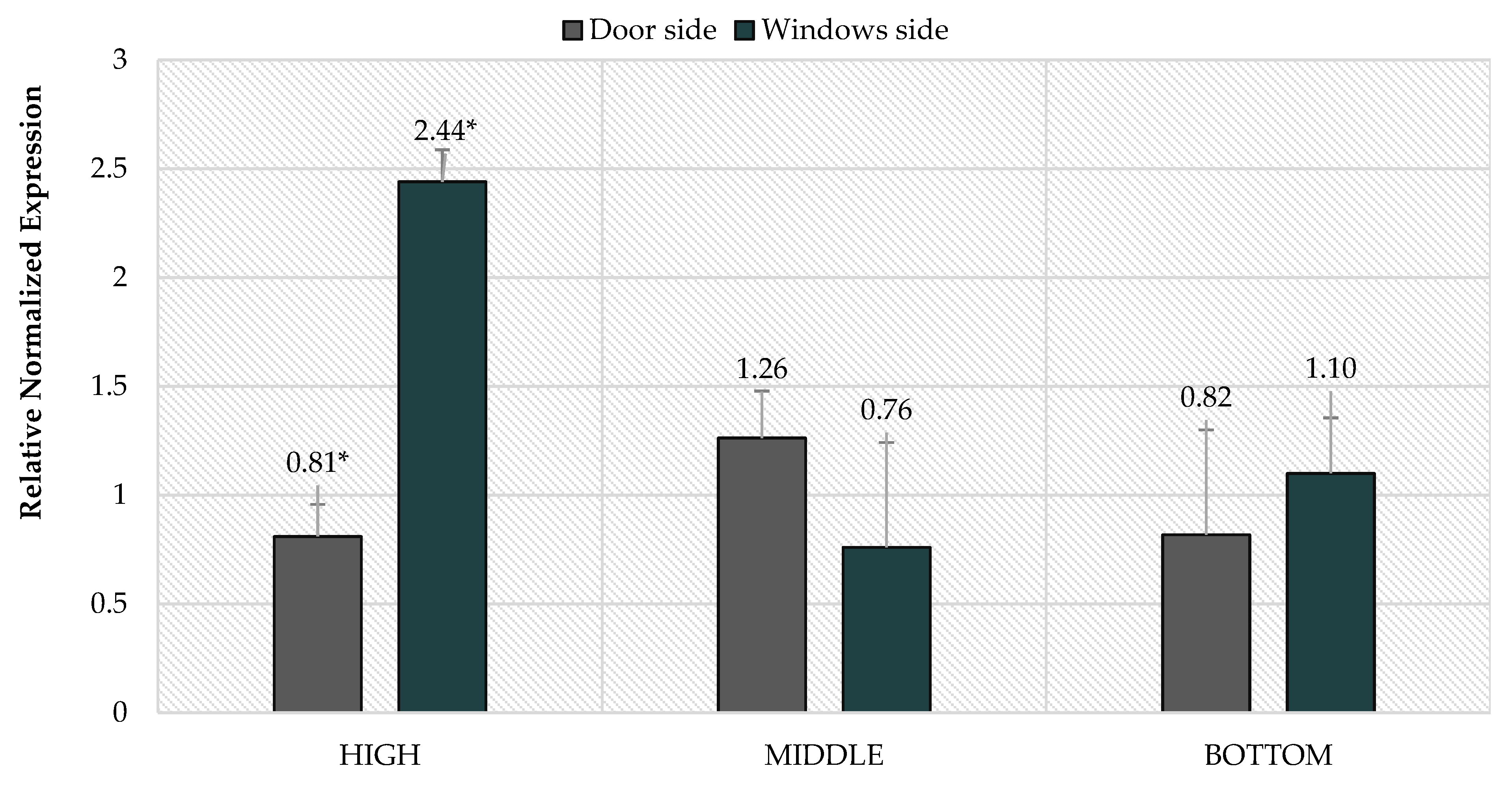
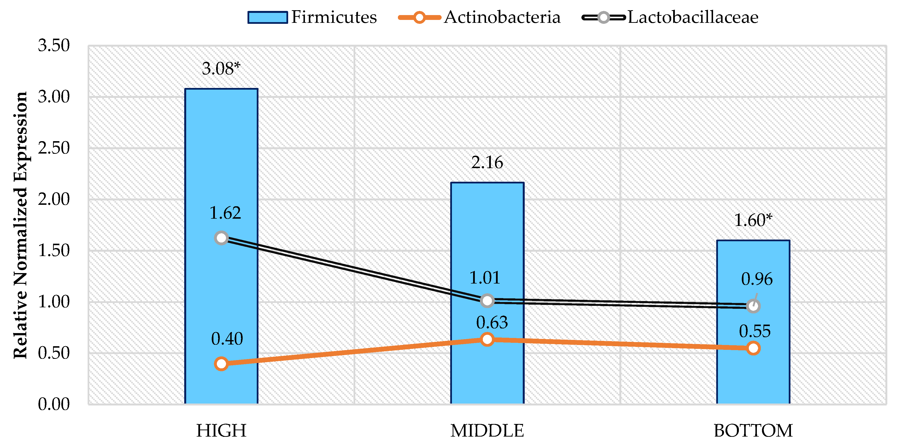
2.3. Assessment of the Housing Environment

Throughout the experimental period, the maintenance conditions were assessed, the differences in temperature between the arrangement of the cages (top, middle and bottom of the cages, as well as on the door and window side) were examined (Table 2). The temperature was measured every 2 days (18 measurements×6 points). The arrangement of the cages in the room is shown in the diagram below (Figure 1). Additionally, the room humidity was measured at the same points, but its range was constant-SD (Standard Deviation) = 5.25.

2.4. DNA (Deoxyrybonucleic acid) Isolation

A genomic bacteria AX Mini (A&A Biotechnology, Gdansk, Poland) was used for DNA isolation. The quality of DNA obtained as a result of isolation was verified using the NanoDrop 2000 spectrophotometer (Thermo Scientific, Wilmington, NC, USA). The average DNA content was 90–110 µg/µL. The level of impurities in the samples was as follows: for parameter 260/230:2.0–2.2 and for parameter 260/280:1.8–2.0 (in accordance with the guidelines from Thermo Scientific). When high levels of impurity or low-quality DNA were found, the samples were isolated again or purified using a clean-up concentrator (A&A Biotechnology, Gdansk, Poland).

2.5. Real-Time PCR Analysis

Bio-Rad CFX Connect 96 Touch apparatus was used to perform real-time PCR (Polymerase Chain Reaction) analysis. Bio-Rad SsoAdvanced™ Universal SYBR® Green Supermix kit (Bio-Rad Laboratories, Inc., Hercules CA, USA) at a volume of 10 μL was applied in 3 technical repetitions (Table 3). A NTC test (no template control) was additionally performed for each gene. The strategy of RT–PCR analysis involved the amplification of genes specific for the examined phyla in the presence of the reference gene for all bacteria (Table 4) [10,11].
In order to determine the performance of individual genes, a standard curve was drawn for the genes under the study. A sample dilution of 10−3 was selected for analysis (from the 10−1 to 10−7 series). The analysis was performed in accordance with a 40 cycles protocol: polymerase activation and DNA denaturation 95 °C (3 min), denaturation 95 °C (15 s), annealing 60.5 °C (15 s), extension and plate reading at 72 °C (40 s). The melting curves analysis for the samples was performed at temperatures ranging from 65 °C (5 s) to 95 °C (0.5 °C increments in 2 s).
The data obtained were then processed using the CFX Maestro software v. 1.1 (Bio-Rad Laboratories, Inc., Hercules CA, USA). The sample with a DNA level of 100 μg/μL and impurities at a level consistent with the standards was an arbitrary calibrator. The efficiency of individual primers was normal (in accordance with the standards established by Bio-Rad) and amounted to 89.4% for Firmicutes, 93.6% for Actinobacteria, 97.7% for Lactobacillaceae; Universal—94.4. CFX Maestro calculated the results from the number of the reference gene matrix and the differences at the relative normalized expression (RNE = ΔΔCq) phylum’s level, taking into account the amplification efficiency of individual genes [10].

2.6. Statistical Analysis
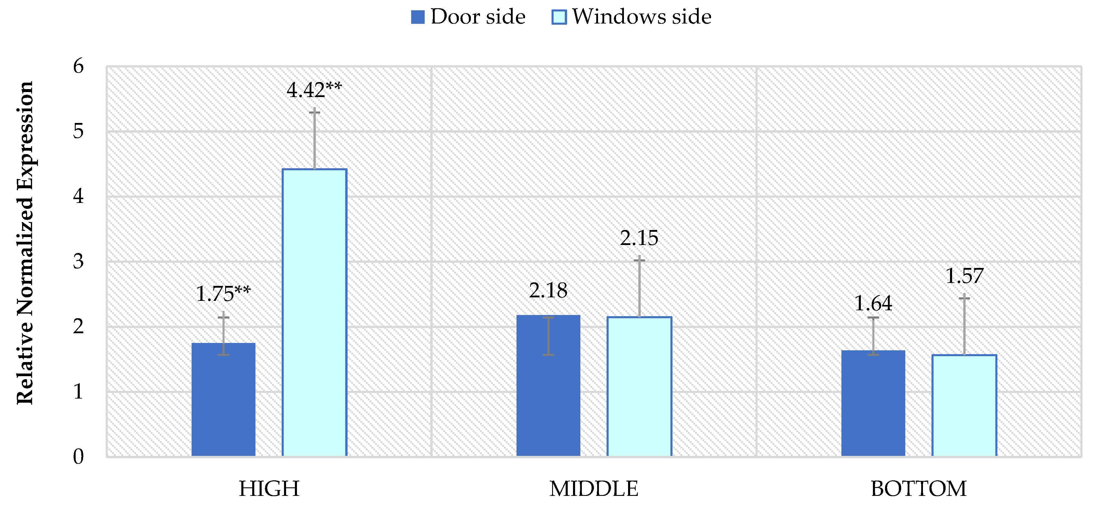
The results were analyzed with the use of the Statistica software (v. 13.3, StatSoft Inc., Tulsa, OK, USA). The data distribution was checked with the Shapiro–Wilk test. The data relating to the bodyweight of the chickens were analyzed using the ANOVA (analysis of variance) test, while the remaining data (no normal distribution) using the Wilcoxon pair test and the Friedman ANOVA (p > 0.05). The differences in the case of the ANOVA test were determined using Tukey’s test.

3. Results

The weighing performed on the 36th day of life showed that the group with the addition of 5% standard rapeseed meal and 5% fermented rapeseed meal (D) had the highest body weight. On the other hand, the lowest body weight was found in individuals from the group with the addition of 5% of standard rapeseed meal (Table 5). Additionally, the performed statistical analysis in terms of the arrangement of cages (level and side-window/door) did not show any significant statistical differences.
The analysis of the RT–PCR results showed that the group fed with 10% standard rapeseed meal (C) had a significantly higher RNE level of Firmicutes compared to the group fed with 10% fermented rapeseed meal and 10% standard (E). In addition, this level was generally the highest also compared to the other nutritional groups; similar trends are also visible in the comparison of the level of Actinobacteria and the Lactobacillaceae family. The lowest level of the Actinobacteria phylum was characteristic for the control group (A), and the highest, the aforementioned group C. The lowest level of the Lactobacillaceae family was characteristic for group B—5% addition of standard rapeseed meal (Figure 2).
The analysis of the level of selected bacterial phyla also showed differences depending on the arrangement of the cages. The animals placed in the highest cages differed in the level of the Firmicutes phylum depending on the side of the location—doors or windows. In the window-up zone, animals were characterized by a much higher level (about 3 times) of this phylum compared to the up-door zone (Figure 3). Additionally, the temperature in the window-top sector was on average higher by 1.05 °C, and on the door side by 0.4 °C. Similar results were also obtained in the case of the Lactobacillaceae family, where the difference between the RNE level was more than twofold (Figure 4). Significant differences were also observed for the Actinobacteria phylum (Figure 5). The most significant differences in the level of the phyla were between the top-window and top-door zones. The highest RNE level was found in the middle cages in the middle-door zone, while the lowest in the upper part in the top-window zone.
However, when analyzing only the position of the cages in terms of height, significant differences were shown only in the level of the Firmicutes phylum (Figure 6). The highest level of this phylum was characterized by the groups staying in the upper cages and the lowest in the lowest cages. There is also a downward trend in RNE levels in the Lactobacillaceae family. On the other hand, in the case of the Actinobacteria phylum, there is a slight upward trend between individuals housed in the upper and lower cages. However, the highest level occurred in the middle frames. On the other hand, when analyzing the level of the tested bacteria broken down into the window and door zones only, no significant differences in the RNE level were found.

4. Discussion

The microbial composition of the skin and the digestive tract is important because of its role in regulating the immune system and overall health status maintenance [16,17]. The skin microbiota changes with age but is somewhat stable in adult animals. In the case of animals, the main factors influencing the composition of their skin microbiome are environment, diet, livelihood, and geographical area [6,18,19]. In the experiment carried out on adult animals, it was shown that depending on the feed supplement obtained; there were differences in the level of the Firmicutes phylum on the skin surface. The group with the addition of 10% rapeseed meal (C) had the highest level of this phylum, while the group with the addition of 10% standard rapeseed meal and 10% fermented rapeseed meal had the lowest level of this phylum. Statistical differences were observed only in the case of Firmicutes phylum. Such a phenomenon, diet affecting microbiota of the skin, has been observed previously for felines and humans [20,21,22] but not for poultry.
The housing conditions and animal welfare of livestock, including poultry, can influence the microbiological composition of the organism. Research and analyses conducted by, among others, Hoffman et al. [8] or Ross et al. [6] indicate that the environment in which animals are kept may affect the skin microbiota. In the conducted study, there were observed statistical differences between groups on the basis of the cage arrangement. The highest level of the Firmicutes phylum has been observed in the case of individuals located the highest from the side of the windows; similar results were obtained in the case of the Lactobacillaceae family; in addition, the highest temperature was recorded in this place. Taking into account the sensitivity of bacteria to changing environmental conditions, the bacteria on the skin surface are the most sensitive to changes, because even despite the constant internal temperature of the host, the temperature of the skin surface also depends on the environment in which it lives [23,24,25]. However, for this phylum and family, no significant differences were found in the lower and mid-level cages, which may have been due to a more stable holding temperature.
On the other hand, the greatest differentiation occurred at the level of the Actinobacteria phylum. Its highest level occurred in the cages arranged in the middle part from the side of the door, and an equally high-level occurred in the upper part of the cages on the same side. The upper part from the window side had the highest temperature in the room, and the middle part from the same side was also slightly higher than the general temperature, which could have influenced the higher level of this phylum from the door side, where the temperature was usually lower. Similar relationships also occurred at the lower level; in this part, the temperature was generally slightly lower on the window side and slightly higher on the door side. This indicates that this phylum is better kept at a slightly lower temperature than the aforementioned bacteria and allows the conclusion that its level, as in the case of the others, is also dependent on the temperature on the skin surface [26,27,28]. As can be seen, despite the presence of the animals in the same room, the studied phyla appeared at different levels, depending on the arrangement of the cages. This indicates that the housing system in the case of poultry can significantly affect the microbiological composition of its skin. Both Firmicutes and Actinobacteria phyla are characterized by bacteria that have a positive effect on the condition of the skin and are relatively pathogenic. In the Actinobacteria phylum, these include, but are not limited to, C. amicolatum, C. striatum, C. jeikeium, C. urealyticum, and C. xerosis. In the case of the Firmicutes phyla, these are, for example, Staphylococcus epidermidis. Disturbance of homeostasis in the composition of the microbiota may result in an increased risk of skin diseases [16,17,27,28].
Due to the lack of similar studies, it is not possible to unequivocally assess the impact of individual factors included in the experiment. Most of the studies performed on broilers were focusing only on microbiota associated with the digestive tract. There is a clearly visible lack of studies focused on chicken skin microbiota; thus given issue requires further research and analysis. In addition, in the future, this type of research should be performed using sequencing to detect as many bacteria on the skin as possible, as was the case with humans.

5. Conclusions

The conducted experiment showed that the diet had a significant effect on broilers’ skin microbiota in terms of Firmicutes phylum. Additionally, environmental factors related to the keeping of animals—the location of cages—also had a significant impact on levels of selected bacteria. It has been shown that, depending on their arrangement, there may be changes in the level of Firmicutes (including the Lactobacillaceae family) and Actinobacteria phyla, depending on the ambient temperature. These changes indicate that normalizing the conditions in the room where animals are located may positively impact their skin microbiota and thus their health.

Author Contributions

Conceptualization, M.M., K.W. and P.C.; methodology M.M., P.C.; formal analysis, P.C., K.W. and M.M.; investigation, S.S., J.S. and P.C.; data curation, P.C., K.W.; writing—original draft preparation, P.C. and M.M.; writing—review and editing, J.S. and K.W.; visualization, P.C., J.S., S.S.; supervision, K.C.; project administration, K.C. All authors have read and agreed to the published version of the manuscript.

Funding

This research received no external funding.

Institutional Review Board Statement

All animals that qualified for the study were subjected to standard procedures without any harm or discomfort, and therefore, the study did not require the consent of the Local Ethical Commission for Animal Experiments at the Institute of Immunology and Experimental Therapy of the Polish Academy of Sciences in Wroclaw, Poland (Act of 15 January 2015 on protection animals used for scientific or educational purposes).

Informed Consent Statement

Not applicable.

Data Availability Statement

The data presented in this study are available on request from the corresponding author. The data are not publicly available due to privacy.

Acknowledgments

The authors would like to thank Magdalena Wołoszyńska (Department of Genetics, Wroclaw University of Environmental and Life Sciences, 51-630 Wroclaw, Poland) for the access to the equipment.

Conflicts of Interest

All authors declare no conflict of interest.

References

  1. FAO. 2017. Available online: http://www.fao.org/poultry-production-products/production/en/ (accessed on 9 January 2021).
  2. Cholewińska, P.; Górniak, W.; Wojnarowski, K. Impact of selected environmental factors on microbiome of the digestive tract of ruminants. BMC Vet. Res. 2021, 17, 1–10. [Google Scholar] [CrossRef]
  3. Weeks, C.A.; Lambton, S.L.; Williams, A.G. Implications for welfare, productivity and sustainability of the variation in reported levels of mortality for laying hen flocks kept in different housing systems: A meta-analysis of ten studies. PLoS ONE 2016, 11, e0146394. [Google Scholar] [CrossRef] [Green Version]
  4. Atterbury, R.J.; Gigante, A.M.; Lozano, M.D.L.S.R.; Medina, R.D.M.; Robinson, G.; Alloush, H.; Allen, V.M. Reduction of salmonella contamination on the surface of chicken skin using bacteriophage. Virol. J. 2020, 17, 1–8. [Google Scholar] [CrossRef]
  5. Jablonski, N.G. Skin: A Natural History; University of California Press: Berkeley, CA, USA, 2008. [Google Scholar]
  6. Ross, A.A.; Rodrigues Hoffmann, A.; Neufeld, J.D. The skin microbiome of vertebrates. Microbiome 2019, 7, 79. [Google Scholar] [CrossRef] [Green Version]
  7. Nagase, S.; Ogai, K.; Urai, T.; Shibata, K.; Matsubara, E.; Mukai, K.; Okamoto, S. Distinct skin microbiome and skin physiological functions between bedridden older patients and healthy people: A single-center study in Japan. Front. Med. 2020, 7, 101. [Google Scholar] [CrossRef]
  8. Hoffmann, A.R. The cutaneous ecosystem: The roles of the skin microbiome in health and its association with inflammatory skin conditions in humans and animals. Adv. Vet. Dermatol. 2017, 8, 71–83. [Google Scholar]
  9. Naji, S.A.H.; AI-Zanili, I.F.B.; AI-Gharawi, J. The effect of feed wetting and fermentation on the intestinal flora, humoral and cellular immunity of broiler chicks. Int. J. Adv. Res. 2015, 3, 87–94. [Google Scholar]
  10. Brandwein, M.; Katz, I.; Katz, A.; Kohen, R. Beyond the gut: Skin microbiome compositional changes are associated with BMI. Hum. Microbiome J. 2019, 13, 100063. [Google Scholar] [CrossRef]
  11. Górniak, W.; Cholewińska, P.; Szeligowska, N.; Wołoszyńska, M.; Soroko, M.; Czyż, K. Effect of intense exercise on the level of bacteroidetes and Firmicutes phyla in the digestive system of thoroughbred racehorses. Animals 2021, 11, 290. [Google Scholar] [CrossRef] [PubMed]
  12. Dowd, S.E.; Callaway, T.R.; Wolcott, R.D.; Sun, Y.; McKeehan, T.; Hagevoort, R.G.; Edrington, T.S. Evaluation of the bacterial diversity in the feces of cattle using 16S rDNA bacterial tag-encoded FLX amplicon pyrosequencing (bTEFAP). BMC Microbiol. 2008, 8, 1–8. [Google Scholar] [CrossRef] [Green Version]
  13. De Gregoris, T.B.; Aldred, N.; Clare, A.S.; Burgess, J.G. Improvement of phylum-and class-specific primers for real-time PCR quantification of bacterial taxa. J. Microbiol. Methods 2011, 86, 351–356. [Google Scholar] [CrossRef]
  14. Blackwood, C.B.; Oaks, A.; Buyer, J.S. Phylum-And class-specific PCR primers for general microbial community analysis. Appl. Environ. Microbiol. 2005, 71, 6193–6198. [Google Scholar] [CrossRef] [Green Version]
  15. Walter, J.; Hertel, C.; Tannock, G.W.; Lis, C.M.; Munro, K.; Hammes, W.P. Detection of lactobacillus, pediococcus, leuconostoc, and weissella species in human feces by using group-specific PCR primers and denaturing gradient gel electrophoresis. Appl. Environ. Microbiol. 2001, 67, 2578–2585. [Google Scholar] [CrossRef] [Green Version]
  16. Yan, D.; Issa, N.; Afifi, L. The Role of the skin and gut microbiome in psoriatic disease. Curr. Dermatol. Rep. 2017, 6, 94–103. [Google Scholar] [CrossRef]
  17. Xiao, Y.; Yun, X.; Weidong, Z.; Jinggang, C.; Kaifeng, L.; Hua, Y. Microbial community mapping in intestinal tract of broiler chicken. Poult. Sci. 2017, 96, 1387–1393. [Google Scholar] [CrossRef]
  18. Henderson, G.; Cox, F.; Ganesh, S.; Jonker, A.; Young, W.; Global Rumen Census Collaborators; Janssen, P.H. Rumen microbial community composition varies with diet and host, but a core microbiome is found across a wide geographical range. Sci. Rep. 2015, 5, 14567. [Google Scholar] [CrossRef] [PubMed]
  19. Oakley, B.B.; Morales, C.A.; Line, J.; Berrang, M.E.; Meinersmann, R.J.; Tillman, G.E.; Wise, M.G.; Siragusa, G.R.; Hiett, K.L.; Seal, B.S. The poultry-associated microbiome: Network analysis and farm-to-fork characterizations. PLoS ONE 2013, 8, e57190. [Google Scholar] [CrossRef] [PubMed] [Green Version]
  20. Older, C.E.; Diesel, A.B.; Lawhon, S.D.; Queiroz, C.R.; Henker, L.C.; Rodrigues Hoffmann, A. The feline cutaneous and oral microbiota are influenced by breed and environment. PLoS ONE 2019, 14, e0220463. [Google Scholar] [CrossRef] [PubMed] [Green Version]
  21. Morvan, P.Y.; Vallee, R. Evaluation of the effects of stressful life on human skin microbiota. Appl. Microbiol. Open Access 2018, 4, 1–8. [Google Scholar]
  22. Lehtimäki, J.; Karkman, A.; Laatikainen, T.; Paalanen, L.; Von Hertzen, L.; Haahtela, T.; Ruokolainen, L. Patterns in the skin microbiota differ in children and teenagers between rural and urban environments. Sci. Rep. 2017, 7, 1–11. [Google Scholar] [CrossRef] [PubMed] [Green Version]
  23. Shawkey, M.D.; Mills, K.L.; Dale, C. Microbial diversity of wild bird feathers revealed through culture-based and culture-independent techniques. Microb. Ecol. 2005, 50, 40–47. [Google Scholar] [CrossRef] [PubMed]
  24. Romanovsky, A.A. Skin temperature: Its role in thermoregulation. Acta Physiol. 2014, 210, 498–507. [Google Scholar] [CrossRef] [PubMed]
  25. Theurer, M.E.; White, B.J.; Anderson, D.E.; Miesner, M.D.; Mosier, D.A.; Coetzee, J.F.; Amrine, D.E. Effect of transportation during periods of high ambitne temperature on physiologic ang behavioral indices of beef heifers. Am. J. Vet. Res. 2013, 74, 481–490. [Google Scholar] [CrossRef] [PubMed]
  26. Jutte, L.S.; Merrick, M.A.; Ingersoll, C.D.; Edwards, J.E. The relationship between intramuscular temperature, skin temperature, and adipose thickness during cryotherapy and rewarming. Arch. Phys. Med. Rehabil. 2001, 82, 845–850. [Google Scholar] [CrossRef] [PubMed]
  27. Li, W.; Ma, Z. FBA Ecological guild: Trio of firmicutes-bacteroidetes alliance against actinobacteria in human oral microbiome. Sci. Rep. 2020, 10, 287. [Google Scholar] [CrossRef] [Green Version]
  28. Zhao, Y.; Qian, L.; Yuquan, W.; Hongyang, C.; Xu, Z.; Xueqin, W.; Si, S.; Zimin, W. Effect of actinobacteria agent inoculation methods on cellulose degradation during composting based on redundancy analysis. Bioresour. Technol. 2016, 219, 196–203. [Google Scholar] [CrossRef] [PubMed]
Figure 1. Diagram presenting arrangement of individual digestibility cages.
Agriculture 11 00287 g001
Figure 2. Level of relative normalized expression (RNE) of Firmicutes, Actinobacteria phyla and Lactobacillaceae family depending on the nutritional supplement. * p = 0.048. (A)—control (no rapeseed meal); (B)—addition of 5% of standard rapeseed meal; (C)—addition of 10% of standard rapeseed meal; (D)—addition of 5% standard rapeseed meal and 5% fermented rapeseed meal; (E)—addition of 10% standard rapeseed meal and 10% fermented rapeseed meal.
Agriculture 11 00287 g002
Figure 3. Level of relative normalized expression (RNE) of Firmicutes depending on the cage setting. ** p = 0.00427.
Agriculture 11 00287 g003
Figure 4. Level of relative normalized expression (RNE) of Lactobacillaceae family depending on the cage setting. * p = 0.0428.
Agriculture 11 00287 g004
Figure 5. Level of relative normalized expression (RNE) of Actinobacteria phylum depending on the cage setting. ** p = 0.00427; * p = 0.046.
Agriculture 11 00287 g005
Figure 6. The level of studied phyla and families depending on the height of the cages. * p = 0.038.
Agriculture 11 00287 g006
Table 1. Nutritional composition of experimental diets.
MixtureGroupDry Matter (%)Ash (%)Protein (%)Fat (%)Fiber (%)Gross Energy (kcal)
Starter
(Day: 1–7)
A87.944.4621.907.637.244275.29
B87.903.9920.777.457.394252.56
C87.704.1722.787.817.674306.74
D87.634.4122.377.727.534293.92
E87.994.8324.077.417.104243.74
Grower 1
(Day: 8–28)
A89.176.3319.988.117.624268.95
B89.035.9519.257.806.924247.90
C89.376.4119.838.276.614246.39
D89.165.9020.678.606.554294.93
E89.595.9220.237.877.414308.50
Grower 2
(Day: 28–35)
A89.766.5820.858.906.014356.83
B89.905.9820.779.896.174398.55
C89.586.0220.229.628.114364.70
D89.746.4020.619.026.314358.05
E89.576.6220.908.868.614327.62
Table 2. Average differences in room temperature depending on the arrangement of cages (°C) in relation to the general room temperature.
Cage PositionTop/WindowTop/DoorMiddle/DoorMiddle/WindowBottom/WindowBottom/Door
Mean+1.05+/−0.42+/−0.31+0.61−0.65−0.44
SD 0.300.280.340.150.150.35
SD-Standard devation.
Table 3. PCR mix components.
ComponentVolume in 10 μL of Reaction
SsoAdvanced™ Universal SYBR® Green Supermix5 μL
Starter (Foward + Reverse)1 μL (0.8 μM)
Matrix DNA2 μL (0.04 − 0.015 × 10⁻⁴)
Sterile water2 μL
PCR-Polymerase Chain Reaction.
Table 4. Primers used during RT–PCR.
NameForward (5′–3′)Reverse (5′–3′)Source
Universal Eubacterial genes530F (5′-GTC CCA GCM GCN GCG G)1100R (5′-GGG TTN CGN TCG TTG)[12]
Firmicutes928F-Firm (5′-TGA AAC TYA AAG GAA TTG ACG)1040FirmR (5′-ACC ATG CAC CTG TC)[13]
ActinobacteriaAct1159R TCCGAGTTRACCCCGGCEub338F ACGGGCGGTGTGTACA[14]
Lactobacillaceaelac1 forward (5′-AGC AGT AGG GAA TCT TCC A)Lac2Seq (5′-ATTTCACCGCTACACATG)[15]
RT-PCR-Real Time Polymerase Chain Recation.
Table 5. Bodyweight of broiler chickens at 36 days of age (g).
GroupABCDE
Mean1729.92 a1615.83 A1707.29 A1920.76 B,b1642.57 A
SD79.1766.05129.6883.9833.18
p > 0.05—a,b; p > 0.01—A,B.
Publisher’s Note: MDPI stays neutral with regard to jurisdictional claims in published maps and institutional affiliations.

