Article

Production and Molecular Identification of Interspecific Hybrids between Phaius mishmensis (Lindl. and Paxton) Rchb. f. and Phaius tankervilliae (Banks) Blume

by
Tasanai Punjansing
1,
Maliwan Nakkuntod
2,
Somjit Homchan
2,
Phithak Inthima
1,3 and
Anupan Kongbangkerd
1,3,*
1
Plant Tissue Culture Research Unit, Department of Biology, Faculty of Science, Naresuan University, Phitsanulok 65000, Thailand
2
Genetic and Molecular Biology Research Unit, Department of Biology, Faculty of Science, Naresuan University, Phitsanulok 65000, Thailand
3
Center of Excellence in Research for Agricultural Biotechnology, Faculty of Agriculture, Natural Resources and Environment, Naresuan University, Phitsanulok 65000, Thailand
*
Author to whom correspondence should be addressed.
Submission received: 18 February 2021 / Revised: 29 March 2021 / Accepted: 30 March 2021 / Published: 1 April 2021

Abstract

:
This study aimed at assessing the hybridization feasibility and evaluating genetic fidelity of the hybrid seedlings originated from Phaius mishmensis (Lindl. and Paxton) Rchb. f. and P. tankervilliae (Banks) Blume. Intra- and interspecific hybridization between Phaius mishmensis (Lindl. and Paxton) Rchb. f. and P. tankervilliae (Banks) Blume were examined to establish the primary hybrid, observe their cross ability and identify the F1 hybrids using sequence-characterized amplified region (SCAR) markers. Self-incompatibility and cross ability of P. mishmensis and P. tankervilliae were tested before starting the breeding program. Results showed that they were self-compatible orchids. The interspecific hybridization between P. mishmensis and P. tankervilliae was achieved with the highest pod setting (80%), seed germination percentage (94.8%) and the rate of protocorm development into mature seedlings (stage 6) (10.6%), but the smallest size of embryo with width 46.5 μm, length 67.3 μm was also observed when P. mishmensis was taken as the female parent. A comparative study on leaf morphology and anatomy of plantlets regenerated from intra- and interspecific hybrids of P. mishmensis and P. tankervilliae showed a transitional character to the parental species. Herein, the presence of interspecific hybrids between P. mishmensis and P. tankervilliae, as well as their reciprocal cross, was verified using Pmis524 SCAR markers developed by the decamer primer.

1. Introduction

Orchidaceae is one of the largest families of flowering plants, with 26,000 species comprising up to 10% of all angiosperms, including five subfamilies and approximately 750 recognized genera. The Calanthe group, a member of Orchidaceae family in the Epidendroideae subfamily and Collabienae tribe has 200 known species. Recently, around 216 species of Calanthe group have been identified from all over the world [1]. Thirty-one species have been identified and recorded in Thailand from three genera of the Calanthe group, including Calanthe R.Br., Phaius Lour. and Cephalantheropsis Guillemin [2]. Phaius, a terrestrial orchid distributed in Asia, Africa, Northern Australia, and the Pacific Islands, has been classified into 45 species [1]. Six Phaius species, including Phaius tankervilliae (Banks) Blume and Phaius mishmensis (Lindl. and Paxton) Rchb.f., are of interest in Thailand for developing new hybrids through breeding programs due to their distinctive colorful flowers and long blooming period [3]. Many orchids are listed as rare species [4]. The Phaius species, especially P. mishmensis, is being over-collected from natural habitats for commercial purposes because of its attractive floricultural characteristics. This species is currently listed as rare in nature, with a rapid decrease in plant numbers and risk of extinction [5]. An alternative method for their rapid propagation and protection is needed. Generally, natural propagation of orchids via symbiotic seed germination using mycorrhizal association is time-consuming and produces a low number of new plants [6]. The Phaius is generally propagated by stem cutting and clump division. However, these conventional propagation techniques are still producing a low number of new plants. Nowadays, plant tissue culture has been adopted for assisting plant propagation and conservation and also for breeding purposes [7]. Because of the potential demand for commercialization, the Calanthe group has been continuously improved, with new varieties developed through intraspecific, interspecific, and intergeneric cross-pollination breeding programs. However, only a few members of the Thai Calanthe group are being studied for hybrid improvement. The Phaius genus is also being used for breeding improvement to develop new hybrids by self-pollination, interspecific cross-pollination and intergeneric-cross-pollination, but there are no reports on in vitro germination of Phaius hybrid seeds. Some success has been recorded in the development of hybrids, such as Phaius longicornu × Phaius grandiflora “Masoko” hybrid, Calanthe dominii hybrid from Calanthe masuca × Calanthe fucata [8] and “Kryptonite”, a native hybrid from Calanthe rozel × P. tankervilliae [9]. However, visualizing the phenotypic traits of hybrids necessitates a long cycle of development and flowering time. As a result, features of the hybrid flower cannot be examined in a short period, demanding the development of simple, rapid and effective hybrid validation techniques. The use of molecular markers has become a viable alternative for hybrid detection. Several studies have used randomly amplified polymorphic DNA (RAPD) markers [10]. However, the use of RAPD markers is limited due to low reproducibility [10]. To enhance the reliability of RAPD, sequence-characterized amplified region (SCAR) markers were developed to overcome the problem of unstable reproducibility, which predominantly occurs in RAPD markers [11]. SCAR markers have been successfully used for authentication of several species like Commiphora [12], Artemisia [13], Pueraria tuberosa [14], and Nicotiana tabacum [15]. SCAR markers were also introduced to identify the orchid Paphiopedilum species and their hybrids [16]. In this study, establishment of the F1 hybrid via interspecific cross-pollination between P. mishmensis and P. tankervilliae was performed. In vitro asymbiotic seed germination protocol of Phaius and hybrids was also employed to compare and analyze important morphological and anatomical features between parents and hybrids. Furthermore, leaf morphology and anatomy were also observed and compared for hybrid confirmation. Hybridization success was determined using molecular SCAR markers to describe their differences between parental plant and their hybrids and determine their putative interspecific hybrids.

2. Materials and Methods

2.1. Plant Materials

Phaius mishmensis (Lindl. and Paxton) Rchb. f. (PmisRK01) and Phaius tankervilliae (Banks) Blume (PtanRK01) plants used in this study were kindly provided by Dr. Charun Maknoi (Ban Rom Klao-Phitsanulok Botanical Garden, Phitsanulok, Thailand).

2.2. Self-Incompatibility and Crossability Test

Hand pollination was performed to access self-incompatibility in the two Phaius species. Their flowers, which bloomed during 72 h after anthesis, were selected for pollination treatment by transferring pollen onto the top of the recently opened flowers’ stigma. When fully open, 40 flowers were self-pollinated in both P. mishmensis and P. tankervilliae. After all the flowers had withered, pods in each treatment were selected for further calculation of pod setting percentage. The index of self-incompatibility (ISI) was applied for breeding-system determination [17].

2.3. Comparative Characteristics among Intra- and Interspecific Hybridizations

For the breeding program, hand pollination was applied for 4 different types including 2 intra- and 2 interspecific cross in P. mishmensis and P. tankervilliae. Each cross breeding type was conducted for 10 crosses. Crossing potential was determined by the percentage of pod setting, embryo formation, seed viability and seed germination derived from each pollination treatment. The pod setting was observed and recorded at the twelfth week after pollination.
Embryo formation and seed viability were monitored under a compound light source microscope (Olympus BX43) (Olympus, Hamburg, Germany) and camera adaptor Olympus (DP21) using digital imaging. Fifty seeds derived from each pollination treatment were randomly counted to observe the seed forming embryo. Seeds with embryos were classified as seed setting, while the seeds without embryos were classified as no seed setting. Seed forming embryo was observed and counted. The counting and observing process was repeated five times. Growth parameters were measured by width, length and diameter of seed and embryo size were also recorded. One hundred seeds, tested for viability using the tetrazolium (TZ) staining method, were observed and randomly counted. A solution of 1% TZ was added and the seeds were incubated in the dark at 40 °C for 24 h [18]. Seeds with stained embryos (red) were classified as viable, while the stainless embryos (white) were classified as nonviable. Assessment of seed viability was repeated three times.
Mature seeds of intra- and interspecific hybrids were examined for in vitro seed germination efficiency. The seeds were cultured on modified medium [19] supplemented with 150 mL/L of coconut water, 50 g/L of potato extract and 8 g/L agar. The pH of the medium was adjusted to 5.2 before autoclaving. Seed germination was observed and recorded after 16 weeks of culture under dark condition. Observation and recording data for interpretation of seed germination were performed once every week and photographed using digital imaging under a stereo microscope. Each treatment consisted of 100 seeds and the counting for germinated seeds was independently repeated three times. Seed germination stages were classified following Miyoshi and Mii (1995) [20]. There were 6 developmental stages started from stage 1 (no development of seed); stage 2 (embryo enlargement and rupture from the testa); stage 3 (germination with appearance of rhizoids); stage 4 (apical meristem production); stage 5 (emergence of the leaf) and stage 6 (emergence of the root). Data were analyzed for statistical differences by one-way ANOVA using Duncan’s multiple range test (DMRT).

2.4. Morphological and Anatomical Analysis

Comparisons of leaf morphology and anatomy of young seedlings derived from Phaius parents and F1 hybrids were performed. The samples used in this study were randomly measured from in vitro 6-month-old Phaius hybrid seedlings. Leaf structure and components were observed under a compound light microscope (Olympus BX43, Humburg, Germany). Growth and development of leaf anatomy as leaf length and width, leaf area, leaf thickness, stomatal density, stomata size (length and width) and number of subsidiary cells per stomatal complex were observed, counted and recorded under a compound light microscope.

2.5. Development and Validation of Sequence-Characterized Amplified Region (SCAR) Markers for Phaius Hybrid Identifications

Genomic DNA extraction: Genomic DNA was extracted from fresh young leaves of Phaius species and putative hybrids using the modified Doyle and Doyle CTAB method (1990) [21]. Quality and quantity of DNA were determined by a Multimode Microplate Reader (Synergy H1 Biotex, Winooski, VT, USA) and 0.8% agarose gel electrophoresis, respectively.
RAPD screening: RAPD fingerprinting was carried out with 20 RAPD primers from Punjansing et al. (2017) [22]. The PCR was performed in a total volume of 25 μL containing 100 ng DNA, 1X PCR buffer, 4 mM MgCl2, 0.4 mM dNTP, 0.4 mM RAPD primer and 1 unit Taq DNA polymerase (RBC Bioscience, Chung Ho, Taiwan). The reaction was adjusted to 25 μL final volume with ddH2O. The PCR condition started with an initial denaturation step at 94 °C for 5 min, followed by 35 cycles of denaturation at 94 °C for 1 min, annealing at 40 °C for 2 min and extension at 72 °C for 2 min, with a further extension at 72 °C for 5 min. The amplified products were then examined using 1.5% agarose gel electrophoresis. Polymorphic bands were chosen and excised from the agarose gel and purified using the PureDirex PCR Clean-up & Gel Extraction Kit (GeneDirex, Keelung, Taiwan) for DNA cloning.
DNA Cloning: Purified DNA fragments were ligated to plasmid pTZ57R/T cloning vector (Thermo Scientific, Waltham, MA, USA) at 16 °C for 12 h. The ligated plasmid was transformed into the CaCl2 competent cell, Escherichia coli DH5α using the heat shock method. Recombinant clones were selected on LB agar plates containing 100 μg/mL of ampicillin, 20 mg/mL of X-gal solution and 100 μg/mL of IPTG using the blue-white screening method. Three white colonies were chosen on each plate and the insertion was verified using T7/SP6 primers. Positive plasmids were extracted using PureDirex Plasmid miniPREP Kit (Bio-Helix) and sequenced by Macrogen Company (Seoul, Korea) with Universal Primers M13F and M13R.
SCAR primer design: The BLAST program (ncbi.nlm.nih.gov/BLAST, accessed on 16 February 2021) was employed to examine nucleotide similarity of obtained query sequences against subject sequences on the National Center for Biotechnology Information (NCBI) nucleotide database. Representative sequences of each species were utilized for specific SCAR primer design using website IDTDNA (https://sg.idtdna.com/site, accessed on 16 February 2021). The primers were rechecked by the Primer 3 program (http://bioinfo.ut.ee/primer3–0.4/primer3/, accessed on 16 February 2021) and synthesized by Macrogen Company (Seoul, Korea).
The PCR product of 754 base pairs of RAPD primer was deposited to GenBank (accession number: MW55252) and used to design SCAR primer.
The specificity of 5 pairs of SCAR primers (Pmis466, Pmis524, Pmis364, Pmis565 and Ptan285) developed from P. mishmensis and P. tankervilliae was tested with 6 species of Phaius, P. tankervalliae (Banks) Blume (PtanRK01), P. tankervalliae (Banks) Blume var. alba (PtanaRK01), P. mishmensis (Lindl. and Paxton) Rchb.f. (PmisRK01), P. flavus (Blume) Lindl. (PflRK01), P. indochainesis Seidenf. and Ormerod (PinRK01), P. takeoi (Hayata) H. J. Su (PtakRK01) and 2 species of Cephalantheropsis, Ce. obcordata (Lindl.) Ormerod (CeobRK01) and Ce. longipes Hook.f. Ormerod (Celo01). In terms of PCR condition, annealing temperatures ranged from 50 to 60 °C depending on each primer pair.

3. Results

3.1. Self-Incompatibility and Cross Ability

To test for self-incompatibility, the intraspecific hybridization process was additionally done in P. mishmensis and P. tankervilliae. The index of self-incompatibility (ISI) in P. mishmensis and P. tankervilliae, calculated by the formula of Rodger and Ellis (2016) [17], was 0.04 and 0.5, respectively suggesting that these plants were self-compatible orchids.

3.2. Intra- and Interspecific crossing Potential

Feasibility of intra- and interspecific hybridization by hand cross-pollination between P. mishmensis and P. tankervilliae was studied and examined. The highest pod setting (80%) was obtained in interspecific pollination between P. mishmensis and P. tankervilliae, while their reciprocal cross produced the lowest pod setting (25.0%). No significant differences in pod setting percentage of intraspecific pollination of P. mishmensis (62.5%) and P. tankervilliae (60.0%) were noticed. Characteristics of hybrid seeds obtained from both intra- and interspecific cross-pollination of P. mishmensis and P. tankervilliae indicated different performances. In intraspecific hybrids of these two Phaius species, P. tankervilliae had higher percentage of embryo formation (100%) and seed viability (99.8%) than P. mishmensis, whereas lowest embryo formation (9.3%) and seed viability (8.2%) were recorded from interspecific hybrids between P. tankervilliae (♀) and P. mishmensis (♂). By comparison, results showed that higher embryo formation (48.3%) and seed viability (44.6%) were detected when P. mishmensis was taken as the female parent (Table 1).

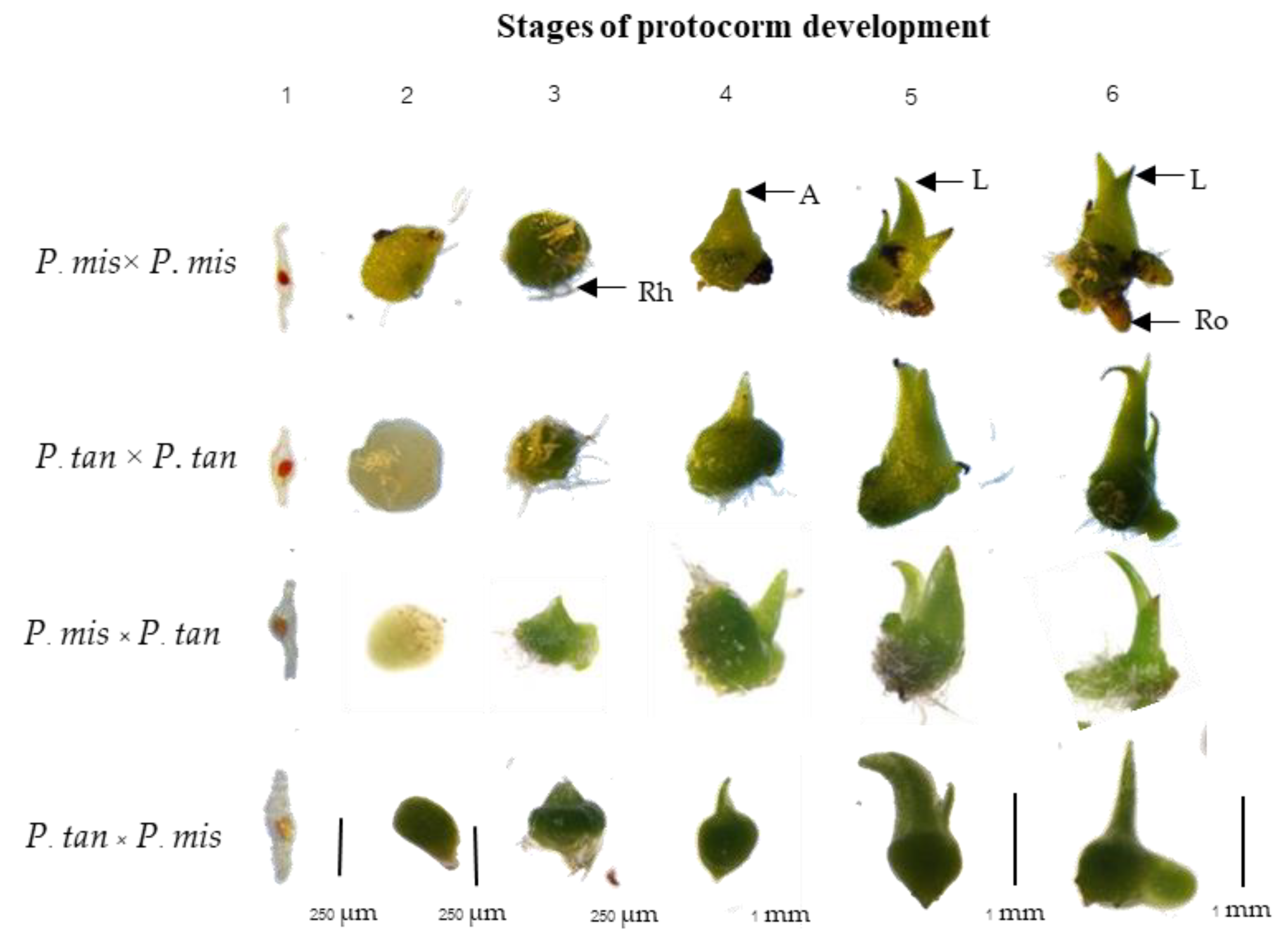
3.3. Comparative Characteristics of Pod and Embryo among Inter- and Intraspecific Hybridizations

Observations on average time for fully ripe pods (as indicated by pod dehiscence) varied from 50 to 147 days (after pollination) depending on the type of hybridization. The longest period (147 days) for pod ripening was noticed from intraspecific hybrids of P. tankervilliae followed by interspecific hybrids between P. tankervilliae (♀) and P. mishmensis (♂) (105 days), while the shortest period for pod ripening was observed in intraspecific hybrids of P. mishmensis (50 days) and interspecific hybrids between P. mishmensis (♀) and P. tankervilliae (♂) (60 days) (Table 2).
Average pod sizes as described by length and width received from both intra- and interspecific pollination were significantly different. Besides, pod weight from different pollination types was also significantly different and related to pod sizes. The highest pod weight was found in intraspecific hybrids of P. tankervilliae (10 g), while the lowest was recorded in intraspecific hybrids of P. mishmensis (4.1 g). When considering the interspecific hybridization, results showed no significant differences in pod weight of interspecific hybrids between P. tankervilliae and P. mishmensis and their reciprocal cross. However, they gave lower pod weight than intraspecific hybrids of P. tankervilliae. By contrast, pod weight from interspecific hybrids between P. tankervilliae and P. mishmensis was higher than intraspecific hybrids of P. mishmensis. Furthermore, the longer (145.7 µm) and wider (87.7 µm) embryo size were obtained from intraspecific hybrids of P. tankervilliae, whereas shorter (67.3 µm) and narrower (46.5 µm) embryo size were detected in interspecific hybrids between P. mishmensis (♀) and P. tankervilliae (♂).
The highest germination (94.8%) was found in the interspecific hybrid between P. mishmensis (♀) and P. tankervilliae (♂), whereas lower seed germination was recorded from intraspecific hybridization of P. mishmensis (72.2%) and P. tankervilliae (70.8%), while interspecific hybridization between P. tankervilliae (♀) and P. mishmensis (♂) (77.3%) showed no significant difference (Table 1).
Protocorm development stages during in vitro germination are illustrated in Figure 1. Results showed that the percentage of protocorm development from stage 1 to stage 3 was significantly different in almost all breeding types. Percentage of protocorm development in stages 4 and 5 were not significantly different for all breeding types. Hybrid seeds derived from interspecific hybrids between P. mishmensis (♀) and P. tankervilliae (♂) developed slowly into swelling protocorms, with the lowest first development stage (5.1%) and subsequently returned higher results for development stages 2–5 than the other breeding types. The highest protocorm development in stage 6 (10.6%) was observed in interspecific hybrids between P. mishmensis (♀) and P. tankervilliae (♂) (Table 3).

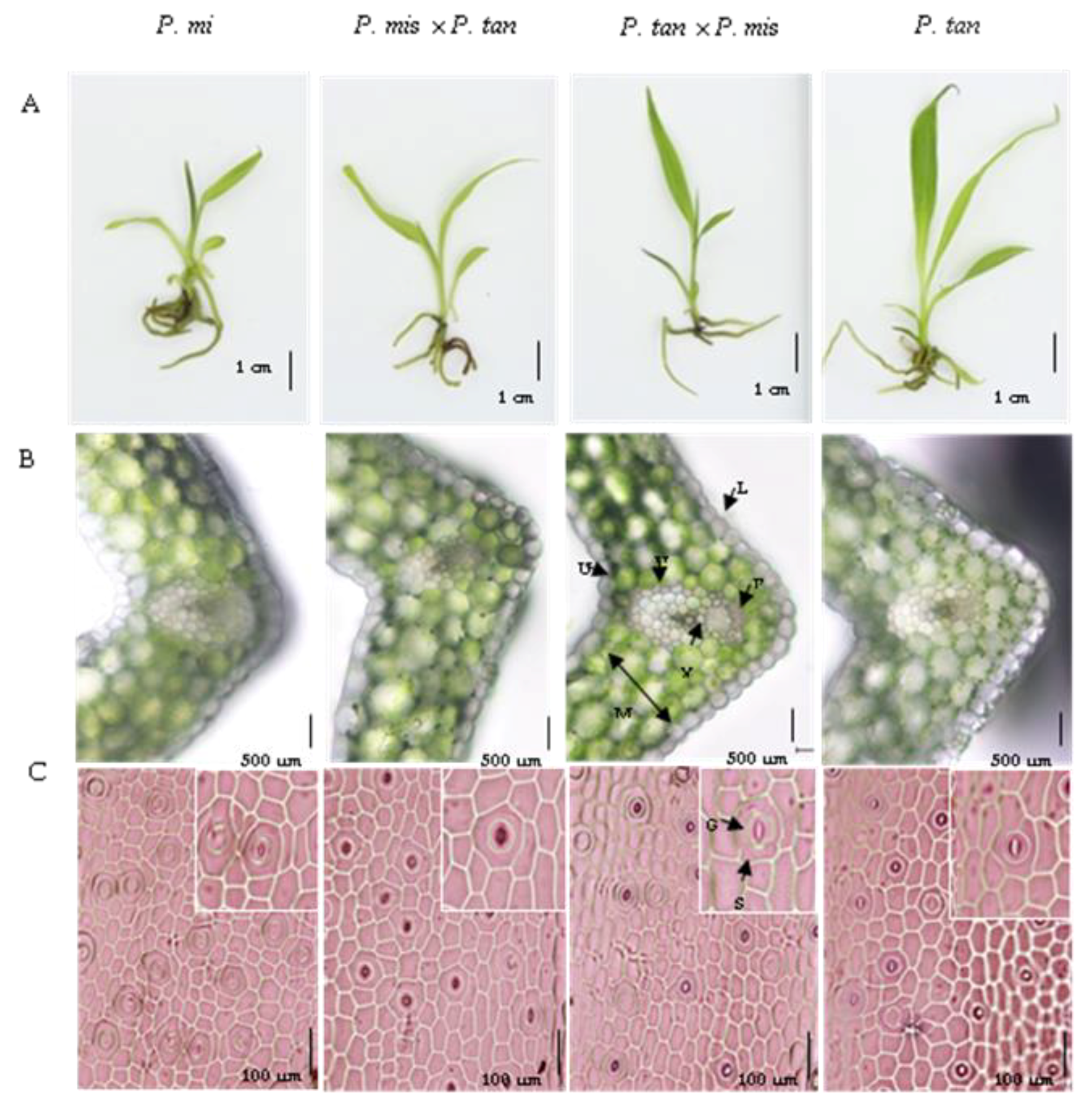
3.4. Morphology and Leaf Anatomy of Young Hybrid Seedlings

There were significant differences in leaf area among intra- and interspecific hybrids between P. mishmensis and P. tankervilliae. Intraspecific hybrids of P. tankervilliae showed higher leaf area than P. mishmensis, while interspecific hybrids using P. tankervilliae as the female parent gave higher leaf area than their reciprocal cross. Seedling leaf size from intra- and interspecific hybrids of P. mishmensis and P. tankervilliae seedlings showed significant differences in length and width, while leaf width was different in all breeding types. Furthermore, the thickest leaf (294.5 µm) was found in intraspecific hybrids of P. mishmensis, while no significant difference in leaf blade thickness of other intra- and interspecific pollination types was detected. The highest stomatal density (35.2 µm) was observed in intraspecific hybrids of P. mishmensis, while the lowest stomatal density (24.8 µm) was found in interspecific hybrids between P. tankervilliae (♀) and P. mishmensis (♂). However, stomatal density of intraspecific hybrids of P. tankervilliae and interspecific hybrids between P. mishmensis (♀) and P. tankervilliae (♂) were not significantly different. The highest stomatal length (44.2 µm) was observed in interspecific hybrids between P. mishmensis (♀) and P. tankervilliae (♂), while the lowest stomatal length (34.0 µm) was found in intraspecific hybrids of P. mishmensis. Likewise, the highest stomatal width (38.2 µm) was recorded in intraspecific hybrids of P. tankervilliae, while the lowest (32.8 µm) was obtained in intraspecific hybrids of P. mishmensis. The highest number of subsidiary cells per stomatal complex (4.1) was found in interspecific hybrids between P. mishmensis (♀) and P. tankervilliae (♂), while the lowest number (3.4) was found in interspecific hybrids between P. tankervilliae (♀) and P. mishmensis (♂). No significant difference in the number of subsidiary cells per stomatal complex was noticed in intraspecific hybrids of P. mishmensis (3.8) and P. tankervilliae (3.8) (Table 4, Figure 2).

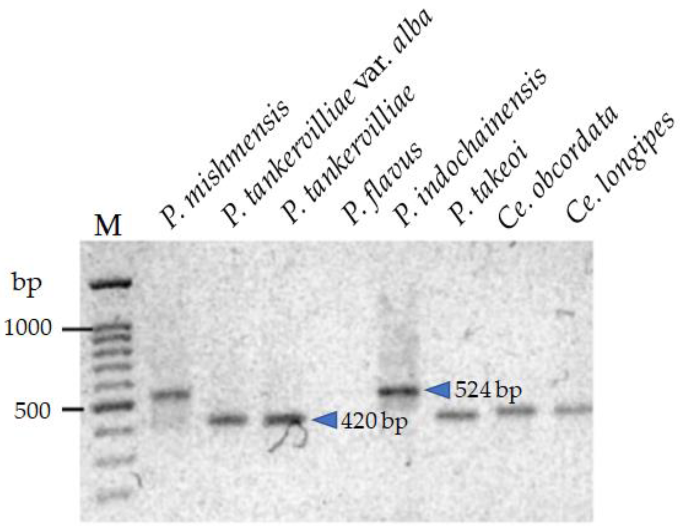
3.5. Development of Sequence-Characterized Amplified Region (SCAR) Markers for Identification of Phaius Hybrids

For RAPD primer screening, total genomic DNA of 10 orchid species in the Calanthe group comprising Calanthe (C. cardioglossa Schltr. (CcaRK01), C. masuca (D. Don) Lindl. (CmaRK01), C. lyroglosa Rchb.f. (ClyRK01), C. triplicata (Willemet) Ames (CtrRK01)), Phaius (P. tankervalliae (Banks) Blume (PtanRK01), P. tankervalliae (Banks) Blume var. alba (PtanaRK01), P. mishmensis (Lindl. and Paxton) Rchb.f. (PmisRK01), P. flavus (Blume) Lindl. (PflRK01)) and Cephalantheropsis (Ce. obcordata (Lindl.) Ormerod (CeobRK01) and Ce. longipes Hook.f. Ormerod (Celo01)) were amplified using 20 selected RAPD primers. OPX12, OPX13, OPY02 and OPY20, were successfully amplified in all species mentioned above, with sizes ranging from 200 to 1800 bp. These primers generated 51 polymorphic bands out of 70 (72.86%) with a mean of 18 amplicons per primer.
The 4 primers of RAPD markers showed specific DNA bands useful for the development of SCAR markers, except bands from the primerOPY20. In total, Only 7 specific bands were extracted from agarose gel, and all were successfully cloned. Recombinant DNA from each clone was sequenced and blasted with nucleotide BLAST. The 3 different sizes of DNA sequences were generated; 382bp from C. masuca with primer OPX12 and 570 and 600 bp from P. mishmensis and P. tankervilliae with primer OPY02, respectively. These sequences were used to design 13 primers for SCAR marker development.
Five out of 13 SCAR primer pairs developed from P. mishmensis, including Pmis524 (524 bp), were successfully amplified. These primers were used to validate with six Phaius species comprising P. mishmensis, P. tankervilliae, P. tankervilliae var. alba, P. flavus, P. indochinensis and P. takeoi, and two species of the genus Cephalantheropsis, Ce. obcordata and Ce. longipes. Two fragment sizes of 524 bp and 420 bp were generated from Pmis524. Interestingly, the 524 bp fragment was only found in P. mishmensis and P. indochinensis, while the 420 bp fragment was found in all other species, except for P. flavus, which showed none of the fragments (Figure 3). Therefore, Pmis524 SCAR primer was chosen to identify interspecific hybrids between P. mishmensis and P. tankervilliae.

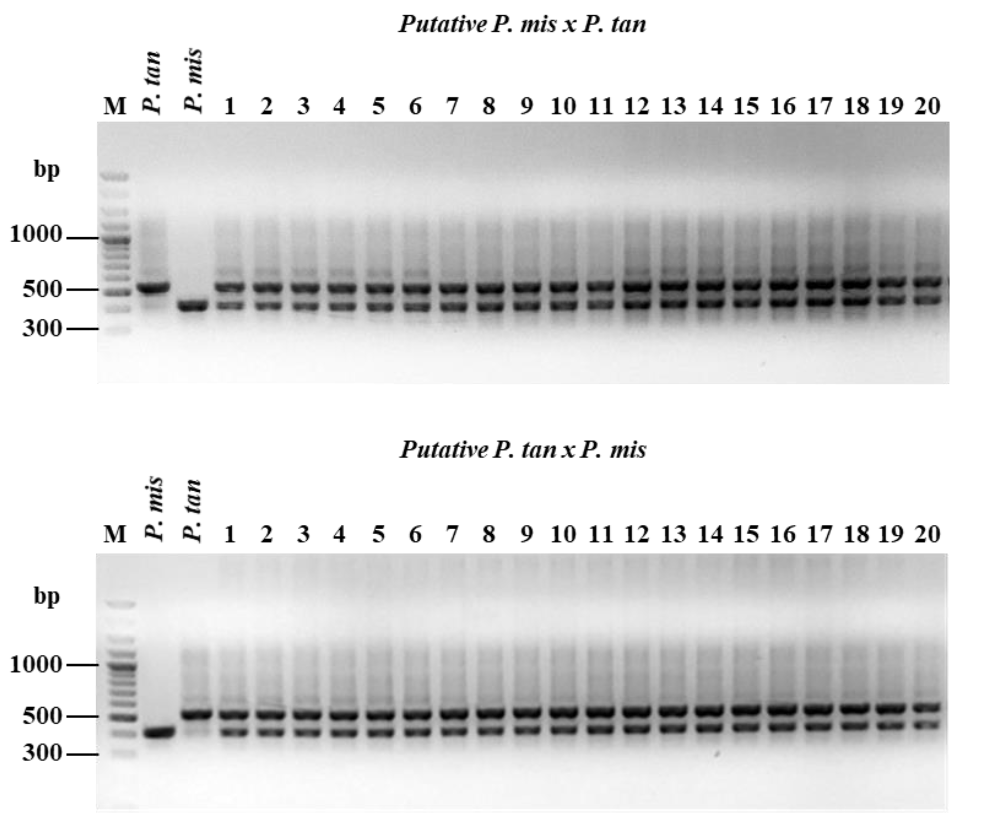
3.6. Confirmation of SCAR Primers with Hybrids Determination and Confirmation of Putative Hybrids were Conducted Using Pmis524 SCAR Primer Pairs

(Pmis524forward5′CTATGAAAGGCAGTCTCCAG3′, Pmis 524 reverse 5′GAACAATGAAGCTGAGTACG3′) with 55 °C of annealing temperature. The PCR was performed under the same condition of RAPD screening as mentioned above. The 21 putative hybrids from P. mishmensis (♀) × P. tankervilliae (♂) and another 21 putative hybrids from reciprocal cross-pollination of both species were randomly selected from seedlings under tissue culture condition. The PCR amplification showed the specific fragment of P. mishmensis and P. tankervilliae shown in Figure 4. This suggests that maternal and paternal species showed different fragment size; hence, 21 putative hybrids should present both fragment sizes. From the results, all hybrids exhibited two fragment sizes in both direct and reciprocal crosses of P. mishmensis and P. tankervilliae (Figure 4). Overall, Pmis524 was the perfect single-SCAR primer for detecting hybrids.

4. Discussion

Interspecific and intergeneric hybrids are feasible in the Orchidaceae family [22,23]. Terrestrial orchids are now being improved to produce new varieties for commercialization. The Calanthe group, including the Phaius genus, has been used in breeding programs to improve the form, color, shape and size of hybrids [24]. In this study, the development of intraspecific and interspecific hybridization between P. mishmensis and P. tankervilliae including their reciprocal cross was successful. This suggests that both species are polyphyletic, with genetic similarity indicated by molecular taxonomy [25,26]. Moreover, these two species are closely related in adjacent chromosome numbers of P. tankervilliae (2n = 48) [27] and P. mishmensis (2n = 44) [28]. Self-incompatibility and cross ability calculated by the index of self-incompatibility (ISI) indicated that P. mishmensis and P. tankervilliae are self-compatible orchids. Previous studies showed that cross-pollination between P. tankervilliae (♀) and P. flavus (♂) and their reciprocal cross, as well as self-pollinated P. tankervilliae, formed capsules with successful seed development, indicating cross compatibility among Phaius species and self-compatibility within species [9]. Pod setting provides a common measure of reproductive success in orchid breeding [29]. The highest percentage of pod setting was observed in P. mishmensis (♀) × P. tankervilliae (♂) whereas the lowest was found in their reciprocal cross. Lower pod setting percentage in the successful cross may be due to the incompatibility effect [30]. The lowest embryo formation and seed viability percentage detected in interspecific hybrids between P. tankervilliae (♀) × P. mishmensis (♂) might be due to the occurrence of either pre- or post-zygotic barriers within the Phaius genus for reproductive isolation between species and lead to unsuccessful hybridizations [31]. Similar results on the incompatibility effect [32,33] were also reported in Dendrobium [34] and Octomeria [35].
Protocorm development stages during in vitro seed germination of intraspecific and interspecific hybridization between P. mishmensis × P. tankervilliae and their reciprocal cross were different. The highest embryo formation and seed viability percentage were obtained from the intraspecific hybrids of P. tankervilliae, whereas the highest percentage of seed germination, as well as faster and higher percentage of protocorm development into the complete seedling (stage 6), was observed from interspecific hybrids between P. mishmensis (♀) × P. tankervilliae (♂) than from their intraspecific hybrids. This might be due to an inbreeding depression [36]. Similar results presented in previous studies reported that orchid seeds from cross-pollination provided higher germination rates and protocorm development than from self-pollination [37,38,39,40].
Orchid hybrids usually demonstrate transitional features from their parental species and some characteristics of one parent may predominate [41]. In this study, different leaf size and anatomy of in vitro young hybrid seedlings of P. mishmensis and P. tankervilliae were indicated by leaf length, width, area, thickness, stomatal density and stomatal size. Results showed that interspecific hybrids and their reciprocal crosses were evenly matched to their parental species. Similar results were also observed in Epipactis [42], Epidendrum [43] and Habenaria [44].
DNA products were amplified using RAPD primers followed by the previous protocol [45] and 20 primer pairs were selected in this study, though these primers only relied on previous detection of 3 orchid species via the RAPD technique. Molecular markers for identifying Phaius species have been previously studied using the SRAP marker technique. These SRAP markers can be used to separate Phaius species from each other, whereas P. mishmensis and P. indochinensis were placed in the same clade [46]. However, our results indicated that the development of SCAR markers to identify interspecific hybrids between P. mishmensis and P. tankervilliae was feasible, effective and successful. SCAR molecular markers have advantages for species and hybrid identification in orchids [47] and bamboo [48]. Application of SCAR markers to confirm hybrids has also been successfully developed in three species of the genus Paphiopedilum, based on the ITS region [16] as well as the use of multiplex PCR to test for hybrids.

5. Conclusions

Herein, an efficient method for interspecific hybridization between P. mishmensis (Lindl. and Paxton) Rchb. f. and P. tankervilliae (Banks) is described. Successful interspecific hybridization between P. mishmensis and P. tankervilliae and their reciprocal cross produced new seedlings that propagated well under in vitro germination when compared to their parental species. Development of the SCAR marker technique for interspecific hybrid identification of Phaius species is an effective and practicable method for molecular hybrid detection. SCAR marker technique can be efficiently used for hybrid detection at the early stage of growing plantlets before flowering stage. This study will benefit the hybrid breeding program of the Thai Calanthe group for further commercial uses in agriculture practices.

Author Contributions

T.P. conducted the experiments and prepared the draft manuscript. M.N. designed the experiments and suggested research results. S.H. revised and edited the manuscript. P.I. suggested research experiments and edited the results. A.K. identified the supporting facilities, discussed the research results and edited and finalized the manuscript. All authors have read and agreed the published version of this manuscript.

Funding

This research was funded by the Capacity Building Program for New Researcher 2018, National Research Council of Thailand (NRCT) (Contract number: NRCT(DPARB)21/2018), and partly funded by budget support from the Faculty of Science, Naresuan University, Thailand.

Institutional Review Board Statement

Not applicable.

Informed Consent Statement

Not applicable.

Data Availability Statement

Not applicable.

Acknowledgments

The authors would like to thank Charun Maknoi for providing plant samples and thanking to Yash Munnalal Gupta and Assistant Professor Kittasak Buddhachat for their technical support.

Conflicts of Interest

The authors declare no conflict of interest.

References

  1. Christenhusz, M.J.; Fay, M.F.; Chase, M.W. Plants of the World: An Illustrated Encyclopedia of Vascular Plants; University of Chicago Press: Chicago, IL, USA, 2017; p. 816. [Google Scholar]
  2. Kurzweil, H. A Precursory study of the Calanthe Group (Orchidaceae) in Thailand. Natl. Parks. Adansonia 2010, 32, 57–107. [Google Scholar] [CrossRef]
  3. Santisuk, T.; Larsen, K. Flora of Thailand; Orchidaceae 1 (Cypripedioideae, Orchidoideae, Vanilloideae); The Forest Herbarium, National Park, Wildlife and plant Conservation Department: Bangkok, Thailand, 2011; p. 12. [Google Scholar]
  4. Santisuk, T.; Chayamarit, K.; Pooma, R.; Sudee, S. Thailand Red Data: Plants; Integrated Promotion Technology: Bangkok, Thailand, 2006; p. 214. [Google Scholar]
  5. Tomonari, H.; Godo, T.; Miyoshi, K.; Ishikawa, K.; Ishikawa, M.; Mii, M. Cryopreservation and low-temperature storage of seeds of Phaius tankervilleae. Plant Biotechnol. Rep. 2009, 3, 103–109. [Google Scholar]
  6. Yam, T.W.; Arditti, J. History of orchid propagation: A mirror of the history of biotechnology. Plant Biotechnol. Rep. 2009, 3, 1–56. [Google Scholar] [CrossRef] [Green Version]
  7. Chugh, S.; Guha, S.; Rao, I.U. Micropropagation of orchids: A review on the potential of different explants. Sci. Hortic. 2009, 122, 507–520. [Google Scholar] [CrossRef]
  8. Lepick, N. NYBG’s orchid show Singapore. Mos Newslett. 2019, 1–15. [Google Scholar]
  9. Devadas, R.; Pamarthi, R.K.; Meitei, A.L.; Pattanayak, S.L.; Sherpa, R. Morphological description of novel Phaius primary hybrid (Orchidaceae). J. Exp. Biol. Agric. Sci. 2019, 7, 138–147. [Google Scholar] [CrossRef]
  10. Pérez, T.; Albornoz, J.; Domínguez, A. An evaluation of RAPD fragment reproducibility and nature. Mol. Ecol. 1998, 7, 1347–1357. [Google Scholar] [CrossRef] [PubMed]
  11. Paran, I.; Michelmore, R.W. Development of reliable PCR-based markers linked to downy mildew resistance genes in lettuce. Theor. Appl. Genet. 1993, 85, 985–993. [Google Scholar] [CrossRef] [PubMed]
  12. Sairkar, P.K.; Sharma, A.; Shukla, N.P. SCAR marker for identification and discrimination of Commiphora wightii and C. myrrha. Mol. Biol. Int. 2016, 1, 1–10. [Google Scholar] [CrossRef]
  13. Lee, M.Y.; Doh, E.J.; Park, C.H.; Kim, Y.H.; Kim, E.S.; Ko, B.S.; Ok, S.E. Development of SCAR marker for discrimination of Artemisia princeps and A. argyi from other Artemisia herbs. Biol. Pharm. Bull. 2006, 29, 629–633. [Google Scholar] [CrossRef] [Green Version]
  14. Devaiah, K.M.; Venkatasubramanian, P. Development of SCAR marker for authentication of Pueraria tuberosa (Roxb. Ex. Willd.) DC. Curr. Sci. 2008, 94, 1306–1309. [Google Scholar]
  15. Julio, E.; Verrier, J.; Dorlhac de Borne, F. Development of SCAR markers linked to three disease resistances based on AFLP within Nicotiana tabacum L. Theor. Appl. Genet. 2006, 112, 335–346. [Google Scholar] [CrossRef] [PubMed]
  16. Yung, W.S.; Liaoa, Y.J.; Hunga, Y.S.; Changa, J.C.; Sung, J.M. Development of ITS sequence based SCAR markers for discrimination of Paphiopedilum armeniacum, P. micranthum, P. delenatii and their Hybrids. Sci. Hortic. 2011, 127, 405–410. [Google Scholar]
  17. Rodger, J.G.; Ellis, A.G. Distinct effects of pollinator dependence and self-incompatibility on pollen limitation in South African biodiversity hotspots. Biol. Lett. 2016, 12, 1–4. [Google Scholar] [CrossRef] [Green Version]
  18. Takao, H.S.; Custódio, C.C.; Seaton, P.T.; Timothy, R.M.; Machado-Neto, N.B. Improved assessment of viability and germination of Cattleya (Orchidaceae) seeds following storage. In Vitro Cell. Dev. Biol. Plant 2012, 48, 27–36. [Google Scholar]
  19. Vacin, E.F.; Went, F.W. Some pH changes in nutrient solutions. Bot. Gazette. 1949, 110, 605–613. [Google Scholar] [CrossRef]
  20. Miyoshi, K.; Mii, M. Enhancement of seed germination and protocorm formation in Calanthe discolor (Orchidaceae) by NaOCl and polyphenol absorbent treatments. Plant Tiss. Cult. Lett. 1995, 12, 267–272. [Google Scholar] [CrossRef]
  21. Doyle, J.J.; Doyle, J.D. Isolation of plant DNA from fresh tissue. Electron. J. Biotechnol. 1990, 12, 13–15. [Google Scholar]
  22. Arditti, J. An history of orchid hybridization, seed germination and tissue culture. Bot. J. Linn. Soc. 1984, 89, 359–381. [Google Scholar] [CrossRef]
  23. Grantham, M.A.; Ford, B.A.; Worley, A.C. Pollination and fruit set in two rewardless slipper orchids and their hybrids (Cypripedium, Orchidaceae): Large yellow flowers outperform small white flowers in the northern tall grass prairie. Plant Biol. 2019, 21, 997–1007. [Google Scholar] [CrossRef]
  24. Udomdee, W.; Kongsawad, P.; Darak, R.; Somrit, P. Collection and evaluation on ex situ conservation of Phaius orchid. Khon Kaen Agr. J. 2014, 3, 529–534. [Google Scholar]
  25. Harrison, D.K.; Kwan, H.; Johnston, M.E.; Harris, W.K. Molecular taxonomy of the Australian swamp orchids (Phaius spp.). Acta Hortic. 2005, 694, 121–124. [Google Scholar] [CrossRef]
  26. Zhai, J.W.; Zhang, G.Q.; Li, L.; Wang, M.; Chen, L.J.; Chung, S.W.; Rodriguez, F.J.; Ortega, J.F.; Lan, S.R.; Xing, F.W.; et al. A new phylogenetic analysis sheds new light on the relationships in the Calanthe alliance (Orchidaceae) in China. Mol. Phylogenet. Evol. 2014, 77, 216–222. [Google Scholar] [CrossRef] [PubMed]
  27. Teoh, S.B. Cytological studies in Malayan members of the Phaius tribe (Orchidaceae). Ii. Meiotic and b-Chromosomes. Caryologia 1980, 33, 483–493. [Google Scholar] [CrossRef]
  28. Jones, N. New species with B chromosomes discovered since 1980. Nucleus 2017, 60, 263–281. [Google Scholar] [CrossRef]
  29. Proctor, H.C. Effect of pollen age on fruit set, fruit weight and seed set in three orchid species. Can. J. Bot. 1998, 76, 420–427. [Google Scholar]
  30. Shiau, Y.J.; Sagare, A.P.; Chen, U.C.; Yang, S.R.; Tsay, H.S. Conservation of Anoectochilus formosanus Hayata by artificial cross pollination and in vitro culture of seeds. Bot. Bull. Acad. Sin. 2002, 43, 123–130. [Google Scholar]
  31. Edmands, S. Does parental divergence predict reproductive compatibility. Trends Ecol. Evol. 2002, 17, 520–527. [Google Scholar] [CrossRef]
  32. Devi, J.; Deka, P.C. Pollen viability, stigma receptivity and cross compatibility of some Indian orchids. J. Orchid Soc. India. 1992, 6, 79–84. [Google Scholar]
  33. Pershinaa, L.A.; Trubacheeva, N.V. Interspecific incompatibility in the wide hybridization of plants and ways to overcome it. Russ. J. Genet. Appl. Res. 2017, 7, 358–368. [Google Scholar] [CrossRef]
  34. Johansen, B. Incompatibility in Dendrodium (Orchidaceae). Bot. J. Linn. Soc. 1990, 103, 165–196. [Google Scholar] [CrossRef]
  35. Barbosa, A.R.; de Melo, M.C.; Borba, E.L. Self-incompatibility and myophily in Octomeria (Orchidaceae, Pleurothallidinae) species. Plant Syst. Evol. 2009, 283, 1–8. [Google Scholar] [CrossRef]
  36. Charlesworth, D.; Charlesworth, B. Inbreeding depression and its evolutionary consequences. Ann. Rev. Ecol. Evol. Syst. 1987, 18, 237–268. [Google Scholar] [CrossRef]
  37. Bellusci, F.; Pellegrino, G.; Musacchio, A. Different levels of inbreeding depression between outcrossing and selfing Serapias species. Biol. Plant 2009, 53, 175–178. [Google Scholar] [CrossRef]
  38. Petit, S.; Jusaitis, M.; Bickerton, D. Effect of pollen load, self-pollination and plant size on seeds and germination in the endangered pink-lipped spider orchid, Caladenia behrii. Aust. J. Bot. 2009, 57, 307–314. [Google Scholar] [CrossRef]
  39. Pakum, W.; Watthana, S.; Srimuang, K.; Kongbangkerd, A. Influence of medium component on in vitro propagation of Thai’s endangered orchid: Bulbophyllum nipondhii Seidenf. Plant Tiss. Cult. Biotechnol. 2016, 25, 37–46. [Google Scholar] [CrossRef] [Green Version]
  40. Kunakhonnuruk, B.; Inthima, P.; Kongbangkerd, A. In vitro propagation of Epipactis flava Seidenf. an endangered rheophytic orchid: A first study on factors affecting asymbiotic seed germination, seedling development and greenhouse acclimatization. Plant Cell Tiss. Organ Cult. 2018, 135, 419–432. [Google Scholar] [CrossRef]
  41. Moccia, M.D.; Widmer, A.; Cozzolino, S. The strength of reproductive isolation in hybridizing food deceptive orchids. Mol. Ecol. 2007, 16, 2855–2866. [Google Scholar] [CrossRef]
  42. Busse, A.J.; Gola, E.M. Morphological variability of Helleborines. I. Diagnostic significance of morphological features in Epipactis helleborine (L.) Crantz, Epipactis atrorubens (Hoffm.) Besser and their hybrid, Epipactis × schmalhausenii Richt. (Orchidaceae, Neottieae). Acta Soc. Bot. Pol. 2010, 79, 207–213. [Google Scholar] [CrossRef] [Green Version]
  43. Devadas, R.; Medhi, R.P.; Das, S.P. Interspecific hybrid developed in Epidendrum orchid from the cross E. radicans Pav. Ex. Lindl. × E. xanthinum Lindl. J. Hortic. Sci. 2010, 5, 144–147. [Google Scholar]
  44. Sinumporn, P.; Narumi-Kawasaki, T.; Fukai, S. Development of interspecific hybrids between Habenaria radiata and Habenaria rhodocheila complex. Adv. Hort. Sci. 2020, 34, 3–10. [Google Scholar]
  45. Punjansing, T.; Chaichanachap, N.; Sanitchon, J.; Pinta, W.; Nakkuntod, M. Genetic diversity of some species in the genus Calanthe analyzed by RAPD markers. Acta Hortic. 2017, 1167, 323–330. [Google Scholar] [CrossRef]
  46. Nutthapornnitchakul, S.; Peyachoknagul, S.; Sangin, P.; Kongbungkerd, A.; Punjansing, T.; Nakkuntod, M. Genetic relationship of orchids in the Calanthe group based on sequence-related amplified polymorphism markers and development of sequence-characterized amplified regions markers for some genus/species identification. Agric. Nat. Resour. 2019, 53, 340–347. [Google Scholar]
  47. Handa, T. Utilization of molecular markers for ornamental plants. J. Am. Soc. Hortic. Sci. 1998, 67, 1197–1199. [Google Scholar] [CrossRef]
  48. Malay, D.; Bhattacharya, S.; Pal, A. Generation and characterization of SCARs by cloning and sequencing of RAPD products: A strategy for species- specific marker development in bamboo. Ann. Bot. 2005, 95, 835–841. [Google Scholar]
Figure 1. Protocorm development stages after asymbiotic seed germination of P. mishmensis and P. tankervilliae and interScheme 1. No development of seed; Stage 2, embryo enlargement and rupture from the testa; Stage 3, appearance of rhizoids (Rh) (germination); Stage 4, apical meristem (A) production; Stage 5, emergence of the leaf (L); Stage 6, emergence of the root (Ro).
Agriculture 11 00306 g001
Figure 2. Characteristics of plantlet (A), leaf cross section (B) and leaf epidermis containing stomata complexes of P. mishmensis, P. tankervilliae and their F1 interspecific hybrids (C): G, guard cell; L, lower epidermis; M, mesophyll; P, phloem; S, subsidiary cell; U, upper epidermis; X, xylem.
Agriculture 11 00306 g002
Figure 3. PCR products showed specific bands from different species using Pmis524 SCAR primer, M: 100 bp DNA ladder (Gene Direx, Jhunan Township, Taiwan).
Agriculture 11 00306 g003
Figure 4. Confirmation of interspecific hybrid by PCR using SCAR primers designed for differentiation of P. mishmensis and P. tankervilliae.
Agriculture 11 00306 g004
Table 1. Pod setting, seed set, seed viability and seed germination of intra- and interspecific hybridization between Phaius mishmensis (M) and P. tankervilliae (T).
Treatments% Pod
Setting (n)
% Embryo Formation% Seed
Viability
% In Vitro Seed Germination
M (♀) × M (♂)62.5 (40)34.0 ± 0.2 c29.9 ± 1.9 c72.2 ± 4.1 b
T (♀) × T (♂)60.0 (40)100.0 ± 0.0 a99.8 ± 0.2 a70.8 ± 3.1 b
M (♀) × T (♂)80.0 (20)48.3 ± 0.7 b44.6 ± 1.0 b94.8 ± 0.8 a
T (♀) × M (♂)25.0 (20)9.3 ± 0.8 d8.2 ± 0.6 d77.3 ± 6.8 b
Values are means ± SE. Different letters within the same column indicate significant differences analyzed by Duncan’s new Multiple Range Test (DMRT) at p < 0.05 and the number in parentheses mean the number of pollinated flowers.
Table 2. Period of pod dehiscence and characteristics of intra- and interspecific hybridization between Phaius mishmensis (M) and Phaius tankervilliae (T).
TreatmentAverage Days of Pod Dehiscence (Days after Pollination)Pod Size (cm)Pod Weight (g)Embryo Size (µm)
WidthLengthWidthLength
M (♀) × M (♂)501.9 ± 0.5 ab4.7 ± 0.4 b4.1 ± 0.8 c55.4 ± 0.4 b94.8 ± 1.5 c
T (♀) × T (♂)1472.4 ± 0.7 a5.3 ± 1.0 ab10.0 ± 3.8 a87.7 ± 1.6 a145.7 ± 1.5 a
M (♀) × T (♂)602.0 ± 0.2 ab5.1 ± 0.4 b6.8 ± 1.7 b46.5 ± 0.6 c67.3 ± 0.9 d
T (♀) × M (♂)1051.6 ± 0.5 b5.4 ± 0.2 a6.2 ± 1.8 bc86.2 ± 0.9 a113.0 ± 6.9 b
Values are means ± SD. Different letters within the same column indicate significant differences analyzed by DMRT at p < 0.05.
Table 3. Protocorm development of intra- and interspecific hybrids of P. mishmensis (M) and P. tankervilliae (T) after cultured on VW medium for 16 weeks.
TreatmentStage of Protocorm Development (%)
123456
M (♀) × M (♂)27.7 ± 4.1 a21.5 ± 1.5 c16.6 ± 1.7 a20.2 ± 2.6 ns10.6 ± 2.1 ns3.1 ± 0.5 b
T (♀) × T (♂)29.1 ± 3.1 a28.0 ± 3.1 b11.1 ± 1.7 c 17.5 ± 2.710.4 ± 1.63.7 ± 0.7 b
M (♀) × T (♂)5.1 ± 0.8 b38.0 ± 1.4 a14.6 ± 1.7 ab 22.0 ± 3.09.5 ± 0.210.6 ± 1.7 a
T (♀) × M (♂)22.6 ± 6.8 a23.1 ± 1.2 bc12.6 ± 1.3 ab 21.1 ± 3.114.8 ± 3.55.5 ± 1.7 b
The number of seeds in each treatment are 500 seeds. Values are means ± SE. Different letters within the column indicate significant differences analyzed by DMRT at p < 0.05.
Table 4. Comparison of leaf morphology and anatomy of Phaius hybrids.
Plants TestedLeaf Size (cm)Leaf Area (cm2)Thickness
of Leaf
(µm)
Stomata Density (Stomata/mm2)Stomatal Size (µm)No. of Subsidiary Cell/Stomata Complex
LengthWidthLengthWidth
M(♀) × M(♂)2.4 ± 0.1 c0.8 ± 0.1 a 1.6 ± 0.1 b 294.5 ± 6.2 a 35.2 ± 0.4 a34.0 ± 0.4 c 32.8 ± 0.6 c3.8 ± 0.1 b
T(♀) × T(♂)4.5 ± 0.2 a0.6 ± 0.1 b 2.5 ± 0.2 a 212.6 ± 7.3 b 27.1 ± 0.5 b43.4 ± 1.2 a 38.2 ± 1.0 a3.8 ± 0.1 b
M(♀) × T(♂)3.7 ± 0.3 b0.7 ± 0.4 b 2.2 ± 0.2 b 222.9 ± 3.3 b 26.4 ± 0.9 bc44.2 ± 0.2 a 37.7 ± 0.8 a4.1 ± 0.1 a
T(♀) × M(♂)4.9 ± 0.2 a0.7 ± 0.4 b 2.3 ± 0.2 a 202.8 ± 7.8 b 24.8 ± 0.3 c40.8 ± 0.5 b 33.1 ± 0.7 b3.4 ± 0.1 c
Values are means ± SE of 3 replicates (7 leaves per replicate) except for stomata size, while number of subsidiary cell/stomata complex are mean ± SD of 30 stomata (measured from 10 leaves, 3 stomata per leaf). Data were derived from leaves at 6 months after germination from plantlets under in vitro culture with VW medium. Different letters within the column indicate significant differences analyzed by DMRT at p < 0.05.
Publisher’s Note: MDPI stays neutral with regard to jurisdictional claims in published maps and institutional affiliations.

