Article

Phenolic Response to Walnut Anthracnose (Ophiognomonia leptostyla) Infection in Different Parts of Juglans regia Husks, Using HPLC-MS/MS

Department of Agronomy, Biotechnical Faculty, University of Ljubljana, Jamnikarjeva 101, SI-1000 Ljubljana, Slovenia
*
Author to whom correspondence should be addressed.
Submission received: 29 June 2021 / Revised: 9 July 2021 / Accepted: 11 July 2021 / Published: 13 July 2021
(This article belongs to the Special Issue Development and Cultivar Improvement of Nut Crops)

Abstract

:
This study compares the individual phenolic response of husk tissues of Juglans regia L., infected to different degrees of severity with walnut anthracnose, which is one of the most serious and widespread walnut diseases worldwide. A comparison among three differently susceptible cultivars, ‘Franquette’, ‘Milotai 10’ (‘M10’), and ‘Milotai intenziv’ (‘M10-37’), is made. In our methodology, high performance liquid chromatography coupled with mass spectrometry is used to identify and quantify the compounds. Our results show that flavanols, flavonols, and naphthoquinones account for more than 95% of the phenolic compounds identified in the walnut husk. The higher total analyzed phenolic content in tissues is more affected by walnut anthracnose confirmed that phenolics play a major role in the plant’s response against pathogens. A difference between cultivars is observed, since French cultivar ‘Franquette’ responds differently to walnut anthracnose infection than Hungarian cultivars ‘M10’ and ‘M10-37’. Naphthoquinones and flavanols have a very similar response to walnut anthracnose infection. The resistance of cultivars may be due to the reaction time of the plant and the speed with which it recognizes the pathogen and responds quickly to the infection by containing it while it has not yet spread. Flavonols may be the most important phenolic compounds in disease control, since they respond more rapidly to infection than flavanols and naphthoquinones. They also play an inhibitory role in the early stages of viral and bacterial infections.

1. Introduction

Juglans regia L. is a highly economically important species for wood and fruit production that is susceptible to anthracnose [1]. Walnut anthracnose is caused by the ascomycetous fungus Ophignomonia leptostyla (Fr.) Ces. et de Not. (anamorph Marssonina juglandis (Lib.) Magn.), which is one of the most serious and widespread walnut diseases in almost all walnut growing areas worldwide [2]. The disease rapidly becomes severe in wet and rainy weather, especially in the spring. Walnut anthracnose can cause damage to the current season’s leaves, twigs, shoots, and husk in the form of irregular necrotic patches often surrounded by small chlorotic halos, which subsequently affect fruit yield [3]. Early infection can lead to fruit deformation and fruit being prematurely dropped. Repeated defoliation caused by walnut anthracnose can have a negative effect on plant growth, sometimes resulting in plant collapse [4,5]. Walnut production is limited by walnut anthracnose, and field losses can reach up to 50% [2].
O leptostyla usually overwinters as ascocarps in fallen leaves and rarely as mycelium on lesions of fruits and twigs. Primary infections occur in early spring, mainly by ascospores. Mitospores are formed in blackish, translucent, punctate acervuli, often arranged in concentric rings on the abaxial leaf, husk, shoot, or twig surface [6]. The spots on the walnut husks are sunken and smaller than those on the leaves [4]. Several secondary cycles occur during the growing season of the walnut [6].
The disease control management of walnut anthracnose is to date mainly based on chemical pesticides. New agronomic, genetic, and biological approaches for controlling the disease have been proposed [2], but an overall solution still needs to be found. The most efficient and green solution, an alternative to chemical management, would be selecting resistant plant genotypes [1]. To identify resistance to walnut anthracnose, several studies have been conducted using nucleotide bidding site profiling approaches. One of the studies divided 132 Juglans genotypes into three groups according to the percentage of necrotic leaf area, depending on whether they were resistant or susceptible to walnut anthracnose [7]. The variation in susceptibility to the disease is significant, but no specific genotype has been found to be highly resistant or immune to the disease. Identification of walnuts resistant to walnut anthracnose in field plantings is costly and complicated because the trees are very large and have a long juvenility, and there is no uniform method for evaluating the trees’ resistance [2,4]. An in vivo approach examining secondary metabolites would be a more appropriate, accurate, and rapid approach for determining resistance in either newly bred or established cultivars. This would result in fewer labor hours, a lesser effect of environmental conditions on the resistance studied, and fewer years and money saved, since trees would not need to reach maturity and take up valuable land and resources. A molecular marker-assisted breeding and identification of walnut genes have been proposed [8], but the method is expensive and complex. A simpler chemical basis-assisted breeding of plants with high performance liquid chromatography (HPLC) or HPLC coupled with mass spectrometry HPLC/MS for quantification, would be easier and more cost-effective.
Resistance to walnut anthracnose may be related to a specific phenolic compound or group of compounds, as reported for some economically important pests and plant diseases in general [9]. A chemical basis for the resistance to scab has been previously reported in pecan (Carya illinoinensis (Wangenh.) K. Koch) [10] and in apple (Malus domestica Borkh.) [11], and chemical response to the infection with bacteria Xanthomonas arboricola pv. juglandis was observed in walnut (Juglans regia L.) [12,13]. In C. illinoinensis, the toxic principles were identified as juglone (5-hydroxy-1,4-naphthoquinone) and hydrojuglone glucoside (4,8-dihydroxy-1-naphthalenyl-β-d-glucopyranoside or hydrojuglone-β-d-glucopyranoside), since higher levels of juglone in certain cultivars correlated with reduced scab disease in nature. Juglone and hydrojuglone glucoside were also shown to inhibit fungal growth in in vitro conditions in concentrations lower than found in the plant itself [14]. In J. regia, in a comparison between healthy husk tissue and husks infected with Xanthomonas arboricola pv. juglandis, the infected tissue contained more hydroxycinnamic acids, gallic acid, quercetin glycosides, and catechin [12]. The reaction of naphthoquinones in response to infection with Xanthomonas arboricola pv. juglandis has unfortunately not been studied in J. regia [12,13], showing no clear picture of the response of all individual phenolic compounds present in the walnut husk.
To the best of our knowledge, the mechanisms of plant response to walnut anthracnose infection are poorly understood and should be further investigated, since the use of pesticides is inefficient and undesirable [7]. The objective of our study was to investigate the phenolic content in different parts of infected husks to better understand the response of the individual phenolics to the disease. The husk was divided into four categories (infected tissue, outer margin of infected tissue, healthy tissue surrounding the infection, and healthy tissue) using three different susceptible cultivars, ‘Franquette’, ‘Milotai 10’ (‘M10’), and ‘M10-37’. Based on previous work, we expected that the infected tissue would have a higher total phenolic content and higher content of certain phenolic compounds compared to other tissues, contributing to the plant response mechanisms. Since total phenolic content does not show a clear picture of a plant’s response and mechanisms in relation to infection, individual groups and individual phenolics were also examined. Individual phenolics provided insights for further understanding which ones may be most important in the plant’s response to walnut anthracnose infection, as well as provide interesting data for simpler chemical-based breeding of plants using HPLC or HPLC/MS for quantification, which is easier and more cost-effective than molecular marker-assisted breeding and identification of walnut genes.

2. Materials and Methods

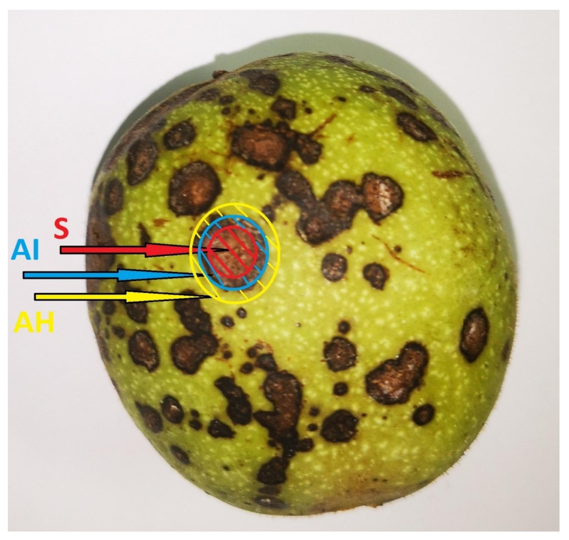
2.1. Plant Materials

Three walnut cultivars were used for the experiment, ‘M10-37’, ‘M10’ and ‘Franquette’. Samples of walnut husk with different disease severity were collected on 10 September 2020 at the Experimental Field for Nut Crops in Maribor, operating under University of Ljubljana, Slovenia (46°34′01″ N; 15°37′51″ E; 275 m a.s.l.). They were obtained from 25-year-old trees growing at a planting density of 10 m × 10 m, all under the same agronomical management, soil, and climatic conditions. Disease severity was assessed based on previous work describing disease severity in walnut husk caused by Xanthomonas arboricola pv. Juglandis [13]. ‘Franquette’ husk displayed weak infection (separated small spots, with a necrotic surface of the husk between 4 and 10%), ‘M10’ husk displayed medium infection (separated larger spots, with a necrotic surface of the husk between 11 and 25%), and ‘M10-37’ husk displayed severe infection (confluent large spots, with a necrotic surface of the husk between 26 and 50%). Samples were collected from three trees for each cultivar, for a total of three biological repetitions per analysis. Samples of fruit were collected from the middle third of the branches on the eastern side of the trees and transported in a cool box to the laboratory of the Department of Agronomy of the Biotechnical Faculty, University of Ljubljana, where they were divided into four categories. The categories were sampled as follows and shown in Figure 1: Inner spot of infected tissue, outer margin of infected tissue, healthy tissue surrounding the infection, and healthy tissue.
All samples were collected from the same husk, except for the healthy tissue, which was collected from a completely healthy husk, from the same tree, but not infected with walnut anthracnose or affected by any other disease or pathogen. Samples were carefully taken with a scalpel, removing all but approximately 0.5 mm of tissue. Samples were immediately frozen with liquid nitrogen, then lyophilized, ground to a powder using liquid nitrogen and a mortar, and stored at −20 °C before further analysis.

2.2. Extraction of the Individual Phenolic Compounds

Briefly, 200 mg of sample was extracted using 80% methanol with 3% formic acid and bi-distillate water at a 1:100 (w/v) tissue:solution ratio. The protocol followed that described by Medic et al. [15].

2.3. HPLC–Mass Spectrometry Analysis of Individual Phenolic Compounds

The phenolics were analyzed on a UHPLC system (Vanquish; Thermo Scientific, Waltham, MA, USA) with a diode array detector at 280 nm to detect hydroxycinnamic acids, hydroxybenzoic acids, flavanols, and naphthoquinones, and at 350 nm to detect flavonols. The recorded spectra were between 200 and 600 nm, and a C18 column (Gemini 150 × 4.60 mm; 3 μm; Phenomenex, Torrance, CA, USA) was used to separate the phenolics, as previously described by Medic et al. [15].
Phenolics were identified using tandem mass spectrometry (MS/MS; LTQ XL; Thermo Scientific, Waltham, MA, USA) with heated electrospray ionization (HESI) operating in negative ion mode, using the parameters as described by Medic et al. [15]. Data was acquired using Xcalibur 2.2 software (Thermo Fischer Scientific Institute, Waltham, MA, USA). Phenolics were fragmented using external standards to identify and quantify known compounds. For identification of the unknown compounds, MS fragmentation and literature data were used. Unknown compounds were quantified using similar standards. The contents of the individual phenolics are given in mg/g dry weight, and their quantification was done according to the most relevant standard.

2.4. Chemicals

The following standards were used to identify and quantify the phenolics: procyanidin B1, quercetin-3-glucoside produced by Fluka Chemie GmbH (Buchs, Switzerland); (+)catechin produced by Roth (Karlsruhe, Germany); 4-O-caffeoylquinic acid, neochlorogenic acid (3-caffeoylquinic acid), quercetin-3-galactoside, quercetin-3-rhamnoside, juglone (5-hydroxy-1,4-naphthoquinone), 1,4-naphthoquinone, gallic acid, (−)epicatechin produced by Sigma-Aldrich Chemie GmbH (Steinheim, Germany); and quercetin-3-arabinofuranoside, quercetin-3-arabinopyranoside, quercetin-3-xyloside produced by Apin Chemicals (Abingdon, UK).
The acetonitrile and formic acid for the mobile phases were HPLC-MS grade produced by Fluka Chemie GmbH (Buchs, Switzerland). The water used for all sample preparation, solutions, and analyses was bi-distilled and purified using a Milli-Q water purification system produced by (Millipore, Bedford, MA, USA).

2.5. Statistical Analysis

The data were collated using Microsoft Excel 2016, and analyzed using R commander. Three repetitions of each methodology were performed, for each cultivar. The data are expressed as means ± standard error (SE). One-way analysis of variance (ANOVA), with Tukey’s tests, was used to determine significant differences between the data. To determine the significance of the differences, statistical means at a 95% confidence level were calculated. The R commander hierarchical clustering (dendrogram) was used to determine the grouping for flavanols and naphthoquinones, using Ward’s method based on Euclidian distance.

3. Results and Discussion

3.1. Identification of Individual Phenolics

Based on the existing literature and the use of standard compounds, a total of 26 phenolic compounds were tentatively identified. Of these 26 phenolics, 10 were identified using standards. Fragmentation of both the standards and the addition of external standards were used to confirm their identities. The remaining 16 phenolics were tentatively identified according to their pseudo molecular ions ([M-H]-) and their specific fragmentation patterns. The 26 phenolic compounds identified and the standards they are expressed, as shown in Table 1.
Most of the phenolic compounds were already identified in J. regia husk by Medic et al. [16], except for juglanin B, which was previously identified in Juglans mandshurica by Huo et al. [17], and tryhidroxytertalone galloyl hexoside and dihydroxytetralone galloyl hexoside previously reported in J. mandshurica by Wang et al. [18]. These compounds are reported here for the first time in the walnut J. regia. They were also quantified for the first time in J. regia or any other Juglans genus.

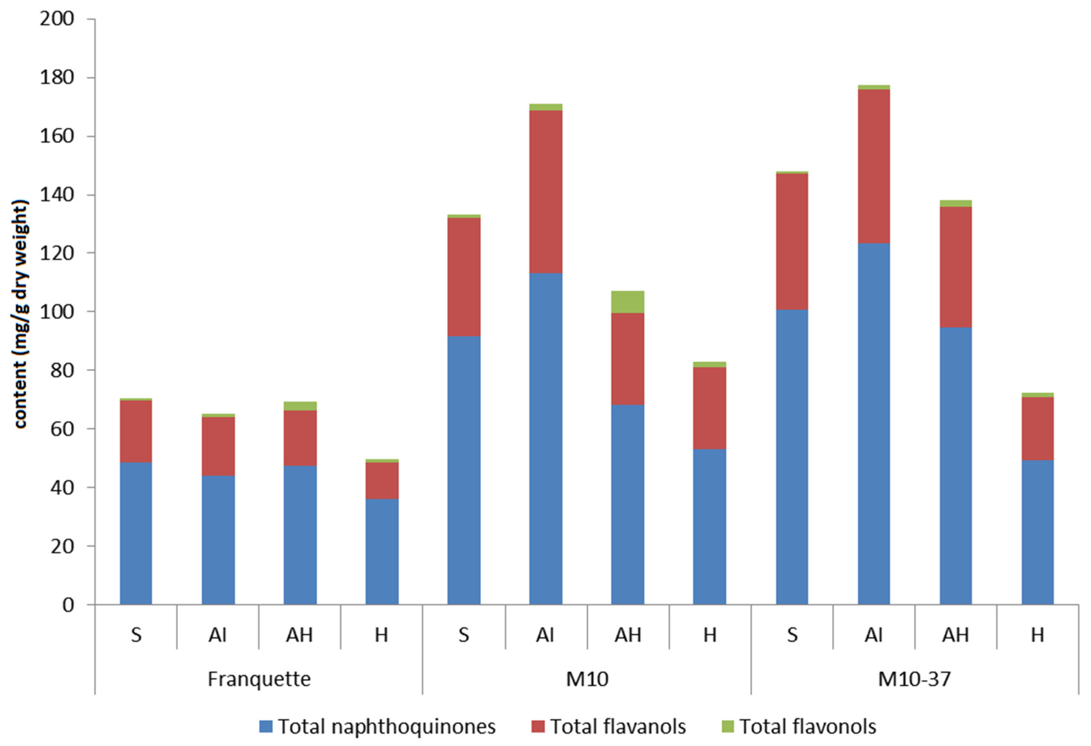
3.2. Phenolic Groups in Response to Walnut Anthracnose Infection

Contrary to prediction and previous reports on scab in M. domestica [11], total analyzed phenolic content (TAPC) was the highest in the outer margin of infected tissue (AI) and not in the inner spot of infected tissue (S) in semi-susceptible cultivar ‘M10’ and the susceptible cultivar ‘M10-37’, as can also be seen in the supplementary material (Table S1).
The resistant cultivar ‘Franquette’ showed little difference in TAPC content between S, AI, and healthy tissue surrounding the infection (AH), but the content in these tissues was greater than the TAPC content in healthy tissue (H), confirming the results of a previous study comparing healthy and infected husks in J. regia [12]. The lower TAPC content in S compared to AI could be explained by the severe infection of cultivars ‘M10-37’ and ‘M10’, since the tissue had already started to die away, the phenolic compounds are thus deteriorating with the rest of the tissue. The higher TAPC content in the tissues more affected by walnut anthracnose confirmed that phenolics play a major role in the plant’s response against pathogens, as mentioned earlier [9,19].
For some economically important pests and diseases of plants in general, it is reported that a specific phenolic group of compounds is responsible for the plant’s response [9,11,12]. Flavanols, flavonols, and naphthoquinones represent more than 95% of the phenolic compounds identified and quantified. The phenolic response of flavanols, flavonols, and naphthoquinones can be seen in Figure 2.
The difference between the cultivars can be clearly seen since the French cultivar ‘Franquette’ reacted differently to infection with walnut anthracnose than the Hungarian cultivars ‘M10’ and ‘M10-37’. This could be due to the country of origin of the cultivars, since it has previously been found that cultivars of the same origin produced similar levels of phenolic compounds compared to cultivars of different origins [15], or due to the fact that some cultivars react faster than others to the infection and contain the infection more quickly before it spreads, while others react more slowly, so that the infection has already spread and the infection is more difficult to control. This could also explain the higher content of TAPC and naphthoquinones in AH compared to AI in ‘Franquette’, while the highest content of TAPC and naphthoquinones in ‘M10’ and ‘M10-37’ is present in AI. Some authors have linked the higher phenolic content, individual phenolic content, or an increase of guaiacol peroxidase, L-phenylalanine ammonia-lyase, dihydroflavanol-4-reductase, and flavanone 3-hydroxylase activity [9,11], to higher pathogen resistance. The resistance of the cultivars may be due to the reaction time of the plant and the speed with which it recognizes the pathogen and responds quickly to the infection by containing it while it has not yet spread [9].
Compared to the TAPC, flavanols, and naphthoquinones with which a difference was spotted between the cultivars’ origin, the content of flavonols did not vary among the cultivars. It can be seen from Figure 3 that naphthoquinones and flavanols have a very similar response to walnut anthracnose infection, and that the response could be related to the cultivar’s origin.
Looking at flavonols, the highest content was observed in AH, and the lowest in S, suggesting that flavonols could be involved in various defense mechanisms, and in earlier stages compared to naphthoquinones and flavanols. Furthermore, flavonols may be the key phenolic compounds in disease control, since they respond faster than flavanols, and naphthoquinones to the infection, previously seen in scab in M. domestica [11]. Flavonols may be the first defense mechanism of the plant for containing the disease, and naphthoquinones and flavanols only the second defense mechanism. We think that future studies on the speed of the plant’s response to infection with the pathogen are necessary, and if the hypothesis proves to be correct, could be useful in the breeding of plants because it would make the breeding process cheaper and faster, since the mechanisms of the plant’s response could be observed in a very juvenile plant. In addition, flavonols have in recent years been associated with many health-promoting properties, with antioxidant properties in human health. However, recently, studies have focused on the flavonols antimicrobial, antifungal, and antiviral potentials, producing interesting results in the control of viral infections [20]. Particular attention was paid to flavanols since they displayed the ability to inhibit SARS-CoV-2 3CLpro, a protease involved in the viral replication cycle [21,22]. Natural plant extracts containing polyphenols have proved to have antifungal and bacterial activity and appear promising natural active substances to control plant diseases [23,24,25]. Similar inhibition of walnut anthracnose is observed in our study in AH tissues, suggesting that flavonols may play an inhibitory role in the early stages of viral and bacterial infections. Since microbial resistance to antibiotics is a major global public health problem [20], new drugs derived from flavonols that exhibit inhibition against viral and bacterial infections could be a much-needed innovation.

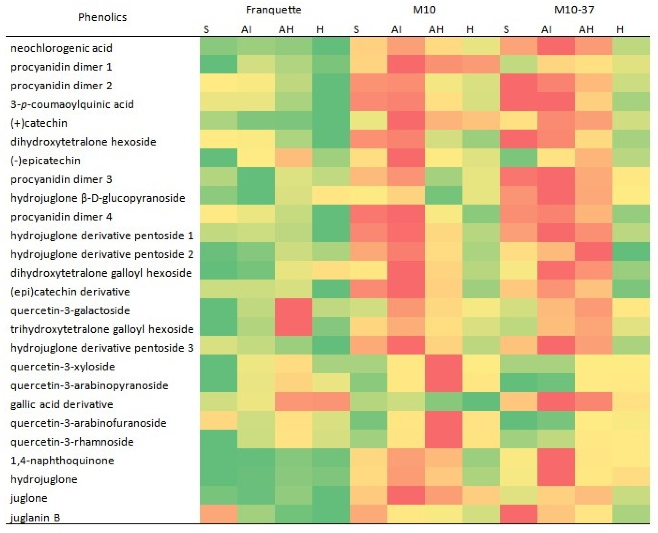
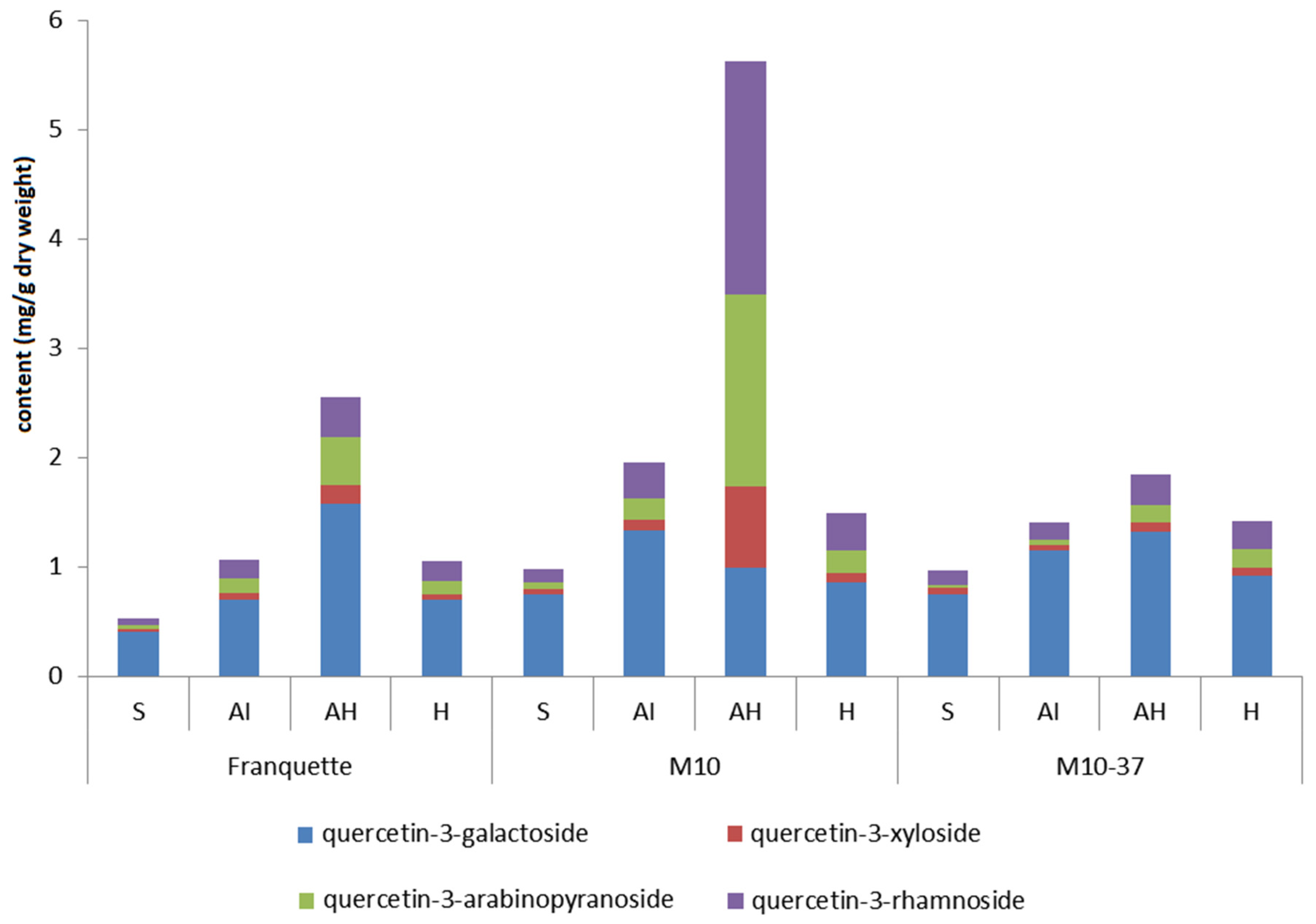
3.3. Individual Phenolic Compounds in Response to Walnut Anthracnose Infection

The reactions of the individual phenolic compounds are presented in the supplementary material and in Figure 4.
It can be seen that the highest contents of individual phenolic compounds are found in cultivars ‘M10’ and ‘M10-37’, with the exception of quercetin-3-galactoside, and tryhidroxytetralone galloyl hexoside, which have the highest content in ‘Franquette’. The content of individual flavonols was higher than previously reported in walnut husk infected with Xanthomonas arboricola pv. juglandis bacteria [12], but similar to the content of individual flavonols in a study of apples with varying severity of fruit infected with apple scab [11]. Interestingly, the highest content of quercetin-3-galactoside was observed in ‘Franquette’, suggesting that this could be a key flavonol in cultivar resistance, while quercetin-3-xyloside, quercetin-3-arabinopyranoside, quercetin-3-arabinofuranoside, and quercetin-3-rhamnoside had a higher content in cultivar ‘M10’. When comparing the resistance of cultivars of the same origin, quercetin-3-xyloside, quercetin-3-arabinopyranoside, quercetin-3-arabinofuranoside, and quercetin-3-rhamnoside were present in higher levels in semi-susceptible cultivar ‘M10’ than in susceptible cultivar ‘M10-37’ against walnut anthracnose, indicating that all quercetin glucosides play a major role in plant defense and its resistance. However, the content of flavonols in healthy tissue does not indicate the resistance of the cultivars, but rather the rapid and strong response of the cultivars in the synthesis of flavonols, as shown in Figure 5.
Juglone content was similar to that previously reported in walnut husk [13]. The hydrojuglone β-d-glucopyranoside content was lower, and the juglone content was similar to that reported in walnut leaves affected by walnut anthracnose [14]. In contrast to individual flavonols, in which the highest content was observed in AH tissue, the highest contents of individual naphthoquinones were observed in either infected tissue (S or AI) as previously reported in walnut leaves infected with walnut anthracnose [14]. The same can be observed when looking at flavanols, as seen in Figure 6.
Once again, the content of naphthoquinones or flavanols in healthy tissue was shown to have no effect on the resistance of cultivars. Interestingly, the newly quantified naphthoquinone juglanin B showed the highest response to infection, with the highest content in the more infected tissues, which was observed in all cultivars. The reaction of individual flavanols was similar to that previously reported in walnut husk after infection with Xanthomonas arboricola pv. juglandis bacteria [12]. The highest content of individual naphthoquinones and flavanols in S and AI may indicate that individual naphthoquinones and flavanols take more time to be synthesized or that they are the second defense mechanism when flavonols do not stop the infection. The reaction is probably the result of higher enzymatic activity, since it induces additional production and accumulation of phenolic compounds that may prevent the bacteria from spreading from infected cells into healthy cells, thus limiting infection [11,12].

4. Conclusions

A total of 26 phenolic compounds were tentatively identified and quantified, with flavanols, flavonols, and naphthoquinones accounting for more than 95% of the phenolic compounds. Juglanin B, tryhidroxytertalone galloyl hexoside, and dihydroxytetralone galloyl hexoside, previously reported in J. mandshurica, are reported here for the first time in the husk of J. regia. They were also quantified for the first time in J. regia or any other Juglans genus. The higher TAPC content in tissues more affected by walnut anthracnose confirmed that phenolics play a major role in the plant’s response against pathogens. A difference between cultivars was observed, since French cultivar ‘Franquette’ responded differently to walnut anthracnose infection than Hungarian cultivars ‘M10’ and ‘M10-37’. Naphthoquinones and flavanols have very similar responses to walnut anthracnose infection. Compared to TAPC, flavanols, and naphthoquinones, with which a difference was found between the cultivars of different origin, the content of flavonols did not vary between cultivars. Flavonols may be the most important phenolic compounds in disease control because they respond more rapidly to infection than flavanols and naphthoquinones, which has been previously observed with apple scab [11]. Flavonols may play an inhibitory role in the early stages of viral and bacterial infections. Since microbial resistance to antibiotics is a major global public health problem [20], new flavonol-derived drugs that exhibit inhibition against viral and bacterial infections could be a much-needed innovation. The highest content of quercetin-3-galactoside was observed in ‘Franquette’, suggesting that this may be a key flavonol in cultivar resistance. All quercetin glucosides play a major role in plant defense and its resistance. However, the content of flavonols in healthy tissues does not indicate cultivar resistance, but rather the reaction time of the plant and the speed with which it recognizes the pathogen and responds quickly to the infection by containing it while it has not yet spread. Among the naphthoquinones, the newly quantified juglanin B showed the highest response to walnut infection. Overall, it may be that naphthoquinones and flavanols either require more time to be synthesized or are the second defense mechanism when flavonols fail to stop the infection. These results not only enrich our understanding of phenolic response and resistance to walnut anthracnose, but also lay the foundation for future breeding of resistant walnut cultivars.

Supplementary Materials

The following are available online at https://0-www-mdpi-com.brum.beds.ac.uk/article/10.3390/agriculture11070659/s1. Table S1: Comparison of individual phenolic compounds in the husk of Juglans regia L. among three differently susceptible cultivars to walnut anthracnose (mean ± SE, in mg/g dry weight).

Author Contributions

Conceptualization, A.M., A.S. and R.V.; Data curation, A.M.; Formal analysis, A.M.; Funding acquisition, M.H.; Investigation, A.M.; Methodology, A.M. and R.V.; Project administration, R.V.; Resources, A.M., M.H. and A.S.; Software, A.M. Supervision, R.V.; Validation, A.S. and R.V.; Visualization, A.M.; Writing—original draft, A.M.; Writing—review and editing, M.H., A.S. and R.V. All authors have read and agreed to the published version of the manuscript.

Funding

This study is part of program P4-0013-0481, which is funded by the Slovenian Research Agency (ARRS).

Institutional Review Board Statement

Not applicable.

Informed Consent Statement

Not applicable.

Data Availability Statement

Part of the data presented in this study are available in supplementary material here. The remaining data presented in this study are available on request from the corresponding author. The remaining data are not publicly available due to privacy.

Conflicts of Interest

The authors declare that they have no known competing financial interests or personal relationships that could appear to have influenced the work reported here.

References

  1. Pollegioni, P.; Van der Linden, G.; Belisario, A.; Gras, M.; Anselmi, N.; Olimpieri, I.; Luongo, L.; Santini, A.; Turco, E.; Scarascia Mugnozza, G.; et al. Mechanisms governing the responses to anthracnose pathogen in Juglans spp. J. Biotechnol. 2012, 159, 251–264. [Google Scholar] [CrossRef]
  2. Yang, H.; Cao, G.; Jiang, S.; Han, S.; Yang, C.; Wan, X.; Zhang, F.; Chen, L.; Xiao, J.; Zhu, P.; et al. Identification of the anthracnose fungus of walnut (Juglans spp.) and resistance evaluation through physiological responses of resistant vs. susceptible hosts. Plant Pathol. 2021, 70, 1219–1229. [Google Scholar] [CrossRef]
  3. Belisario, A.; Scotton, M.; Santori, A.; Onofri, S. Variability in the Italian population of Gnomonia leptostyla, homothallism and resistance of Juglans species to anthracnose. For. Pathol. 2008, 38, 129–145. [Google Scholar] [CrossRef]
  4. Woeste, K.E.; Beineke, W.F. An efficient method for evaluating black walnut for resistance to walnut anthracnose in field plots and the identification of resistant genotypes. Plant Breed. 2001, 120, 454–456. [Google Scholar] [CrossRef]
  5. Arnaudov, V.A.; Gandev, S.I. Susceptibility of some walnut cultivars to Gnomonia Leptostila. Acta Hortic. 2009, 825, 407–412. [Google Scholar] [CrossRef]
  6. Belisario, A.; Forti, E.; Cichello, A.M.; Zoina, A.; Barbieri, E.; Valier, A. Epidemilogical surveys of Gnomonia leptostyla in Juglans regia hedgerow trained orchard. Acta Hortic. 2001, 544, 405–408. [Google Scholar] [CrossRef]
  7. Pollegioni, P.; Van der Linden, G.; Gras, M.; Olimpieri, I.; Anselmi, N.; Scarascia, G. Identification of resistant genotypes to anthracnose (Gnomonia leptostyla Fr. Ces) in Juglans spp. by functional and neutral markers. J. Biotechnol. 2010, 150, 114. [Google Scholar] [CrossRef]
  8. Zhu, Y.; Yin, Y.; Yang, K.; Li, J.; Sang, Y.; Huang, L.; Fan, S. Construction of a high-density genetic map using specific length amplified fragment markers and identification of a quantitative trait locus for anthracnose resistance in walnut (Juglans regia L.). BMC Genom. 2015, 16, 614. [Google Scholar] [CrossRef] [Green Version]
  9. Treutter, D. Significance of flavonoids in plant resistance and enhancement of their biosynthesis. Plant Biol. 2005, 7, 581–591. [Google Scholar] [CrossRef]
  10. Hedin, P.A.; Langhans, V.E.; Graves, C.H. Identification of juglone in pecan as a possible factor of resistance to Fusicladium effusum. J. Agric. Food Chem. 1979, 27, 92–94. [Google Scholar] [CrossRef]
  11. Slatnar, A.; Mikulic Petkovsek, M.; Halbwirth, H.; Stampar, F.; Stich, K.; Veberic, R. Polyphenol metabolism of developing apple skin of a scab resistant and a susceptible apple cultivar. Trees 2012, 26, 109–119. [Google Scholar] [CrossRef]
  12. Mikulic-Petkovsek, M.; Slatnar, A.; Veberic, R.; Stampar, F.; Solar, A. Phenolic response in green walnut husk after the infection with bacteria Xanthomonas arboricola pv. juglandis. Physiol. Mol. Plant Pathol. 2011, 76, 159–165. [Google Scholar] [CrossRef]
  13. Solar, A.; Jakopic, J.; Veberic, R.; Stampar, F. Correlations between Xanthomonas arboricola pv. juglandis severity and endogenous juglone and phenolic acids in walnut. J. Plant Pathol. 2012, 94, 229–235. [Google Scholar] [CrossRef]
  14. Cline, S.; Neely, D. Relationship between juvenile-leaf resistance to anthracnose and the presence of juglone and hydrojuglone glucoside in black walnut. Phytopathology 1984, 74, 185–188. [Google Scholar] [CrossRef]
  15. Medic, A.; Jakopic, J.; Hudina, M.; Solar, A.; Veberic, R. Identification and quantification of the major phenolic constituents in Juglans regia L. peeled kernels and pellicles, using HPLC–MS/MS. Food Chem. 2021, 352, 129404. [Google Scholar] [CrossRef] [PubMed]
  16. Medic, A.; Jakopic, J.; Solar, A.; Hudina, M.; Veberic, R. Walnut (J. regia) agro-residues as a rich source of phenolic compounds. Biology 2021, 10, 535. [Google Scholar] [CrossRef] [PubMed]
  17. Huo, J.-H.; Du, X.-W.; Sun, G.-D.; Dong, W.-T.; Wang, W.-M. Identification and characterization of major constituents in Juglans mandshurica using ultra performance liquid chromatography coupled with time-of-flight mass spectrometry (UPLC-ESI-Q-TOF/MS). Chin. J. Nat. Med. 2018, 16, 525–545. [Google Scholar] [CrossRef]
  18. Wang, T.-M.; Fu, Y.; Yu, W.-J.; Chen, C.; Di, X.; Zhang, H.; Zhai, Y.-J.; Chu, Z.-Y.; Kang, T.-G.; Chen, H.-B. Identification of polar constituents in the decoction of Juglans mandshurica and in the medicated egg prepared with the decoction by HPLC-Q-TOF MS2. Molecules 2017, 22, 1452. [Google Scholar] [CrossRef] [PubMed] [Green Version]
  19. Solar, A.; Colaric, M.; Hudina, M.; Stampar, F. Phenolic content of walnut fruit as affected by cultivar and developmental stage. Acta Hortic. 2005, 705, 231–240. [Google Scholar] [CrossRef]
  20. Barreca, D.; Trombetta, D.; Smeriglio, A.; Mandalari, G.; Romeo, O.; Felice, M.R.; Gattuso, G.; Nabavi, S.M. Food flavonols: Nutraceuticals with complex health benefits and functionalities. Trends Food Sci. Technol. 2021. [Google Scholar] [CrossRef]
  21. Abian, O.; Ortega-Alarcon, D.; Jimenez-Alesanco, A.; Ceballos-Laita, L.; Vega, S.; Reyburn, H.T.; Rizzuti, B.; Velazquez-Campoy, A. Structural stability of SARS-CoV-2 3CLpro and identification of quercetin as an inhibitor by experimental screening. Int. J. Biol. Macromol. 2020, 164, 1693–1703. [Google Scholar] [CrossRef]
  22. Quiles, J.L.; Rivas-García, L.; Varela-López, A.; Llopis, J.; Battino, M.; Sánchez-González, C. Do nutrients and other bioactive molecules from foods have anything to say in the treatment against COVID-19? Environ. Res. 2020, 191, 110053. [Google Scholar] [CrossRef] [PubMed]
  23. Belgacem, I.; Li Destri Nicosia, M.G.; Pangallo, S.; Abdelfattah, A.; Benuzzi, M.; Agosteo, G.E.; Schena, L. Pomegranate peel extracts as safe natural treatments to control plant diseases and increase the shelf-life and safety of fresh fruits and vegetables. Plants 2021, 10, 453. [Google Scholar] [CrossRef] [PubMed]
  24. Belgacem, I.; Pangallo, S.; Abdelfattah, A.; Romeo, F.V.; Cacciola, S.O.; Li Destri Nicosia, M.G.; Ballistreri, G.; Schena, L. Transcriptomic analysis of orange fruit treated with pomegranate peel extract (PGE). Plants 2019, 8, 101. [Google Scholar] [CrossRef] [PubMed] [Green Version]
  25. Pangallo, S.; Destri Nicosia, M.G.; Agosteo, G.E.; Abdelfattah, A.; Romeo, F.V.; Cacciola, S.O.; Rapisarda, P.; Schena, L. Evaluation of a Pomegranate Peel Extract as an Alternative Means to Control Olive Anthracnose. Phytopathology 2017, 107, 1462–1467. [Google Scholar] [CrossRef] [PubMed] [Green Version]
Figure 1. Different parts of walnut tissue were sampled. Red circle (S): inner spot of infected tissue; blue circle (AI): outer margin of infected tissue; yellow circle (AH): healthy tissue surrounding the infection.
Agriculture 11 00659 g001
Figure 2. Contents of three major groups of phenolics identified in different severity of walnut anthracnose-affected tissues of J. regia husk, between three cultivars: Susceptible ‘M10-37’, semi-susceptible ‘M10’ and least susceptible ‘Franquette’ (in mg/g dry weight). S: inner spot of infected tissue; AI: outer margin of infected tissue; AH: healthy tissue surrounding the infection; H: healthy tissue.
Agriculture 11 00659 g002
Figure 3. Dendrogram showing the grouping of naphthoquinones (A) and flavanols (B) between the severities of walnut anthracnose-affected tissues of three cultivars, using Ward’s method (squared Euclidean distance) based on total phenolic compounds. Data are standardized (µ = 0, σ = 1). S: inner spot of infected tissue; AI: outer margin of infected tissue; AH: healthy tissue surrounding the infection; H: healthy tissue.
Agriculture 11 00659 g003
Figure 4. Heat map showing relative content of the phenolic compound identified between cultivars from highest to lowest content between columns. Red: highest concentration; green: lowest concentration. S: inner spot of infected tissue; AI: outer margin of infected tissue; AH: healthy tissue surrounding the infection; H: healthy tissue.
Agriculture 11 00659 g004
Figure 5. Contents of flavonols identified in different severity of walnut anthracnose-affected tissues of J. regia husk, between three cultivars: susceptible ‘M10-37’, semi-susceptible ‘M10’ and least susceptible ‘Franquette’ (in mg/g dry weight). S: inner spot of infected tissue; AI: outer margin of infected tissue; AH: healthy tissue surrounding the infection; H: healthy tissue.
Agriculture 11 00659 g005
Figure 6. Contents of selected naphthoquinones and flavanols identified in different severity of walnut anthracnose-affected tissues of J. regia husk, between three cultivars: susceptible ‘M10-37’, semi-susceptible ‘M10’ and least susceptible ‘Franquette’ (in mg/g dry weight). S: inner spot of infected tissue; AI: outer margin of infected tissue; AH: healthy tissue surrounding the infection; H: healthy tissue.
Agriculture 11 00659 g006
Table 1. Tentative identification of 26 phenolic compounds from the husk of Juglans regia L., and the standard compounds they are expressed as.
PhenolicsRt
(min)
[M − H]
(m/z)
MS2
(m/z)
Expressed as
neochlorogenic acid (3-caffeoylquinic acid)9.36353191, 179, 135neochlorogenic acid
procyanidin dimer 110.38577425, 407, 289procyanidin B1
procyanidin dimer 211.47577425, 407, 289procyanidin B1
3-p-coumaoylquinic acid12.01337163, 191, 1734-O-caffeoylquinic acid
(+) catechin12.22289245, 205, 179(+) catechin
dihydroxytetralone hexoside13.04339159, 177juglone
(−) epicatechin14.53289245, 205, 179(−) epicatechin
procyanidin dimer 315.53577425, 407, 289procyanidin B1
hydrojuglone β-d-glucopyranoside16.26337175juglone
procyanidin dimer 416.89577425, 407, 289procyanidin B1
hydrojuglone derivative pentoside 117.98435303, 285juglone
hydrojuglone derivative pentoside 218.26435303, 285juglone
dihydroxytetralone galloyl hexoside19.06491271, 331juglone
(epi)catechin derivative20.17441289(+) catechin
quercetin-3-galactoside20.53463301quercetin-3-galactoside
trihydroxytetralone galloyl hexoside20.76507331, 271juglone
hydrojuglone derivative pentoside 321.27435303, 285juglone
quercetin-3-xyloside21.56433301quercetin-3-xyloside
quercetin-3-arabinopyranoside21.73433301quercetin-3-arabinopyranoside
gallic acid derivative21.99489271, 313gallic acid
quercetin-3-arabinofuranoside22.21433301quercetin-3-arabinofuranoside
quercetin-3-rhamnoside22.43447301quercetin-3-rhamnoside
1,4-naphthoquinone28.21173111, 155, 129, 1451,4-naphthoquinone
hydrojuglone28.21175131, 147, 157, 115, 103juglone
juglone (5-hydroxy-1,4-naphthoquinone)29.99189161juglone
juglanin B31.37327312, 253juglone
Rt, retention time; [M − H], pseudo-molecular ion identified in negative ion mode.
Publisher’s Note: MDPI stays neutral with regard to jurisdictional claims in published maps and institutional affiliations.

