Article

Exploratory Sampling of Spalting Fungi in the Southern Peruvian Amazon Forest

by
Sarath M. Vega Gutierrez
1,*,
Javier F. Illescas Guevara
2,
Claudia C. Andersen
1,
Jose Koechlin von Stein
3 and
Seri C. Robinson
1
1
Wood Science & Engineering, Oregon State University, Corvallis, OR 97331, USA
2
College of Forestry, Universidad Nacional Agraria la Molina, Lima 15024, Peru
3
Inkaterra, Lima 15074, Peru
*
Author to whom correspondence should be addressed.
Submission received: 20 October 2020 / Revised: 14 December 2020 / Accepted: 17 December 2020 / Published: 20 December 2020

Abstract

:
Most of the research related to Peruvian Amazon fungi is focused on edible mushrooms and pathogens. Other important fungi, such as the spalting type (decay fungi that pigment wood internally), are not broadly studied, as most of them do not produce fruiting bodies and can be difficult to locate. Spalting fungi, however, are of broad economic importance due to their ability to produce pigments that enhance wood aesthetics, resulting in an increased economic value. In order to begin understanding the diversity of spalting fungi within certain regions of the Amazon, a sampling of downed trees and branches (through the opening of the xylem to identify potential pigmenting and zone line producing fungi) was done in the district of Las Piedras, Madre de Dios, Peru. Fungi suspected of causing internal pigment and zone lines were collected, cultured, isolated, and sequenced. The species found belonged to the orders Helotiales, Xylariales, Hypocreales, Russulales, Polyporales, Botryosphaeriales and two specimens of the class Leotiomycetes. The fungi collected produced pigments or zone lines in wild conditions and all of them were capable of wood decomposition. Interestingly, these are the same orders and genera as North American spalting fungi, which may indicate a correlation within species that pigment wood. The results obtained start a specific database of spalted fungi in the Amazon and, with it, help support an effort to increase the forest value of ecosystems primarily used for a few high-valued tree species.

1. Introduction

The Peruvian Amazon rainforest is one of the most mega-diverse places on earth [1]. It is also one of the most endangered ecosystems due to land use changes that include agriculture, urban expansion and mining. These changes have resulted in the loss of forest land [2,3,4]. Diversity studies in the area have examined the effects of these changes on birds [5], mammals, insects, reptiles [6] and fungal fruiting bodies [7]. Unfortunately, little is known about any potential changes in the diversity of spalting fungi in this region. While there is evidence of the presence of spalting-type fungi growing in the Amazon rainforest, research on those that do not form an obvious sporocarp is fairly recent, and has consisted entirely of research into blue-staining fungi [8].
Spalting fungi are wood-decay organisms which produce internal pigmentation in wood [9]. This pigmentation can vary between species, but is generally broken out into three groups. Bleaching fungi that cause a lighter coloration in wood due to the removal of lignin (mostly white-rot Basidiomycetes) [10]. Zone line fungi generate pigment lines in the wood. Most zone lines are black/brown from extracellular melanin, although green, red, yellow, and other colored zone lines do exist [11,12]. The zone-line fungi are primarily white-rot fungi [13,14], along with the Ascomycete genus Xylaria [15]. The third group are made up of rarer fungi that produce internal and/or extracellular pigmentation in wood.
Known and well-studied pigment-type spalting fungi [16,17] include Scytalidium cuboideum (Sacc. And Ellis) Singler and Kang, a fungus that produces a red pigmenting napthoquinone crystal (“Dramada”) [18]. Another spalting fungus, Scytalidium ganodermophthorum Kang, Singler, Y. W. Lee and S. H. Yun (syn. Xylogone ganodermophthora Kang, Singler, Y.W. Lee and S.H. Yun), produces a yellow pigment that remains unidentified [19]. The genus Chlorociboria produces blue-green pigmentation caused by xylindein [17,20]. All of these pigmenting fungi belong to the order Helotiales.
The wood pigmented by these fungi (spalted wood) is part of an ancient art form (used in intarsia and marquetry) that dates back hundreds of years in Europe. From zone-lines to green pigment, artwork that used this wood is still brilliantly pigmented, showing the long-lasting properties of the colors produced by these fungi [21,22,23,24]. Spalted wood is currently popular for woodwork where its unique aesthetics attracts customers [25] that are willing to pay premium prizes for it, classifying spalted wood as a value-added wood product [26].
Most research on spalting fungi has focused on fungal species in temperate forests [27,28] and has overlooked the tropical forests and its vast potential. Recent explorations in the Peruvian Amazon rainforest found a wide variety of spalting fungi in decomposing wood. Colors that range from dark green to lilac have been identified [29]. Previous studies on moderate value Peruvian woods have shown that they can be successfully spalted [8] and therefore, an increased knowledge on potential fungal species that can be used on them could further help increase their market value.
This research establishes the first sequencing of (exclusively) spalting fungi in the southern Peruvian rainforest. The goal of this study is to genetically identify some of the fungal species that are present in the area of Las Piedras, Puerto Maldonado, Peru. It also will have a secondary effect of increasing the value of the forest, through identification of fungal species that can be used for value-added products such as wood art pieces and furniture.

2. Results

A total of 250 samples were collected from the field. From that original number, several isolate attempts were contaminated with bacteria and other fungi such as the ones from the genera Trichoderma spp., and Penicillium spp. From the field laboratory a total of 45 tubes containing possible isolates were shipped to UNALM in Lima, Peru. After further purification, a total of 35 fungal species were exported to the Forest Pathology Laboratories at OSU, USA. It is important to note that the export process from Peru to the USA took two years due to governmental issues. During this time the samples were stored without checking in Peru, and many acquired mites and contamination. This is why so few samples came through to the USA and were able to be sequenced—not due to field isolation techniques. With the continuing isolation process in the USA, only 29 specimens were sequenced.
From the obtained sequences, fungi were classified according to the spalting type that they generated in wood (zone lines and pigments) during the sample collection. The results are shown in Table 1. Each sequence has its own accession number, as well as the similarity percentage to an existing GenBank sequence.
The most common order was Hypocreales, which included the genera Fusarium, Trichoderma, Hypocrea, Cosmospora and Bionectria. The fungi in this order were in samples that contained pigments and zone lines at the moment of the collection. The second most common order that was Xylariales. This order included the genera Xylaria and Neopestalotiopsis. This order was found in samples that contained mostly black zone lines. The third most common order was Helotiales. The genus identified for this order was Scytalidium and it was found in samples that showed colored pigments. Other orders such as Botryosphaeriales, Polyporales and Russulales were identified, and they contained a single genus per order. Several samples were classified as Incertae sedis and the genus Xylogone. The classification of the fungi can be visualized in Figure 1.
Microscopical identification was difficult due to the lack of reproductive structures recorded during the collection (under most circumstances, none existed), and the absence of asexual reproductive tissue in Petri dishes. This was a specific challenge of the research, stemming from the long wait times for export of the fungi to the USA and the limited availability for proper storage of the cultures.

3. Discussion

There are some similarities between the tropical spalting fungi and the temperate forest fungal genera. One of the most common orders was the black zone line producer Xylariales. This order is known in temperate forests for the species Xylaria polymorpha [30,31] and Xylaria hypoxylon. These fungi are prolific black zone line producers (melanin) [32] in nature and under laboratory conditions [11]. Wood colonized by these fungi is commonly used in art pieces, both historically and in modern art, within work ranging from marquetry to turned objects [33]. Previous research on Peruvian spalting fungi was also focused on melanin producing organisms (also known as blue stains) [8]. But these fungi do not produce zone lines, rather, their melanin is excreted as a broad extracellular pigment.
In the Amazon, fungi from the genus Xylaria were found in wood pieces that showed black zone lines, indicating that the production of melanin barriers is a common characteristic within the genus. The results obtained increase the information about the distribution of the genus Xylaria in the Neotropics. Previous studies in the cloud forest of Ecuador have shown the presence of X. guianensis and X. adscendens [34]. The DNA sequences obtained in the present study also report their presence in the southern Amazon rainforest of Peru. Within the order Xylariales the fungus Neopestalotiopsis clavispora was also found. This fungus is known as a plant pathogen [35] and has a worldwide distribution and can be found in decaying wood [36].
In the order Helotiales, the identified Peruvian samples belonged to the genus Scytalidium, and all were found on deeply-pigmented wood. This genus also contains pigment-producing fungi Scytalidium cuboideum and Scytalidium ganodermophthorum. The genus Chlorociboria [27,37] also belongs to this order. The aforementioned species are known for producing red [38], yellow [16] and blue-green pigments [38] respectively, both in wood and in liquid cultures [12,39]. It is hopeful that the Helotiales fungi found in the Peruvian rainforest will also produce extracellular pigments under laboratory conditions, similarly to the spalting fungal species from the temperate forest species, since they are closely related to commonly used spalting fungi and were collected from stained wood [40].
The Hypocreales in the samples included the genera Fusarium, Trichoderma, Hypocrea, Cosmospora and Bionectria, which are likely all culture contaminants. Trichoderma harzanium is a fungus that is used for biocontrol [41], and is abundant in the Neotropical region, expanding across Central and South America [42] similarly to T. atrobrunneum, which is considered part of the complex of species of T. harzanium [43] together with its teleomorph Hypocrea lixii [44]. Species from the genus Fusarium were also found. Fusarium oxysporum have already being used for pigmenting textiles [45] while Fusarium acuminatum and Fusarium flocciferm have been used for promoting pigmentation in wood [46]. The species found in the Peruvian Amazon corresponds to F. solani, which has been reported as a lignin degrading fungus [47]. The genus Fusarium may not be useful for controlled spalting due to its inability to produce significant internal coloration in wood under standard laboratory conditions [46,48] and also because many Fusarium species are considered human pathogens [49]. Other fungal species found within this order were Cosmospora sp. and Bionectria sp., which have been previously reported in Peru [50,51].
Several sequences were assigned to the order Incertia sedis. Three of the isolates belonged to the genus Xylogone, which are mainly fungal parasites. One of the temperate spalting fungi that previously belonged to this genus is S. ganodermophthorum (ex. Xylogone ganodermophthora) until a genetic and morphological study determined that it belonged to the order Helotiales and the genus Scytalidium [19]. Similar to the yellow pigment produced by S. ganodermophthorum [16,52], these unidentified samples were found on wood samples showing brown, lilac and yellow pigmentation, which could indicate that this fungus likely will produce extracellular pigments under laboratory conditions. The other four fungi found in the group without an order did not match a genetic genus. This may indicate that they are new species, but further morphological research is required.
One other potential new species identified was in the order Polyporales (Polyporales sp.). Most of these fungi specialize in the decomposition of lignin [53]. This genus is not unique to the tropics and has a worldwide distribution [54,55]. The wood sample from which this fungus was isolated presented orange zone lines, a rare occurrence that differs from the common black zone lines. The orange lines will require an HPLC or FTIR analysis to identify the compounds within them. It will also be necessary to perform competition testing between the orange zone line fungi and other competing species, such as Trametes versicolor due to the ability of Basidiomycetes to produce zone lines in the presence of an antagonistic white-rot [14]. It is also possible the zone lines were formed due to somatic incompatibility within the same strain, or by a reaction to desiccation [56]. Within the white-rot fungi, an individual from the order Russulales, Peniophora sp. was also identified. Fungi from this genus are lignin decomposers [57,58] and some species have been identified as true endophytes in tropical tree species such as the genus Hevea [59]. This genus is also known for its wide distribution in tropical and temperate forests as a wood decomposer [60].
Finally, a fungus from the order Botryosphaeriales, Lasiodiplodia theobromae (syn. Botryodiplodia theobromae) was identified. This species is common in the tropics around the world and is considered a plant pathogen for fruit trees [61] as well as a soft-rot wood decay fungus [62]. Previous testing on its spalting potential in tropical woods was performed due to its ability to produce an intense black pigmentation [8] which was also observed in the sample where it was isolated.
At the current time, detailed information about the nutritional requirements for the laboratory growth of the identified Peruvian spalting fungi cannot be assessed. But, as there are similarities within the genera level with the North American species, it is possible to apply similar culturing methods to them such as the 2% MEA. The use of this media has been previously determined by Robinson et al. [12], as highly accurate for the evaluation of pigmenting capabilities of temperate forest spalting fungi. Its use with the Peruvian spalting fungi allowed the early identification of specimens of interest. Nevertheless it is important to pursue further research within this topic, as more specific information about this interesting group of fungi needs to be developed.
The Peruvian fungi grew best at temperatures between 18 to 22 °C (room temperature in the laboratory). The observed temperature, besides being within the optimal range for wood decay fungi to grow [63], also correspond to the lower average temperatures in Puerto Maldonado (collection area), but further studies are required to determine the optimal growth temperature and light conditions for the identified tropical fungi.
An interesting observation were the similarities between the composition of wood-decay fungi in the tropics and in the temperate forest. In this study, several genera such as Xylaria, Scytalidium, Peniophora, and Polyporales were identified, but are also known to have a worldwide distribution (as wood-decay fungi). Unfortunately, although the genera were the same that produce excellent spalting in temperate woods, the species identified were not the same. Therefore it is possible that while a genus may have a worldwide distribution, species within the genus are more affected by the different composition of the forests, as tropical forests tend to be formed predominantly by hardwoods (broadleaves) [64,65], while temperate forests are composed mostly by softwoods (conifers) [66]. Temperature, rainfall, and other climatic conditions also likely play a role. Future research is also necessary to determine if some of these species have specific requirements for laboratory pigment production as well as continuing cataloging spalting fungi in the tropics.
The confirmed presence of potential spalting fungi in the Amazon could increase the use of undervalued wood in the region, if education of its global economic benefits could be achieved. As current research in spalting fungi is showcasing the industrial potential of fungal pigments applied into inks [67], dyes [68], textiles [69], solar energy [70] as well as woodcrafts [26]. Should this occur, it could help protect this endangered ecosystem by giving additional value to the decayed wood from the native forests, thus generating more revenue from forestland versus land use change to agriculture or grazing. This preliminary work on spalting fungi in the Amazon enriches our knowledge of the potential biodiversity of the area, and also should help to generally increase the value of the forest.

4. Materials and Methods

4.1. Collection

Samples were collected at Inkaterra Guides Field Station (12°31′52.9″ S 60°02′41.5″ W), Tambopata in the district of Las Piedras, Madre de Dios, Peru; under the permit number 0328-2013-MINAGRI-DGFFS-DGEFFS, issued by the Peruvian Forest Service (SERFOR).
The methodology of the collection consisted on locating and sampling dead logs and branches along all of the trails within the area. Collections were limited to a 20 m radius from the trails, and no new trails were established due to the exploratory nature of the study. The total distance traveled on the trails was 15 km.
All fallen logs and branches over two centimeters in diameter were sampled within the boundaries set forth above. To sample a piece, a longitudinal cut was made with a machete at approximately 45 degrees until the xylem was visible (areas close to the bark or surface of the exposed log were not taken into account). The logs were cut every 30 cm, beginning from the end of the log. Smaller branches and logs that could be rotated were sampled around the entire diameter. Those that were too large to rotate were cut only on the top surface. This method has been previously applied with a high degree of success for finding wood colonized with Chlorociboria spp. [71].
The first cuts had an average size of 15 cm × 15 cm × 9 cm. If in one of these cuts the presence of spalting fungi were confirmed (presence of pigmentation or zone lines) like in Figure 2, the cut was lengthened and widened until the start and end of the spalting area was found. The type of spalting was classified as follows: zone line (thickness, length, depth, color), pigment (width, length, depth, color), or undefined (explain re: combination of zone line and pigment, etc.) Tissue samples of the affected wood were collected from every distinct area. Collected samples were generally around 2 cm × 5 cm × 10 cm, although were smaller in cases where the affected area was smaller. Samples were placed individually in brown paper bags. The bags were labeled with the unique identification code for each sample (multiple samples might be collected from a single log, so notation was used to indicate log number, as well as sample number) and were packed in paper bags back to base camp. Wood species were not identified for this study due to two reasons: first, the wood on which the spalting occurred was often too decayed to do a field ID (and the collection permit did not allow for wood to be removed from the forest to better laboratory sites) and second, the common names of the trees provided by the field guides (still recorded) often were made up of several genera. An example of a collected specimen can be seen in Figure 1.
A GPS waypoint was taken for each specimen using a Garmin GPSMAP 64s marking the location and the type of spalting found. The data obtained allowed the research team to avoid repetitive sampling.

4.2. Fungal Isolation

The samples were processed in the same day of collection at the base camp field laboratory. The laboratory contained only the most basic facilities, and no laminar flow hood was available. As such, successful monoculture isolation of the fungi from the affected wood did not have a high success rate. Tissue isolations consisted of slicing into the interior of the sample with a sterilized scalpel blade. Forceps of 114 mm (sterilized with ethanol and fire from an alcohol lamp) were used to pull a piece of pigmented wood from the whole. For samples with pigments, the pieces for isolation came from only areas that contained the coloration, while in the case of zone lines samples were taken from each side of the zone line. The extracted pieces of less than 0.5 cm of the newly exposed area was taken and introduced into a slant containing 2% malt extract agar (MEA, VWR, Radnor, PA, USA). Slants rather than Petri dishes were used to reduce the probabilities of contaminants in the cultures. This decision was made by previous experience while collecting samples in a similar environment by the same team [71,72].
Samples were transported in sealed slant tubes to the Wood Protection Laboratory at Universidad Nacional Agraria la Molina (UNALM) in Lima, Peru where the purification of the cultures was performed. The viable cultures were transferred onto Petri dishes containing 1.5% potato dextrose agar (PDA). The transfer process was repeated until molds, bacteria, or other fungi accompanying the wanted fungus were removed. As the purpose of the culturing was to propagate unknown fungal species, only a visual evaluation could be used to determine if the fungus growing on the plate was the one of interest. This was done through color matching (e.g., the pigmentation the fungus made in wood had to be similar to the one the culture was making on the plate) [71] or confirmation of zone line production.
Once the cultures were free from contaminants, they were placed onto slants containing 1.5% PDA. Then, the samples were shipped to the Forest Pathology Laboratory at Oregon State University (OSU) for further purification and ITS rDNA identification under the permit 002,822 MINAGRI-DGFFS issued by SERFOR.
In the Forest Pathology Laboratory, monoculture status was confirmed before the fungi were moved to 2% MEA in Petri dishes. This was done to confirm their spalting ability, as most fungi capable of spalting will pigment 2% MEA, but many fungi pigment potato dextrose agar and do not perform similarly on wood [12]. If the fungi did successfully pigment the malt agar, they were prepared for DNA sequencing.

4.3. DNA Sequencing

For DNA sequencing, small amounts of active mycelium of each pure culture were taken and placed in micro-centrifuge tubes of 1.5 mL containing 1 mL of 2% potato broth. The fungi were left to grow for 3 days before performing the DNA isolation. The DNA isolation was performed with the QIAGEN® DNeasy Plant Mini Kit (QIAGEN, Hilden, Germany). The Internal Transcribed Spacer (ITS) region was amplified using the universal ITS1-F and ITS4 primer [73], and a quality check of the amplified DNA was done with the use of 1% agarose gel electrophoresis. PCR was performed using the hot start polymerase in a Bio-Rad PTC-100 following the protocol of Tudor et al. (2014) [74] and cleaned with EXOsap-IT (Thermo Fisher Scientific) [75]. The Samples were sequenced at the Center for Genome Research and Biocomputing (CGRB) at OSU. Results were analyzed using the 4Peaks software (Nucleobytes B.V., North Holland, Netherlands). This software allowed the selection of the best sections of the resulting DNA and mostly consisted in 480–520 bases. The DNA was later compared in the BLAST® webpage to identify the samples collected on October 2016. The parameters used to select possible matches in the nucleotide collection (nr/nt) consisted on the use of highly similar sequences (megablast). Besides this, a range of 95–100% of query cover and 90–100% of the DNA identity were considered, as well as a selection of accession numbers that were from studies performed in South America as this information would provide a more accurate identity.

4.4. Pairwise Sequence Alignment

For fungal sequences that presented the same genus, a pairwise nucleotide sequence alignment performed with EMBOSS Matcher from the European Bioinformatics Institute was utilized. If the sequences had a similarity above 99%, they were considered the same individual. If the percentage was lower than 99%, a number was assigned to the specimen (e.g., Scytalidium sp1.), to indicate that it belonged to the same genus (with a similarity of 80%), but to a different species.

5. Conclusions

Spalting fungi collected and identified from the Peruvian Amazon rainforest belong primarily to the order Helotiales—the order that also holds most of pigment-type spalting fungi of temperate forests. Representatives from several other orders were found, such as the Xylariales, which are known zone line producers. The other fungi identified belong generally to wood-decay groups (white rots and soft rots). These findings show shared genera and orders between spalting fungi in the tropical rainforests of Peru and the temperate rainforests of North America. Follow up testing should determine if these new fungi have similar capabilities as their temperate forest counterparts (in producing extracellular pigments) under laboratory conditions, and could establish if genetic similarities exist. Additionally, the presence of spalting fungi in the Amazon will open the door for further research in the region, as well as the increased value of decayed wood.

Author Contributions

Conceptualization, S.M.V.G. and S.C.R.; Data curation, S.M.V.G.; Funding acquisition, J.K.v.S.; Investigation, S.M.V.G., J.F.I.G. and C.C.A.; Methodology, S.M.V.G.; Resources, J.K.v.S.; Writing—original draft, S.M.V.G.; Writing—review and editing, S.M.V.G. and S.C.R. All authors have read and agreed to the published version of the manuscript.

Funding

This research received no external funding.

Acknowledgments

The Peruvian Forest Service (SERFOR), the Agriculture Ministry of Peru (MINAG), the Wood Protection Laboratory at Universidad Nacional Agraria la Molina in Lima, Peru, the Forest Pathology Laboratory at Oregon State University, Jed Capellazzi and the Center for Genome Research and Biocomputing at Oregon State University.

Conflicts of Interest

The authors declare no conflict of interest.

References

  1. Sánchez, S.; Llosa, G.; Pulido, V.; Del Rio, M.L.; Rueda, D. Perú: Estrategia Nacional Sobre Diversidad Biológica; Consejo Nacional del Ambiente (CONAM): Lima, Peru, 2001. [Google Scholar]
  2. Escobal, J.; Aldana, U. Are nontimber forest products the antidote to rainforest degradation? Brazil nut extraction in Madre De Dios, Peru. World Dev. 2003, 31, 1873–1887. [Google Scholar] [CrossRef]
  3. Scullion, J.J.; Vogt, K.A.; Sienkiewicz, A.; Gmur, S.J.; Trujillo, C. Assessing the influence of land-cover change and conflicting land-use authorizations on ecosystem conversion on the forest frontier of Madre de Dios, Peru. Biol. Conserv. 2014, 171, 247–258. [Google Scholar] [CrossRef]
  4. Swenson, J.J.; Carter, C.E.; Domec, J.-C.; Delgado, C.I. Gold mining in the Peruvian Amazon: Global prices, deforestation, and mercury imports. PLoS ONE 2011, 6, e18875. [Google Scholar] [CrossRef] [Green Version]
  5. Puhakka, L.; Salo, M.; Saaksjarvi, I. Bird Diversity, Birdwatching Tourism and Conservation in Peru: A Geographic Analysis. PLoS ONE 2011, 6. [Google Scholar] [CrossRef] [PubMed]
  6. Duellman, W.E. The Reserva Cuzco Amazonico, Peru: Biological Investigations, Conservation, and Ecotourism; Museum of Natural History, University of Kansas: Lawrence, KS, USA, 1991. [Google Scholar]
  7. Mori del Aguila, T.; Bendayan Acosta, M.E.; Tresierra Ayala, A.; Mildred, G.D.; Ruiz Sanchez, E.; Bardales Farcia, J.; Reategui Amasifuen, R.; Espinoza Campos, F.; Davila Flores, C. Acomicetes y Basidiomicetes Macroscopicos en Bosques de Puerto Almendras (Loreto, Peru). Folia Amaz. 2011, 20, 7–14. [Google Scholar] [CrossRef]
  8. Vega Gutiérrez, S.; Robinson, S. Potential of spalting moderate value wood species in Peru. Int. Wood Prod. J. 2015, 6, 165–168. [Google Scholar] [CrossRef]
  9. Robinson, S.C.; Richter, D.L.; Laks, P.E. Colonization of sugar maple by spalting fungi. For. Prod. J. 2007, 57, 24–32. [Google Scholar]
  10. Robinson, S.C.; Richter, D.L.; Laks, P.E. Inducing and Stimulating Spalting in Sugar Maple; IRG 08-10652; International Research Group on Wood Protection: Stockholm, Sweden, 2008. [Google Scholar]
  11. Robinson, S.C.; Weber, G.; Hinsch, E. Inducing zone lines and melanin formation for decorative purposes on North American western wood species, with emphasis on conifers. Int. Wood Prod. J. 2014, 5, 196–199. [Google Scholar] [CrossRef]
  12. Robinson, S.C.; Richter, D.L.; Laks, P.E. Effects of substrate on laboratory spalting of sugar maple. Holzforschung 2009, 63, 491–495. [Google Scholar] [CrossRef]
  13. Shigo, A.L.; Sharon, E.M. Mapping columns of discolored and decayed tissues in sugar maple, Acer saccharum. Phytopathology 1970, 60, 232–237. [Google Scholar] [CrossRef]
  14. Rayner, A.D.M. Interactions between fungi colonising hardwood stumps and their possible role in determining patterns of colinisation and succession. Ann. Appl. Biol. 1978, 89, 131–134. [Google Scholar] [CrossRef]
  15. Campbell, A.H. Zone lines in plant tissues.1. The black lines formed by Xylaria polymorpha (pers.) grev. in hardwoods. Ann. Appl. Biol. 1933, 20, 123–145. [Google Scholar] [CrossRef]
  16. Robinson, S.C.; Tudor, D.; Zhang, W.R.; Ng, S.; Cooper, P.A. Ability of three yellow pigment producing fungi to colour wood under controlled conditions. Int. Wood Prod. J. 2014, 5, 103–107. [Google Scholar] [CrossRef]
  17. Maeda, M.; Yamauchi, T.; Oshima, K.; Shimomura, M.; Miyauchi, S.; Mukae, K.; Sakaki, T.; Shibata, M.; Wakamatsu, K. Extraction of xylindein from Chlorociboria aeruginosa complex and its biological characteristics. Bull. Nagaoka Univ. Technol. 2003, 25, 105–111. [Google Scholar]
  18. Vega Gutierrez, S.M.; Hazell, K.K.; Simonsen, J.; Robinson, S.C. Description of a Naphthoquinonic Crystal Produced by the Fungus Scytalidium cuboideum. Molecules 2018, 23, 1905. [Google Scholar] [CrossRef] [PubMed] [Green Version]
  19. Kang, H.; Sigler, L.; Lee, J.; Gibas, C.; Yun, S.; Lee, Y. Xylogone ganodermophthora sp. nov., an ascomycetous pathogen causing yellow rot on cultivated mushroom Ganoderma lucidum in Korea. Mycologia 2010, 102, 1167–1184. [Google Scholar] [CrossRef] [Green Version]
  20. Blackburn, G.M.; Ekong, D.E.; Nielson, A.H.; Todd, L. Xylindein. Chimia 1965, 19, 208–212. [Google Scholar]
  21. Otterstedt, A. Investigating Green Marquetry on Bowed-String Instruments. The Leaves Be Greene. Galpin Soc. J. 2001, 54, 330–338. [Google Scholar] [CrossRef]
  22. Blanchette, R.A.; Wilmering, A.M.; Baumeister, M. The use of green-stained wood caused by the fungus Chlorociboria in Intarsia masterpieces from the 15th century. Holzforschung 1992, 46, 225–232. [Google Scholar] [CrossRef]
  23. Michaelsen, H.; Unger, A.; Fischer, C.-H. Blaugrüne Färbung an Intarsienhölzern des 16. bis 18. Jahrhunderts. Restauro 1992, 98, 17–25. [Google Scholar]
  24. Gutierrez, P.T.V.; Robinson, S.C. Determining the Presence of Spalted Wood in Spanish Marquetry Woodworks of the 1500s through the 1800s. Coatings 2017, 7, 188. [Google Scholar] [CrossRef] [Green Version]
  25. Nicholls, D.L. Evaluation of the Retail Market Potential for Locally Produced Paper Birch Lumber in Alaska; USDA: Washington, DC, USA, 2002; pp. 1–19. [Google Scholar]
  26. Vega Gutierrez, P.T.; Robinson, S.C. Complexity of biodegradation patterns in spalted wood and its influense on the perception of US woodturners. Eur. J. Wood Wood Prod. 2020, 172–183. [Google Scholar] [CrossRef]
  27. Ramamurthi, C.S.; Korf, R.P.; Batra, L.R. A revision of the North American species of Chlorociboria (Sclerotiniaceae). Mycologia 1958, 49, 854–863. [Google Scholar] [CrossRef]
  28. Robinson, S.C. Developing fungal pigments for “painting” vascular plants. Appl. Microbiol. Biotechnol. 2012, 93, 1389–1394. [Google Scholar] [CrossRef]
  29. Vega Gutierrez, S.M. Spalting Fungi: Genetic Identification, Material Interactions and Microscopical Characteristics of Extracted Pigments. Ph.D. Thesis, Oregon State University, Corvallis, OR, USA, 2017. [Google Scholar]
  30. Rogers, J.D.; Calla, B.E. Xylaria polymorpha and its allies in the continental United States. Mycologia 1986, 78, 391–400. [Google Scholar] [CrossRef]
  31. Worrall, J.T.; Anagnost, S.E.; Zabel, R.A. Comparison of wood decay among diverse lignicolous fungi. Mycologia 1997, 89, 199–219. [Google Scholar] [CrossRef]
  32. Phillips, L.W. The nature of spalted wood: Analysis of zone line formation between six white rot fungi. Ph.D. Thesis, Brigham Young University, Provo, UT, USA, 1987. [Google Scholar]
  33. Robinson, S.C.; Michaelsen, H.; Robinson, J.C. Spalted Wood. The History, Science and Art of a Unique Material, 1st ed.; Schiffer Publishing, Ltd.: Atglen, PA, USA, 2016; p. 287. [Google Scholar]
  34. Thomas, D.C.; Vandegrift, R.; Ludden, A.; Carroll, G.C.; Roy, B.A. Spatial Ecology of the Fungal Genus Xylaria in a Tropical Cloud Forest. Biotropica 2016, 48, 381–393. [Google Scholar] [CrossRef]
  35. González, P.; Alaniz, S.; Montelongo, M.; Rauduviniche, L.; Rebellato, J.; Silvera-Pérez, E.; Mondino, P. First report of Pestalotiopsis clavispora causing dieback on blueberry in Uruguay. Plant Dis. 2012, 96, 914. [Google Scholar] [CrossRef]
  36. Maharachchikumbura, S.S.; Hyde, K.D.; Groenewald, J.Z.; Xu, J.; Crous, P.W. Pestalotiopsis revisited. Stud. Mycol. 2014, 79, 121–186. [Google Scholar] [CrossRef] [Green Version]
  37. Seaver, F.G. Photographs and descriptions of cup fungi –XXIV. Chlorociboria. Mycologia 1936, 28, 309–394. [Google Scholar]
  38. Robinson, S.C.; Hinsch, E.; Weber, G.; Freitas, S. Method of extraction and resolubilisation of pigments from Chlorociboria aeruginosa and Scytalidium cuboideum, two prolific spalting fungi. Coloration Technol. 2014, 130, 221–225. [Google Scholar] [CrossRef]
  39. Weber, G.L.; Boonloed, A.; Naas, K.M.; Koesdjojo, M.T.; Remcho, V.T.; Robinson, S.C. A method to stimulate production of extracellular pigments from wood-degrading fungi using a water carrier. Curr. Res. Environ. Appl. Mycol. 2016, 6, 218–230. [Google Scholar] [CrossRef]
  40. Robinson, S.C.; Laks, P.E. Wood species affects laboratory colonization rates of Chlorociboria sp. Int. Biodeterior. Biodegrad. 2010, 64, 305–308. [Google Scholar] [CrossRef]
  41. Etebarian, H.; Scott, E.; Wicks, T. Trichoderma harzianum T39 and T. virens DAR 74290 as Potential Biological Control Agents for Phytophthora erythroseptica. Eur. J. Plant Pathol. 2000, 106, 329–337. [Google Scholar] [CrossRef]
  42. Hoyos-Carvajal, L.; Orduz, S.; Bissett, J. Genetic and metabolic biodiversity of Trichoderma from Colombia and adjacent neotropic regions. Fungal Genet. Biol. 2009, 46, 615–631. [Google Scholar] [CrossRef]
  43. Chaverri, P.; Branco-Rocha, F.; Jaklitsch, W.; Gazis, R.; Degenkolb, T.; Samuels, G.J. Systematics of the Trichoderma harzianum species complex and the re-identification of commercial biocontrol strains. Mycologia 2015, 107, 558–590. [Google Scholar] [CrossRef] [Green Version]
  44. Chaverri, P.; Castlebury, L.A.; Samuels, G.J.; Geiser, D.M. Multilocus phylogenetic structure within the Trichoderma harzianum/Hypocrea lixii complex. Mol. Phylogenetics Evol. 2003, 27, 302–313. [Google Scholar] [CrossRef]
  45. Nagia, F.; El-Mohamedy, R.S.R. Dyeing of wool with natural anthraquinone dyes from Fusarium oxysporum. Dye. Pigment. 2007, 75, 550–555. [Google Scholar] [CrossRef]
  46. Robinson, S.C.; Tudor, D.; Cooper, P.A. Feasibility of using red pigment producing fungi to stain wood for decorative applications. Can. J. For. Res. 2011, 41, 1722–1728. [Google Scholar] [CrossRef]
  47. Lozovaya, V.V.; Lygin, A.V.; Zernova, O.V.; Li, S.; Widholm, J.M. Lignin degradation by Fusarium solani f. sp. glyines. Plant Dis. 2006, 90, 77–82. [Google Scholar] [CrossRef] [PubMed] [Green Version]
  48. Robinson, S.C.; Laks, P.E.; Richter, D.L. Stimulating spalting in sugar maple using sub-lethal doses of copper. Eur. J. Wood Wood Prod. 2011, 69, 527–532. [Google Scholar] [CrossRef]
  49. Milus, E.A.; Parson, C.E. Evaluation of foliar fungicides for controlling Fusarium head blight of wheat. Plant Dis. 1994, 78, 697–699. [Google Scholar] [CrossRef]
  50. Gazis, R.; Rehner, S.; Chaverri, P. Species delimitation in fungal endophyte diversity studies and its implications in ecological and biogeographic inferences. Mol. Ecol. 2011, 20, 3001–3013. [Google Scholar] [CrossRef]
  51. Herrera, C.S.; Rossman, A.Y.; Samuels, G.J.; Pereira, O.L.; Chaverri, P. Systematics of the Cosmospora viliuscula species complex. Mycologia 2015, 107, 532–557. [Google Scholar] [CrossRef]
  52. Weber, G.; Chen, H.-L.; Hinsch, E.; Freitas, S.; Robinson, S. Pigments extracted from the wood-staining fungi Chlorociboria aeruginosa, Scytalidium cuboideum, and S. ganodermophthorum show potential for use as textile dyes. Coloration Technol. 2014, 130, 445–452. [Google Scholar] [CrossRef]
  53. Floudas, D.; Binder, M.; Riley, R.; Barry, K.; Blanchette, R.A.; Henrissat, B.; Martínez, A.T.; Otillar, R.; Spatafora, J.W.; Yadav, J.S. The Paleozoic origin of enzymatic lignin decomposition reconstructed from 31 fungal genomes. Science 2012, 336, 1715–1719. [Google Scholar] [CrossRef] [Green Version]
  54. Kotlaba, F. Geographical distribution and ecology of polypores/Polyporales sl. in Czechoslovakia. Academia Praha 1984, 180–184. [Google Scholar]
  55. Mendil, D.; Uluözlü, Ö.D.; Hasdemir, E.; Çaǧlar, A. Determination of trace elements on some wild edible mushroom samples from Kastamonu, Turkey. Food Chem. 2004, 88, 281–285. [Google Scholar] [CrossRef]
  56. Hubert, E.E. The Diagnosis of Decay in Wood; US Government Printing Office: Washington, DC, USA, 1925.
  57. Bannwarth, M.; Bastian, S.; Heckmann-Pohl, D.; Giffhorn, F.; Schulz, G.E. Crystal structure of pyranose 2-oxidase from the white-rot fungus Peniophora sp. Biochemistry 2004, 43, 11683–11690. [Google Scholar] [CrossRef]
  58. Okamoto, K.; Imashiro, K.; Akizawa, Y.; Onimura, A.; Yoneda, M.; Nitta, Y.; Maekawa, N.; Yanase, H. Production of ethanol by the white-rot basidiomycetes Peniophora cinerea and Trametes suaveolens. Biotechnol. Lett. 2010, 32, 909–913. [Google Scholar] [CrossRef] [PubMed]
  59. Martin, F. The Ecological Genomics of Fungi; Wiley Blackwell: Ames, IA, USA, 2014. [Google Scholar]
  60. Sexton, C.M.; Smith, S.M.; Morrell, J.J.; Kropp, B.R.; Corden, M.E.; Graham, R.D. Identity and distribution of Basidiomycotina colonizing Douglas-fir poles during three years of air-seasoning. Mycol. Res. 1992, 96, 321–330. [Google Scholar] [CrossRef]
  61. Mohali, S.; Burgess, T.; Wingfield, M. Diversity and host association of the tropical tree endophyte Lasiodiplodia theobromae revealed using simple sequence repeat markers. For. Pathol. 2005, 35, 385–396. [Google Scholar] [CrossRef]
  62. Encinas, O.; Daniel, G. Decay capacity of different strains of the blue stain fungus Lasiodiploida theobromae on various wood species. Mater. Org. 1996, 30, 239–258. [Google Scholar]
  63. Zabel, R.A.; Morrell, J.J. Wood Microbiology. Decay and Its Prevention; Harcourt Brace Jovanovich, Academic Press, Inc.: New York, NY, USA, 1992. [Google Scholar]
  64. Louman, B. Silvicultura de Bosques Latifoliados Húmedos con Énfasis en América Central; CATIE: Turrialba, Costa Rica, 2001; Volume 46. [Google Scholar]
  65. Pennington, T.D.; Reynel, C.; Daza, A. Illustrated Guide to the Trees of Peru; David Hunt: St. Louis, MO, USA, 2004. [Google Scholar]
  66. Schweingruber, F.H. Tree Species in North America. In Trees and Wood in Dendrochronology; Springer: Berlin/Heidelberg, Germany, 1993; pp. 208–342. [Google Scholar]
  67. Vega Gutierrez, S.M.; He, Y.; Cao, Y.; Stone, D.; Walsh, Z.; Malhotra, R.; Chen, H.-L.; Chang, C.-H.; Robinson, S.C. Feasibility and Surface Evaluation of the Pigment from Scytalidium cuboideum for Inkjet Printing on Textiles. Coatings 2019, 9, 266. [Google Scholar] [CrossRef] [Green Version]
  68. Robinson, S.C.; Vega Gutierrez, S.M.; Cespedes Garcia, R.A.; Iroume, N.; Vorland, N.R.; McClelland, A.; Huber, M.; Stanton, S. Potential for carrying dyes derived from spalting fungi in natural oils. J. Coat. Technol. Res. 2017, 14, 1107–1113. [Google Scholar] [CrossRef]
  69. Palomino Agurto, M.E. Wood-Rotting Fungal Pigments as Colorant Coatings on Oil-Based Textile Dyes: A Detailed View of the Interaction between Fungal Pigments and Some Commercial Fabrics. Master’s Thesis, Oregon State University, Corvallis, OR, USA, 2018. [Google Scholar]
  70. Giesbers, G.; Van Schenck, J.; Vega Gutierrez, S.; Robinson, S.; Ostroverkhova, O. Fungi-Derived Pigments for Sustainable Organic (Opto) Electronics. MRS Adv. 2018, 1–6, 2059–8521. [Google Scholar] [CrossRef] [Green Version]
  71. Vega Gutierrez, S.M.; Kramer, O.E.; Andersen, C.C.; Vega Gutierrez, P.T.; Robinson, S.C. A Method for Citizen Scientists to Catalogue Worldwide Chlorociboria spp. Distribution. Challenges 2018, 9, 11. [Google Scholar] [CrossRef] [Green Version]
  72. Vega Gutierrez, S.M.; Robinson, S.C. Initial investigations into the spalting potential of Peruvian hardwoods. In Proceedings of the IRG45—International Research Group on Wood Protection, St. George, Utah, 11–15 May 2014. [Google Scholar]
  73. White, T.J.; Bruns, T.; Lee, S.; Taylor, J. Amplification and direct sequencing of fungal ribosomal RNA genes for phylogenetics. PCR Protoc. 1990, 18, 315–322. [Google Scholar]
  74. Tudor, D.; Margaritescu, S.; Sánchez-Ramírez, S.; Robinson, S.C.; Cooper, P.A.; Moncalvo, J.M. Morphological and molecular characterization of the two known North American Chlorociboria species and their anamorphs. Fungal Biol. 2014, 118, 732–742. [Google Scholar] [CrossRef]
  75. Dugan, K.A.; Lawrence, H.S.; Hares, D.R.; Fisher, C.L.; Budowle, B. An improved method for post-PCR purification for mtDNA sequence analysis. J. Forensic Sci. 2002, 47, 1–8. [Google Scholar] [CrossRef] [Green Version]
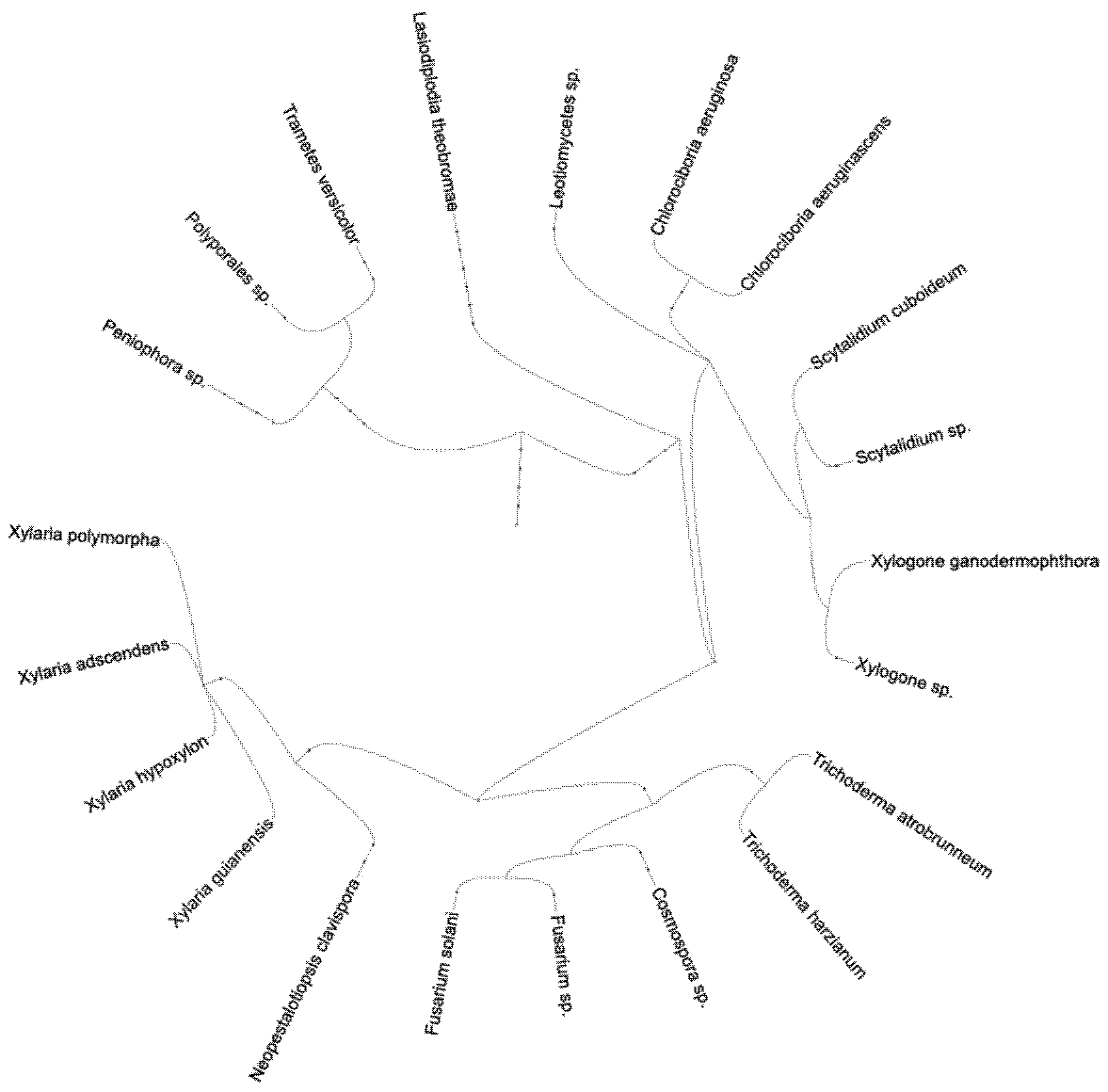
Figure 1. Phylogenetic tree of the species found in this study.
Challenges 11 00032 g001
Figure 2. Specimen collected in the field station. The orange zone-lines made this a sample of interest.
Challenges 11 00032 g002
Table 1. Fungi identified from the Peruvian Amazon rainforest from the region of Madre de Dios. As all these fungi were pulled from spalted wood, they are assumed to be, at least preliminarily, capable of some degree of spalting.
Spalting Type ObservedOrderGenusSpeciesGenBank Accession NumberGenBank Accession Number-CompareID % Similarity
Brown pigmentIncertae sedisXylogoneXylogone sp1. Arx and T. NilssonMW340804KT264505.196%
Yellow pigmentHelotialesScytalidiumScytalidium sp1. PesanteMW340805HQ631037.196%
Orange pigmentHelotialesScytalidiumScytalidium sp2. PesanteMW340806KR093920.196%
Black pigmentBotryosphaerialesLasiodiplodiaLasiodiplodia theobromae (Pat.) Griffon and Maubl.MW340807KU507483.1100%
Purple pigmentHypocrealesFusariumFusarium solani (Mart.) Sacc.MW340808JX282606.1100%
Yellow pigmentHypocrealesTrichodermaTrichoderma atrobrunneum F.B. Rocha, P. Chaverri and W. JaklitschMW340833FJ442677.1100%
Purple pigmentHelotialesScytalidiumScytalidium sp2. PesanteMW340806KR093920.197%
Orange pigmentXylarialesNeopestalotiopsisNeopestalotiopsis clavispora (G.F. Atkinson) Maharachch, K.D. Hyde and CrousMW340817KX721071.1100%
Lime green pigmentIncertae sedis Fungal sp1.MW340811KT996091.1100%
Lilac pigmentIncertae sedisXylogoneXylogone sp2. Arx and T. NilssonMW340823KU512708.192%
Yellow pigmentIncertae sedisXylogoneXylogone sp2. Arx and T. NilssonMW340823KU512708.192%
Orange pigmentHelotialesScytalidiumScytalidium sp4. PesanteMW340832KR093920.192%
Green and purple pigmentHypocrealesFusariumFusarium sp. LinkMW340819KU950729.1100%
Yellow pigmentLeotiomycetesLeotiomyceteLeotiomycetes sp1. O.E. Erikss. and WinkaMW340814KF638554.1100%
Purple pigmentLeotiomycetesLeotiomyceteLeotiomycetes sp2. O.E. Erikss. and WinkaMW340816KF638554.196%
Black zone lineXylarialesXylariaXylaria guianensis (Mont.) Fr.MW340809AM993100.199%
Orange zone lineIncertae sedis Fungal sp2.MW340824KT996094.199%
Black zone lineXylarialesXylariaXylaria hypoxylon (L.) Grev.MW340812KP143687.199%
Orange zone lineHypocrealesTrichodermaTrichoderma harzianum RifaiMW340813KC576745.199%
Black zone lineRussulalesPeniophoraPeniophora sp. CookeMW340815KJ832046.199%
Orange zone lineHypocrealesHypocreaHypocrea lixii Pat.MW340829FJ442252.1100%
Black zone lineIncertae sedis Fungal sp2.MW340824 KT996094.199%
Brown zone lineHypocrealesCosmosporaCosmospora sp. RabenhMW340827KJ676175.1100%
Black zone lineXylarialesXylariaXylaria adscendens (Fr.) Fr.MW340826KP133288.196%
Orange zone linePolyporales Polyporales sp. GäumMW340822LN997757.191%
Black zone lineHypocrealesBionectriaBionectria sp. SpegMW340821HM770964.199%
Black zone lineXylarialesNeopestalotiopsisNeopestalotiopsis clavispora (G.F. Atkinson) Maharachch, K.D. Hyde and CrousMW340831KX721071.1100%
Black zone lineIncertae sedis Fugal sp3.MW340828KM265525.1100%
Publisher’s Note: MDPI stays neutral with regard to jurisdictional claims in published maps and institutional affiliations.

