Biocompatibility of α-Al2O3 Ceramic Substrates with Human Neural Precursor Cells


 ,
,
Abstract
:1. Introduction
2. Materials and Methods
2.1. Ceramic Substrate Surface Modification
2.2. XRD Spectroscopy
2.3. Brunauer–Emmett–Teller (BET) Analysis
2.4. Wettability
2.5. NPC Culture
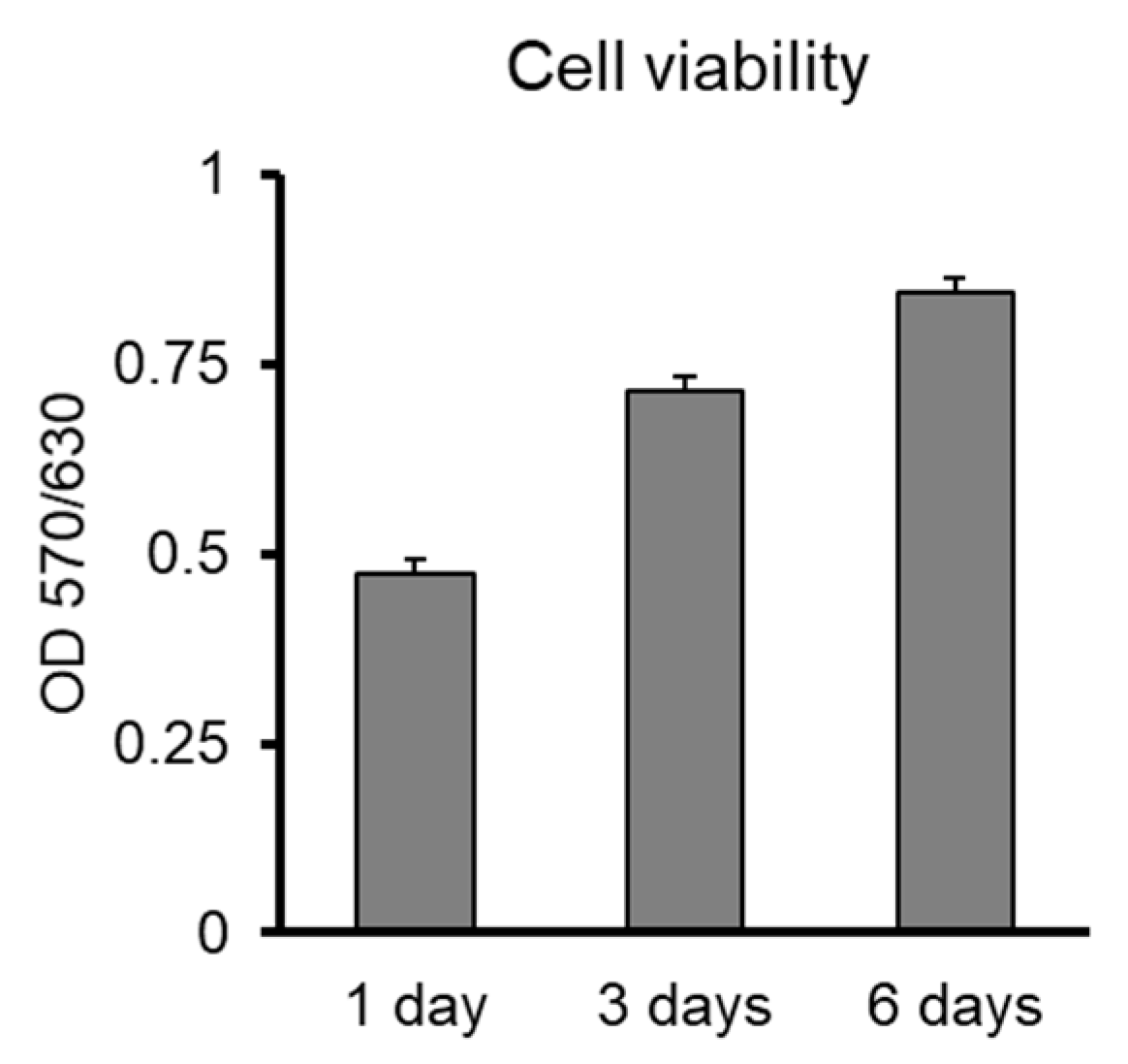
2.6. MTT Cell Viability Assay
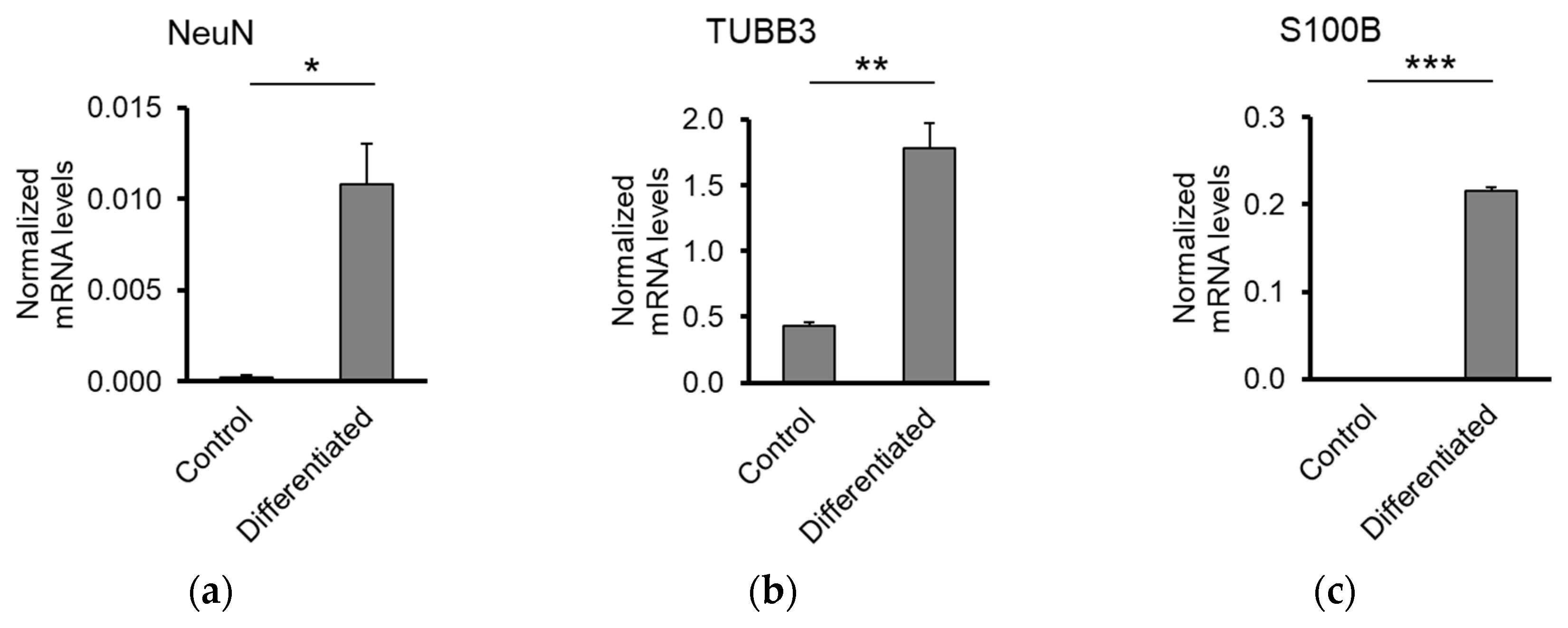
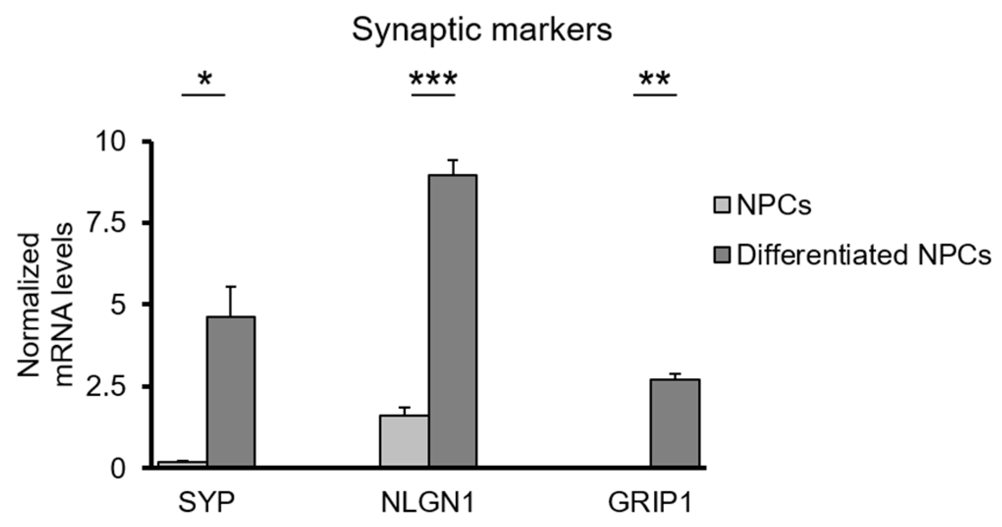
2.7. RT-qPCR
2.8. Statistical Analysis
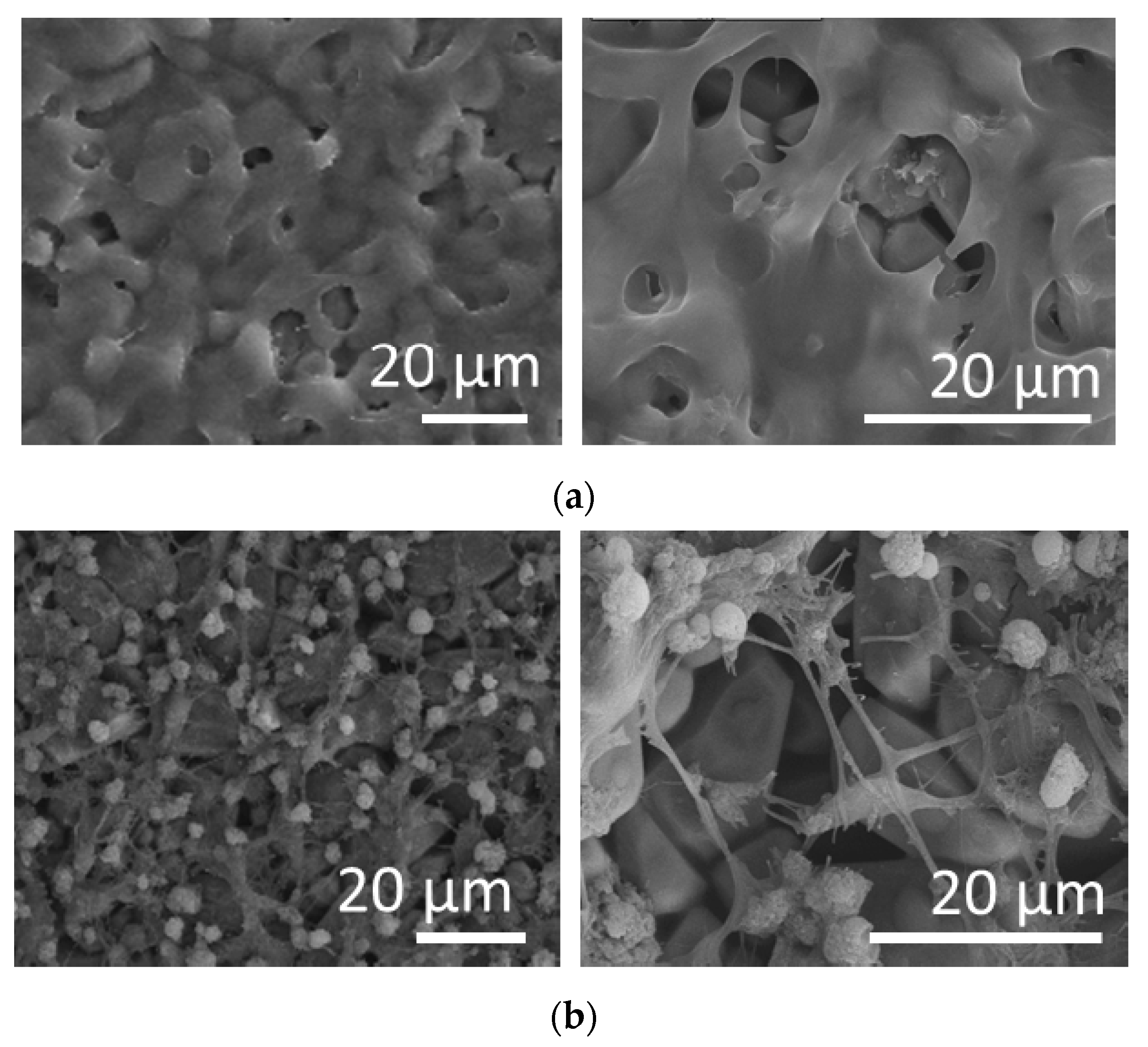
2.9. Scanning Electron Microscopy (SEM)
3. Results
3.1. Substrate Characterization
3.1.1. Surface Topology and Nanoporosity
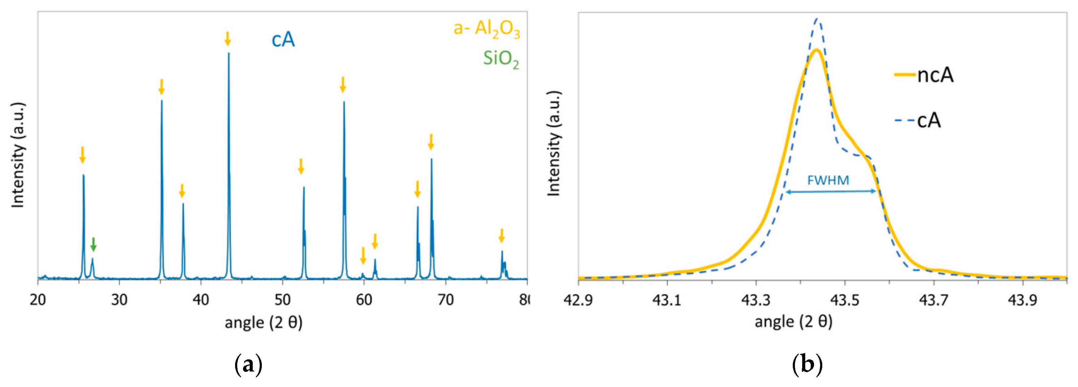
XRD Spectroscopy
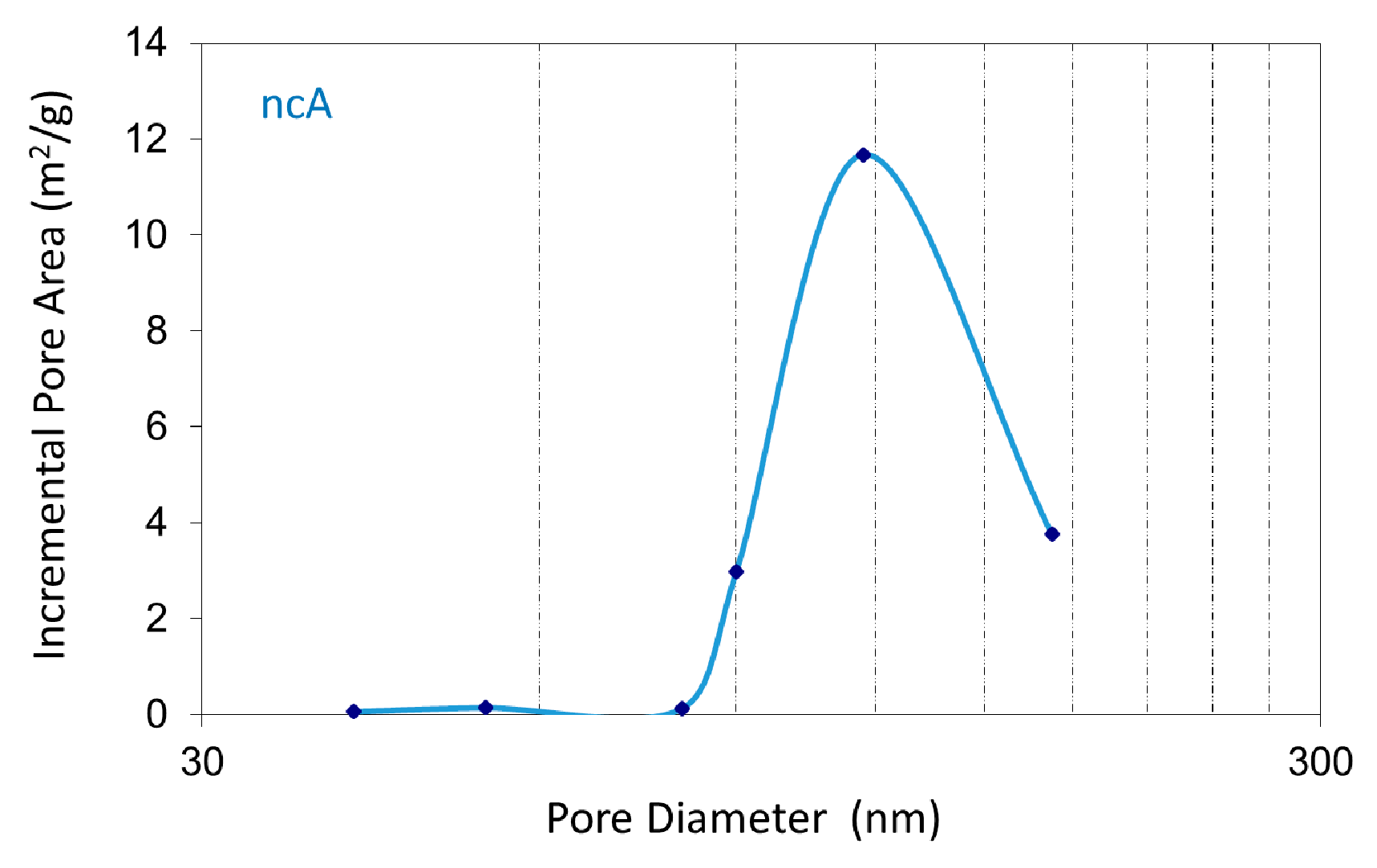
BET Analysis
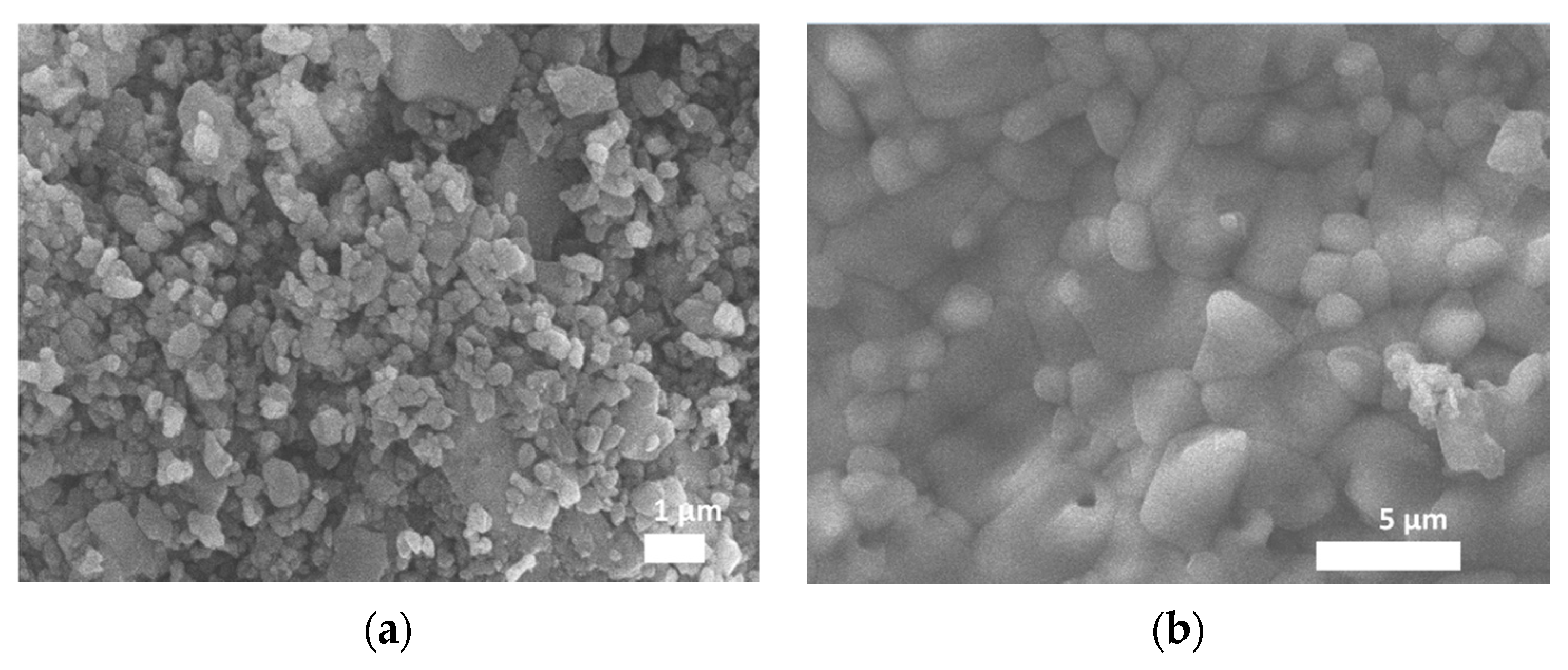
Scanning Electron Microscopy (SEM)
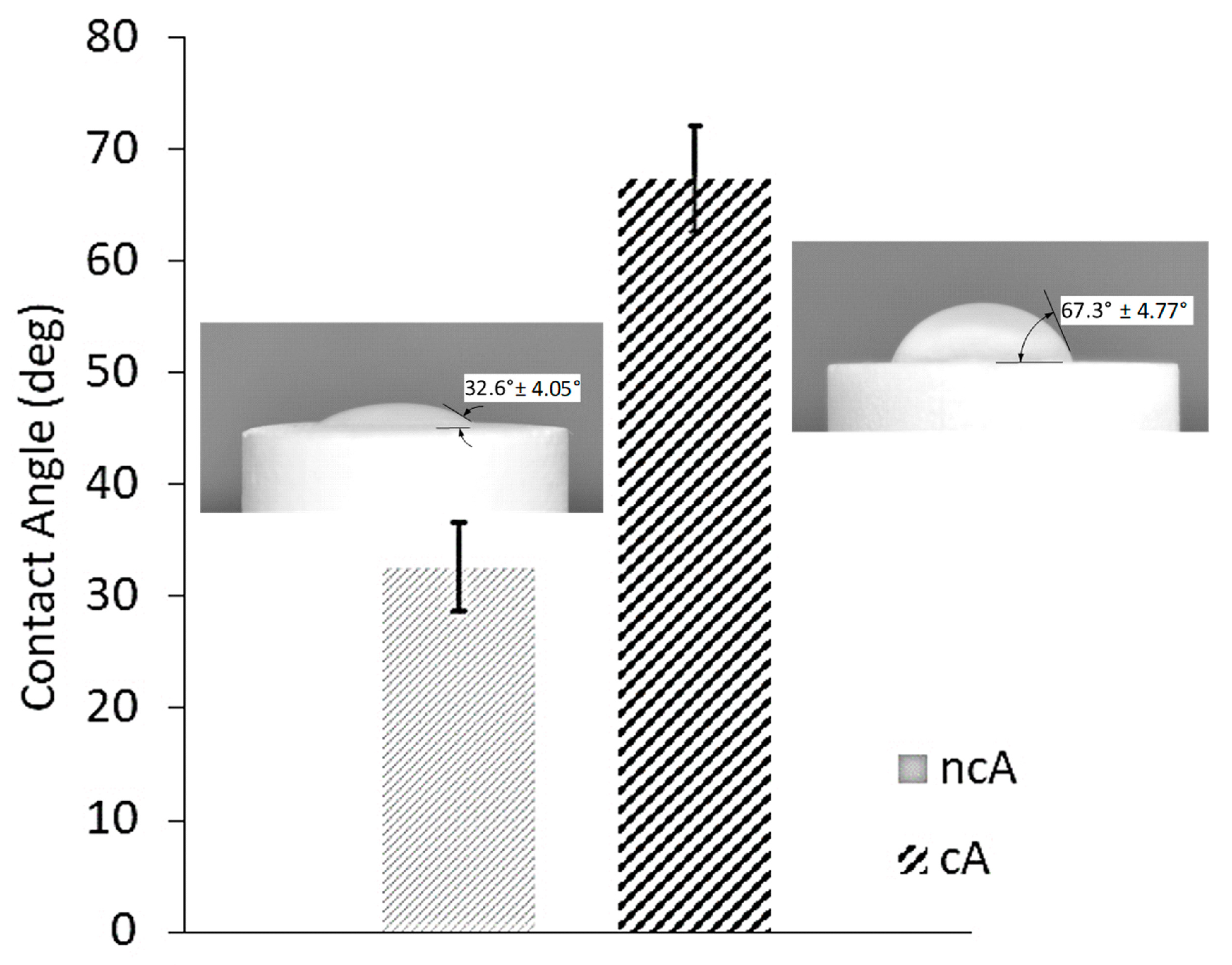
3.1.2. Wettability
3.2. Biological Assays
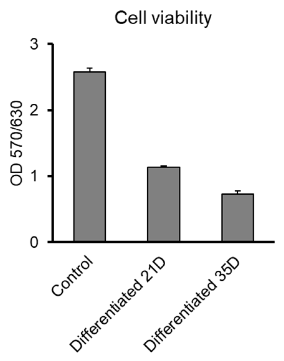
3.2.1. Biocompatibility of Al2O3 Ceramic Substrates with Human NPCs
3.2.2. Functionality of the Neuronal Network on Al2O3 Ceramic Substrates
4. Discussion
Supplementary Materials
Author Contributions
Funding
Acknowledgments
Conflicts of Interest
References
- Kato-Negishi, M.; Morimoto, Y.; Onoe, H.; Takeuchi, S. Millimeter-sized neural building blocks for 3D heterogeneous neural network assembly. Adv. Healthc. Mater. 2013, 2, 1564–1570. [Google Scholar] [CrossRef] [PubMed]
- Zhuang, P.; Sun, A.X.; An, J.; Chua, C.K.; Chew, S.Y. 3D neural tissue models: From spheroids to bioprinting. Biomaterials 2018, 154, 113–133. [Google Scholar] [CrossRef]
- Fuhrer, E.; Backer, A.; Kraft, S.; Gruhl, F.J.; Kirsch, M.; MacKinnon, N.; Korvink, J.G.; Sharma, S. 3D Carbon scaffolds for neural stem cell culture and magnetic resonance imaging. Adv. Healthc. Mater. 2018, 7. [Google Scholar] [CrossRef] [Green Version]
- Guo, W.; Qiu, J.; Liu, J.; Liu, H. Graphene microfiber as a scaffold for regulation of neural stem cells differentiation. Sci. Rep. 2017, 7, 5678. [Google Scholar] [CrossRef] [PubMed]
- Mou, X.; Wang, S.; Guo, W.; Ji, S.; Qiu, J.; Li, D.; Zhang, X.; Zhou, J.; Tang, W.; Wang, C.; et al. Localized committed differentiation of neural stem cells based on the topographical regulation effects of TiO2 nanostructured ceramics. Nanoscale 2016, 8, 13186–13191. [Google Scholar] [CrossRef]
- Patel, B.B.; Sharifi, F.; Stroud, D.P.; Montazami, R.; Hashemi, N.N.; Sakaguchi, D.S. 3D Microfibrous scaffolds selectively promotes proliferation and glial differentiation of adult neural stem cells: A platform to tune cellular behavior in neural tissue engineering. Macromol. Biosci. 2019, 19, e1800236. [Google Scholar] [CrossRef] [Green Version]
- Wang, S.; Guan, S.; Xu, J.; Li, W.; Ge, D.; Sun, C.; Liu, T.; Ma, X. Neural stem cell proliferation and differentiation in the conductive PEDOT-HA/Cs/Gel scaffold for neural tissue engineering. Biomater. Sci. 2017, 5, 2024–2034. [Google Scholar] [CrossRef] [PubMed]
- Kourgiantaki, A.; Tzeranis, D.S.; Karali, K.; Georgelou, K.; Bampoula, E.; Psilodimitrakopoulos, S.; Yannas, I.V.; Stratakis, E.; Sidiropoulou, K.; Charalampopoulos, I.; et al. Neural stem cell delivery via porous collagen scaffolds promotes neuronal differentiation and locomotion recovery in spinal cord injury. NPJ Regen. Med. 2020, 5, 12. [Google Scholar] [CrossRef] [PubMed]
- Ma, Q.; Yang, L.; Jiang, Z.; Song, Q.; Xiao, M.; Zhang, D.; Ma, X.; Wen, T.; Cheng, G. Three-dimensional stiff graphene scaffold on neural stem cells behavior. ACS Appl. Mater. Interfaces 2016, 8, 34227–34233. [Google Scholar] [CrossRef]
- Tang-Schomer, M.D.; White, J.D.; Tien, L.W.; Schmitt, L.I.; Valentin, T.M.; Graziano, D.J.; Hopkins, A.M.; Omenetto, F.G.; Haydon, P.G.; Kaplan, D.L. Bioengineered functional brain-like cortical tissue. Proc. Natl. Acad. Sci. USA 2014, 111, 13811–13816. [Google Scholar] [CrossRef] [Green Version]
- Boccaccini, A.R.; Blaker, J.J. Bioactive composite materials for tissue engineering scaffolds. Expert Rev. Med. Devices 2005, 2, 303–317. [Google Scholar] [CrossRef]
- Jones, J.R. Review of bioactive glass: From hench to hybrids. Acta Biomater. 2013, 9, 4457–4486. [Google Scholar] [CrossRef]
- Baino, F.; Novajra, G.; Vitale-Brovarone, C. Bioceramics and Scaffolds: A winning combination for tissue engineering. Front. Bioeng. Biotechnol. 2015, 3, 202. [Google Scholar] [CrossRef] [Green Version]
- Rahmati, M.; Mozafari, M. Biocompatibility of alumina-based biomaterials—A review. J. Cell. Physiol. 2019, 234, 3321–3335. [Google Scholar] [CrossRef]
- Swan, E.E.; Popat, K.C.; Desai, T.A. Peptide-immobilized nanoporous alumina membranes for enhanced osteoblast adhesion. Biomaterials 2005, 26, 1969–1976. [Google Scholar] [CrossRef]
- Adiga, S.P.; Jin, C.; Curtiss, L.A.; Monteiro-Riviere, N.A.; Narayan, R.J. Nanoporous membranes for medical and biological applications. WIREs Nanomed. Nanobiotechnol. 2009, 1, 568–581. [Google Scholar] [CrossRef] [Green Version]
- Gultepe, E.; Nagesha, D.; Sridhar, S.; Amiji, M. Nanoporous inorganic membranes or coatings for sustained drug delivery in implantable devices. Adv. Drug Deliv. Rev. 2010, 62, 305–315. [Google Scholar] [CrossRef]
- Santos, A.; Sinn Aw, M.; Bariana, M.; Kumeria, T.; Wang, Y.; Losic, D. Drug-releasing implants: Current progress, challenges and perspectives. J. Mater. Chem. B 2014, 2, 6157–6182. [Google Scholar] [CrossRef]
- Kuhn, P.T. The Effect of Wettability and Stiffness on Stem Cell Behavior at Biointerfaces. Ph.D. Thesis, Rijksuniversiteit Groningen, Groningen, The Netherlands, 2016. [Google Scholar]
- THERMANSYS. T-Ceramics. Available online: https://www.thermansys.com/gallery/files/Brochures/Machinable_Ceramics/t-ceramics-machinables-January-2017.pdf (accessed on 5 May 2020).
- Chen, H.; Muros-Cobos, J.L.; Amirfazli, A. Contact angle measurement with a smartphone. Rev. Sci. Instrum. 2018, 89, 035117. [Google Scholar] [CrossRef]
- Lorenz, C.; Lesimple, P.; Bukowiecki, R.; Zink, A.; Inak, G.; Mlody, B.; Singh, M.; Semtner, M.; Mah, N.; Aure, K.; et al. Human iPSC-Derived neural progenitors are an effective drug discovery model for neurological mtDNA Disorders. Cell Stem Cell 2017, 20, 659–674. [Google Scholar] [CrossRef] [Green Version]
- Yao, X.; Peng, R.; Ding, J. Cell-material interactions revealed via material techniques of surface patterning. Adv. Mater. 2013, 25, 5257–5286. [Google Scholar] [CrossRef]
- Alizadeh, A.; Moztarzadeh, F.; Ostad, S.N.; Azami, M.; Geramizadeh, B.; Hatam, G.; Bizari, D.; Tavangar, S.M.; Vasei, M.; Ai, J. Synthesis of calcium phosphate-zirconia scaffold and human endometrial adult stem cells for bone tissue engineering. Artif. Cells Nanomed. Biotechnol. 2016, 44, 66–73. [Google Scholar] [CrossRef]
- An, S.H.; Matsumoto, T.; Miyajima, H.; Nakahira, A.; Kim, K.H.; Imazato, S. Porous zirconia/hydroxyapatite scaffolds for bone reconstruction. Dent. Mater. 2012, 28, 1221–1231. [Google Scholar] [CrossRef]
- Piconi, C.; Condo, S.G.; Kosmač, T. Alumina- and zirconia-based ceramics for load-bearing applications. In Advanced Ceramics for Dentistry; Shen, J.Z., Kosmač, T., Eds.; Butterworth-Heinemann: Oxford, UK, 2014; pp. 219–253. [Google Scholar]
- Xifre-Perez, E.; Ferre-Borull, J.; Pallares, J.; Marsal, L.F. Mesoporous alumina as a biomaterial for biomedical applications. Open Mater. Sci. 2015, 2, 13–32. [Google Scholar] [CrossRef]
- Catelas, I.; Petit, A.; Zukor, D.J.; Marchand, R.; Yahia, L.; Huk, O.L. Induction of macrophage apoptosis by ceramic and polyethylene particles in vitro. Biomaterials 1999, 20, 625–630. [Google Scholar] [CrossRef]
- Takami, Y.; Yamane, S.; Makinouchi, K.; Otsuka, G.; Glueck, J.; Benkowski, R.; Nose, Y. Protein adsorption onto ceramic surfaces. J. Biomed. Mater. Res. 1998, 40, 24–30. [Google Scholar] [CrossRef]
- Morterra, C.; Magnacca, G. A case study: Surface chemistry and surface structure of catalytic aluminas, as studied by vibrational spectroscopy of adsorbed species. Catal. Today 1996, 27, 497–532. [Google Scholar] [CrossRef]
- Ferraz, N.; Hong, J.; Santin, M.; Karlsson Ott, M. Nanoporosity of alumina surfaces induces different patterns of activation in adhering monocytes/macrophages. Int. J. Biomater. 2010, 2010, 402715. [Google Scholar] [CrossRef]
- Koutsonikolas, D.E.; Kaldis, S.P.; Sklari, S.D.; Pantoleontos, G.; Zaspalis, V.T.; Sakellaropoulos, G.P. Preparation of highly selective silica membranes on defect-free γ-Al2O3 membranes using a low temperature CVI technique. Microporous Mesoporous Mater. 2010, 132, 276–281. [Google Scholar] [CrossRef]
- Sklari, S.; Pagana, A.; Nalbandian, L.; Zaspalis, V.T. Ceramic membrane materials and process for the removal of As(III)/As(V) ions from water. J. Water Process Eng. 2015, 5, 42–47. [Google Scholar] [CrossRef]
- Gupta, A.K.; Coburn, J.M.; Davis-Knowlton, J.; Kimmerling, E.; Kaplan, D.L.; Oxburgh, L. Scaffolding kidney organoids on silk. J. Tissue Eng. Regen. Med. 2019, 13, 812–822. [Google Scholar] [CrossRef] [PubMed]
- Lancaster, M.A.; Renner, M.; Martin, C.A.; Wenzel, D.; Bicknell, L.S.; Hurles, M.E.; Homfray, T.; Penninger, J.M.; Jackson, A.P.; Knoblich, J.A. Cerebral organoids model human brain development and microcephaly. Nature 2013, 501, 373–379. [Google Scholar] [CrossRef]
- Lancaster, M.A.; Knoblich, J.A. Organogenesis in a dish: Modeling development and disease using organoid technologies. Science 2014, 345, 1247125. [Google Scholar] [CrossRef]
- Antill-O’Brien, N.; Bourke, J.; O’Connell, C.D. Layer-By-Layer: The Case for 3D bioprinting neurons to create patient-specific epilepsy models. Materials 2019, 12, 3218. [Google Scholar] [CrossRef]
- Luo, Y. Three-dimensional scaffolds. In Principles of Tissue Engineering, 5th ed.; Lanza, R., Langer, R., Vacanti, J.P., Atala, A., Eds.; Academic Press: Cambridge, MA, USA, 2020. [Google Scholar] [CrossRef]
- Nikolova, M.P.; Chavali, M.S. Recent advances in biomaterials for 3D scaffolds: A review. Bioact. Mater. 2019, 4, 271–292. [Google Scholar] [CrossRef]


| Property | Value | Property | Value |
|---|---|---|---|
| Max. Operating Temperature, °C | 1750 | Dielectric Strength, AC-KV/mm | 16.9 |
| Porosity, % vol. | <0.5 | Dielectric Constant, 1 MHz | 9.8 |
| Density, gr/cm3 | 3.8 | Compressive Strength, MPa. | 2600 |
| Color | Ivory | Flexural Strength, MPa | 380 |
| Thermal Conductivity at 20 °C, W/mK | 30 | Elastic Modulus, GPa | 375 |
| Thermal Conductivity at 800 °C, W/mK | 8 | Shear Modulus, GPa | 152 |
| Coefficient of Thermal Expansion, 10–6/°C | 8.4 | Hardness, kg/mm2 | 1440 |
| Target Gene | Forward Primer | Reverse Primer |
|---|---|---|
| NeuN | 5′-CCAAGCGGCTACACGTCT-3′ | 5′-GCTCGGTCAGCATCTGAG-3′ |
| TUBB3 | 5′-CCAAGGGTCACTACACGGAG-3′ | 5′-ATGATGCGGTCGGGATACTC-3′ |
| S100B | 5′-ATGTCTGAGCTGGAGAAGG-3′ | 5′-CTCATGTTCAAAGAACTCGTG-3′ |
| SYP | 5′-CTGTGACCTCGGGACTCAAC-3′ | 5′-CATAGTCAGGCTGGTAGCCG-3′ |
| NLGN1 | 5′-CCTTTCCAGCTGGGCTGTTA-3′ | 5′-TCTGGGGGTCGTCTGGTATT-3′ |
| GRIP1 | 5′-CCGTTGTCAAATTCTGAGGCG-3′ | 5′-TACCGTCAGACCCAGGGTAG-3′ |
| Sample | Calcination Temperature (°C) | Surface Area (m2/gr) | Crystallite Size (nm) |
|---|---|---|---|
| ncA | 800 | 5.56 | 42.28 |
| cA | 1650 | - | 42.29 |
© 2020 by the authors. Licensee MDPI, Basel, Switzerland. This article is an open access article distributed under the terms and conditions of the Creative Commons Attribution (CC BY) license (http://creativecommons.org/licenses/by/4.0/).
Share and Cite
Asimakopoulou, A.; Gkekas, I.; Kastrinaki, G.; Prigione, A.; Zaspalis, V.T.; Petrakis, S. Biocompatibility of α-Al2O3 Ceramic Substrates with Human Neural Precursor Cells. J. Funct. Biomater. 2020, 11, 65. https://0-doi-org.brum.beds.ac.uk/10.3390/jfb11030065
Asimakopoulou A, Gkekas I, Kastrinaki G, Prigione A, Zaspalis VT, Petrakis S. Biocompatibility of α-Al2O3 Ceramic Substrates with Human Neural Precursor Cells. Journal of Functional Biomaterials. 2020; 11(3):65. https://0-doi-org.brum.beds.ac.uk/10.3390/jfb11030065
Chicago/Turabian StyleAsimakopoulou, Akrivi, Ioannis Gkekas, Georgia Kastrinaki, Alessandro Prigione, Vasileios T. Zaspalis, and Spyros Petrakis. 2020. "Biocompatibility of α-Al2O3 Ceramic Substrates with Human Neural Precursor Cells" Journal of Functional Biomaterials 11, no. 3: 65. https://0-doi-org.brum.beds.ac.uk/10.3390/jfb11030065