Hardness, an Important Indicator of Bone Quality, and the Role of Collagen in Bone Hardness

 ,
,
Abstract
:1. Introduction
2. Materials and Methods
Bone Sample Preparation
3. Results & Discussion
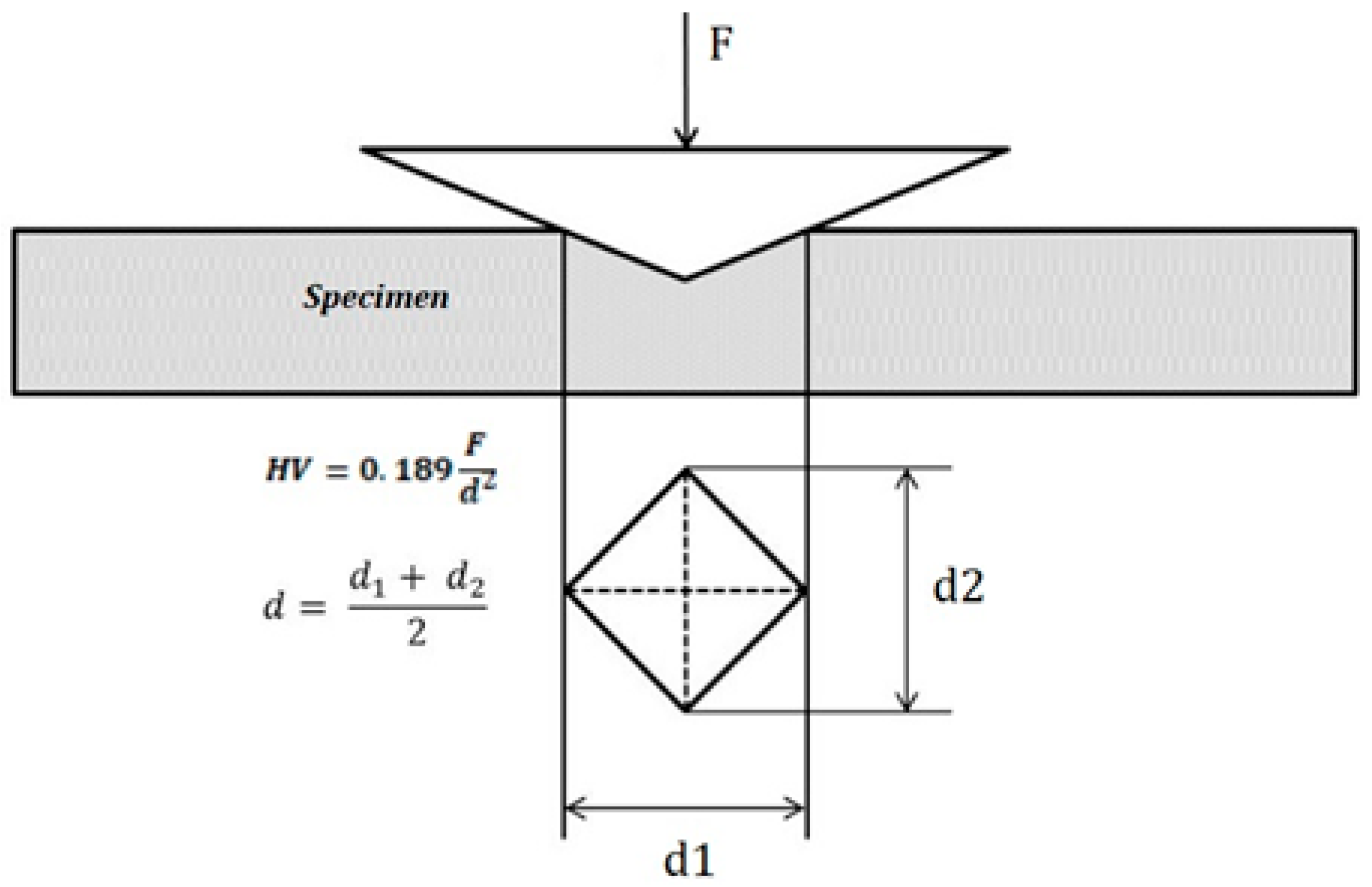
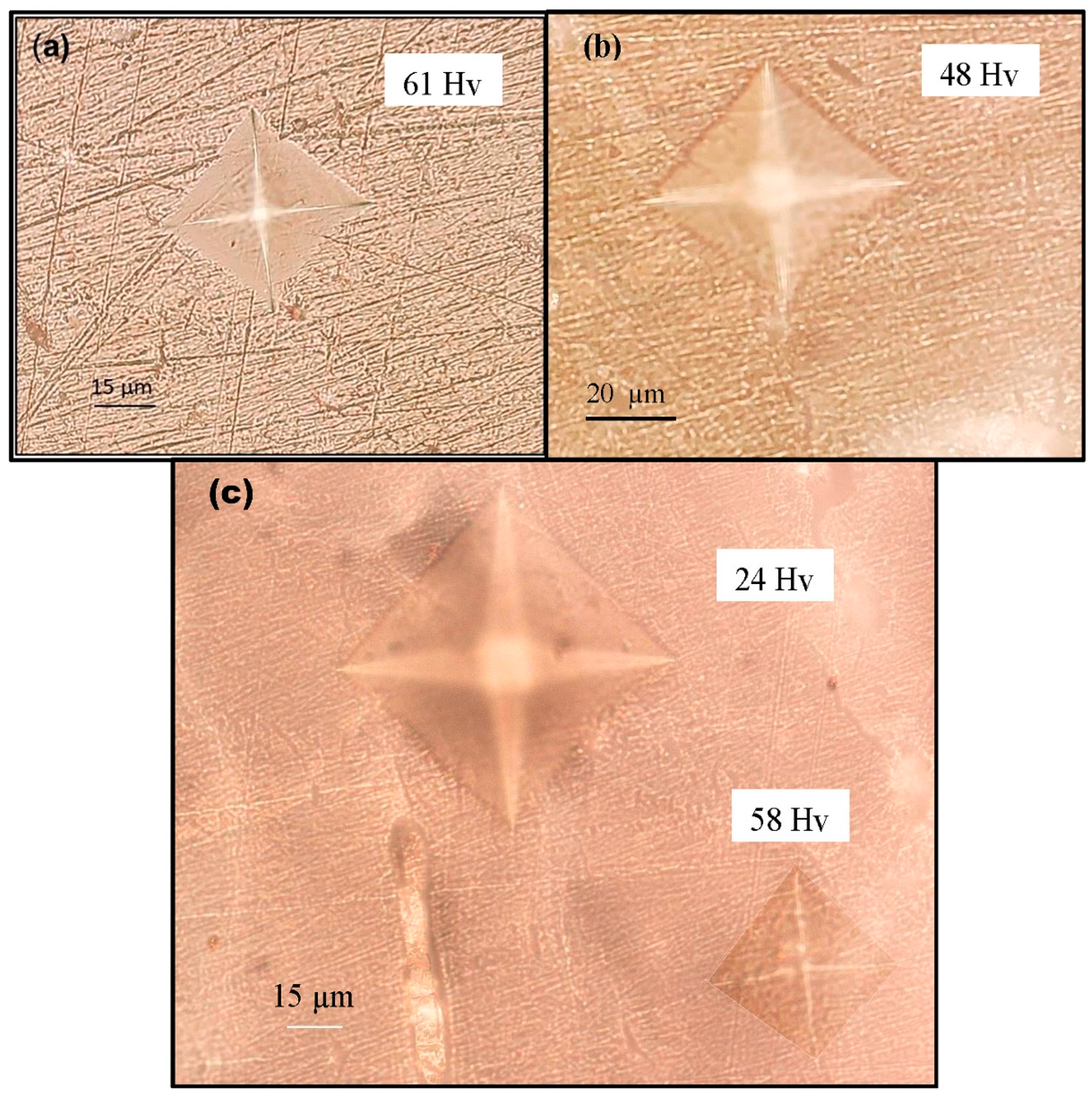
3.1. Vickers Microhardness Testing
3.2. Effect of Chemical Treatment on Bone Hardness
4. Conclusions
Author Contributions
Funding
Conflicts of Interest
References
- Rho, J.Y.; Kuhn-Spearing, L.; Zioupos, P. Mechanical properties and the hierarchical structure of bone. Med. Eng. Phys. 1998, 20, 92–102. [Google Scholar] [CrossRef]
- Weiner, S.; Wagner, H.D. The material bone: Structure-mechanical function relations. Annu. Rev. Mater. Sci. 1998, 28, 271–298. [Google Scholar] [CrossRef]
- Fratzl, P.; Weinkamer, R. Nature’s hierarchical materials. Prog. Mater. Sci. 2007, 52, 1263–1334. [Google Scholar] [CrossRef] [Green Version]
- Gautieri, A.; Vesentini, S.; Redaelli, A.; Buehler, M.J. Hierarchical structure and nanomechanics of collagen microfibrils from the atomistic scale up. Nano Lett. 2011, 11, 757–766. [Google Scholar] [CrossRef]
- Landis, W.J. The strength of a calcified tissue depends in part on the molecular structure and organization of its constituent mineral crystals in their organic matrix. Bone 1995, 16, 533–544. [Google Scholar] [CrossRef]
- Tai, K.; Dao, M.; Suresh, S.; Palazoglu, A.; Ortiz, C. Nanoscale heterogeneity promotes energy dissipation in bone. Nat. Mater. 2007, 6, 454–462. [Google Scholar] [CrossRef]
- Currey, J.D. The effect of porosity and mineral content on the Young’s modulus of elasticity of compact bone. J. Biomech. 1988, 21, 131–139. [Google Scholar] [CrossRef]
- Burstein, A.H.; Zika, J.M.; Heiple, K.G.; Klein, L. Contribution of collagen and mineral to the elastic-plastic properties of bone. JBJS 1975, 57, 956–961. [Google Scholar] [CrossRef]
- Martin, R.B.; Boardman, D.L. The effects of collagen fiber orientation, porosity, density, and mineralization on bovine cortical bone bending properties. J. Biomech. 1993, 26, 1047–1054. [Google Scholar] [CrossRef]
- Evans, G.P.; Behiri, J.C.; Currey, J.D.; Bonfield, W. Microhardness and Young’s modulus in cortical bone exhibiting a wide range of mineral volume fractions, and in a bone analogue. J. Mater. Sci. Mater. Med. 1990, 1, 38–43. [Google Scholar] [CrossRef]
- Huja, S.S.; Beck, F.M.; Thurman, D.T. Indentation properties of young and old osteons. Calcif. Tissue Int. 2006, 78, 392–397. [Google Scholar] [CrossRef] [PubMed]
- Mulder, L.; Koolstra, J.H.; den Toonder, J.M.P.; van Eijden, T.M.G.J. Relationship between tissue stiffness and degree of mineralization of developing trabecular bone. J. Biomed. Mater. Res. Part A 2008, 84, 508–515. [Google Scholar] [CrossRef] [PubMed] [Green Version]
- Knott, L.; Bailey, A.J. Collagen cross-links in mineralizing tissues: A review of their chemistry, function, and clinical relevance. Bone 1998, 22, 181–187. [Google Scholar] [CrossRef]
- Boivin, G.; Bala, Y.; Doublier, A.; Farlay, D.; Ste-Marie, L.G.; Meunier, P.J.; Delmas, P.D. The role of mineralization and organic matrix in the microhardness of bone tissue from controls and osteoporotic patients. Bone 2008, 43, 532–538. [Google Scholar] [CrossRef]
- Viguet-Carrin, S.; Garnero, P.; Delmas, P.D. The role of collagen in bone strength. Osteoporos. Int. 2006, 17, 319–336. [Google Scholar] [CrossRef]
- Wang, J.; Yin, B.; Liu, G.; Li, S.; Zhang, X.; Hu, Z.; Wu, W.; Zhang, Y. Microhardness distribution of the tibial diaphysis and test site selection for reference point indentation technique. Medicine 2019, 98, e16523. [Google Scholar] [CrossRef]
- Hoc, T.; Henry, L.; Verdier, M.; Aubry, D.; Sedel, L.; Meunier, A. Effect of microstructure on the mechanical properties of Haversian cortical bone. Bone 2006, 8, 466–474. [Google Scholar] [CrossRef]
- Currey, J.D.; Brear, K. Hardness, Young’s modulus and yield stress in mammalian mineralized tissues. J. Mater. Sci. Mater. Med. 1990, 1, 14–20. [Google Scholar] [CrossRef]
- Paschalis, E.P.; Shane, E.; Lyritis, G.; Skarantavos, G.; Mendelsohn, R.; Boskey, A.L. Bone fragility and collagen cross-links. J. Bone Miner. Res. 2004, 19, 2000–2004. [Google Scholar] [CrossRef]
- Saito, M.; Marumo, K.M.S.K.M. Collagen cross-links as a determinant of bone quality: A possible explanation for bone fragility in aging, osteoporosis, and diabetes mellitus. Osteoporos. Int. 2010, 21, 195–214. [Google Scholar] [CrossRef]
- Labonte, D.; Lenz, A.K.; Oyen, M.L. On the relationship between indentation hardness and modulus, and the damage resistance of biological materials. Acta Biomater. 2017, 57, 373–383. [Google Scholar] [CrossRef] [PubMed]
- Boskey, A.L.; Coleman, R. Aging and bone. J. Dent. Res. 2010, 89, 1333–1348. [Google Scholar] [CrossRef] [PubMed]
- Wu, W.W.; Zhu, Y.B.; Chen, W.; Li, S.; Yin, B.; Wang, J.Z.; Zhang, X.J.; Liu, G.B.; Hu, Z.S.; Zhang, Y.Z. Bone Hardness of Different Anatomical Regions of Human Radius and its Impact on the Pullout Strength of Screws. Orthop. Surg. 2019, 11, 270–276. [Google Scholar] [CrossRef]
- Yin, B.; Guo, J.L.; Wang, J.Z.; Li, S.; Liu, Y.K.; Zhang, Y.Z. Bone Material Properties of Human Phalanges Using Vickers Indentation. Orthop. Surg. 2019, 11, 487–492. [Google Scholar] [CrossRef] [Green Version]
- Rodriguez-Florez, N.; Oyen, M.L.; Shefelbine, S.J. Insight into differences in nanoindentation properties of bone. J. Mech. Behav. Biomed. Mater. 2013, 18, 90–99. [Google Scholar] [CrossRef] [PubMed]
- Hengsberger, S.; Kulik, A.; Zysset, P. A combined atomic force microscopy and nanoindentation technique to investigate the elastic properties of bone structural units. Eur. Cell Mater. 2001, 1, 12–17. [Google Scholar] [CrossRef]
- Tang, B.; Ngan, A.H.W.; Lu, W.W. An improved method for the measurement of mechanical properties of bone by nanoindentation. J. Mater. Sci. Mater. Med. 2007, 18, 1875–1881. [Google Scholar] [CrossRef] [Green Version]
- Rho, J.Y.; Pharr, G.M. Effects of drying on the mechanical properties of bovine femur measured by nanoindentation. J. Mater. Sci. Mater. Med. 1999, 10, 485–488. [Google Scholar] [CrossRef]
- Bembey, A.K.; Oyen, M.L.; Bushby, A.J.; Boyde, A. Viscoelastic properties of bone as a function of hydration state determined by nanoindentation. Philos. Mag. 2006, 86, 5691–5703. [Google Scholar] [CrossRef]
- Zysset, P.K.; Guo, X.E.; Hoffler, C.E.; Moore, K.E.; Goldstein, S.A. Elastic modulus and hardness of cortical and trabecular bone lamellae measured by nanoindentation in the human femur. J. Biomech. 1999, 32, 1005–1012. [Google Scholar] [CrossRef]
- Zysset, P.K.; Guo, X.E.; Hoffler, C.E.; Moore, K.E.; Goldstein, S.A. Mechanical properties of human trabecular bone lamellae quantified by nanoindentation. Technol. Health Care 1998, 6, 429–432. [Google Scholar] [CrossRef] [PubMed]
- Fan, Z.; Swadener, J.G.; Rho, J.Y.; Roy, M.E.; Pharr, G.M. Anisotropic properties of human tibial cortical bone as measured by nanoindentation. J. Orthop. Res. 2002, 20, 806–810. [Google Scholar] [CrossRef]
- Rho, J.Y.; Tsui, T.Y.; Pharr, G.M. Elastic properties of human cortical and trabecular lamellar bone measured by nanoindentation. Biomaterials 1997, 18, 1325–1330. [Google Scholar] [CrossRef]
- Franzoso, G.; Zysset, P.K. Elastic anisotropy of human cortical bone secondary osteons measured by nanoindentation. J. Biomech. Eng. 2009, 131, 021001. [Google Scholar] [CrossRef] [PubMed] [Green Version]
- Giambini, H.; Wang, H.J.; Zhao, C.; Chen, Q.; Nassr, A.; An, K.N. Anterior and posterior variations in mechanical properties of human vertebrae measured by nanoindentation. J. Biomech. 2013, 46, 456–461. [Google Scholar] [CrossRef] [PubMed] [Green Version]
- Renders, G.A.P.; Mulder, L.; Van Ruijven, L.J.; Van Eijden, T.M.G.J. Degree and distribution of mineralization in the human mandibular condyle. Calcif. Tissue Int. 2006, 79, 190–196. [Google Scholar] [CrossRef] [PubMed]
- Gong, J.K.; Arnold, J.S.; Cohn, S.H. Composition of trabecular and cortical bone. Anat. Record 1964, 149, 325–331. [Google Scholar] [CrossRef] [PubMed]
- Hodgskinson, R.; Currey, J.D.; Evans, G.P. Hardness, an indicator of the mechanical competence of cancellous bone. J. Orthop. Res. 1989, 7, 754–758. [Google Scholar] [CrossRef]
- Currey, J.D. Effects of differences in mineralization on the mechanical properties of bone. Philosop. Trans. R. Soc. Lond. B Biol. Sci. 1984, 304, 509–518. [Google Scholar]
- Wang, X.; Bank, A.R.; TeKoppele, M.J.; Agrawal, M.C. The role of collagen in determining bone mechanical properties. J. Orthop. Res. 2001, 19, 1021–1026. [Google Scholar] [CrossRef]
- Kruzic, J.J.; Kim, D.K.; Koester, K.J.; Ritchie, R.O. Indentation techniques for evaluating the fracture toughness of biomaterials and hard tissues. J. Mech. Behav. Biomed. Mater. 2009, 2, 384–395. [Google Scholar] [CrossRef] [PubMed]
- Zysset, P.K. Indentation of bone tissue: A short review. Osteop. Int. 2009, 20, 1049–1055. [Google Scholar] [CrossRef] [PubMed]
- Ziv, V.; Wagner, H.D.; Weiner, S. Microstructure-microhardness relations in parallel-fibered and lamellar bone. Bone 1996, 18, 417–428. [Google Scholar] [CrossRef]
- Currey, J.D. How well are bones designed to resist fracture? J. Bone Miner. Res. 2003, 18, 591–598. [Google Scholar] [CrossRef] [PubMed]
- Ritchie, R.O.; Kinney, J.H.; Kruzic, J.J.; Nalla, R.K. A fracture mechanics and mechanistic approach to the failure of cortical bone. Fatigue Fract. Eng. Mater. Struct. 2005, 28, 345–371. [Google Scholar] [CrossRef]
- Oyen, M.L. Nanoindentation hardness of mineralized tissues. J. Biomech. 2006, 39, 2699–2702. [Google Scholar] [CrossRef]
- Hengsberger, S.; Kulik, A.; Zysset, P.K. Nanoindentation discriminates the elastic properties of individual human bone lamellae under dry and physiological conditions. Bone 2002, 30, 178–184. [Google Scholar] [CrossRef]
- Bembey, A.K.; Bushby, A.J.; Boyde, A.; Ferguson, V.L.; Oyen, M.L. Hydration effects on the micro-mechanical properties of bone. J. Mater. Res. 2006, 21, 1962–1968. [Google Scholar] [CrossRef]
- Nyman, J.S.; Roy, A.; Shen, X.; Acuna, R.L.; Tyler, J.H.; Wang, X. The influence of water removal on the strength and toughness of cortical bone. J. Biomech. 2006, 39, 931–938. [Google Scholar] [CrossRef] [Green Version]
- Granke, M.; Does, M.D.; Nyman, J.S. The role of water compartments in the material properties of cortical bone. Calcif. Tissue Int. 2015, 97, 292–307. [Google Scholar] [CrossRef] [Green Version]
- Samuel, J.; Sinha, D.; Zhao, J.C.G.; Wang, X. Water residing in small ultrastructural spaces plays a critical role in the mechanical behavior of bone. Bone 2014, 59, 199–206. [Google Scholar] [CrossRef] [PubMed] [Green Version]
- Timmins, P.A.; Wall, J.C. Bone water. Calcif. Tissue Int. 1977, 23, 1–5. [Google Scholar] [CrossRef] [PubMed]
- Morgan, E.F.; Bayraktar, H.H.; Keaveny, T.M. Trabecular bone modulus–density relationships depend on anatomic site. J. Biomech. 2003, 36, 897–904. [Google Scholar] [CrossRef]
- Dall’Ara, E.; Ohman, C.; Baleani, M.; Viceconti, M. The effect of tissue condition and applied load on Vickers hardness of human trabecular bone. J. Biomech. 2007, 40, 3267–3270. [Google Scholar] [CrossRef] [PubMed]
- Currey, J.D.; Zioupos, P.; Davies, P.; Casinos, A. Mechanical properties of nacre and highly mineralized bone. Proc. R. Soc. Lond. B 2001, 268, 107–111. [Google Scholar] [CrossRef] [Green Version]
- Todoh, M.; Tadano, S.; Imari, Y. Effect of heat denaturation of collagen matrix on bone strength. In IFMBE, Proceedings of the 13th International Conference on Biomedical Engineering, Singapore, 3–6 December 2008; Springer: Berlin/Heidelberg, Germany, 2009; pp. 2034–2037. [Google Scholar]
- Bozec, L.; Odlyha, M. Thermal denaturation studies of collagen by microthermal analysis and atomic force microscopy. Biophys. J. 2011, 101, 228–236. [Google Scholar] [CrossRef] [PubMed] [Green Version]
- Currey, J.D. Role of collagen and other organics in the mechanical properties of bone. Osteop. Int. 2003, 14, 29–36. [Google Scholar]
- Boskey, A.L.; Wright, T.M.; Blank, R.D. Collagen and bone strength. J. Bone Miner. Res. 1999, 14, 330–335. [Google Scholar] [CrossRef]
- Currey, J. Structural heterogeneity in bone: Good or bad? J. Musculoskelet. Neuronal Interact. 2005, 5, 317. [Google Scholar]
- Fantner, G.E.; Birkedal, H.; Kindt, J.H.; Hassenkam, T.; Weaver, J.C.; Cutroni, J.A.; Bosma, B.L.; Bawazer, L.; Finch, M.M.; Cidade, G.A.; et al. Influence of the degradation of the organic matrix on the microscopic fracture behavior of trabecular bone. Bone 2004, 35, 1013–1022. [Google Scholar] [CrossRef]
- Jain, R.; Kochhar, R.; Dewan, R.; Malhotra, N.; Srivastava, S. Comparative evaluation of the effect of two commonly used irrigants on bone: An ex-vivo study. Endodontology 2015, 27, 8. [Google Scholar]
- Kerbl, F.M.; DeVilliers, P.; Litaker, M.; Eleazer, P.D. Physical effects of sodium hypochlorite on bone: An ex vivo study. J. Endod. 2012, 38, 357–359. [Google Scholar] [CrossRef] [PubMed]
- Tartari, T.; Bachmann, L.; Maliza, A.G.A.; Andrade, F.B.; Duarte, M.A.H.; Bramante, C.M. Tissue dissolution and modifications in dentin composition by different sodium hypochlorite concentrations. J. Appl. Oral Sci. 2016, 24, 291–298. [Google Scholar] [CrossRef]
- Christensen, C.E.; McNeal, S.F.; Eleazer, P. Effect of lowering the pH of sodium hypochlorite on dissolving tissue in vitro. J. Endod. 2008, 34, 449–452. [Google Scholar] [CrossRef] [PubMed]
- Dumitriu, D.; Dobre, T. Effects of temperature and hypochlorite concentration on the rate of collagen dissolution. J. Endod. 2015, 41, 903–906. [Google Scholar] [CrossRef] [PubMed]


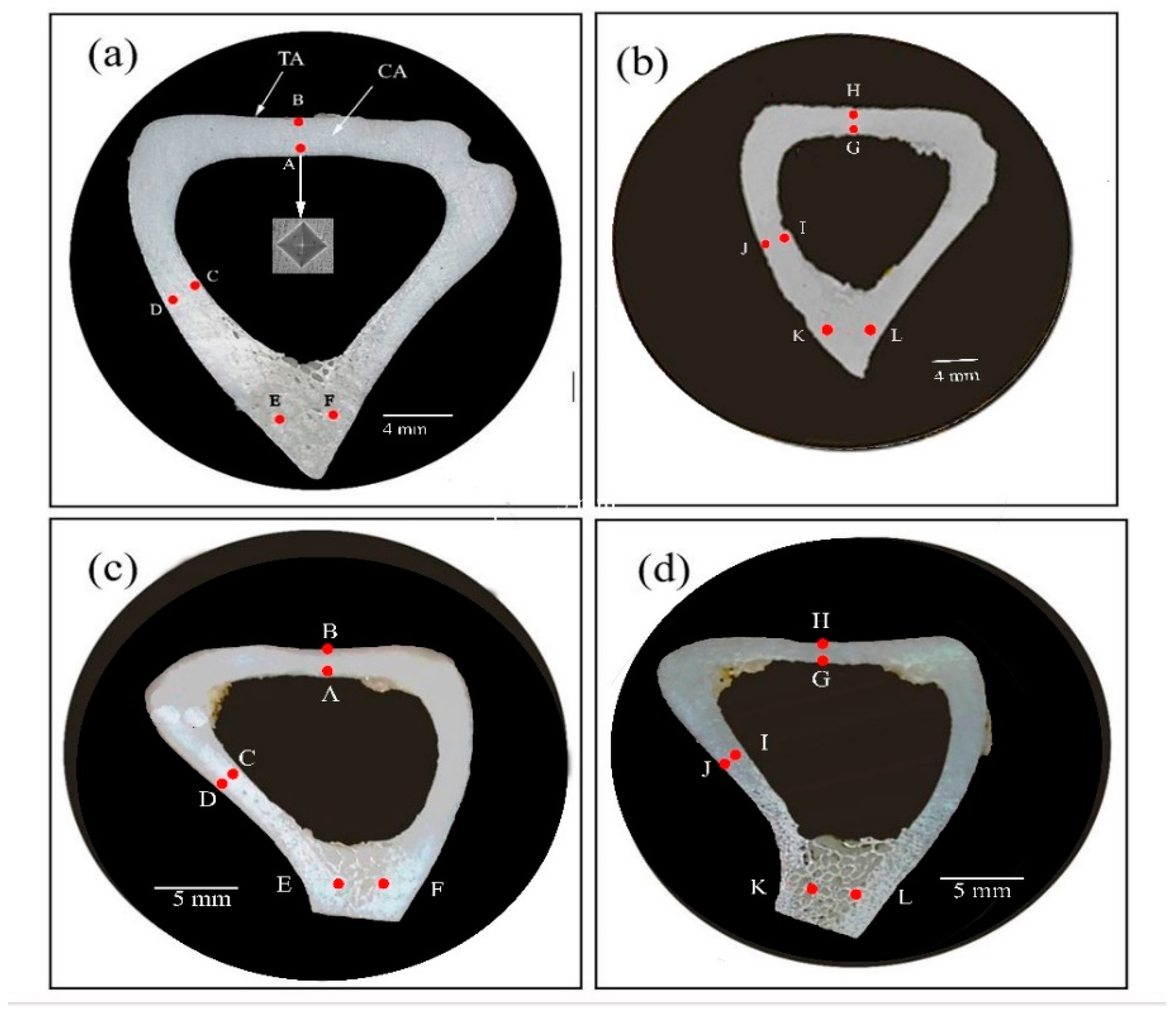
| Location | Microhardness (Hv) G I (a & b) | Location | Microhardness (Hv) G II (c & d) |
|---|---|---|---|
| A | 56.7 ± 4.5 | A | 48.0 ± 4.7 |
| B | 69.8 ± 3.2 | B | 58.1 ± 3.9 |
| C | 53.3 ± 4.8 | C | 49.8 ± 3.3 |
| D | 77.7± 6.3 | D | 58.6 ± 4.4 |
| E | 65.8 ± 5.6 | E | 48.6 ± 4.6 |
| F | 62.9 ± 4.1 | F | 53.0 ± 5.3 |
| G | 55.0 ± 3.0 | G | 56.2 ± 3 |
| H | 66.3 ± 5.3 | H | 62.5 ± 5.0 |
| I | 53.7 ± 4.2 | I | 57.3 ± 2.6 |
| J | 67.2 ± 6.7 | J | 63.7 ± 4.5 |
| K | 62.2 ± 5.6 | K | 46.4 ± 3.8 |
| L | 56.1 ± 4.3 | L | 48.3 ± 4.1 |
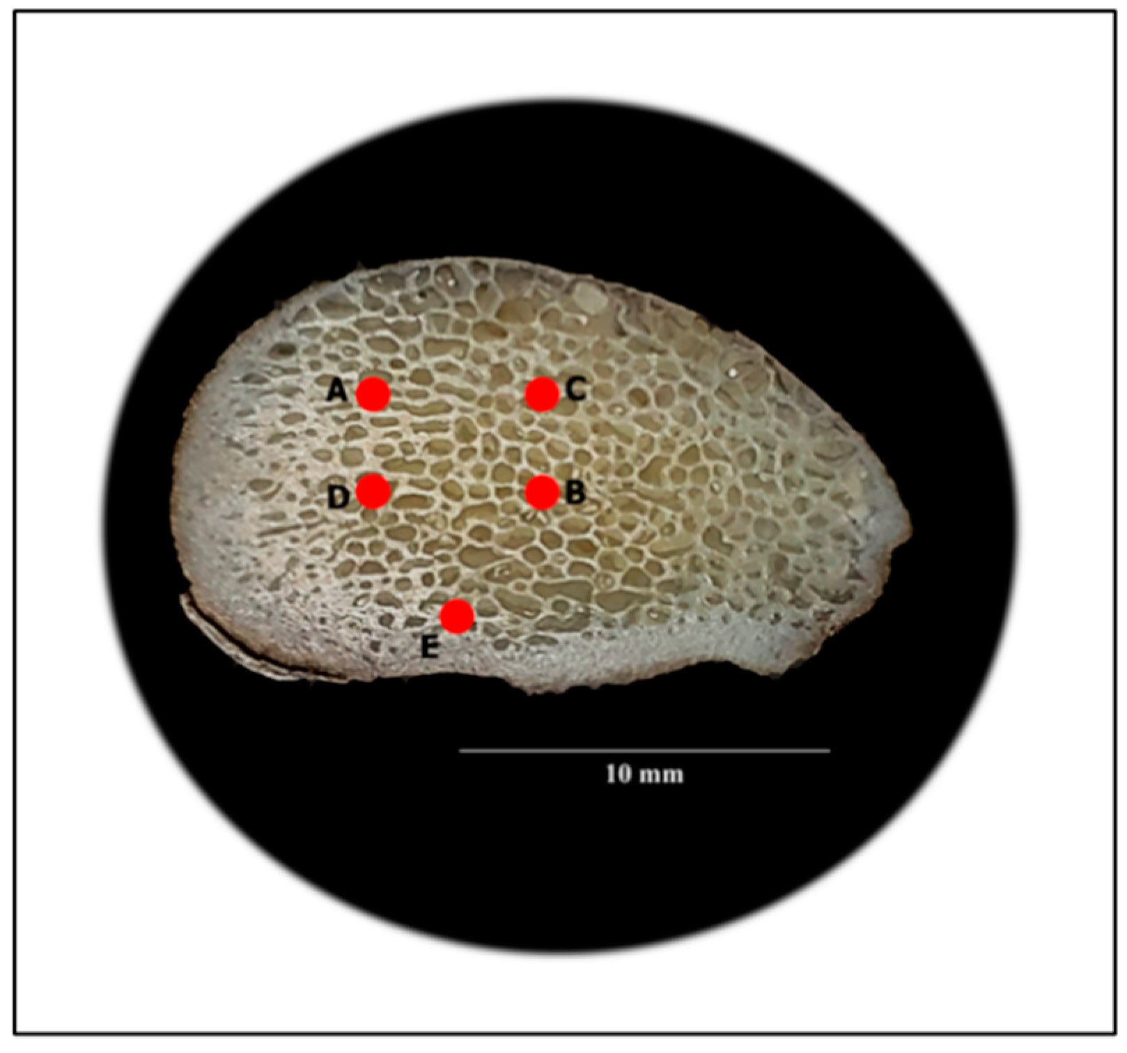
| Location | Microhardness (Hv) |
|---|---|
| A | 66.8 ± 5.6 |
| B | 52.2 ± 5.8 |
| C | 76 ± 6.3 |
| D | 65.2 ± 4.2 |
| E | 65 ± 5.5 |
Publisher’s Note: MDPI stays neutral with regard to jurisdictional claims in published maps and institutional affiliations. |
© 2020 by the authors. Licensee MDPI, Basel, Switzerland. This article is an open access article distributed under the terms and conditions of the Creative Commons Attribution (CC BY) license (http://creativecommons.org/licenses/by/4.0/).
Share and Cite
Ibrahim, A.; Magliulo, N.; Groben, J.; Padilla, A.; Akbik, F.; Abdel Hamid, Z. Hardness, an Important Indicator of Bone Quality, and the Role of Collagen in Bone Hardness. J. Funct. Biomater. 2020, 11, 85. https://0-doi-org.brum.beds.ac.uk/10.3390/jfb11040085
Ibrahim A, Magliulo N, Groben J, Padilla A, Akbik F, Abdel Hamid Z. Hardness, an Important Indicator of Bone Quality, and the Role of Collagen in Bone Hardness. Journal of Functional Biomaterials. 2020; 11(4):85. https://0-doi-org.brum.beds.ac.uk/10.3390/jfb11040085
Chicago/Turabian StyleIbrahim, Ahmed, Nicole Magliulo, James Groben, Ashley Padilla, Firas Akbik, and Z. Abdel Hamid. 2020. "Hardness, an Important Indicator of Bone Quality, and the Role of Collagen in Bone Hardness" Journal of Functional Biomaterials 11, no. 4: 85. https://0-doi-org.brum.beds.ac.uk/10.3390/jfb11040085