
Annealed ZnO/Al2O3 Core-Shell Nanowire as a Platform to Capture RNA in Blood Plasma

 , ,
, ,  and
and 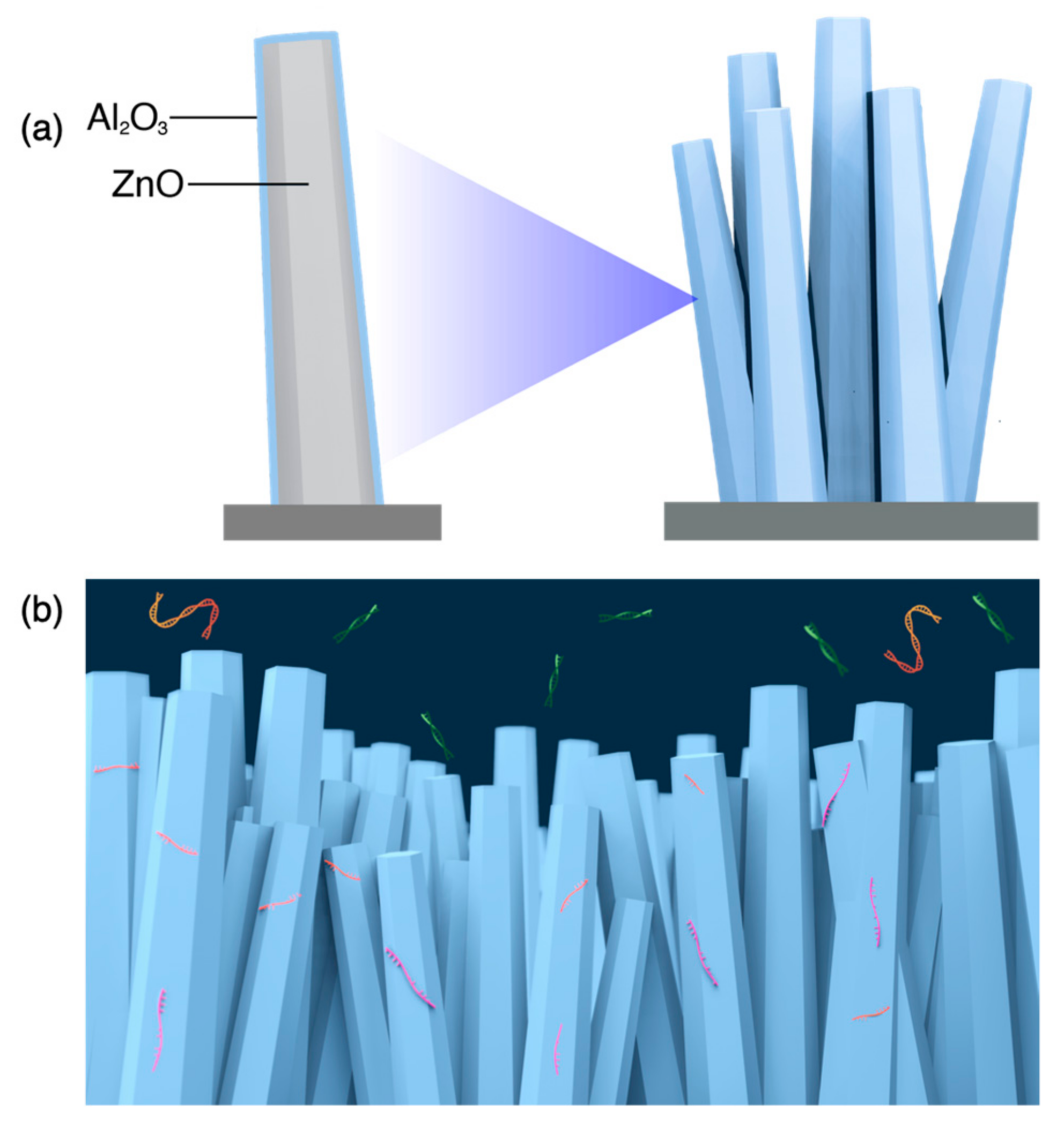
Abstract
:1. Introduction
2. Materials and Methods
2.1. Preparation of Blood Plasma Samples
2.2. Preparation of Nucleic Acid Samples
2.3. Fabrication of ZnO/Al2O3 Core-Shell Nanowire
2.4. Structural Characterization
2.5. Capture Experiment on a Nanowire for Circulating Nucleic Acids
2.6. Real-Time Quantification of Nucleic Acid
3. Results
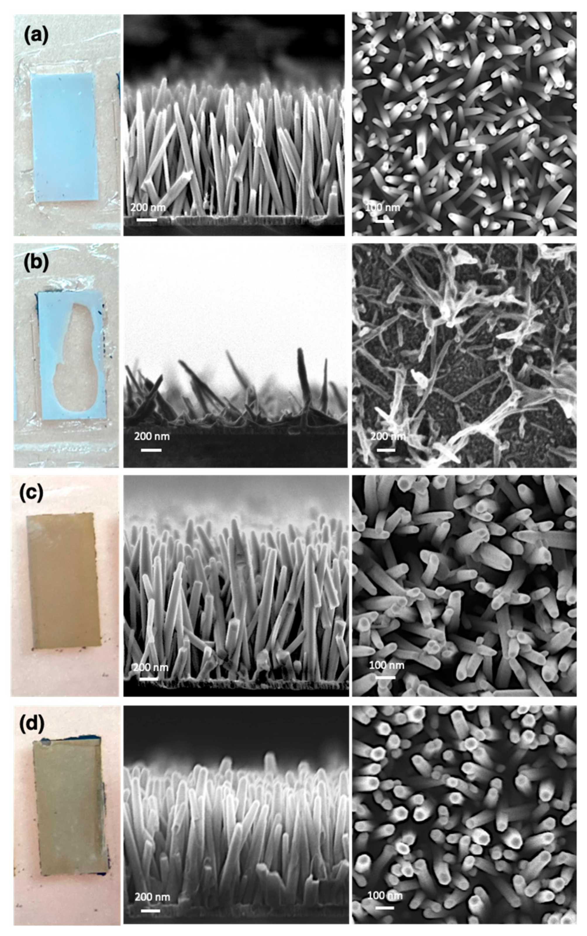
3.1. Compatibility of the Nanowire with Blood Plasma Samples
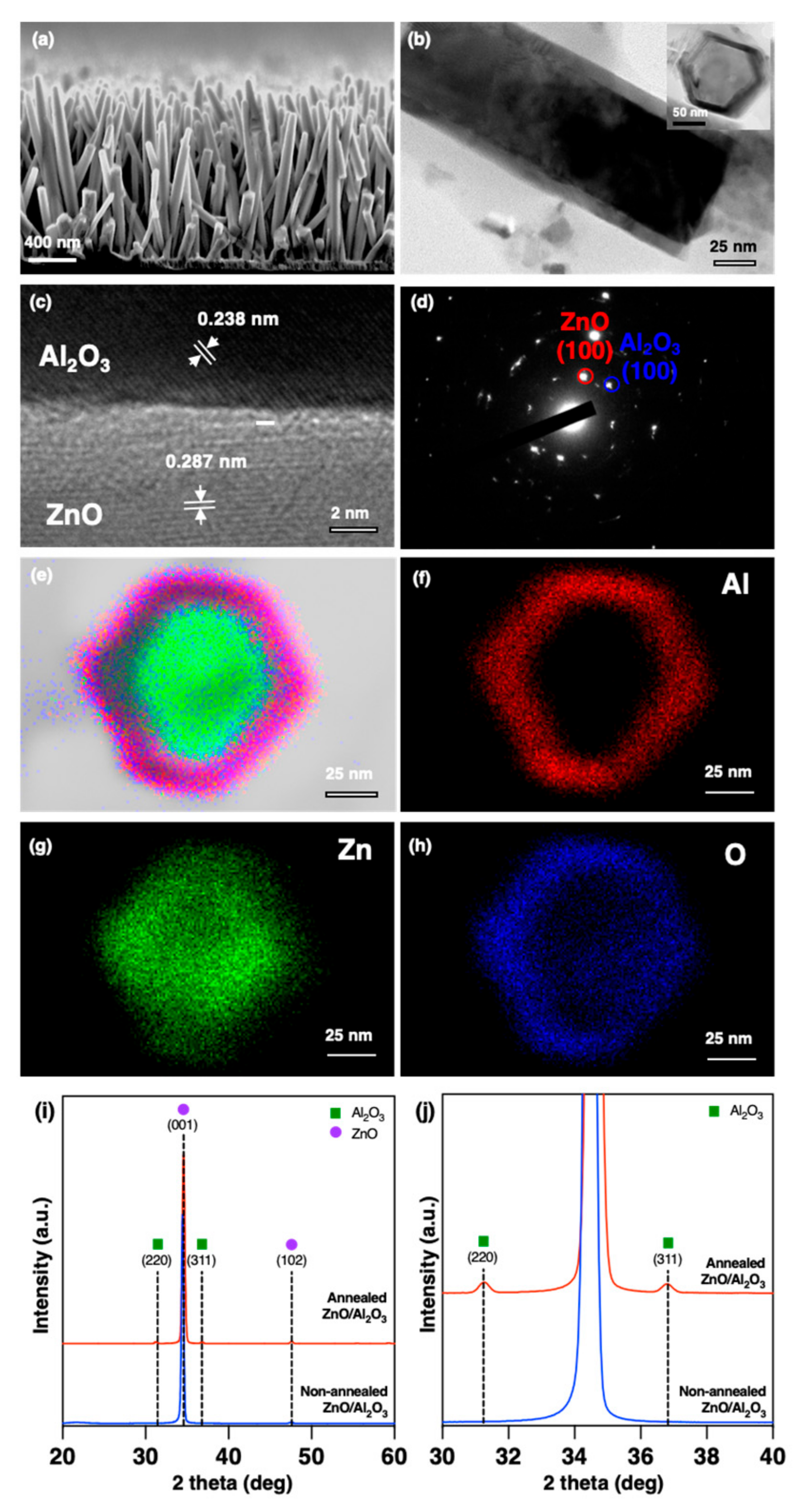
3.2. Morphological and Structural Analyses
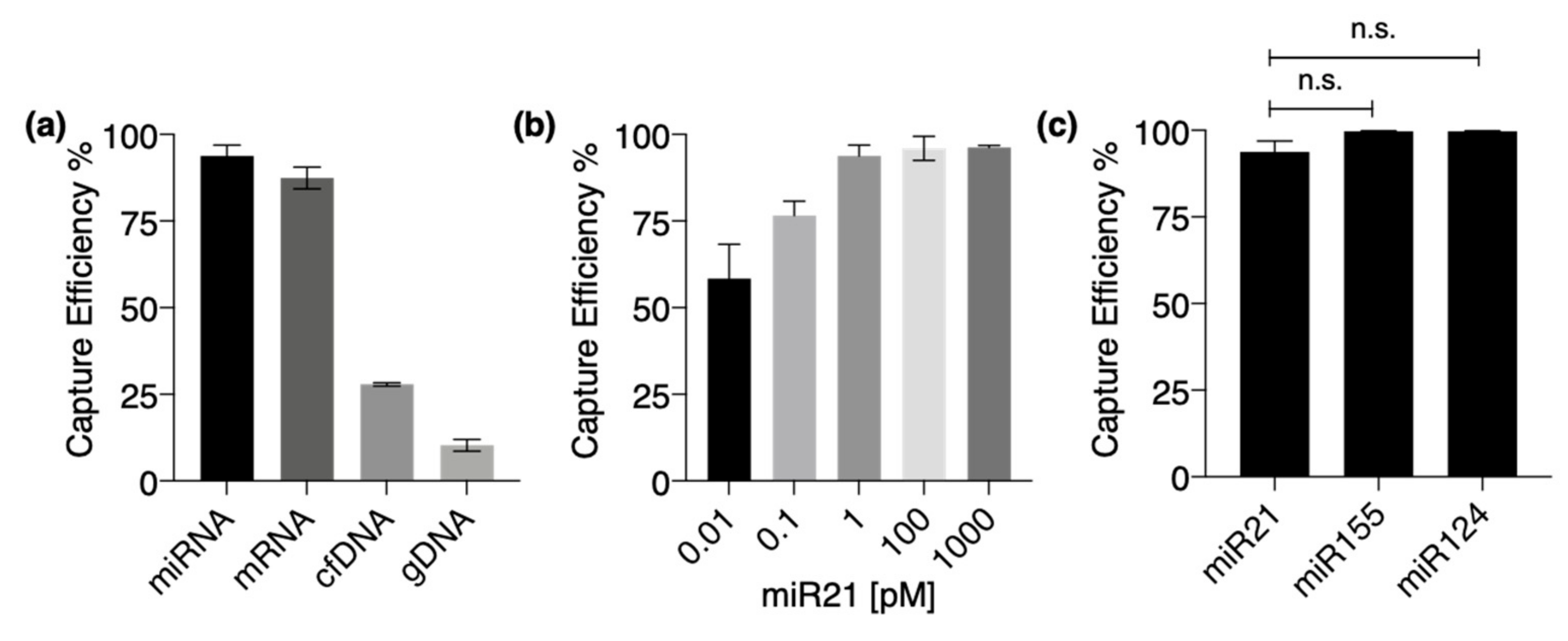
3.3. Nucleic Acid Capture on Annealed ZnO/Al2O3 Core-Shell Nanowire Structure
4. Conclusions
Author Contributions
Funding
Institutional Review Board Statement
Informed Consent Statement
Acknowledgments
Conflicts of Interest
References
- Wang, F.; Nazarali, A.J.; Ji, S. Circular RNAs as potential biomarkers for cancer diagnosis and therapy. Am. J. Cancer Res. 2016, 6, 1167. [Google Scholar]
- Kulcheski, F.R.; Christoff, A.P.; Margis, R. Circular RNAs are miRNA sponges and can be used as a new class of biomarker. J. Biotechnol. 2016, 238, 42–51. [Google Scholar] [CrossRef] [PubMed]
- Hombach, S.; Kretz, M. Non-coding RNAs: Classification, biology and functioning. In Non-Coding RNAs in Colorectal Cancer; Springer: Cham, Switzerland, 2016; pp. 3–17. [Google Scholar]
- Ullah, M.; Akbar, A. Clinical relevance of RNA editing to early detection of cancer in human. Stem Cell Res. Ther. 2020, 7, 66. [Google Scholar]
- Bremnes, R.M.; Sirera, R.; Camps, C. Circulating tumour-derived DNA and RNA markers in blood: A tool for early detection, diagnostics, and follow-up? Lung Cancer 2005, 49, 1–12. [Google Scholar] [CrossRef]
- Cella, L.N.; Blackstock, D.; Yates, M.A.; Mulchandani, A.; Chen, W. Detection of RNA viruses: Current technologies and future perspectives. Crit. Rev. Eukaryot. Gene Expr. 2013, 23, 125–137. [Google Scholar] [CrossRef] [PubMed]
- Seo, G.; Lee, G.; Kim, M.J.; Baek, S.-H.; Choi, M.; Ku, K.B.; Lee, C.-S.; Jun, S.; Park, D.; Kim, H.G.; et al. Rapid Detection of COVID-19 Causative Virus (SARS-CoV-2) in Human Nasopharyngeal Swab Specimens Using Field-Effect Transistor-Based Biosensor. ACS Nano 2020, 14, 5135–5142. [Google Scholar] [CrossRef] [PubMed] [Green Version]
- Spencer, S.M.; Lin, L.; Chiang, C.F.; Peng, Z.; Hesketh, P.; Salon, J.; Huang, Z. Direct and rapid detection of RNAs on a novel RNA microchip. ChemBioChem 2010, 11, 1378–1382. [Google Scholar] [CrossRef] [PubMed]
- Xu, Y.; Zheng, Z. Direct RNA detection without nucleic acid purification and PCR: Combining sandwich hybridization with signal amplification based on branched hybridization chain reaction. Biosens. Bioelectron. 2016, 79, 593–599. [Google Scholar] [CrossRef]
- Monis, P.T.; Giglio, S. Nucleic acid amplification-based techniques for pathogen detection and identification. Infect. Genet. Evol. 2006, 6, 2–12. [Google Scholar] [CrossRef]
- Tanner, N.A.; Zhang, Y.; Evans, T.C., Jr. Visual detection of isothermal nucleic acid amplification using pH-sensitive dyes. Biotechniques 2015, 58, 59–68. [Google Scholar] [CrossRef] [Green Version]
- Streit, S.; Michalski, C.W.; Erkan, M.; Kleeff, J.; Friess, H. Northern blot analysis for detection and quantification of RNA in pancreatic cancer cells and tissues. Nat. Protoc. 2009, 4, 37–43. [Google Scholar] [CrossRef]
- Tsanov, N.; Samacoits, A.; Chouaib, R.; Traboulsi, A.-M.; Gostan, T.; Weber, C.; Zimmer, C.; Zibara, K.; Walter, T.; Peter, M. smiFISH and FISH-quant–a flexible single RNA detection approach with super-resolution capability. Nucleic Acid Res. 2016, 44, e165. [Google Scholar] [CrossRef] [PubMed] [Green Version]
- Gaspar, I.; Ephrussi, A. Strength in numbers: Quantitative single-molecule RNA detection assays. Wiley Interdiscip. Rev. Dev. Biol. 2015, 4, 135–150. [Google Scholar] [CrossRef] [PubMed]
- Tian, W.; Li, P.; He, W.; Liu, C.; Li, Z. Rolling circle extension-actuated loop-mediated isothermal amplification (RCA-LAMP) for ultrasensitive detection of microRNAs. Biosens. Bioelectron. 2019, 128, 17–22. [Google Scholar] [CrossRef]
- El-Mogy, M.; Lam, B.; Haj-Ahmad, T.A.; McGowan, S.; Yu, D.; Nosal, L.; Rghei, N.; Roberts, P.; Haj-Ahmad, Y. Diversity and signature of small RNA in different bodily fluids using next generation sequencing. BMC Genet. 2018, 19, 408. [Google Scholar] [CrossRef] [PubMed]
- Lee, W.G.; Kim, Y.-G.; Chung, B.G.; Demirci, U.; Khademhosseini, A. Nano/Microfluidics for diagnosis of infectious diseases in developing countries. Adv. Drug Deliv. Rev. 2010, 62, 449–457. [Google Scholar] [CrossRef] [PubMed] [Green Version]
- Diercks, A.H.; Ozinsky, A.; Hansen, C.L.; Spotts, J.M.; Rodriguez, D.J.; Aderem, A. A microfluidic device for multiplexed protein detection in nano-liter volumes. Anal. Biochem. 2009, 386, 30–35. [Google Scholar] [CrossRef] [Green Version]
- Perozziello, G.; Candeloro, P.; Gentile, F.; Nicastri, A.; Perri, A.; Coluccio, M.L.; Adamo, A.; Pardeo, F.; Catalano, R.; Parrotta, E. Microfluidics & nanotechnology: Towards fully integrated analytical devices for the detection of cancer biomarkers. RSC Adv. 2014, 4, 55590–55598. [Google Scholar]
- Kanai, Y.; Ohmuro-Matsuyama, Y.; Tanioku, M.; Ushiba, S.; Ono, T.; Inoue, K.; Kitaguchi, T.; Kimura, M.; Ueda, H.; Matsumoto, K. Graphene Field Effect Transistor-Based Immunosensor for Ultrasensitive Noncompetitive Detection of Small Antigens. ACS Sens. 2020, 5, 24–28. [Google Scholar] [CrossRef]
- De Moraes, A.C.; Kubota, L.T. Recent Trends in Field-Effect Transistors-Based Immunosensors. Chemosensors 2016, 4, 20. [Google Scholar] [CrossRef] [Green Version]
- Zhu, Z.; Yasui, T.; Liu, Q.; Nagashima, K.; Takahashi, T.; Shimada, T.; Yanagida, T.; Baba, Y. Fabrication of a Robust In(2)O(3) Nanolines FET Device as a Biosensor Platform. Micromachines 2021, 12, 642. [Google Scholar] [CrossRef] [PubMed]
- Musa, M.; Yasui, T.; Zhu, Z.; Nagashima, K.; Ono, M.; Liu, Q.; Takahashi, H.; Shimada, T.; Arima, A.; Yanagida, T.; et al. Oxide Nanowire Microfluidic Devices for Capturing Single-stranded DNAs. Anal. Sci. 2021, 20P421. [Google Scholar] [CrossRef] [PubMed]
- Tan, H.J.; Zainal, Z.; Talib, Z.A.; Lim, H.N.; Shafie, S.; Tan, S.T.; Tan, K.B.; Bahrudin, N.N. Synthesis of high quality hydrothermally grown ZnO nanorods for photoelectrochemical cell electrode. Ceram. Int. 2021, 47, 14194–14207. [Google Scholar] [CrossRef]
- Angub, M.C.M.; Vergara, C.J.T.; Husay, H.A.F.; Salvador, A.A.; Empizo, M.J.F.; Kawano, K.; Minami, Y.; Shimizu, T.; Sarukura, N.; Somintac, A.S. Hydrothermal growth of vertically aligned ZnO nanorods as potential scintillator materials for radiation detectors. J. Lumin. 2018, 203, 427–435. [Google Scholar] [CrossRef]
- Ma, C.; Zhao, C.; Fan, X.; Liu, Z.; Liu, J. Preparation of non-stoichiometric Al2O3 film with broadband antireflective by magnetron sputtering. Chem. Phys. Lett. 2021, 764, 138299. [Google Scholar] [CrossRef]
- Gonullu, M.P.; Ates, H. The characteristic evolution of TiO2/Al2O3 bilayer films produced by ALD: Effect of substrate type and wide range annealing temperature. Superlattices Microstruct. 2020, 142, 106529. [Google Scholar] [CrossRef]
- Zhou, G.; Wang, L.; Wang, X.; Yu, Y.; Mutzke, A. Effect of bias voltage on microstructure and optical properties of Al2O3 thin films prepared by twin targets reactive high power impulse magnetron sputtering. Vacuum 2019, 166, 88–96. [Google Scholar] [CrossRef]


Publisher’s Note: MDPI stays neutral with regard to jurisdictional claims in published maps and institutional affiliations. |
© 2021 by the authors. Licensee MDPI, Basel, Switzerland. This article is an open access article distributed under the terms and conditions of the Creative Commons Attribution (CC BY) license (https://creativecommons.org/licenses/by/4.0/).
Share and Cite
Takahashi, H.; Yasui, T.; Klamchuen, A.; Khemasiri, N.; Wuthikhun, T.; Paisrisarn, P.; Shinjo, K.; Kitano, Y.; Aoki, K.; Natsume, A.; et al. Annealed ZnO/Al2O3 Core-Shell Nanowire as a Platform to Capture RNA in Blood Plasma. Nanomaterials 2021, 11, 1768. https://0-doi-org.brum.beds.ac.uk/10.3390/nano11071768
Takahashi H, Yasui T, Klamchuen A, Khemasiri N, Wuthikhun T, Paisrisarn P, Shinjo K, Kitano Y, Aoki K, Natsume A, et al. Annealed ZnO/Al2O3 Core-Shell Nanowire as a Platform to Capture RNA in Blood Plasma. Nanomaterials. 2021; 11(7):1768. https://0-doi-org.brum.beds.ac.uk/10.3390/nano11071768
Chicago/Turabian StyleTakahashi, Hiromi, Takao Yasui, Annop Klamchuen, Narathon Khemasiri, Tuksadon Wuthikhun, Piyawan Paisrisarn, Keiko Shinjo, Yotaro Kitano, Kosuke Aoki, Atsushi Natsume, and et al. 2021. "Annealed ZnO/Al2O3 Core-Shell Nanowire as a Platform to Capture RNA in Blood Plasma" Nanomaterials 11, no. 7: 1768. https://0-doi-org.brum.beds.ac.uk/10.3390/nano11071768