
Ag-Modified Porous Perovskite-Type LaFeO3 for Efficient Ethanol Detection


1
School of Materials and Chemistry, University of Shanghai for Science & Technology, Shanghai 200093, China
2
NEST Lab, Department of Physics, Department of Chemistry, College of Sciences, Shanghai University, Shanghai 200444, China
3
Shanghai Yaolu Instrument & Equipment Co., Ltd., Shanghai 200444, China
*
Authors to whom correspondence should be addressed.
†
These authors contributed equally to this work.
Nanomaterials 2022, 12(10), 1768; https://0-doi-org.brum.beds.ac.uk/10.3390/nano12101768
Submission received: 2 May 2022
/
Revised: 20 May 2022
/
Accepted: 20 May 2022
/
Published: 22 May 2022
(This article belongs to the Special Issue Advanced Nanomaterials and Nanodevices for VOCs Gas Sensor)
Abstract
:Perovskite (ABO3) nanosheets with a high carrier mobility have been regarded as the best candidates for gas-sensitive materials arising from their exceptional crystal structure and physical–chemical properties that often exhibit good gas reactivity and stability. Herein, Ag in situ modified porous LaFeO3 nanosheets were synthesized by the simple and efficient graphene oxide (GO)-assisted co-precipitation method which was used for sensitive and selective ethanol detection. The Ag modification ratio was studied, and the best performance was obtained with 5% Ag modification. The Ag/LaFeO3 nanomaterials with high surface areas achieved a sensing response value (Rg/Ra) of 20.9 to 20 ppm ethanol at 180 °C with relatively fast response/recovery times (26/27 s). In addition, they showed significantly high selectivity for ethanol but only a slight response to other interfering gases. The enhanced gas-sensing performance was attributed to the combination of well-designed porous nanomaterials with noble metal sensitization. The new approach is provided for this strategy for the potential application of more P-type ABO3 perovskite-based gas-sensitive devices.
1. Introduction
ABO3 perovskite-type oxides have been widely applied in various fields, such as catalysts [1,2], Li-O2 batteries [3,4], magnetic materials [5] and gas sensors [6,7,8]. ABO3 consists of a metal cation with a strong thermal stability at the large ionic radius A-site and a B-O octahedral structure providing the active site. Its positive crystal structure and physical–chemical properties tend to give superior gas reactivity and stability, which offer huge potential for the detection of VOCs [9,10]. As a classical ABO3 material, LaFeO3 has superior p-type electron conductivity, outstanding oxygen ion mobility and catalytic activity rendering it a suitable candidate for gas-sensing applications [11,12,13,14]. However, it still has a few difficulties to overcome in order to suit practical requirements, such as a poor gas sensitivity and higher operating temperatures. Theoretically, the response value of a p-type MOS would be the square root of an n-type MOS if they have the same dimensions and morphology [15]. In particular, a high operating temperature increases electrical consumption and reduces gas sensitivity caused by the desorption of target gases and surface oxygen ions [16]. Nevertheless, broadening the gas-sensitive properties of LaFeO3 perovskite materials is of great importance.
Operating temperature, sensitivity, response/recovery time, selectivity and stability are all critical factors in the application of semiconductor gas sensor materials, which in turn are closely tied to the morphology, size, surface area to volume ratio, modification and so on [17,18]. Porous nanosheets have demonstrated excellent physical, chemical, mechanical, electronic and surface properties that hold promise and appeal [19,20]. In recent years, porous sensitive nanomaterials have increasingly been used owing to their higher surface area to volume ratio, active surface oxygen vacancy concentration and excellent oxygen storing capacity [21,22]. To date, the graphene oxide (GO) template method has been adopted for the preparation of various single metal oxide nanosheets, such as ZnO [23], TiO2 [24], SnO2 [25], Co3O4 [26], as well as others. Nevertheless, few reports have been devoted to the preparation of porous perovskite-type polymetallic oxide nanomaterials. In addition, noble metal modifications are common means of reducing resistance and increasing sensitivity. Notably, Ag modification is an appealing approach to achieve a better sensing performance on account of its excellent chemically and electronically sensitive catalytic properties [27,28]. With the sensitized atoms also generating further adsorption sites for atmospheric oxygen, the target gas molecules contribute meaningfully to the exchange on the base surface and the adsorbate [29]. Therefore, well-designed, non-toxic, porous nanomaterials combined with active noble metal sensitization is a highly promising approach.
In this work, ultra-thin, porous, Ag-modified LaFeO3 nanosheets with perovskite structures were successfully synthesized via the graphene oxide (GO) template method and subsequent thermal treatment. The effect of the Ag-modified content on the morphology, structure and ethanol gas-sensitive performance of the product was also investigated. In addition, an exploration of the mechanism of Ag modification on its gas-sensitive performance enhancement was carried out. Finally, a possible mechanism for the enhanced gas-sensitive performance of Ag/LaFeO3 was explored.
2. Materials and Methods
2.1. Materials
Lanthanum(III) nitrate hexahydrate (La(NO3)3·6H2O), ferric nitrate nonahydrate (Fe(NO3)3·9H2O), sodium hydroxide (NaOH, 98%), ethylene glycol (EG, 99%) and ethanol (EtOH, 99.7%) were purchased from Sigma-Aldrich (Burlington, MA, USA). Graphene oxide (GO) was purchased from Changzhou Sixth Element Material Technology Co., Ltd. (Changzhou, China). All chemical reagents were available in analytical grade and suitable for use without further purification.
2.2. Methods
Synthesis of ultrathin porous LaFeO3 nanosheets: As part of the typical procedure, GO (80 mg) was added into 168 mL ethylene glycol with 32 mL deionized water to form a clear solution with an ultrasound for 30 min. Then, 21.6 mg La(NO3)3·6H2O and 20.2 mg Fe(NO3)3·9H2O were added into the ethylene glycol solution followed by the addition of 140 mg NaOH with an ultrasound for around 30 min. Then, the mixture was stirred at room temperature for 4 h. Finally, the precipitate was collected, washed with ethanol and deionized water three times and freeze-dried for 24 h. After drying, the powder was calcined in air at a heating rate of 2 °C/min and kept at a certain temperature for 2 h to obtain the ultrathin porous LaFeO3 nanosheets.
Synthesis of ultrathin porous Ag/LaFeO3 nanosheets: Ag/LaFeO3 nanosheets were synthesized via a similar procedure to the synthesis of LaFeO3 nanosheets except that a certain amount of AgNO3 solution was added in the former before centrifugation. The Ag/La ratios of the prepared powers were 0, 0.01, 0.05 and 0.10. The as-generated Ag-modified LaFeO3 nanosheets are herein denoted as Ag/LaFeO3-0, Ag/LaFeO3-1, Ag/LaFeO3-5 and Ag/LaFeO3-10, respectively.
2.3. Characterization
LaFeO3 nanosheets and Ag/LaFeO3 nanosheets were analyzed via the following characterization methods. The morphology of the samples was visualized by FE-SEM (FEI, Quanta FEG 450, Houston, TX, USA). Transmission electron microscope (TEM) images and high-resolution transmission electron microscope (HRTEM) images were accessed by a Tecnai G220S-Twin transmission electron microscope operated at 120 and 200 kV accelerating voltages to explore the morphology and microstructure of the samples. Prior to measurement, samples were sonicated and dispersed in ethanol, then immersed in a copper grid with a lacey carbon film and dried at room temperature. An X-ray diffractometer (Bruker, D8 Advance, Bremen, Germany) with Cu-Kα (λ = 0.15418 nm) recorded the phases of the solid powders. Moreover, the XRD patterns were collected over a 2θ range of 20–70° in 5° min−1 steps at room temperature. The chemical state of the surface of the samples was appraised by X-ray photoelectron spectroscopy (XPS, Thermo Scientific, Waltham, MA, USA, ESCALAB 250) with Mg Kα radiation. The C1s photoelectron peak (284.6 eV) was used as a reference to calibrate the binding energy. TG/DSC was measured from 25 to 1000 °C in the air at a heating rate of 5 °C/min with a thermal analyzer (TG, STA449C). Brunauer–Emmett–Teller (BET) surface area and pore size distribution investigations were carried out on an ASIC-2 gas adsorption analyzer. (N2 as the adsorbate and operation temperature = −196 °C).
2.4. Gas Sensor Fabrication and Measurement
The samples were mixed with deionized water at a certain ratio and grated into a paste which was applied to the ceramic tube elements where a thin sensing film was formed. Each end of the ceramic tube was fitted with a pair of gold electrodes attached to two platinum wires on each electrode. The ceramic tubes were inserted into a Ni-Cr heating wire resulting in an indirectly heated gas sensor (Figure S1). The design and details of the ceramic sensor were as reported previously in the literature [30]. In this paper, the gas-sensing performances of the gas sensors were measured with the commercial CGS-8 gas-sensing measurement system by recording the change of resistance of the gas sensors.
3. Results and Discussion
3.1. Characterization of the Sensing Materials
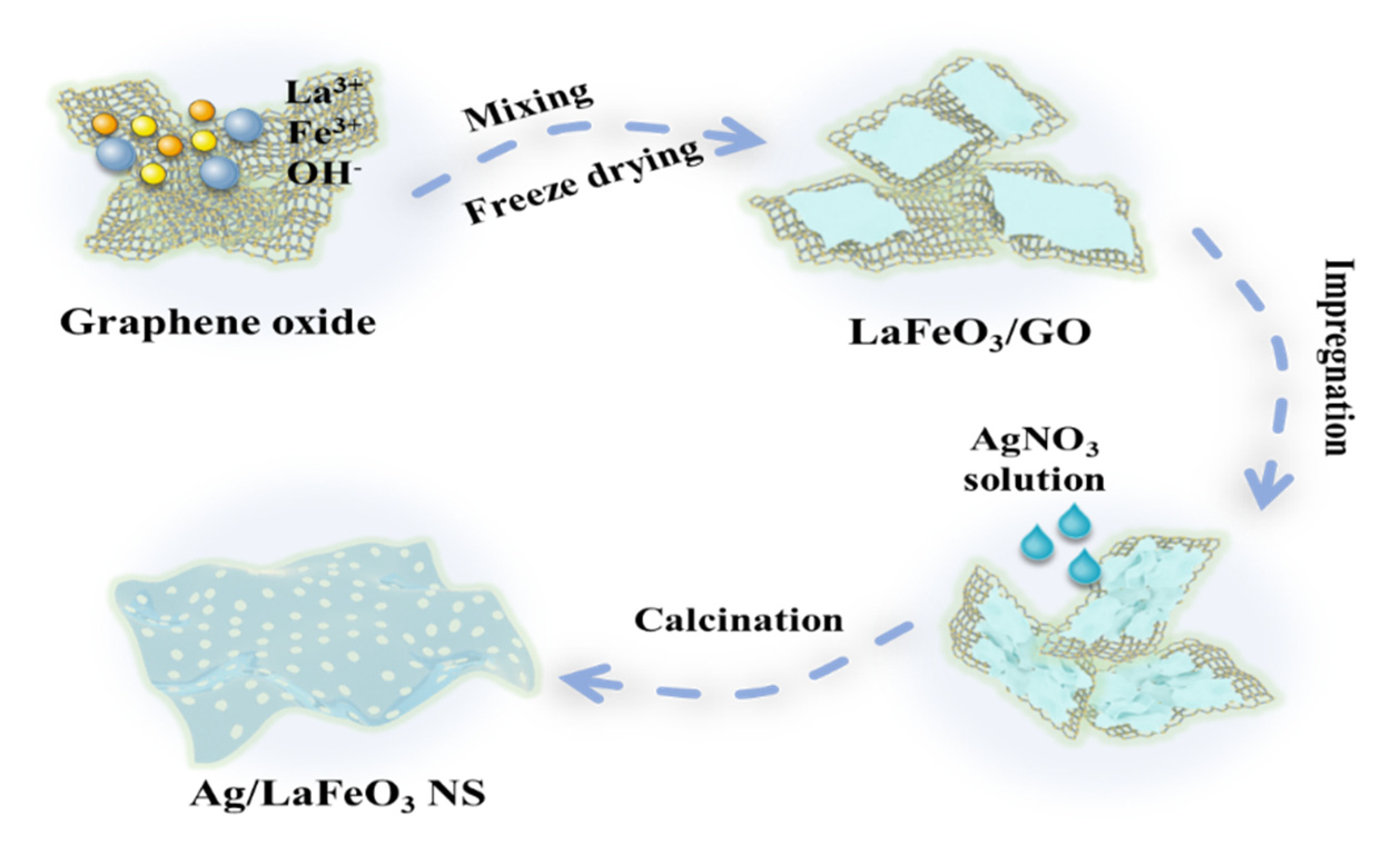
The synthesis process of the porous Ag/LaFeO3 nanosheets is described in Scheme 1. The precursor LaFeO3/GO was synthesized by the graphene oxide-assisted co-precipitation method. Subsequently, the precursor was impregnated with different concentrations of AgNO3 solution. Then, the Ag/LaFeO3 nanosheets were obtained by heat treatment. Usually, the high calcination temperature is prone to sintering, which accounts for the drop in the active specific surface area, while the low temperature does not lead to crystallinity. As a gas-sensitive material, LaFeO3 requires both a high specific surface area and a certain degree of crystallinity, which requires the study of the appropriate calcination temperature.
To investigate the thermal behavior of the LaFeO3/GO template hybrid precursors, TG tests were carried out under an air atmosphere in the range of 50~1000 °C with a heating rate of 5 °C·min−1, as shown in Figure S2a. According to the TG curves, the thermal performance of the LaFeO3/GO nanocomposites improved significantly with the rise in temperature. The first loss of weight until 110 °C (5.4%) was mainly responsible for the removal of adsorbed water from the sample. The second stage of weight loss (16.8%) could be attributed to the loss of ethylene glycol and the degradation of the residual organic functional groups remaining on the graphene oxide template. The weight loss in the third stage (19.7%) was due to the combustion of the residual nitrates. The weight loss in the fourth stage (38%) was recognized as the combustion of graphene oxide [31]. Therefore, it was reasonable to choose the calcination temperature of 500–800 °C.
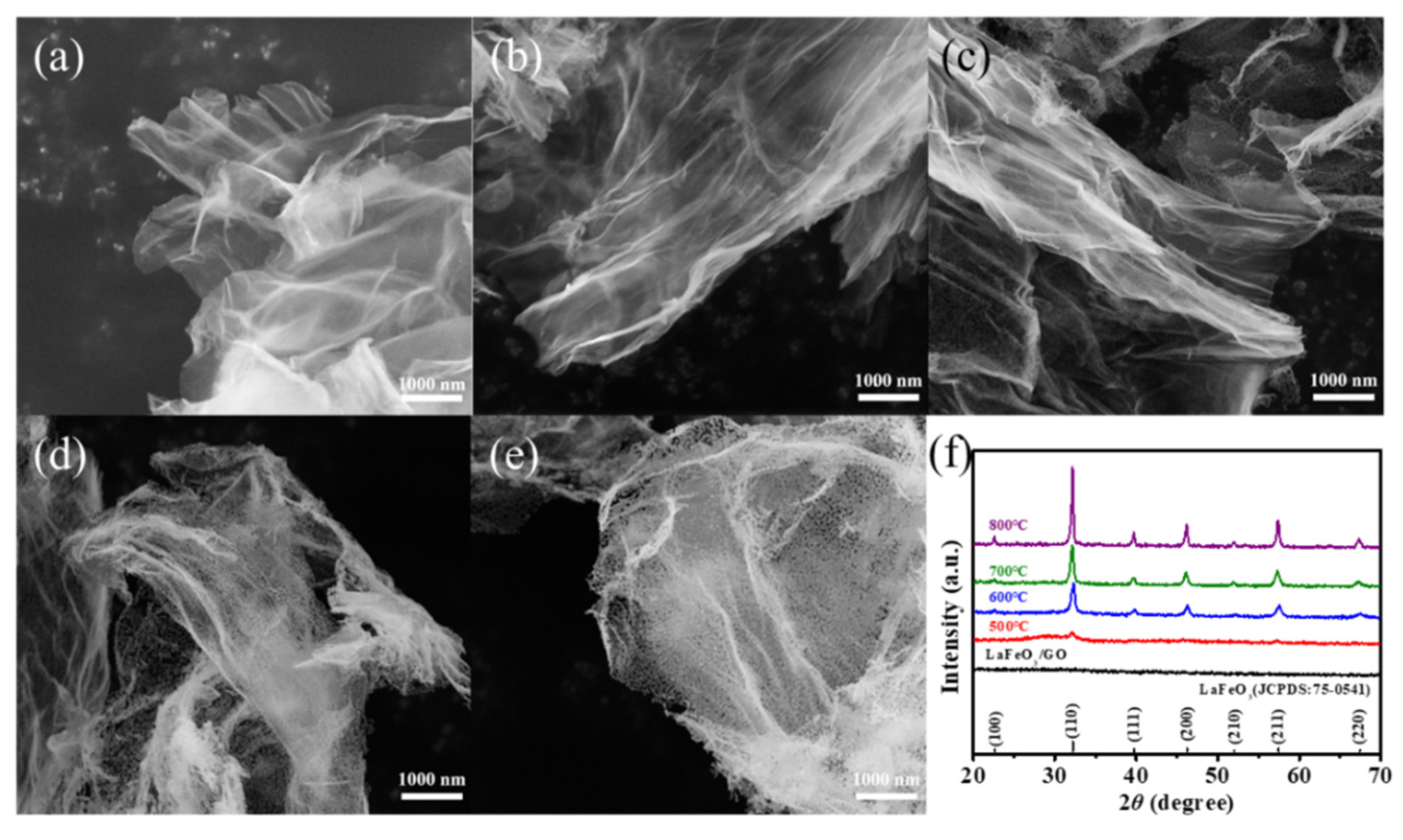
The morphology of LaFeO3 calcined at different temperatures was characterized. Figure 1a–e are the SEM images of LaFeO3/GO and LaFeO3 obtained after the calcination of LaFeO3/GO at 500–800 °C, respectively. All these products were ultrathin porous nanosheets with rolled-up edges due to the surface tension, which was caused by the GO template. It was found that the LaFeO3/GO intact ultrathin nanosheets gradually turned into porous nanosheets composed of particles with the increase in calcination temperature. In Figure 1d–e, the porous nanosheets assembled by nanoparticles can be clearly observed. In addition, the pore size of the ultrathin nanosheets increased due to the gradual increase in nanoparticles.
The XRD patterns of the LaFeO3/GO and LaFeO3 materials are given in Figure 1f. Among them, the nanosheets of LaFeO3/GO showed an amorphous structure. Correspondingly, in the Raman spectrum in Figure S2b, the peaks of the curves corresponding to LaFeO3/GO at around 1347 cm−1 and 1588 cm−1 could be attributed to the D and G bands of the graphene plane [32]. In addition, the diffraction peaks of LaFeO3 calcined at four different temperatures matched well with the standard LaFeO3 (JCPDS: 75-0541) perovskite structure [33]. This implies that all the prepared LaFeO3 sensing materials had a perovskite crystal structure with orthorhombic phases when calcined at 500 °C and higher. The XRD patterns showed that the crystallinity of the prepared LaFeO3 nanosheets increased significantly with the increase in calcination temperature. The working temperature had a significant effect on the oxygen adsorbed on the sensing materials surface, and it was a major factor affecting the gas-sensitive performance.
The morphologies of Ag/LaFeO3 nanosheets (Ag/LaFeO3-0, Ag/LaFeO3-1, Ag/LaFeO3-5, and Ag/LaFeO3-10) were characterized by SEM. As shown in Figure 2a, the layer sizes of the Ag/LaFeO3-0 nanosheets were approximately 5 μm after calcination at 700 °C, which was determined by the size of the sacrificial GO template. Figure 2b–d shows the SEM patterns of Ag/LaFeO3-1, Ag/LaFeO3-5 and Ag/LaFeO3-10, respectively. There were nano rod-like shadows attached to the nanosheets, which tended to grow with Ag modifications. The XRD patterns provided information on the crystallinity and the perovskite phase of the samples. As shown in Figure 2e, all four prepared samples of Ag-modified ultrathin LaFeO3 nanosheets showed a distinct LaFeO3 perovskite phase, and the main peaks appearing in the plots corresponded to the diffraction of (100), (110), (200), (210), (211) and (221) crystal planes, which matched perfectly with the standard JCPDS card 75-0541 [33]. Only weak Ag2O (JCPDS: 19-1155) diffraction peaks were detected in the Ag/LaFeO3-10 sample corresponding to the peak of (101) crystal plane diffraction due to the low Ag content and high dispersion of the samples [29]. The Ag/LaFeO3 samples were further studied by Raman spectroscopy. As illustrated in Figure 2f, four Raman peaks at 299, 417, 628 and 1316 cm−1 mapped to the orthogonal LaFeO3 structure [31]. The 299 cm−1 (Ag mode) peak was correlated with La-O, and the 417 cm−1 peak denoted the in-plane Fe-O (B3g) vibrational mode. Moreover, the 628 and 1316 cm−1 peaks were associated with the two-photon scattering of O2− [34].
The response of the LaFeO3 samples to 50 ppm ethanol at an operating temperature of 140–220 °C is shown in Figure S3. From the response curves, it is clear that LaFeO3 calcined at 700 °C had the best response at 180 °C. Therefore, the calcination temperature was chosen to be 700 °C for the subsequent material study. In addition, the gas-sensitive properties of Ag/LaFeO3-5 materials were better than those of Ag/LaFeO3-0, Ag/LaFeO3-1 and Ag/LaFeO3-10. Therefore, the following discussions will focus on the Ag/LaFeO3-5 material.
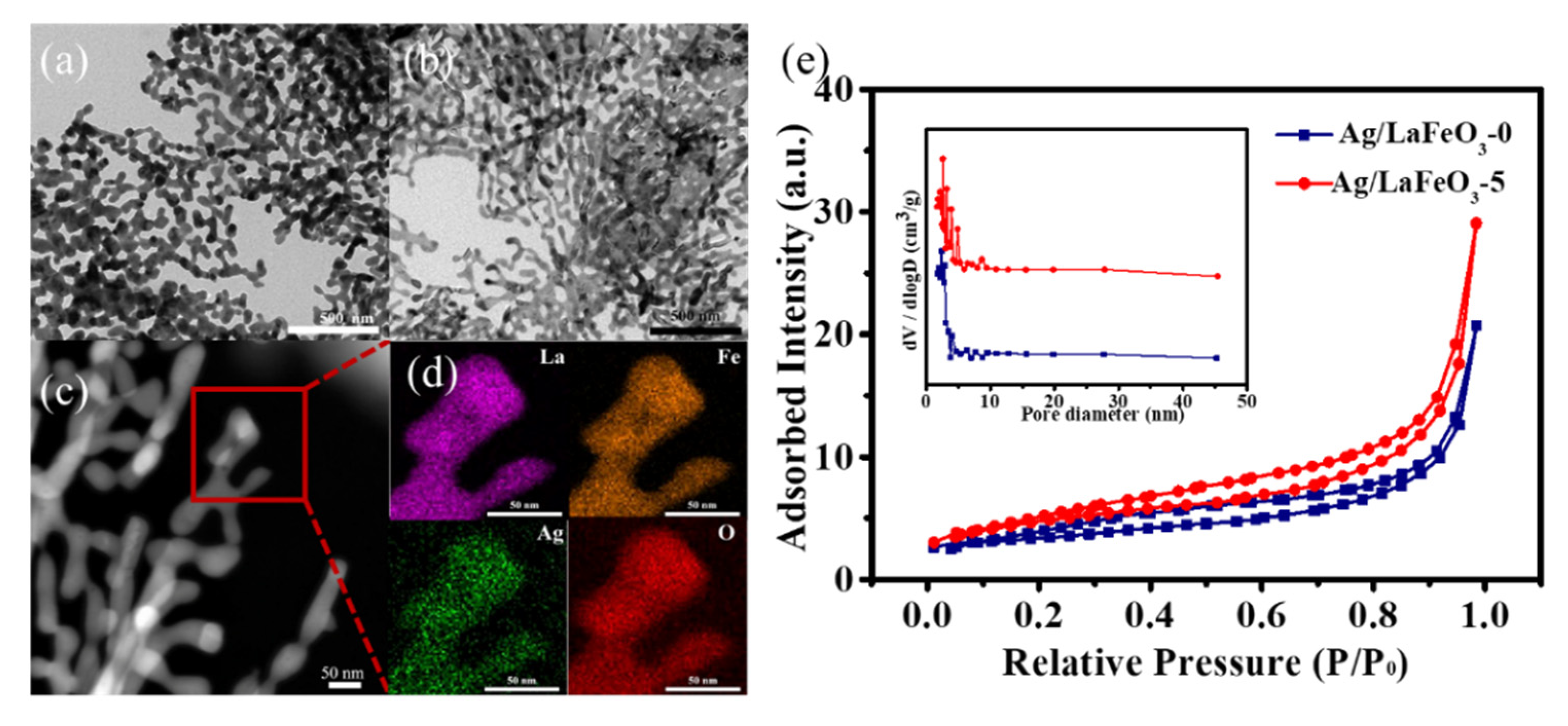
The porous structure and morphologies of the Ag/LaFeO3-0 and Ag/LaFeO3-5 samples were further characterized by TEM. The nanosheets were formed by many irregular dendritic nanocrystals. These dendritic nanocrystals were uniformly distributed and the Ag modification did not change their microscopic morphology (Figure 3a,b). To further determine the elemental distribution of these “dendrites”, the STEM-EDS method was used. From Figure 3c,d, it can be clearly observed that the La, Fe, Ag and O elements were well distributed in the three-dimensional space of the Ag/LaFeO3-5 sample, indicating that the Ag elements were homogeneously dispersed on the whole LaFeO3 sample.
The pore structure and specific surface area of the Ag/LaFeO3 ultrathin nanosheets were investigated using nitrogen adsorption and desorption isotherms. A significant hysteresis in the type IV adsorption isotherm at higher relative pressures indicated the presence of mesoporous structures (Figure 3e). The BET specific surface area of Ag/LaFeO3-5 (16.45 m2/g) was larger than that of Ag/LaFeO3-0 (11.31 m2/g), indicating that the Ag modification had a positive effect on the porous morphology. The pore diameters of Ag/LaFeO3-5 and Ag/LaFeO3-0 were calculated based on the BJH method to be about 6.19 nm and 6.44 nm, respectively. The large specific surface area and abundant mesopores allowed sufficient space to promote the accelerated diffusion of gas molecules and thus improve the gas-sensitive performance.
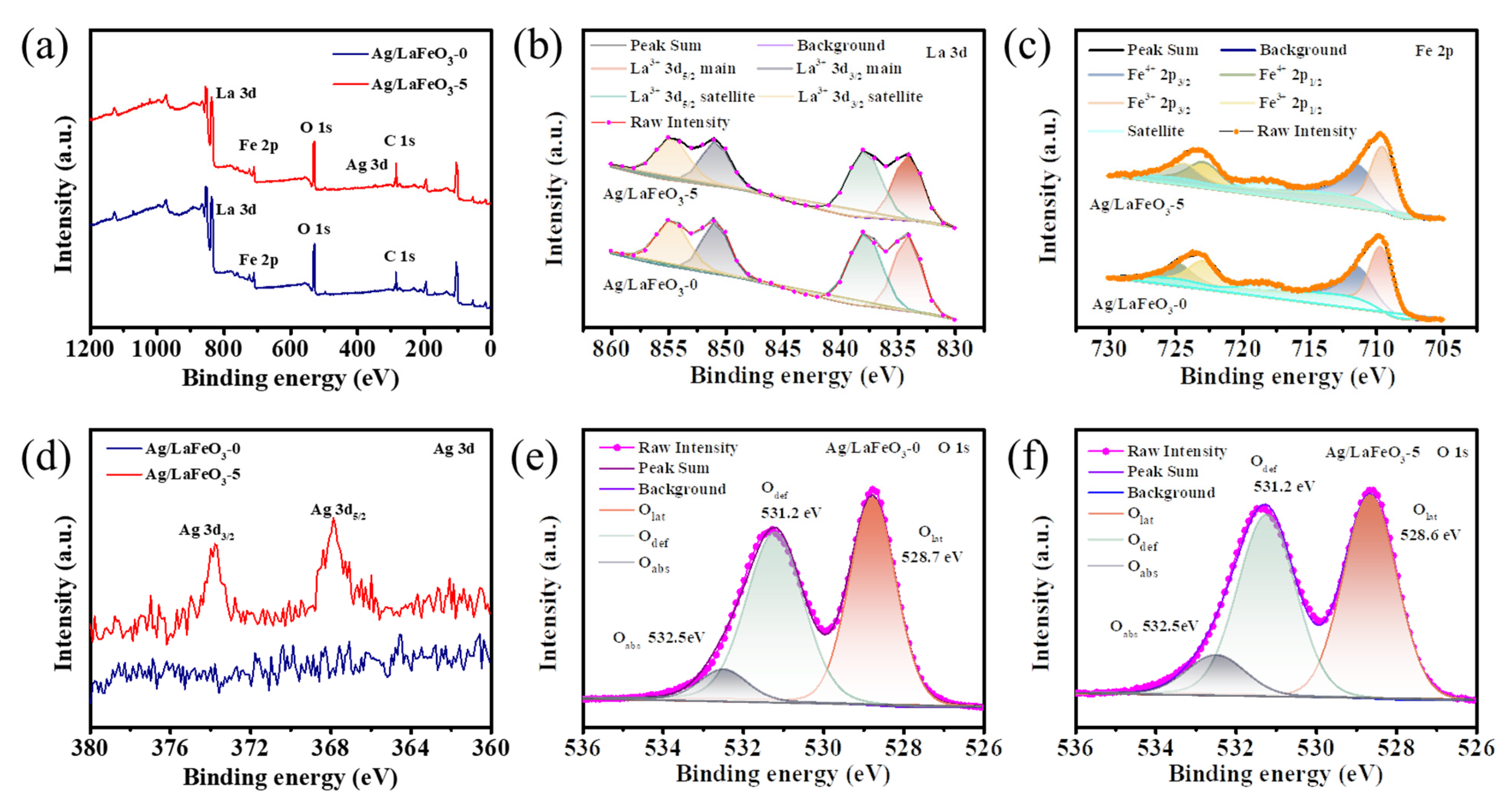
To understand the surface chemical composition and chemical state of the Ag/LaFeO3-0 and Ag/LaFeO3-5 samples, XPS analysis was used for characterization. The full spectra of the XPS measurement verifying the existence of La, Fe, O and C elements in the Ag/LaFeO3-0 and Ag/LaFeO3-5 samples are shown in Figure 4a. Among them, Ag elemental was detected in the Ag/LaFeO3-5 sample. As shown in Figure 4b, the peaks at 834.1 eV and 850.7 eV corresponded to La 3d5/2 and La 3d3/2, respectively. The two peaks at 837.8 eV and 854.5 eV corresponded to the La 3d5/2 and La 3d3/2 satellite peaks, respectively. The typical double peak could be attributed to the splitting of the 3d5/2 and 3d3/2 spin orbits of the La3+ ion, with nuclear holes and electrons, which shifted from the O 2p valence band to the vacant La 4f orbital. The La 3d spectrum indicated that the lanthanide ion was in the La3+ valence state [10,11,29]. Further, in the high-resolution Fe 2p spectra (Figure 4c), peaks at 711.4 and 724.9 eV of binding energy could be attached to 2p3/2 and 2p1/2 of Fe4+, respectively, while the 709.6 eV and 722.8 eV peaks could be ascribed to 2p3/2 and 2p1/2 of Fe3+ [10,12,13]. The Ag element (Figure 4d) appeared at binding energies of 367.3 eV and 373.3 eV, respectively, corresponding to the Ag 3d5/2 and Ag 3d3/2 double peaks of Ag2O [35,36,37,38]. This suggests that certain Ag species in the Ag/LaFeO3-5 were available in the oxidized state of Ag2O. The O 1s spectrum could be an inverse product of three peaks, each corresponding to a different kind of surface chemical state as shown in Figure 4e,f. The binding energy of 528.9 eV was tied to lattice oxygen (Olat), the shaded area of 531.2 eV was dedicated to defect oxygen (Odef), and the surface adsorbed oxygen molecules (Oabs) counted on a weak peak at 532.4 eV [6]. The lattice oxygen could be attributed to La-O and Fe-O in the LaFeO3 lattice. The approximate relative percentage of each oxygen species on the surface of Ag/LaFeO3-0 and Ag/LaFeO3-5 are summarized in Table S1. The Olat percentage of Ag/LaFeO3-0 and Ag/LaFeO3-5 were 46.71% and 46.25%, respectively. Furthermore, compared with the Odef percentage of the Ag/LaFeO3-5 (44.28%), the Ag/LaFeO3-0 (46.55%) showed an increased Odef percentage. On the contrary, the comparative percentage of Oads for the Ag/LaFeO3-5 nanosheets was 9.47%, exhibiting a slightly higher oxygen adsorption percentage than the Ag/LaFeO3-0 nanocomposite (6.74%). Based on the above analysis, the Ag/LaFeO3-5 could deliver more sorbent oxygen species than Ag/LaFeO3-0 nanosheets, which played a significant function in the response with the target gas and could widely enhance the gas-sensing capability of the sensor.
3.2. Ethanol-Sensing Performance
The operating temperature is correlated with the carrier concentration and surface reaction activation energy of gas-sensitive materials, which is one of the major factors that affect the gas-sensitive performance. To explore the best working temperature of the four Ag/LaFeO3 nanosheets, a series of tests were conducted on ethanol at 50 ppm over a temperature range of 140–220 °C (Figure 5a). Apparently, the response value first tended to increase and then decrease as the operating temperature increased. The initial growth in response values was associated with the activation of the gas molecules/sensing material and the acceleration of electron conduction. With a further increase in temperature, the adsorption rate fell far short of the desorption rate and fewer gas molecules were trapped on the interface of the material, which led to smaller response values. At the same time, the sensing response of all Ag/LaFeO3 samples showed a similar trend, with their sensitivity reaching a maximum at 180 °C, respectively. The modification of Ag increased the sensitivity of the LaFeO3 sensor to ethanol, presenting a trend of an increasing and then decreasing response as the Ag content increased. In this case, the number of surface defects (i.e., active sites) did not always increase with the amount of Ag modification [37]. At 5% Ag modification, the concentration of surface defects attained a maximum value, where the corresponding sensor sensitivity was optimal. Sensing performance suffered at a higher Ag-modified amount caused by the aggregation of Ag nanoparticles and the reduction in catalytic active sites [39]. Accordingly, 180 °C was considered the best operating temperature for further gas sensitivity testing.
The sensitivity of the four Ag/LaFeO3 sensors was investigated. Seven interfering gases, including formaldehyde, ethylene glycol, ammonia, toluene, acetone, xylene and methanol were selected as the interfering gases. As the image shows, the results confirmed that the four sensors showed some similarity in selectivity, and the response values of these sensors were higher for ethanol than for other gases (Figure 5b). Therefore, it was tentatively concluded that the gas sensors prepared by Ag-modified ultrathin LaFeO3 nanosheets had a significant selectivity to ethanol. Moreover, the Ag/LaFeO3-5-based gas sensors showed a better selectivity for ethanol gas than the other three sensors. Hence, Ag/LaFeO3-5 nanosheets were considered a promising method for the selective detection of ethanol gas in complex environments. It was also vital to selectively and accurately detect the presence of ethanol from the gas mixture. When the Ag/LaFeO3-5 sensors were exposed to an artificial atmosphere with an ethanol mixture, the sensing signal remained close to the response when ethanol vapor alone was present (Figure 5c). Therefore, the Ag/LaFeO3-5 sensors demonstrated a good interference immunity for the detection of ethanol.
Figure 5d demonstrates that the Ag/LaFeO3-5 sensor exhibited a dynamic response in ethanol from 5 to 100 ppm. The response values of the sensors showed a step-growth as the ethanol concentration in the chamber increased from 5 ppm to 100 ppm. In addition, the gas sensor was tested for ethanol vapor atmospheres of 5, 10, 20, 50 and 100 ppm, corresponding to response values of 3.9, 9.9, 20.9, 30.5 and 50.6, respectively. Furthermore, the inset c clearly shows that the sensor exhibited a good linearity in the range of 5 to 100 ppm for ethanol vapor concentrations. The fitted curve was available as a function y = 0.49x + 0.46, where the value of R2 was 0.98. Thus, the results showed that none of the Ag/LaFeO3-5-based sensors saturated at ethanol gas concentrations below 100 ppm. Considering the performance of sensitive materials to test for low concentrations of ethanol and to obtain detection limits, the gas-sensing potential of these gas sensors was measured over the concentration ranges of 300~1000 ppb of ethanol gas (Figure 5e). Significantly responsive to low gas concentrations (Ra/Rg = 1.6), the sensor showed considerable promise for the capture of trace amounts of ethanol.
Response and recovery times perform a critical role in the monitoring ability of gas sensors. Generally, the faster the response/recovery time, the better the gas sensor performance will be. A graph of the recovery curve of the response to 20 ppm ethanol vapor based on the Ag/LaFeO3-5 sensors is given in Figure 5f. The response/recovery time of the ethanol gas sensors was 26/27 s, which could meet the demand for gas detection in real life. Repeatability and stability are important considerations in routine applications. Figure 5g shows four dynamic cycles of response and recovery for 20 ppm ethanol. Noticeably, the response and recovery resistance of the gas sensor did not vary visibly after four cycles of measurement, which demonstrated a good repeatability. Long-term tests on these gas sensors have revealed that their performance could be maintained over one week with response values of around 95% (Figure 5h). Therefore, the Ag/LaFeO3-5 sensor had a good repeatability and stability. Figure 5i demonstrates the impact of humidity on the response of the alcohol sensor, and it was found that the gas response value tended to decrease as the humidity in the ethanol atmosphere increased.
A comparison of the performance of the Ag/LaFeO3-5 sensor in this work with other ethanol-based sensors previously reported is summarized in Table 1. Most LaFeO3-based gas sensors responded to C2H5OH at high temperatures. However, the Ag-LaFeO3-5 sensors showed a much warmer gas response at lower working temperatures. Consequently, taking the gas response and the operating temperature into account, Ag-LaFeO3-5-based sensors have a relatively advanced commercial application compared to other P-type sensors reported in the literature.
3.3. Gas-Sensing Mechanism
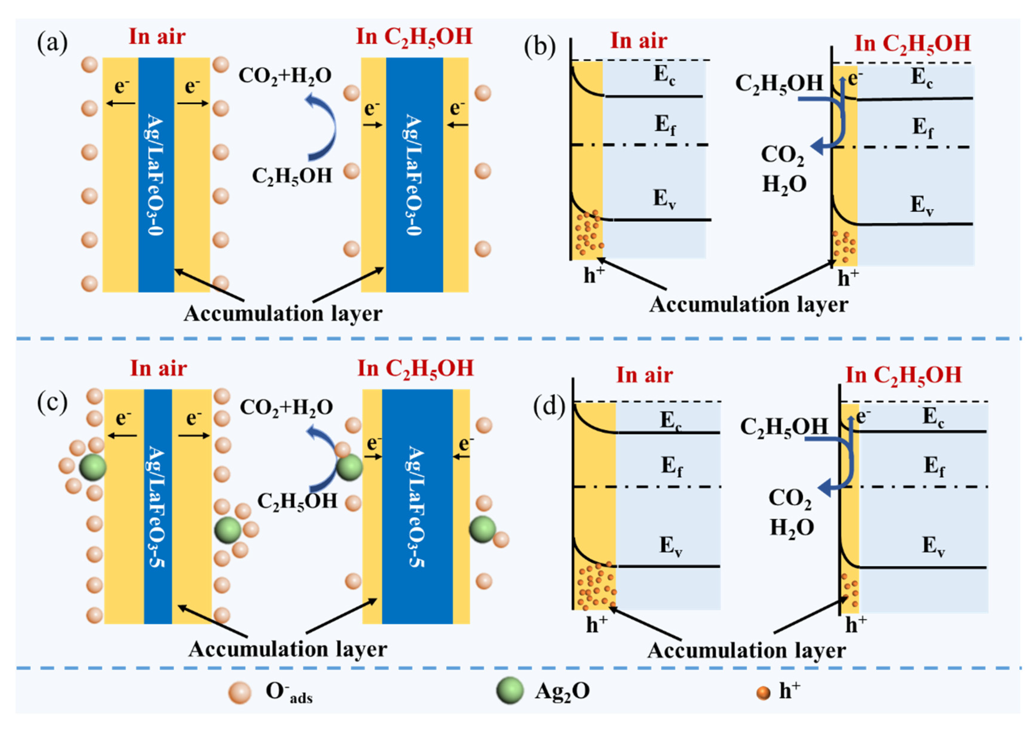
As a classic p-type semiconductor, the holes are the main carriers of LaFeO3. Its gas-sensitive mechanism focuses on gas adsorption. As illustrated in Figure 6a, when the gas sensor is exposed to air, oxygen molecules are potentially attracted to the surface of the LaFeO3 material, thus leading to the formation of adsorbed oxygen anion species through the conductivity of trapping free electrons. This step creates an increase in the carrier, which means that a high potential barrier and a deep cavity buildup layer are formed, as described as follows [10,11,44]:
O2 (gas) ↔ O2 (ads)
O2 (ads) + e− ↔ O2− (ads) (T < 100 °C)
O2− (ads) + 2e− ↔ 2O− (ads) (100 °C < T < 300 °C)
O− (ads) + e− → O2− (ads) (T > 300 °C)
As mentioned above, the optimum operating temperature for the Ag/LaFeO3-based sensor was 180 °C. The oxygen anion attached to the Ag/LaFeO3 material surface was primarily present in the form of O−. When ethanol vapor was injected into the chamber to act as a reducing gas, the oxygen species reacted with the ethanol gas while the electrons captured by the oxygen anion were freed, which returned to the conductive band of the Ag/LaFeO3. The electrons were annihilated by holes, resulting in an extension in the resistance of the sensitive material (Figure 6b),as described in Equations (5)–(6) [11,45]:
CH3CH2OH + 6O− → 2CO2 + 3H2O + 6e−
h+ + e− = null
As shown in Figure 6b,c, when the Ag/LaFeO3-5 sensors were exposed to air, a larger number of oxygen molecules were attached to the surface of the material, which allowed more free electron binding in the conduction band to form O−, and the hole accumulation layer expanded compared to the pure sample. Equally, when the Ag/LaFeO3-5 material surface was exposed to ethanol gas, the O− interacted with further ethanol molecules trapped on the material surface, releasing additional collected electrons into the conduction band, greatly narrowing the hole accumulation layer and substantially increasing the resistance [29,46]. In addition, this outstanding sensing performance could be credited to the chemical and electronic sensitivity of Ag nanoparticles [39,47]. As well as being an efficient catalyst for the oxidation of ethanol, the Ag NPs distributed on the LaFeO3 surface are also potent catalysts for the adsorption–desorption reaction of oxygen [38,48]. To investigate the gas-sensing mechanism in depth, some studies have been performed based on the density flooding theory (DFT) with theoretical calculations explaining that the selectivity of LaFeO3-based sensors for ethanol can probably be attributed to the much higher adsorption energy of ethanol gas on the sensor surface than other gases [13,49]. It was also suggested that the lowest unoccupied molecular orbital (LUMO) energy values of various volatile organic compounds could reflect the gas-sensing sensitivity [50]. The high adsorption energy of ethanol on the LaFeO3-based sensor surface combined with the low LUMO energy value of C2H5OH may help to explain the selective detection of ethanol in this study. As a result, the Ag/LaFeO3-5 sensor offered a higher ethanol gas response compared with pure LaFeO3. Nevertheless, more efforts are still expected to further clarify the details of the current mechanism of the Ag/LaFeO3 system owing to the complexity of the gas-sensing mechanism.
4. Conclusions
In conclusion, the ultrathin two-dimensional LaFeO3 nanosheets were successfully prepared using the GO template method. The XRD and SEM results showed that the LaFeO3 nanosheets obtained by calcination at 700 °C were well crystalline and had an ultrathin two-dimensional porous nanosheet morphology. The Ag-modified ultra-thin porous LaFeO3 nanosheets were investigated, and the LaFeO3 nanosheets calcined at 700 °C were surface modified with Ag to obtain gas-sensitive materials with a higher gas-sensitive response to ethanol. By investigating the effect of Ag modification on the gas-sensitive performance, the Ag/LaFeO3-5 nanosheets were shown to achieve a response of 30.5 at the optimum working temperature of 180 °C towards 50 ppm ethanol, which was about 2.4 times higher than that of the gas-sensitive material without Ag modification (Ag/LaFeO3-0). Therefore, Ag-modified LaFeO3 has considerable prospects for the potential development of simple and economical alcohol gas sensors for practical applications.
Supplementary Materials
The following supporting information can be downloaded at: https://0-www-mdpi-com.brum.beds.ac.uk/article/10.3390/nano12101768/s1, Figure S1: The schematic structure of the gas sensor, Figure S2: (a) TG curves of LaFeO3/GO, (b) Raman spectra of the LaFeO3 samples with different calcination temperatures, Figure S3: Response of LaFeO3 samples toward 50 ppm of EtOH at operation temperature ranging from 140 to 220 °C. Table S1: Fitting results of O 1s XPS spectra of Ag/LaFeO3-0 and Ag/LaFeO3-5.
Author Contributions
Conceptualization, J.Y. and D.W.; methodology, J.Y., C.W. and Q.Y.; software, J.Y.; validation, J.Y., C.W. and Q.Y.; investigation, X.Y., J.Y. and C.W.; resources, D.W.; data curation, J.Y., X.Y. and C.W.; writing—original draft preparation, J.Y.; writing—review and editing, D.W., J.Y. and Q.Y.; visualization, J.Y.; supervision, D.W.; project administration, D.W. and Y.C.; funding acquisition, Y.C. All authors have read and agreed to the published version of the manuscript.
Funding
This research was funded by the National Natural Science Foundation of China (62071300), Science and Technology Commission of Shanghai Municipality (19ZR1435200, 21ZR1444200, 20490761100, YDZX20213100003002).
Data Availability Statement
Data is contained within the article or Supplementary Materials.
Acknowledgments
We greatly appreciate the financial support from the National Natural Science Foundation of China (62071300), Science and Technology Commission of Shanghai Municipality (19ZR1435200, 21ZR1444200, 20490761100, YDZX20213100003002).
Conflicts of Interest
The authors declare no conflict of interest.
References
- Chang, H.; Bjørgum, E.; Mihai, O.; Yang, J.; Lein, H.L.; Grande, T.; Raaen, S.; Zhu, Y.-A.; Holmen, A.; Chen, D. Effects of Oxygen Mobility in La–Fe-Based Perovskites on the Catalytic Activity and Selectivity of Methane Oxidation. ACS Catal. 2020, 10, 3707–3719. [Google Scholar] [CrossRef]
- Zhu, Y.; Zhou, W.; Shao, Z. Perovskite/Carbon Composites: Applications in Oxygen Electrocatalysis. Small 2017, 13, 1603793. [Google Scholar] [CrossRef] [PubMed]
- Kim, Y.S.; Lee, G.-H.; Sung, M.-C.; Kim, D.-W. Orthorhombically distorted perovskite SeZnO3 nanosheets as an electrocatalyst for lithium-oxygen batteries. Chem. Eng. J. 2020, 406, 126896. [Google Scholar] [CrossRef]
- Tan, P.; Liu, M.; Shao, Z.; Ni, M. Recent Advances in Perovskite Oxides as Electrode Materials for Nonaqueous Lithium-Oxygen Batteries. Adv. Energy Mater. 2017, 7, 1602674. [Google Scholar] [CrossRef]
- Shen, S.-Y.; Zheng, H.; Zheng, P.; Wu, Q.; Deng, J.-X.; Ying, Z.-H.; Zheng, L. Microstructure, magnetic properties of hexagonal barium ferrite powder based on calcination temperature and holding time. Rare Met. 2018, 40, 981–986. [Google Scholar] [CrossRef]
- Qin, W.; Yuan, Z.; Shen, Y.; Zhang, R.; Meng, F. Phosphorus-doped porous perovskite LaFe1-xPxO3-δ nanosheets with rich surface oxygen vacancies for ppb level acetone sensing at low temperature. Chem. Eng. J. 2021, 431, 134280. [Google Scholar] [CrossRef]
- Bulemo, P.M.; Kim, I.-D. Recent advances in ABO3 perovskites: Their gas-sensing performance as resistive-type gas sensors. J. Korean Ceram. Soc. 2019, 57, 24–39. [Google Scholar] [CrossRef] [Green Version]
- Shingange, K.; Swart, H.C.; Mhlongo, G.H. Design of porous p-type LaCoO3 nanofibers with remarkable response and selectivity to ethanol at low operating temperature. Sens. Actuators B Chem. 2020, 308, 127670. [Google Scholar] [CrossRef]
- Fergus, J.W. Perovskite Oxides for Semiconductor-based Gas Sensors. Sens. Actuators B Chem. 2007, 123, 1169–1179. [Google Scholar] [CrossRef]
- Gu, J.; Zhang, B.; Li, Y.; Xu, X.; Sun, G.; Cao, J.; Wang, Y. Synthesis of spindle-like Co-doped LaFeO3 porous microstructure for high performance n-butanol sensor. Sens. Actuators B Chem. 2021, 343, 130125. [Google Scholar] [CrossRef]
- Xiang, J.; Chen, X.; Zhang, X.; Gong, L.; Zhang, Y.; Zhang, K. Preparation and characterization of Ba-doped LaFeO3 nanofibers by electrospinning and their ethanol sensing properties. Mater. Chem. Phys. 2018, 213, 122–129. [Google Scholar] [CrossRef]
- Zhang, G.; Song, X.-Z.; Wang, X.-F.; Liu, N.; Li, X.; Wei, Z.; Qian, G.; Wang, Z.; Yu, S.; Tan, Z. LnFeO3(Ln=La, Nd, Sm) Derived from Bimetallic Organic Frameworks for Gas Sensor. J. Alloys Compd. 2022, 902, 163803. [Google Scholar] [CrossRef]
- Cao, E.; Wu, A.; Wang, H.-H.; Zhang, Y.; Hao, W.; Sun, L. Enhanced Ethanol Sensing Performance of Au and Cl Comodified LaFeO3 Nanoparticles. ACS Appl. Nano Mater. 2019, 2, 1541–1551. [Google Scholar] [CrossRef]
- Qin, J.; Cui, Z.; Yang, X.; Zhu, S.; Li, Z.; Liang, Y. Synthesis of three-dimensionally ordered macroporous LaFeO3 with enhanced methanol gas sensing properties. Sens. Actuators B Chem. 2014, 209, 706–713. [Google Scholar] [CrossRef]
- Li, L.; Zhang, C.; Zhang, R.; Gao, X.; He, S.; Liu, M.; Li, X.; Chen, W. 2D ultrathin Co3O4 nanosheet array deposited on 3D carbon foam for enhanced ethanol gas sensing application. Sens. Actuators B Chem. 2017, 244, 664–672. [Google Scholar] [CrossRef]
- Wang, J.; Ren, Y.; Liu, H.; Li, Z.; Liu, X.; Deng, Y.; Fang, X. Ultrathin 2D NbWO6 Perovskite Semiconductor Based Gas Sensors with Ultrahigh Selectivity under Low Working Temperature. Adv. Mater. 2021, 34, 2104958. [Google Scholar] [CrossRef]
- Wang, D.; Deng, L.; Cai, H.; Yang, J.; Bao, L.; Zhu, Y.; Wang, X. Bimetallic PtCu Nanocrystal Sensitization WO3 Hollow Spheres for Highly Efficient 3-Hydroxy-2-butanone Biomarker Detection. ACS Appl. Mater. Interfaces 2020, 12, 18904–18912. [Google Scholar] [CrossRef]
- Wang, D.; Wan, K.; Zhang, M.; Li, H.; Wang, P.; Wang, X.; Yang, J. Constructing hierarchical SnO2 nanofiber/nanosheets for efficient formaldehyde detection. Sens. Actuators B Chem. 2018, 283, 714–723. [Google Scholar] [CrossRef]
- Wang, D.; Huang, S.; Li, H.; Chen, A.; Wang, P.; Yang, J.; Wang, X.; Yang, J. Ultrathin WO3 nanosheets modified by g-C3N4 for highly efficient acetone vapor detection. Sens. Actuators B Chem. 2018, 282, 961–971. [Google Scholar] [CrossRef]
- Chen, Z.; Wang, D.; Wang, X.; Yang, J. Preparation and Formaldehyde Sensitive Properties of N-GQDs/SnO2 Nanocomposite. Chin. Chem. Lett. 2020, 31, 2063–2066. [Google Scholar] [CrossRef]
- Wang, X.-Y.; Guo, H.; Shi, J.-A.; Biao, Y.; Li, Y.; Han, G.-Y.; Zhang, S.; Qian, K.; Zhou, W.; Lin, X.; et al. MgO intercalation and crystallization between epitaxial graphene and Ru(0001). Rare Met. 2022, 41, 304–310. [Google Scholar] [CrossRef]
- Chen, Z.-L.; Wang, D.; Wang, X.-Y.; Yang, J.-H. Enhanced formaldehyde sensitivity of two-dimensional mesoporous SnO2 by nitrogen-doped graphene quantum dots. Rare Met. 2021, 40, 1561–1570. [Google Scholar] [CrossRef]
- Liu, F.; Wang, X.; Chen, X.; Song, X.; Tian, J.; Cui, H. Porous ZnO Ultrathin Nanosheets with High Specific Surface Areas and Abundant Oxygen Vacancies for Acetylacetone Gas Sensing. ACS Appl. Mater. Interfaces 2019, 11, 24757–24763. [Google Scholar] [CrossRef] [PubMed]
- AbdelHamid, A.; Yu, Y.; Yang, J.; Ying, J.Y. Generalized Synthesis of Metal Oxide Nanosheets and Their Application as Li-Ion Battery Anodes. Adv. Mater. 2017, 29, 1701427. [Google Scholar] [CrossRef] [PubMed]
- Zhao, H.; Zhu, Y.; Li, F.; Hao, R.; Wang, S.; Guo, L. A Generalized Strategy for the Synthesis of Large-Size Ultrathin Two-Dimensional Metal Oxide Nanosheets. Angew. Chem. Int. Ed. 2017, 56, 8766–8770. [Google Scholar] [CrossRef] [PubMed]
- Peng, J.; Liao, J.; Yang, X.; Feng, W. Fiber-optic Dual Fabry-Pérot Interferometric Carbon Monoxide Sensor with Polyaniline/Co3O4/graphene Oxide Sensing Membrane. Chin. Chem. Lett. 2020, 31, 2145–2149. [Google Scholar] [CrossRef]
- Postica, V.; Vahl, A.; Santos-Carballal, D.; Dankwort, T.; Kienle, L.; Hoppe, M.; Cadi-Essadek, A.; De Leeuw, N.H.; Terasa, M.-I.; Adelung, R.; et al. Tuning ZnO Sensors Reactivity toward Volatile Organic Compounds via Ag Doping and Nanoparticle Functionalization. ACS Appl. Mater. Interfaces 2019, 11, 31452–31466. [Google Scholar] [CrossRef] [Green Version]
- Zhou, Y.; Chen, G.; Yu, Y.; Yan, C.; Sun, J.; He, F. Synthesis of metal oxide nanosheets through a novel approach for energy applications. J. Mater. Chem. A 2015, 4, 781–784. [Google Scholar] [CrossRef]
- Chen, M.; Wang, H.; Hu, J.; Zhang, Y.; Li, K.; Zhang, D.; Zhou, S.; Zhang, J.; Zhu, Z.; Liu, Q. Near-Room-Temperature Ethanol Gas Sensor Based on Mesoporous Ag/Zn–LaFeO3 Nanocomposite. Adv. Energy Mater. 2019, 6, 1801453. [Google Scholar]
- Wan, K.; Wang, D.; Wang, F.; Li, H.; Xu, J.; Wang, X.; Yang, J. Hierarchical In2O3@SnO2 Core−Shell Nanofiber for High Efficiency Formaldehyde Detection. ACS Appl. Mater. Interfaces 2019, 11, 45214–45225. [Google Scholar] [CrossRef]
- Zhang, Y.; Lin, Y.; Chen, J.; Zhang, J.; Zhu, Z.; Liu, Q. A high sensitivity gas sensor for formaldehyde based on silver doped lanthanum ferrite. Sens. Actuators B Chem. 2013, 190, 171–176. [Google Scholar] [CrossRef]
- Ren, X.; Yang, H. Gen, S.; Zhou, J.; Yang, T.; Zhang, X.; Cheng, Z.; Sun, S. Controlled Growth of LaFeO3 Nanoparticles on Reduced Graphene Oxide for Highly Efficient Photocatalysis. Nanoscale 2016, 8, 752–756. [Google Scholar] [CrossRef] [PubMed]
- Zhang, Z.; Zhang, S.; Jiang, C.; Guo, H.; Qu, F.; Shimakawa, Y.; Yang, M. Integrated Sensing Array of the Perovskite-type LnFeO3 (Ln=La, Pr, Nd, Sm) to Discriminate Detection of Volatile Sulfur Compounds. J. Hazard. Mater. 2021, 413, 125380. [Google Scholar] [CrossRef] [PubMed]
- Gong, S.; Xie, Z.; Li, W.; Wu, X.; Han, N.; Chen, Y. Highly active and humidity resistive perovskite LaFeO3 based catalysts for efficient ozone decomposition. Appl. Catal. B Environ. 2018, 241, 578–587. [Google Scholar] [CrossRef]
- Tjeng, L.H.; Meinders, M.B.J.; van Elp, J.; Ghijsen, J.; Sawatzky, G.A. Electronic Structure of Ag2O. Phys. Rev. B Condens. Matter Mater. Phys. 1990, 41, 3190. [Google Scholar] [CrossRef]
- Kaushik, V.K. XPS core level spectra and Auger parameters for some silver compounds. J. Electron. Spectrosc. Relat. Phenom. 1991, 56, 273–277. [Google Scholar] [CrossRef]
- Yang, C.; Yang, Y.; Zhang, C.; Yu, H.; Wang, T.; Shi, K.; Zhang, Z.; Wang, D.; Dong, X. High selectivity of Ag-doped Fe2O3 hollow nanofibers in H2S detection at room operating temperature. Sens. Actuators B Chem. 2021, 341, 129919. [Google Scholar] [CrossRef]
- Chen, M.; Zhang, Y.; Zhang, J.; Li, K.; Lv, T.; Shen, K.; Zhu, Z.; Liu, Q. Facile Lotus-Leaf-Templated Synthesis and Enhanced Xylene Gas Sensing Properties of Ag-LaFeO3 Nanoparticles. J. Mater. Chem. C 2018, 6, 6138–6145. [Google Scholar] [CrossRef]
- Ren, Y.; Xie, W.; Li, Y.; Ma, J.; Li, J.; Liu, Y.; Zou, Y.; Deng, Y. Noble Metal Nanoparticles Decorated Metal Oxide Semiconducting Nanowire Arrays Interwoven into 3D Mesoporous Superstructures for Low-Temperature Gas Sensing. ACS Central Sci. 2021, 7, 1885–1897. [Google Scholar] [CrossRef]
- Lu, J.; Jia, N.; Cheng, L.; Liang, K.; Huang, J.; Li, J. rGO/CoTiO3 nanocomposite with enhanced gas sensing performance at low working temperature. J. Alloys Compd. 2018, 739, 227–234. [Google Scholar] [CrossRef]
- Tian, M.; Miao, J.; Cheng, P.; Mu, H.; Tu, J.; Sun, J. Layer-by-layer nanocomposites consisting of Co3O4 and reduced graphene (rGO) nanosheets for high selectivity ethanol gas sensors. Appl. Surf. Sci. 2019, 479, 601–607. [Google Scholar] [CrossRef]
- Hao, P.; Qiu, G.; Song, P.; Yang, Z.; Wang, Q. Construction of porous LaFeO3 microspheres decorated with NiO nanosheets for high response ethanol gas sensors. Appl. Surf. Sci. 2020, 515, 146025. [Google Scholar] [CrossRef]
- Phan, T.T.N.; Dinh, T.T.M.; Nguyen, M.D.; Li, D.; Phan, C.N.; Pham, T.K.; Nguyen, C.T.; Pham, T.H. Hierarchically structured LaFeO3 with hollow core and porous shell as efficient sensing material for ethanol detection. Sens. Actuators B Chem. 2021, 354, 131195. [Google Scholar] [CrossRef]
- Shi, Y.; Liu, T.; Zhao, Y.; Su, J.; Zeb, S.; Nie, Y.; Qin, C.; Wang, B.; Jiang, X. Tunable oxygen vacancies of cobalt oxides for efficient gas sensing application. Sens. Actuators B Chem. 2021, 350, 130860. [Google Scholar] [CrossRef]
- Chao, J.; Chen, Y.; Xing, S.; Zhang, D.; Shen, W. Facile fabrication of ZnO/C nanoporous fibers and ZnO hollow spheres for high performance gas sensor. Sens. Actuators B Chem. 2019, 298, 126927. [Google Scholar] [CrossRef]
- Wei, W.; Guo, S.; Chen, C.; Sun, L.; Chen, Y.; Guo, W.; Ruan, S. High sensitive and fast formaldehyde gas sensor based on Ag-doped LaFeO3 nanofibers. J. Alloys Compd. 2016, 695, 1122–1127. [Google Scholar] [CrossRef]
- Lei, M.; Gao, M.; Yang, X.; Zou, Y.; Alghamdi, A.; Ren, Y.; Deng, Y. Size-Controlled Au Nanoparticles Incorporating Mesoporous ZnO for Sensitive Ethanol Sensing. ACS Appl. Mater. Interfaces 2021, 13, 51933–51944. [Google Scholar] [CrossRef]
- Ma, J.; Li, Y.; Zhou, X.; Yang, X.; Alharthi, F.A.; Alghamdi, A.A.; Cheng, X.; Deng, Y. Au Nanoparticles Decorated Mesoporous SiO2 –WO3 Hybrid Materials with Improved Pore Connectivity for Ultratrace Ethanol Detection at Low Operating Temperature. Small 2020, 16, e2004772. [Google Scholar] [CrossRef]
- Cao, E.; Wang, H.; Wang, X.; Yang, Y.; Hao, W.; Sun, L.; Zhang, Y. Enhanced ethanol sensing performance for chlorine doped nanocrystalline LaFeO3-δ powders by citric sol-gel method. Sens. Actuators B Chem. 2017, 251, 885–893. [Google Scholar] [CrossRef]
- Dai, Z.; Lee, C.-S.; Kim, B.-Y.; Kwak, C.-H.; Yoon, J.-W.; Jeong, H.-M.; Lee, J.-H. Honeycomb-like Periodic Porous LaFeO3 Thin Film Chemiresistors with Enhanced Gas-Sensing Performances. ACS Appl. Mater. Interfaces 2014, 6, 16217–16226. [Google Scholar] [CrossRef]
Scheme 1.
The schematic diagram of the synthesis of Ag/LaFeO3 nanosheets by the GO template method.

Figure 1.
SEM micrographs of LaFeO3 at different calcination temperatures. (a) LaFeO3/GO; (b) LaFeO3/GO calcined at 500 °C; (c) LaFeO3/GO calcined at 600 °C; (d) LaFeO3/GO calcined at 700 °C; (e) LaFeO3/GO calcined at 800 °C; (f) XRD patterns of the samples.
Figure 1.
SEM micrographs of LaFeO3 at different calcination temperatures. (a) LaFeO3/GO; (b) LaFeO3/GO calcined at 500 °C; (c) LaFeO3/GO calcined at 600 °C; (d) LaFeO3/GO calcined at 700 °C; (e) LaFeO3/GO calcined at 800 °C; (f) XRD patterns of the samples.

Figure 2.
SEM images of (a) Ag/LaFeO3-0; (b) Ag/LaFeO3-1; (c) Ag/LaFeO3-5; (d) Ag/LaFeO3-10; (e) XRD patterns of the Ag/LaFeO3 samples with different calcination temperatures; (f) Raman spectra of the Ag/LaFeO3 samples.
Figure 2.
SEM images of (a) Ag/LaFeO3-0; (b) Ag/LaFeO3-1; (c) Ag/LaFeO3-5; (d) Ag/LaFeO3-10; (e) XRD patterns of the Ag/LaFeO3 samples with different calcination temperatures; (f) Raman spectra of the Ag/LaFeO3 samples.

Figure 3.
TEM of (a) Ag/LaFeO3-0; and (b) Ag/LaFeO3-5; (c,d) the STEM-EDS elemental mapping images of Ag/LaFeO3-5; (e) BET characterization of Ag/LaFeO3-0 and Ag/LaFeO3-5, and the inset shows the pore size distribution of the corresponding composites.
Figure 3.
TEM of (a) Ag/LaFeO3-0; and (b) Ag/LaFeO3-5; (c,d) the STEM-EDS elemental mapping images of Ag/LaFeO3-5; (e) BET characterization of Ag/LaFeO3-0 and Ag/LaFeO3-5, and the inset shows the pore size distribution of the corresponding composites.

Figure 4.
(a) XPS survey spectra of Ag/LaFeO3-0 and Ag/LaFeO3-5, high-resolution core-level XPS spectra of; (b) La 3d; (c) Fe 2p; (d) Ag 3d; (e) O 1s of Ag/LaFeO3-0; and (f) O 1s of Ag/LaFeO3-5.
Figure 4.
(a) XPS survey spectra of Ag/LaFeO3-0 and Ag/LaFeO3-5, high-resolution core-level XPS spectra of; (b) La 3d; (c) Fe 2p; (d) Ag 3d; (e) O 1s of Ag/LaFeO3-0; and (f) O 1s of Ag/LaFeO3-5.

Figure 5.
(a) The response of Ag/LaFeO3-0--Ag/LaFeO3-5 nanomaterials to 50 ppm ethanol in the range of 140–220 °C; (b) selectivity of the four Ag/LaFeO3 sensors for different gases at 180 °C (concentrations of 50 ppm for all gas); (c) sensor response values for Ag/LaFeO3-5 at 180 °C for a mixture of vapors (20 ppm concentration of these gases); (d) response curves of the Ag/LaFeO3-5 sensor for EtOH vapors ranging from 5 ppm to 100 ppm concentration, the inset exhibits a direct linearity correlation between the sensing response values and EtOH vapor levels; (e) dynamic response curve of Ag/LaFeO3-5-based sensors under a low concentration of EtOH exposed at 180 °C; (f) sensing curves and (g) reproducibility of the Ag/LaFeO3-5 sensor for 20 ppm EtOH vapors; (h) the long-term stability of sensitive materials; (i) response of Ag/LaFeO3-5-based sensors to 20 ppm EtOH under different RH%.

Figure 6.
Schematic diagrams of the mechanism for the C2H5OH sensing on the (a,b) Ag/LaFeO3-0 nanosheets; (c,d) Ag/LaFeO3-5 nanosheets.
Figure 6.
Schematic diagrams of the mechanism for the C2H5OH sensing on the (a,b) Ag/LaFeO3-0 nanosheets; (c,d) Ag/LaFeO3-5 nanosheets.


Table 1.
Comparison of sensor-based ethanol gas detection characteristics with different detection materials.
Table 1.
Comparison of sensor-based ethanol gas detection characteristics with different detection materials.
| Materials | T (°C) | Concentration (ppm) | Response (Rg/Ra) | Tres/Trec (s) | Ref. |
|---|---|---|---|---|---|
| rGO/CoTiO3 | 195 | 50 | 9 | 2/5 | [40] |
| rGO/Co3O4 | 200 | 100 | 21 | [41] | |
| Ba-doped LaFeO3 | 210 | 100 | 31.3 | 40/42 | [11] |
| NiO@LaFeO3 | 240 | 10 | 14.7 | 2/9 | [42] |
| LaFeO3 | 300 | 143 | 14.5 | 23/29 | [43] |
| Ag/LaFeO3-5 | 180 | 20 | 21 | 26/27 | This work |
Publisher’s Note: MDPI stays neutral with regard to jurisdictional claims in published maps and institutional affiliations. |
© 2022 by the authors. Licensee MDPI, Basel, Switzerland. This article is an open access article distributed under the terms and conditions of the Creative Commons Attribution (CC BY) license (https://creativecommons.org/licenses/by/4.0/).
Share and Cite
MDPI and ACS Style
Yu, J.; Wang, C.; Yuan, Q.; Yu, X.; Wang, D.; Chen, Y. Ag-Modified Porous Perovskite-Type LaFeO3 for Efficient Ethanol Detection. Nanomaterials 2022, 12, 1768. https://0-doi-org.brum.beds.ac.uk/10.3390/nano12101768
AMA Style
Yu J, Wang C, Yuan Q, Yu X, Wang D, Chen Y. Ag-Modified Porous Perovskite-Type LaFeO3 for Efficient Ethanol Detection. Nanomaterials. 2022; 12(10):1768. https://0-doi-org.brum.beds.ac.uk/10.3390/nano12101768
Chicago/Turabian StyleYu, Jiejie, Cong Wang, Quan Yuan, Xin Yu, Ding Wang, and Yang Chen. 2022. "Ag-Modified Porous Perovskite-Type LaFeO3 for Efficient Ethanol Detection" Nanomaterials 12, no. 10: 1768. https://0-doi-org.brum.beds.ac.uk/10.3390/nano12101768
Note that from the first issue of 2016, this journal uses article numbers instead of page numbers. See further details here.