Article

Graphene Oxide Synthesis from Agro Waste

by
Thirunavukkarasu Somanathan
1,2,*,
Karthika Prasad
1,3,
Kostya (Ken) Ostrikov
3,
Arumugam Saravanan
1 and
Vemula Mohana Krishna
1
1
Department of Nanoscience, School of Basic Sciences, Vels University, Pallavaram, Chennai, Tamil Nadu 600117, India
2
Center for Advanced Research and Development (CARD), Vels University, Pallavaram, Chennai, Tamil Nadu 600117, India
3
Institute of Future Environments, Nanotechnology and Molecular Science, School of Chemistry, Physics and Mechanical Engineering, Queensland University of Technology, Brisbane, QLD 4000, Australia
*
Author to whom correspondence should be addressed.
Nanomaterials 2015, 5(2), 826-834; https://0-doi-org.brum.beds.ac.uk/10.3390/nano5020826
Submission received: 6 April 2015 / Revised: 9 May 2015 / Accepted: 12 May 2015 / Published: 20 May 2015
(This article belongs to the Special Issue Nanomaterials for Energy and Sustainability Applications)

Abstract

:
A new method of graphene oxide (GO) synthesis via single-step reforming of sugarcane bagasse agricultural waste by oxidation under muffled atmosphere conditions is reported. The strong and sharp X-ray diffraction peak at 2θ = 11.6° corresponds to an interlayer distance of 0.788 nm (d002) for the AB stacked GOs. High-resolution transmission electron microscopy (HRTEM) and selected-area electron diffraction (SAED) confirm the formation of the GO layer structure and the hexagonal framework. This is a promising method for fast and effective synthesis of GO from sugarcane bagasse intended for a variety of energy and environmental applications.

Graphical Abstract

1. Introduction

Graphene and related materials have attracted a great deal of recent interest owing to their unique structure and physical, chemical, thermal, and other properties [1]. Graphene’s two-dimensional (2D) hexagonal lattice consists of a network of sp2-bonded carbon atoms and represents the “thinnest material” which is stable in its free form [2]. Excellent electronic properties [3], thermal conductivity [4], and high surface area, combined with unusual mechanical properties and good dispersion performance, make graphene a promising candidate for structural modification of composite materials and several other applications [5,6,7,8].
Graphene oxide (GO) has also attracted major interdisciplinary attention because of the broad range of envisaged applications across several scientific and engineering fields including physics, chemistry, biology, and medicine [9,10,11,12,13]. The original method to synthesize GO is based on the addition of potassium chlorate to a slurry of graphite in fuming nitric acid [14]. This synthetic protocol can be improved by using concentrated sulfuric acid as well as fuming nitric acid and adding the chlorate in multiple aliquots over the course of the reaction [15]. In the commonly used Hummers’ method [16], graphite is oxidized throughout treatment of KMnO4 and NaNO3 in concentrated H2SO4 acid.
All these procedures involve the generation of NO2, N2O4, and ClO2, toxic gases which are also explosive. In the past few years, various techniques such as mechanical exfoliation techniques, chemical vapor deposition (CVD) techniques and other chemical techniques have been developed for the synthesis of graphene and graphene oxide materials. However, many of these techniques are highly sophisticated and expensive [17]. Most of the commercially available GO samples were synthesized by Hummers' method or a modified version of it. The cost of the commercially available graphene oxide per gram is approximately $200. In comparison with the Hummers' method, our method is more environment friendly as it avoids toxic gas emission during synthesis. The synthesis technique reported here is a quite efficient asset demonstrating a new procedure for the production of graphene oxide from agro waste in simple steps. This simple and low-cost process could lead to new opportunities for cost-effective production of GO [18,19].
Here we report on the synthesis of graphene oxide by directly oxidizing sugarcane bagasse under muffled atmosphere. The GO produced by this method is called SOMA-GO (sugarcane oxidized under muffled atmosphere for graphene oxide). Furthermore, the structural characteristics of the obtained product are confirmed by X-ray diffraction, Fourier transmission infrared spectroscopy, field emission scanning electron microscopy, high-resolution transmission electron microscopy, and Raman spectroscopy.

2. Experimental Section

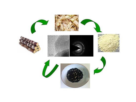
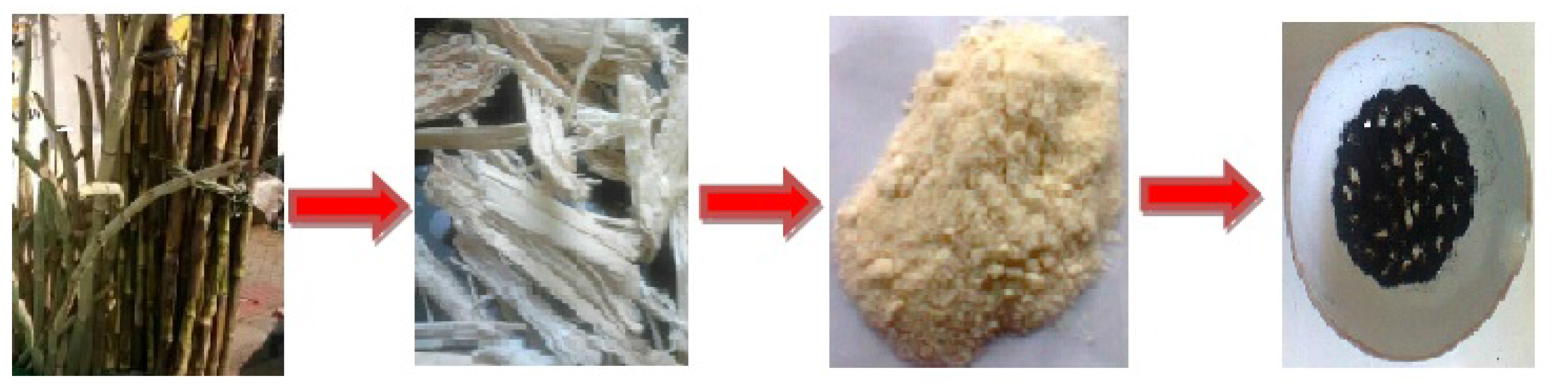
2.1. Synthesis of GO from Agro Waste

Sugarcane bagasse is a type of agricultural waste (agro waste). After juice was extracted, the remaining fiber was taken. The fiber was crushed and ground well in order to produce powder. This crushing and separating process was repeated several times to obtain fine powder. About 0.5 g of ground sugarcane bagasse powder mixed with 0.1 g of ferrocene was placed in a crucible and put directly into a muffle furnace at 300 °C for 10 min under atmospheric conditions. Next, the as-produced black solid was collected at room temperature. The black solid product was subjected to further analysis (Scheme 1).
Scheme 1. Schematic representation of graphene oxide synthesis from agro waste.
Nanomaterials 05 00826 g006

2.2. Characterization of GO

The X-ray diffraction (XRD) pattern was obtained using a Bruker D8 Advance X-ray diffractometer (Berlin, Germany). This device is equipped with a Ni filter, and generates monochromated Cu-Kα radiation (λ = 0.154 nm) operated at 40 kV accelerating voltage and 30 mA current. The samples were scanned in step mode with a 2° min−1 scan rate. Fourier transform infrared spectroscopy (FT-IR), Avatar 370, Thermo Nicolet (Berlin, Germany) was carried out to study the bonding configuration in the synthesized graphene oxide. High resolution transmission electron microscopy (HRTEM) was performed using a JEOL JEM-3000F microscope (Tokyo, Japan) operating at 300 kV to study the microstructure of the GO structures. Field emission scanning electron microscopy was carried out using a JEOL JSM 7800F revealed the morphological features of the GO structure.

3. Results and Discussion

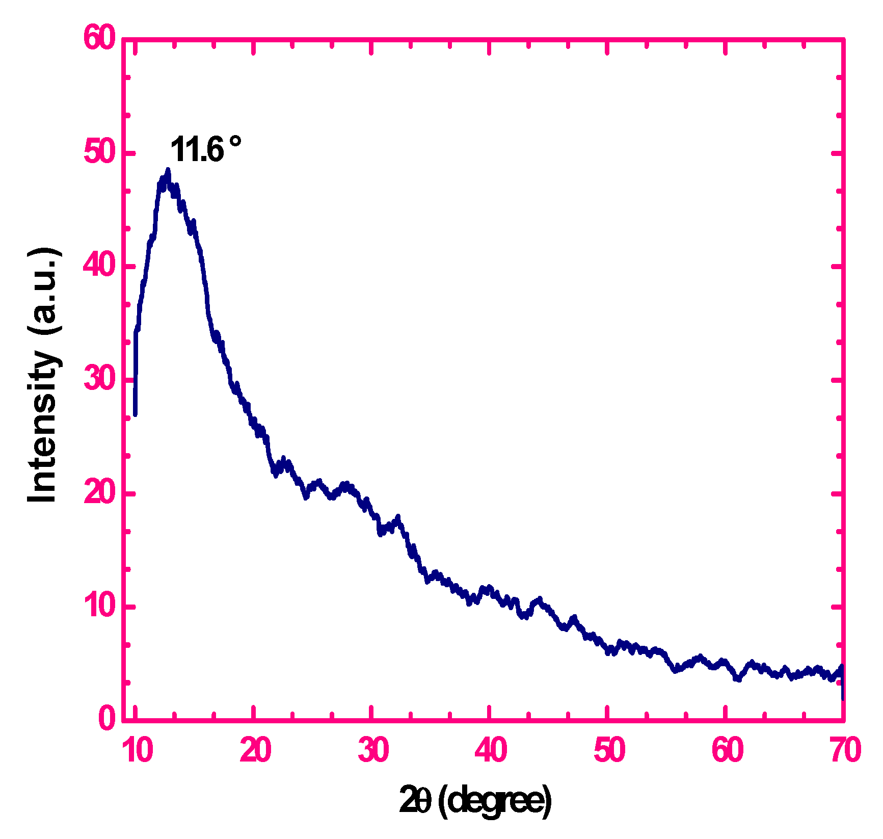
3.1. X-Ray Diffraction Measurements

The structural and chemical analysis of sugarcane bagasse is clearly described by previous investigators [20,21,22]. In this study, we have presented the XRD pattern of the GO which is shown in Figure 1. The peak at 2θ = 11.6° indicates that the agricultural sugarcane bagasse is fully oxidized into graphene oxide with the interlayer distance of 0.79 nm. The 2θ peaks of the GO obtained by the single-step oxidation of sugarcane bagasse are consistent with the results of previous studies [23,24]. This XRD pattern can be attributed to well graphitized two-dimensional structures made of GO sheets.
Figure 1. X-ray diffraction (XRD) pattern of graphene oxide.
Nanomaterials 05 00826 g001

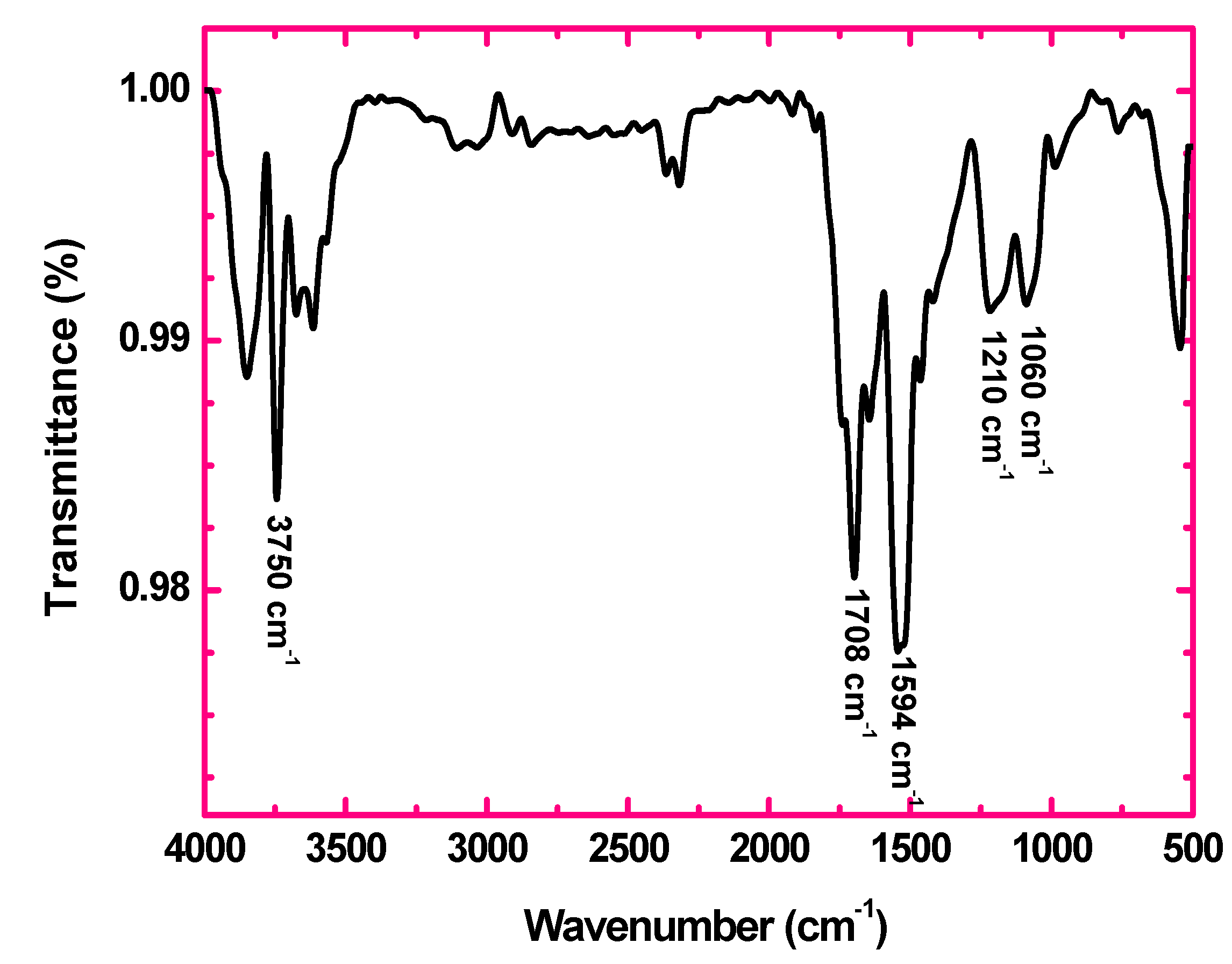
3.2. FT-IR Spectroscopy

The FT-IR spectrum of GO shown in Figure 2 confirms the introduction of oxygen containing groups such as functional hydroxyl, epoxy and carboxylic groups upon oxidation of sugarcane bagasse. The strong band at 1708 cm−1 is attributed to stretching vibration modes of C=O in carboxylic acid and carbonyl groups. The peak at 1594 cm−1 is assigned to the skeletal vibrations of un-oxidized graphitic domains. The band at 1060 cm−1 is assigned to C–O (epoxy) groups while the band at 1210 cm−1 is usually attributed to C–OH stretching vibrations [25,26,27]. The strong peak around 3500–4000 cm−1 can be attributed to the O–H stretching vibrations of the C–OH groups and water [27].
Figure 2. Fourier transform infrared spectroscopy (FT-IR) spectrum of graphene oxide.
Nanomaterials 05 00826 g002

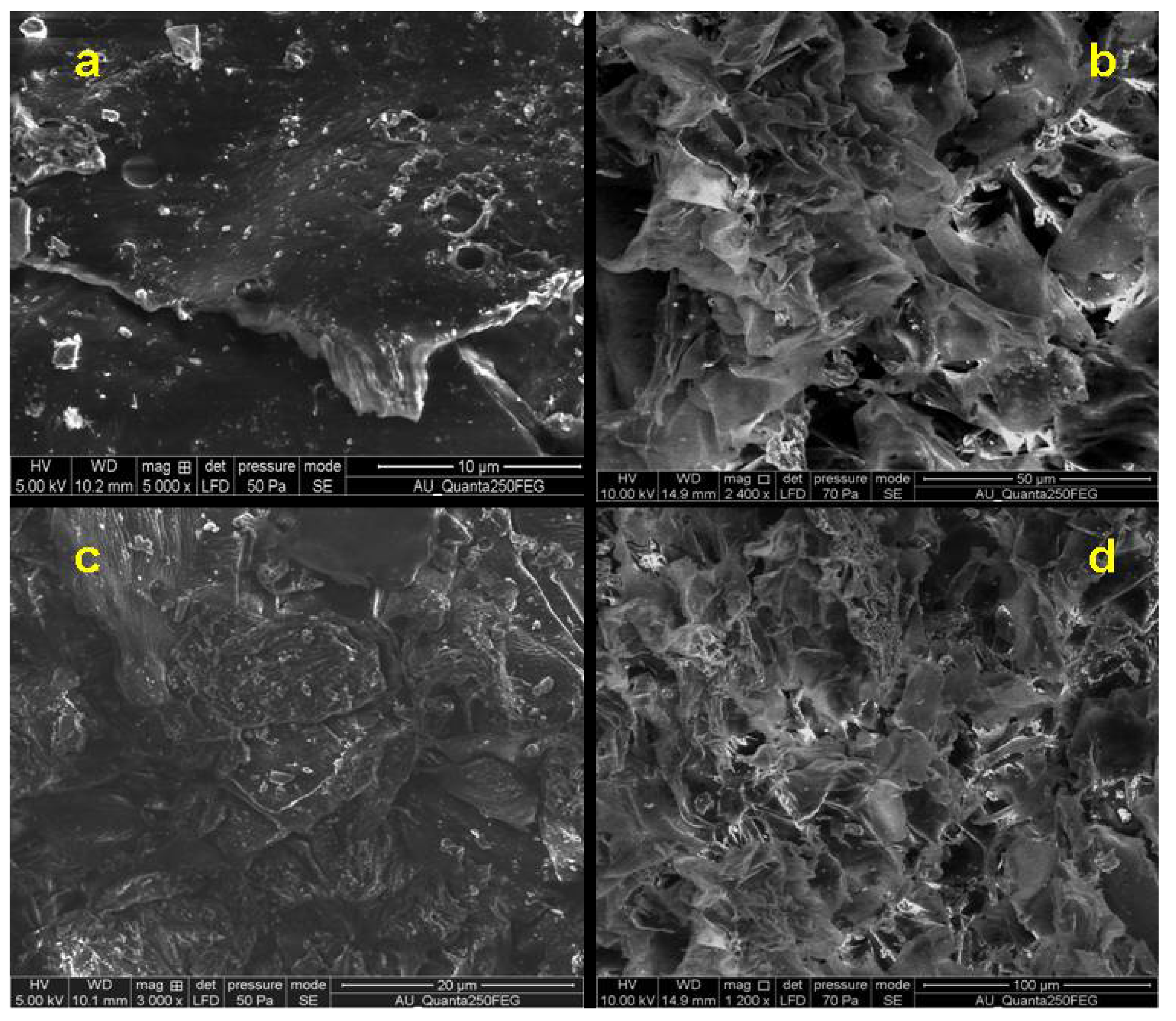
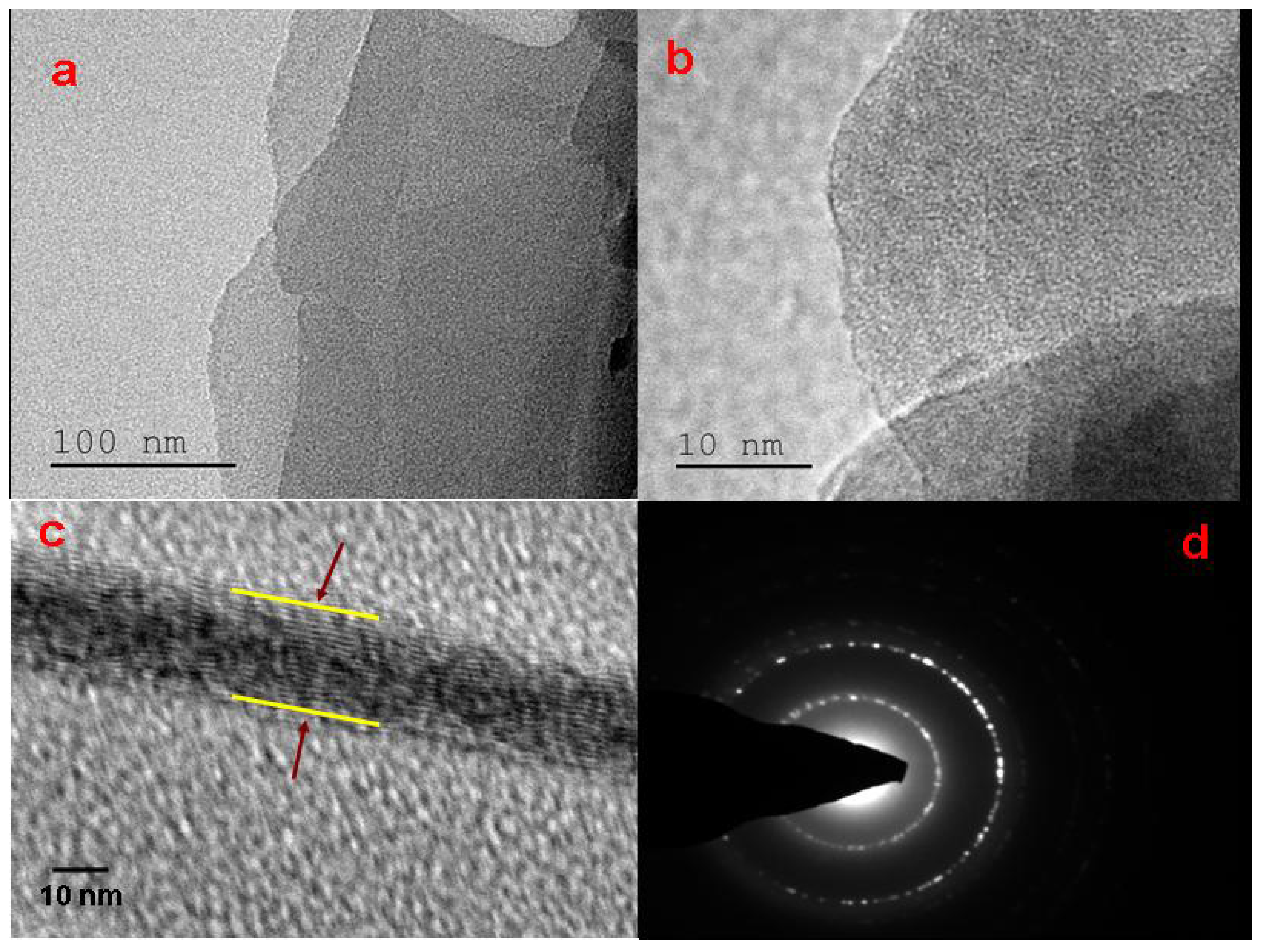
3.3. SEM and HRTEM Analysis

The surface morphology of the graphene oxide (GO) produced by sugarcane bagasse oxidation is identified by FESEM. The SEM image of the GO shown in Figure 3a,c clearly reveals its sheet-like structure. The morphology of the synthesised GO resembles flake-like structures as shown in Figure 3b,d. Quite similar textures have been reported by other authors [28].
HRTEM micrographs of GO are shown in Figure 4. Bends wrinkles, and edges on graphene oxide nanosheets are seen in several places in Figure 4a,b. Various defects and functional groups containing sp3 hybridized carbon atoms may also be introduced during the oxidation process. Figure 4c shows that the flakes contain a few layers of graphene oxide. The selected area of electron diffraction (SAED) pattern shown in Figure 4d indicates the presence of a polycrystalline structure corresponding to graphene oxide [29,30,31,32,33]. Thus, transmission electron microscopy (TEM) analysis confirms the presence of graphene oxide in the oxidized carbon samples.
Figure 3. FESEM images of GO obtained from sugarcane bagasse.
Nanomaterials 05 00826 g003
Figure 4. High-resolution transmission electron microscopy (HRTEM) images of GO obtained from sugarcane bagasse.
Nanomaterials 05 00826 g004

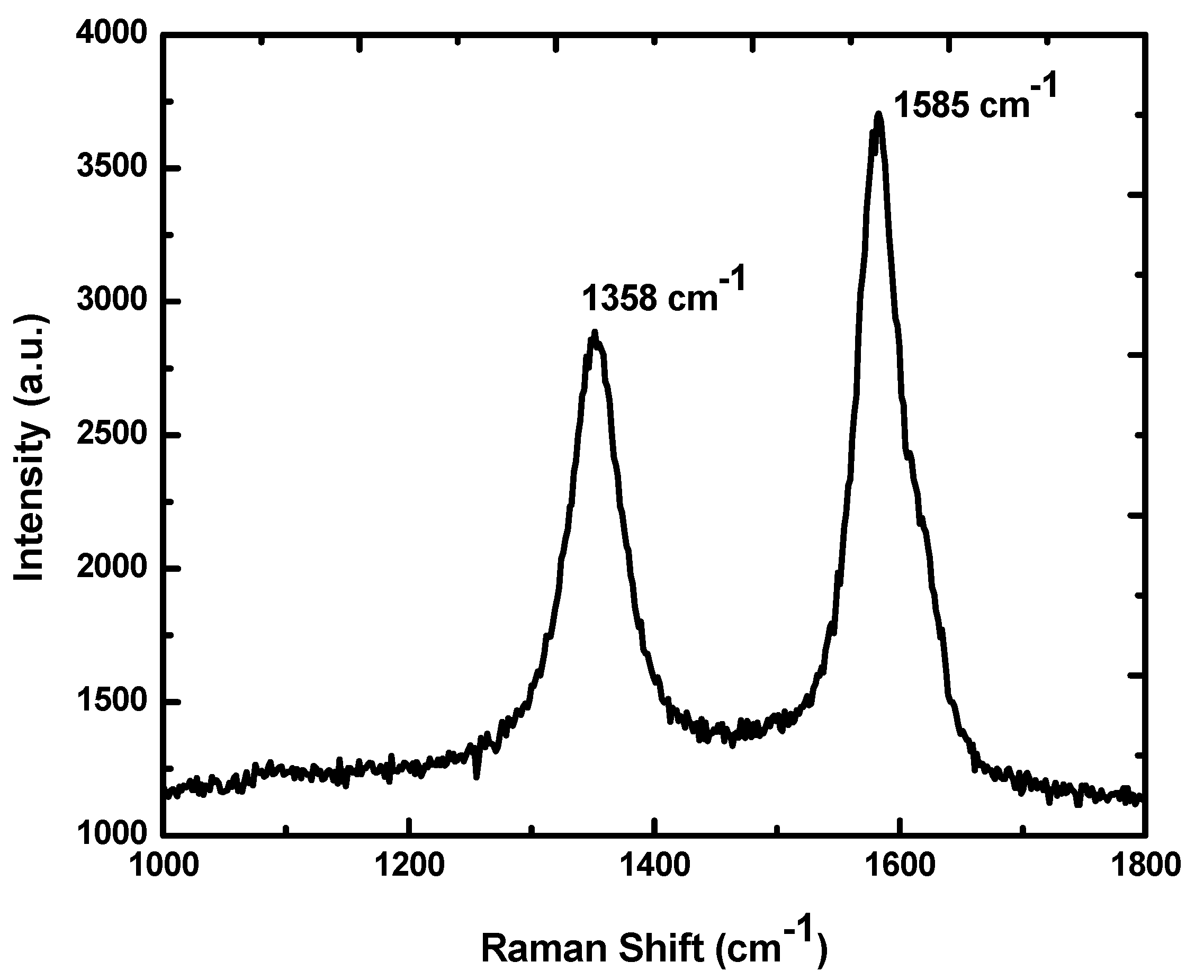
3.4. Raman Spectroscopy

The Raman spectrum of graphene oxide synthesized from sugarcane bagasse is shown in Figure 5. The spectrum of GO displays two prominent peaks at 1358 and 1585 cm−1, which are attributed to the local defects (found at the edges of graphene sheets) and the sp2 graphitized structure, respectively. The G peak formation is due to the double degenerated zone center E2g mode [34,35]. The functionality and reduction in particle size of sp2 plane domains are possibly due to the extensive oxidation of sugarcane bagasse [24]. The two D and G bands at 1358 cm−1 and 1585 cm−1 [36] indicate the clear sp2 carbon hybridization in the observed multi-layer stacks [31]. Thus, ID/IG peak intensity ratios are assigned to lower defects/disorders. Raman spectra show an intensity ratio of ID/IG at 0.76 for GO which is in line with previous investigations [24,37,38].
Figure 5. Raman spectrum of GO obtained from sugarcane bagasse.
Nanomaterials 05 00826 g005

4. Conclusions

A simple and rapid method is shown here to effectively convert solid sugarcane bagasse waste into value-added graphene oxide. The produced graphene oxide presents a well-graphitized structure. In comparison with the commonly used methods, our method is more environment friendly as it avoids toxic gas emission during the synthesis. This simple and low-cost synthesis process could lead to new opportunities for cost-effective production of graphene-based materials for gas sensors, energy storage, and other functional devices.

Acknowledgments

Thirunavukkarasu Somanathan thanks the Department of Science and Technology (DST) for the Fast Track Young Scientist Award and financial support (SR/FT/CS-111/2011). Karthika Prasad acknowledges support from the SEF Postgraduate Research Scholarship (Queensland University of Technology, Australia). This work was partially supported by the Australian Research Council.

Author Contributions

The manuscript was prepared using contributions from all authors. Thirunavukkarasu Somanathan designed the work, conducted the synthesis of graphene oxide from agro waste, analysed the results and led the manuscript preparation. Karthika Prasad prepared agro waste materials and conducted the synthesis and characterization experiments. Kostya (Ken) Ostrikov contributed to the analysis of the results and manuscript writing. Arumugam Saravanan and Vemula Mohana Krishna conducted FE-SEM and HRTEM observation and analyzed the results. All authors have given approval to the final version of the manuscript.

Conflicts of Interest

The authors declare no conflict of interest.

References

  1. Chae, H.K.; Siberio-Perez, D.Y.; Kim, J.; Go, Y.; Eddaoudi, M.; Matzger, A.J.; O’Keeffe, M.; Yaghi, O.M. A route to high surface area, porosity and inclusion of large molecules in crystals. Nature 2004, 427, 523–527. [Google Scholar] [CrossRef] [PubMed]
  2. Novoselov, K.; Geim, A.; Morozov, S.; Jiang, D.; Zhang, Y.; Dubonos, S.; Grigorieva, I.; Firsov, A. Electric field effect in atomically thin carbon films. Science 2004, 306, 666–669. [Google Scholar] [CrossRef] [PubMed]
  3. Schwierz, F. Graphene transistors. Nat. Nanotechnol. 2010, 5, 487–496. [Google Scholar] [CrossRef] [PubMed]
  4. Balandin, A.A.; Ghosh, S.; Bao, W.; Calizo, I.; Teweldebrhan, D.; Miao, F.; Lau, C.N. Superior thermal conductivity of single-layer graphene. Nano Lett. 2008, 8, 902–907. [Google Scholar] [CrossRef] [PubMed]
  5. Song, H.; Zhang, L.; He, C.; Qu, Y.; Tian, Y.F.; Lv, Y. Graphene sheets decorated with SnO2 nanoparticles: In situ synthesis and highly efficient materials for cataluminescence gas sensors. J. Mater. Chem. 2011, 21, 5972–5977. [Google Scholar] [CrossRef]
  6. Si, Y.C.; Samulski, E.T. Exfoliated graphene separated by platinum nanoparticles. Chem. Mater. 2008, 20, 6792–6797. [Google Scholar] [CrossRef]
  7. Lu, G.; Park, S.; Yu, K.; Ruoff, R.S.; Ocola, L.E.; Rosenmann, D.; Chen, J. Toward practical gas sensing with highly reduced graphene oxide: A new signal processing method to circumvent run-to-run and device-to-device variations. ACS Nano 2011, 5, 1154–1164. [Google Scholar] [CrossRef] [PubMed]
  8. Lee, C.; Wei, X.; Kysar, J.W.; Hone, J. Measurement of the elastic properties and intrinsic strength of monolayer grapheme. Science 2008, 321, 385–388. [Google Scholar] [CrossRef] [PubMed]
  9. Georgakilas, V.; Otyepka, M.; Bourlinos, A.B.; Chandra, V.; Kim, N.; Kemp, K.C.; Hobza, P.; Zboril, R.; Kim, K.S. Functionalization of graphene: Covalent and non-covalent approaches, derivatives and applications. Chem. Rev. 2012, 112, 6156–6214. [Google Scholar] [CrossRef] [PubMed]
  10. Novoselov, K.S.; Fal’ko, V.I.; Colombo, L.; Gellert, P.R.; Schwab, M.G.; Kim, K. A roadmap for grapheme. Nature 2012, 490, 192–200. [Google Scholar] [CrossRef] [PubMed]
  11. Chung, C.; Kim, Y.K.; Shin, D.; Ryoo, S.R.; Hong, B.H.; Min, D.H. Biomedical applications of graphene and graphene oxide. Acc. Chem. Res. 2013, 46, 2211–2224. [Google Scholar] [CrossRef] [PubMed]
  12. Mao, H.Y.; Laurent, S.; Chen, W.; Akhavan, O.; Imani, M.; Ashkarran, A.A.; Mahmoudi, M. Graphene: Promises, facts, opportunities, and challenges in nanomedicine. Chem. Rev. 2013, 113, 3407–3424. [Google Scholar] [CrossRef] [PubMed]
  13. Eigler, S.; Hu, Y.; Ishii, Y.; Hirsch, A. Controlled functionalization of graphene oxide with sodium azide. Nanoscale 2013, 5, 12136–12139. [Google Scholar] [CrossRef] [PubMed]
  14. Brodie, B.C. On the atomic weight of graphite. Philos. Trans. R. Soc. Lond. 1859, 149, 249–259. [Google Scholar] [CrossRef]
  15. Staudenmaier, L. Verfahren zur Darstellung der Graphitsäure. Ber. Dtsch. Chem. Ges. 1898, 31, 1481–1487. (In German) [Google Scholar] [CrossRef]
  16. Hummers, W.S.; Offeman, R.E. Preparation of graphitic oxide. J. Am. Chem. Soc. 1958, 80, 1339. [Google Scholar] [CrossRef]
  17. Guo, S.; Dong, S. Graphene nanosheet: Synthesis, molecular engineering, thin film, hybrids, and energy and analytical applications. Chem. Soc. Rev. 2011, 40, 2644–2672. [Google Scholar] [CrossRef] [PubMed]
  18. Seo, D.H.; Rider, A.E.; Kumar, S.; Randeniya, L.K.; Ostrikov, K. Vertical graphene gas- and bio-sensors via catalyst-free, reactive plasma reforming of natural honey. Carbon 2013, 60, 221–228. [Google Scholar] [CrossRef]
  19. Seo, D.H.; Han, Z.J.; Kumar, S.; Ostrikov, K. Structure-controlled, vertical grapheme-based, binder-free electrodes from plasma-reformed butter enhance super capacitor performance. Adv. Energy Mater. 2013, 10, 1316–1323. [Google Scholar] [CrossRef]
  20. Chandel, A.K.; Antunes, F.A.F.; Anjos, V.; Bell, M.J.V.; Rodrigues, L.N.; Polikarpov, I.; de Azevedo, E.R.; Bernardinelli, O.D.; Rosa, C.A.; Pagnocca, F.C.; et al. Multi-scale structural and chemical analysis of sugarcane bagasse in the process of sequential acid-base pretreatment and ethanol production by Scheffersomycesshehatae and Saccharomyces cerevisiae. Biotechnol. Biofuels 2014, 7. [Google Scholar] [CrossRef]
  21. Rezende, C.A.; de Lima, M.A.; Maziero, P.; de Azevedo, E.R.; Garcia, W.; Polikarpov, I. Chemical and morphological characterization of sugarcane bagasse submitted to a delignification process for enhanced enzymatic digestibility. Biotechnol. Biofuels 2011, 11, 4–54. [Google Scholar]
  22. Velmurugan, R.; Muthukumar, K. Utilization of sugarcane bagasse for bioethanol production: Sono-assisted acid hydrolysis approach. Bioresour. Technol. 2011, 102, 7119–7123. [Google Scholar] [CrossRef] [PubMed]
  23. Muralidharan, M.N.; Ansari, S. Thermally reduced graphene oxide/thermoplastic polyurethane nanocomposites as photomechanical actuators. Adv. Mater. Lett. 2013, 4, 927–932. [Google Scholar]
  24. Kellici, S.; Acord, J.; Ball, J.; Reehal, H.S.; Morgan, D.; Saha, B. A single rapid route for the synthesis of reduced graphene oxide with antibacterial activities. RSC Adv. 2014, 4, 14858–14861. [Google Scholar] [CrossRef]
  25. Marcano, D.C.; Kosynkin, D.V.; Berlin, J.M.; Sinitskii, A.; Sun, Z.; Slesarev, A.; Alemany, L.B.; Lu, W.; Tour, J.M. Improved synthesis of graphene oxide. ACS Nano 2010, 4, 4806–4814. [Google Scholar] [CrossRef] [PubMed]
  26. Xu, Y.; Bai, H.; Lu, G.; Li, C.; Shi, G. Flexible graphene films via the filtration of water-soluble noncovalent functionalized graphene sheets. J. Am. Chem. Soc. 2008, 130, 5856–5857. [Google Scholar] [CrossRef] [PubMed]
  27. Verma, S.; Mungse, H.P.; Kumar, N.; Choudhary, S.; Jain, S.L.; Sain, B.; Khatri, O.P. Graphene oxide: An efficient and reusable carbocatalyst for aza-Michael addition of amines to activated alkenes. Chem. Commun. 2011, 47, 12673–12675. [Google Scholar] [CrossRef]
  28. Satish, B.; Venkata Rao, K.; Shilpa Chakra, C.H.; Tejaswi, T. Synthesis and characterization of graphene oxide and its antimicrobial activity against klebseilla and staphylococus. Int. J. Adv. Biotechnol. Res. 2013, 4, 142–146. [Google Scholar]
  29. Sun, Z.; Yan, Z.; Yao, J.; Beitler, E.; Zhu, Y.; Tour, J.M. Growth of graphene from solid carbon sources. Nature 2010, 468, 549–552. [Google Scholar] [CrossRef] [PubMed]
  30. Roy, M.; Kusurkar, T.S.; Maurya, S.K.; Meena, S.K.; Singh, S.K.; Sethy, N.; Bhargava, K.; Sharma, R.K.; Goswami, D.; Sarkar, S.; et al. Graphene oxide from silk cocoon: A novel magnetic fluorophore for multi-photon imaging. 3 Biotech 2014, 4, 67–75. [Google Scholar]
  31. Bo, Z.; Shuai, X.; Mao, S.; Yang, H.; Qian, J.; Chen, J.; Yan, J.; Cen, K. Green preparation of reduced graphene oxide for sensing and energy storage applications. Sci. Rep. 2014, 4, 1–5. [Google Scholar]
  32. Mao, S.; Pu, H.; Chen, J. Graphene oxide and its reduction: Modeling and experimental progress. RSC Adv. 2012, 2, 2643–2662. [Google Scholar] [CrossRef]
  33. Mao, S.; Yu, K.; Cui, S.; Bo, Z.; Lu, G.; Chen, J. A new reducing agent to prepare single-layer, high-quality reduced graphene oxide for device applications. Nanoscale 2011, 3, 2849–2853. [Google Scholar] [CrossRef] [PubMed]
  34. Vidano, R.P.; Fischbach, D.B.; Willis, L.J.; Loehr, T.M. Observation of Raman band shifting with excitation wavelength for carbons and graphites. Solid State Commun. 1981, 39, 341–344. [Google Scholar] [CrossRef]
  35. Tuinstra, F.; Koenig, J.L. Raman spectrum of graphite. J. Chem. Phys. 1970, 53, 1126–1130. [Google Scholar] [CrossRef]
  36. Chun, O.W.; Chen, M.L.; Zhang, K.; Zhang, F.J. The effect of thermal and ultrasonic treatment on the formation of graphene-oxide nanosheets. J. Korean Phys. Soc. 2010, 56, 1097–1102. [Google Scholar] [CrossRef]
  37. Kaniyoor, A.; Ramaprabhu, S. A Raman spectroscopic investigation of graphite oxide derived graphene. AIP Adv. 2012, 2. [Google Scholar] [CrossRef]
  38. Eigler, S.; Dotzer, C.; Hirsch, A. Visualization of defect densities in reduced graphene oxide. Carbon 2012, 50, 3666–3673. [Google Scholar] [CrossRef]

