Enzyme (Single and Multiple) and Nanozyme Biosensors: Recent Developments and Their Novel Applications in the Water-Food-Health Nexus

 ,
,  , , and
Abstract
:1. Introduction
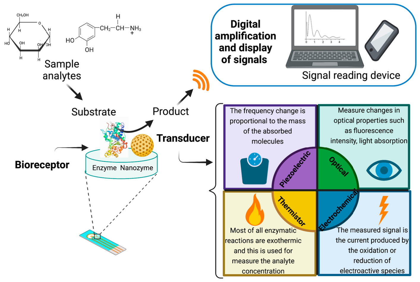
2. Biosensors
3. Enzyme Based Biosensors
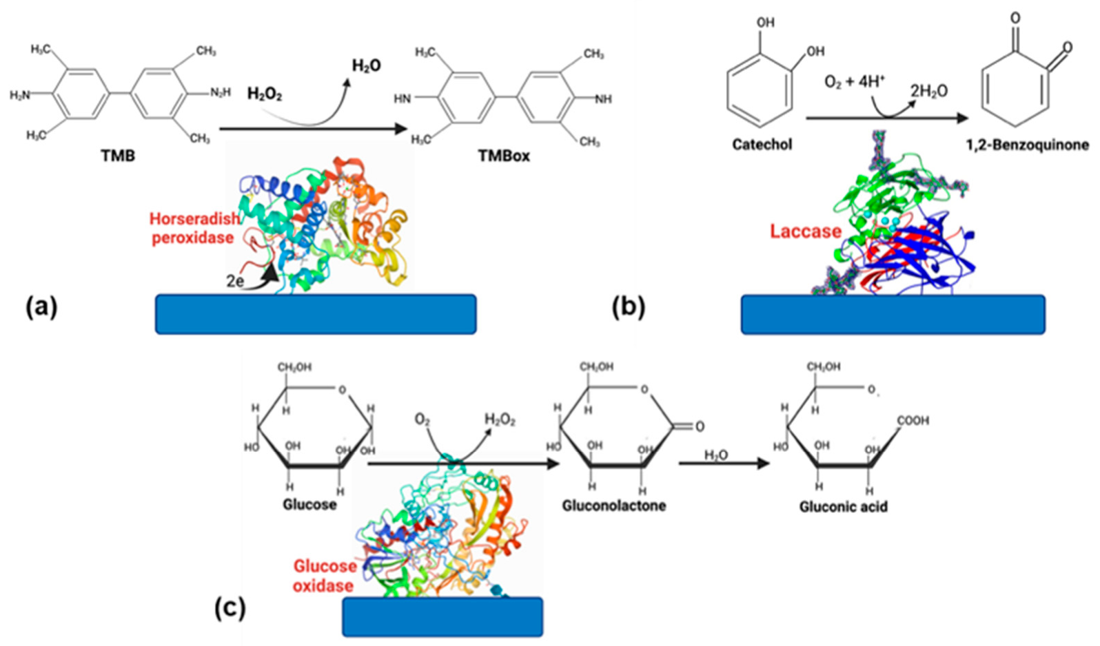
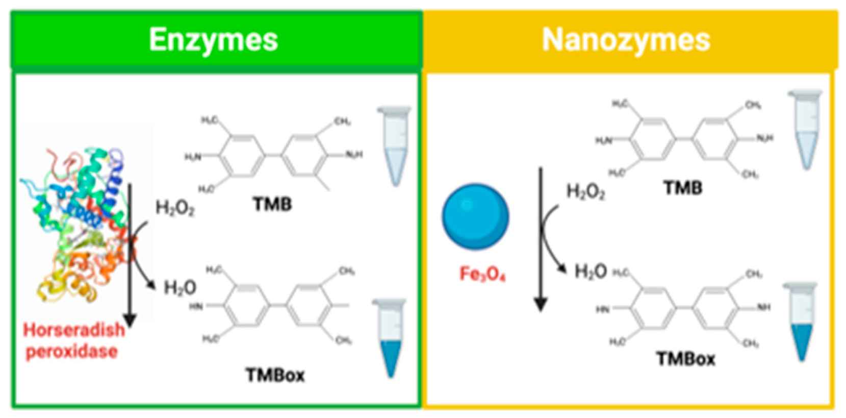
3.1. Horseradish Peroxidase

| Material | Transduction System | Application | Linear Range with a lineal Correlation | Limit of Detection (LOD) | Ref. | |
|---|---|---|---|---|---|---|
| Glass plate covered with fluorine-doped tin oxide (FTO)Copper (I) sulfide (Cu2S) and fluorine-doped tin oxide modified glass slide | Photoelectrochemical | Health | 1,4-dihydroxybenzene (DHB) | 10 nmol L−1 up to 1 mmol L−1 (R = 0.998) | 4.0 nmol L−1 | [36] |
| Encapsulated DNA nanoflowers of magnesium pyrophosphate crystals | Colorimetric | Health | Rapid screening of cancer-derived exosomes | 5.0 × 10³ to 5.0 × 10⁶ particles/μL (R2 = 0.9846) | 3.32 × 10³ particles/μL | [37] |
| Polydimethylsiloxane (PDMS) deposited into a polystyrene tube | Chemiluminescent | Health | Quantification of H2O2 as the oxidizing agent | 0.06−10 μM (R2 = 0.999) | 0.02 μM | [4] |
| Modified multi walled carbon nanotube by γ-aminobutyric acid | Electrochemical | Food, health, environmental | Detection of hydrogen peroxide | 2.0 × 10⁻⁷ M to 2.81 × 10⁻⁴ M (R2 = 0.998) | 0.13 μM | [30] |
| 3D-printed graphene/polylactic (PLA) electrode with gold nanoparticles | Electrochemical | Environmental and biomedical fields. | Hydrogen peroxide detection | 25–100 µM (R = 0.996) | 11.1 µM | [38] |
| HRP-encapsulated protein nanoparticles in an Au electrode surface | Electrochemical | Clinical applications | Hydrogen peroxide detection | 0.01–100 μM | 0.01 µM | [39] |
| Modified platinum electrode covered with poly(4,7-bis(5-bromothiophen-2-yl) benzothiadiazole) | Electrochemical | Health | 17β- estradiol | 0.1 to 200 mM (R2 = 0.99) | 105 nM | [40] |
| Tungsten microwire modified with AuNPs and 3-mercaptopropionic acid | Electrochemical | Health | Determination of hydrogen peroxide | 5 nM to 5 µM (R = 0.999) | 800 pM | [41] |
| Modified acrylic microspheres | Electrochemical | Food | Chilli hotness determination | 0.75–24.94 μM (R2 = 0.992) | 0.39 µM | [42] |
3.2. Glucose Oxidase
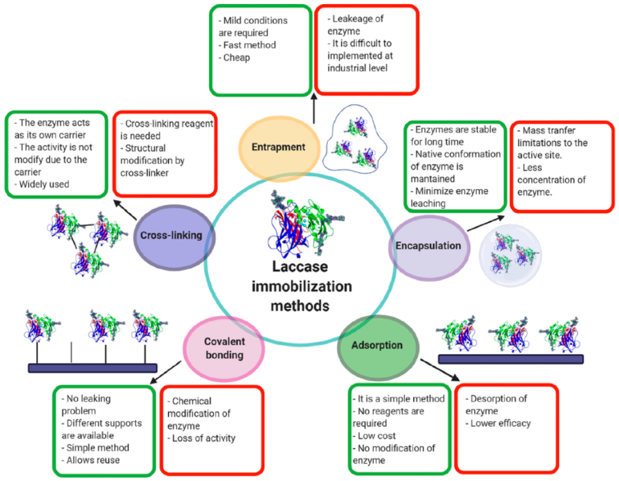
3.3. Laccase
| Material | Transduction System | Application | Linear Range with a Lineal Correlation | Limit of Detection | Ref. | |
|---|---|---|---|---|---|---|
| Laccase hybrid microflowers synthesized with Cu3(PO4)2⋅3H2O | Optical | Health, clinical diagnosis application | Quantification of epinephrine | 1–400 μM (R2 = 0.999) | 0.6 μM | [56] |
| Carbon dots bio functionalized with 3-(aminopropyl)-triethoxysilane | Optical | Health. Clinical diagnosis application. Diagnosis of Alzheimer’s and Parkinson’s diseases. | Detection of dopamine | 0–30 μM (R2 = 0.995) | 41.2 nM | [55] |
| Multi-walled Carbon Nanotubes modified glassy carbon electrode | Electrochemical | Diagnosis of Alzheimer’s and Parkinson’s diseases. | Dopamine detection | 0.1 μmol/dm3 to 10 μmol/dm3 and from 10 µmol/dm3 to 50 µmol/dm3 | 3.63 μA·dm3/μmol and 1.33 μA·dm3/μmol | [10] |
| Fe3O4@SiO2 microspheres stabilized onto glassy carbon electrode | Electrochemical | Health | Dopamine detection | 1.5–75 μmol L−1 (R = 0.9980) | 0.177 μmol L−1 | [57] |
| Glassy carbon electrode layered with multi-walled carbon nanotubes using a film of botryosphaeran | Electrochemical | Health | Dopamine and spironolactone detection | 2.99–38.5 μmol L−1 (R2 = 0.995) | 0.127 μmol L−1 | [54] |
| Carbon paper electrodes with layered two-dimensional molybdenum disulfide (MoS2) in flowers (MoS2-F) and ribbons (MoS2-R) | Electrochemical | Synthetic urine sample | Dopamine detection | 0.1 to 0.5 µM and from 1 to 5 µM (R2 = 0.993) | 10 nM | [58] |
| 6,9-bis(4-hexylthiophen-2-yl)-11H- indeno[2,1-b]quinoxalin-11-one (M1)) polymerized on electrode surface. | Electrochemical | Environmental applications | Catechol in water | 005–0.175 mM (R2 = 0.994) | 9.86 μM | [59] |
| Screen-printed carbon electrodes modified with carboxyl functionalized multi-wallet carbon nanotubes | Electrochemical | Environmental application | Phenolics detection | [14] | ||
3.4. Other Enzymes
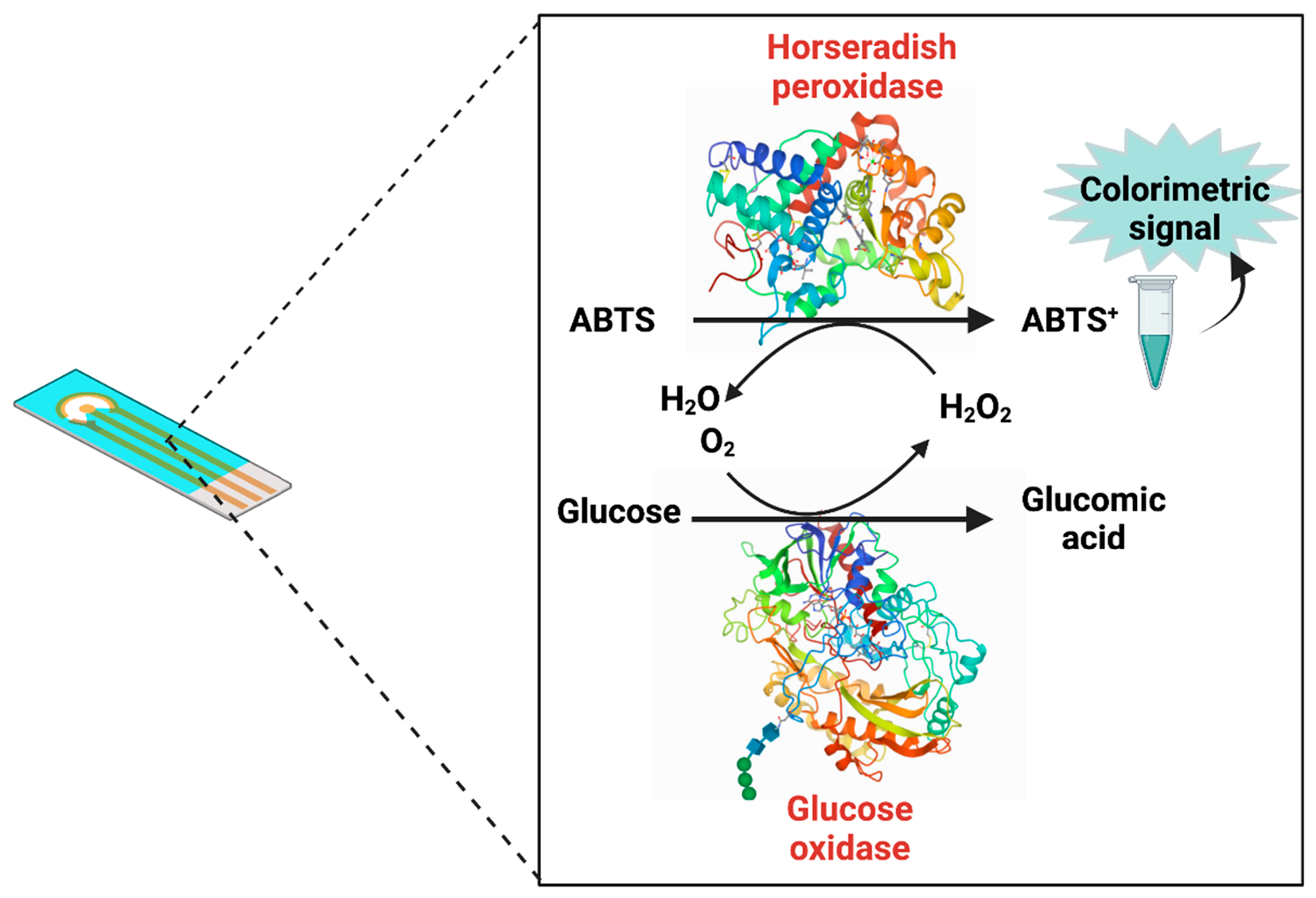
4. Bi-Enzyme Biosensors
| Enzymes | Transduction System | Material | Application | Detection Range with a Linear Correlation | Limit of Detection (LOD) | Ref. |
|---|---|---|---|---|---|---|
| Glucose oxidase and horseradish peroxidase | Electrochemical | Carbon nanotubes modified glassy carbon electrode | Glucose detection | 0.022 to 7.0 mM (R = 0.998) | 7 μM | [77] |
| Glucose oxidase and horseradish peroxidase | Polynoradrenalin/Polyaniline electrode | Glucose | 0.50 μM–0.42 mM | 0.08 μM | [71] | |
| Electrochemical | Cr (III) | 0.01–3.8 µM | ||||
| Cr (VI)) | 0.50–6.0 nM | 0.20 nM | ||||
| HRP and lactate oxidase | Electrochemical | Electrochemical lactate biosensor | Determination of lactate | 30.4 μM −243.9 μM | 22.6 µM | [78] |
| Laccase and tyrosinase | Graphite screen printed electrode modified with ferrocene | Phenol | (R2 = 0.9994) | 2 μM | [79] | |
| Electrochemical | Gallic acid | (R2 = 0.9977) | 50 μM | |||
| Caffeic acid | (R2 = 0.9992) | 24 μM | ||||
| Catechin | (R2 = 0.9930) | 40 μM | ||||
| Alcohol oxidase and horseradish peroxidase | Electrochemical | Carbon nanotube matrix | Methyl salicylate determination in plants | 22.95 μM and 0.98 μM | [80] | |
| D-amino acid oxidase and horseradish peroxidase | Electrochemical | Multi-walled carbon nanotubes and gold nanoparticles modified screen-printed electrode | The total content of D-amino acids | 0.020 to 2.0 mM (R = 0.994) | 18 μm | [81] |
| d-amino acid oxidase and hemoglobin | Electrochemical | MnO2 nanoparticles enriched poly thiophene | Dopamine | 0.04–9.0 μM (R2 = 0.994) | 12.801 μA/μM and 41 nM | [82] |
| Cholesterol oxidase and horseradish peroxidase | Electrochemical | Poly(thionine)-modified glassy carbon electrode | Cholesterol | 25–125 μM (R = 0.99) | 6.3 μM | [83] |
| Acetylcholinesterase and choline oxidase | Optical | Gold nanorods | Dichlorvos | 0.1 to 500 μg/L (R2 = 0.963) | 8.1 × 10⁻³ μg/L | [84] |
| Demeton | 1to 500 μg/L (R2 = 0.963) | 0.32 μg/L | ||||
| Glucose oxidase and lactate oxidase | Electrochemical | Flexible electrode array with gold nanoparticles and Prussian blue | Glucose and lactate detection | 60 μM-1000 μM (glucose) 5 mM–20 mM (lactate) | [74] | |
| Urease and penicillinase | Electrochemical | Ta2O5 | Urea and penicillin detection | 1 mM–25 mM (urea) 0.1 mM–5 mM (penicillin) | [85] |
5. Nanozyme Biosensors
5.1. Peroxidase-like Activity
5.2. Oxidase-like Activity
5.3. Laccase-like Activity
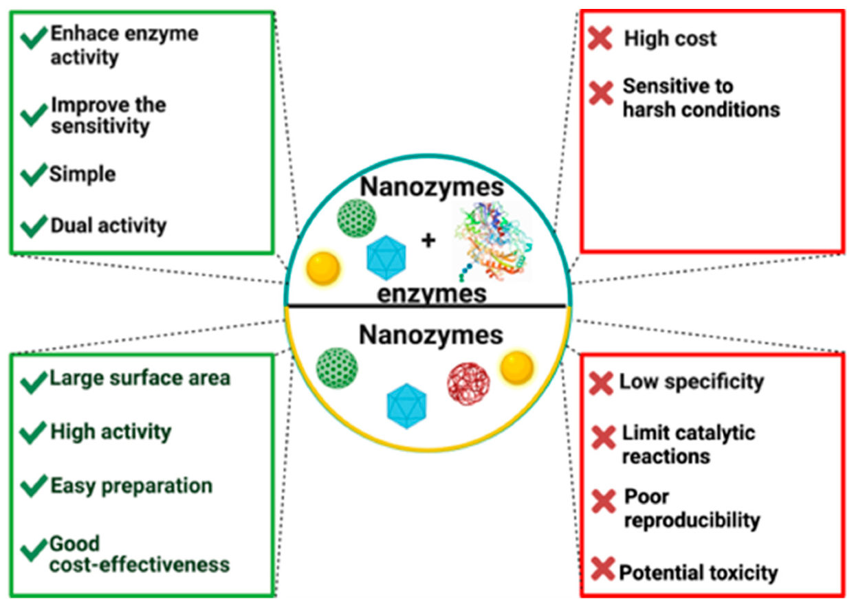
6. Nanozymes-Enzymes Pool
| Enzyme-like Activity | Nanozyme | Enzyme | Transduction System | Application | Range | Limit of Detection | Ref. |
|---|---|---|---|---|---|---|---|
| Peroxidase-like activity | Carbon microfibers modified by hemin and gold nanoparticles | Alcohol oxidase and glucose oxidase | Electrochemical | Detection of ethanol | 0.01–0.15 mM | 0.005 mM | [121] |
| Detection of glucose | 0.1–0.9 mM | 0.05 mM | |||||
| Ceria nanomaterials | Glucose oxidase | Optical | Detection of H2O2 | 10 μM–50 mM | 2 μM | [122] | |
| Mn (II)/CeO2 nanorods nanocomposites | Detection of glucose | 10 μM–100 mM | 8.6 μM | ||||
| Silver nanoparticles decorated on reduced graphene oxide sheets (AgNPs@rGO) nanocomposite | Glucose oxidase | Optical | Colorimetric glucose biosensor colorimetric glucose biosensor | 125 μM to 1 mM | 40 μM | [119] | |
| Graphene oxide | Acetylcholinesterase and choline oxidase | Optical | Colorimetric detection of organophosphourus pesticides | 1–200 ng/mL | 2 ppb | [118] | |
| Peroxidase-like activity | Cobalt oxide supported ordered mesoporous carbon (CoO-OMC) | Glucose oxidase | Optical | Colorimetric detection of glucose | 0.1–5.0 mM | 68 μM | [120] |
| Bimetallic PtRu nanoparticles (nPtRu) | Alcohol oxidase and methylamine oxidase | Electrochemical | Food analysis ethanol detection | 25–200 µM | 3 µM | [123] | |
| Methylamine detection | 20–600 µM | 2.5 µM | |||||
| Metallic cobalt nanoparticles encapsulated in metal–organic frameworks derived carbon | Glucose oxidase | Optical | Colorimetric detection of glucose | 0.25 to 30 μM | 156 nM | [104] | |
| Prussian Blue | Lactate oxidase | Electrochemical | Detection of lactate | [124] | |||
| Peroxidase-like activity | Au nanoparticle/polyluminol | Glucose oxidase | Optical | Detection of glucose | 10–1000 μM | 10 μM | [125] |
| Pt-Ru nanozymes | Glucose oxidase | Optical | Colorimetric and fluorometric glucose detection | 0.25–3.0 mM | 0.988 and 138 μM | [108] |
7. Conclusions and Future Perspectives
Author Contributions
Funding
Acknowledgments
Conflicts of Interest
References
- Ali, J.; Najeeb, J.; Ali, M.A.; Aslam, M.F.; Raza, A. Biosensors: Their Fundamentals, Designs, Types and Most Recent Impactful Applications: A Review. J. Biosens. Bioelectron. 2017, 8, 1–9. [Google Scholar] [CrossRef]
- Meshram, B.; Agrawal, A.; Adil, S.; Ranvir, S.; Sande, K. Biosensor and its Application in Food and Dairy Industry: A Review. Int. J. Curr. Microbiol. Appl. Sci. 2018, 7, 3305–3324. [Google Scholar] [CrossRef]
- Bhalinge, P.; Kumar, S.; Jadhav, A.; Suman, S.; Gujjar, P.; Perla, N. Biosensors: Nanotools of Detection—A Review. Int. J. Healthc. Biomed. Res. 2016, 04, 26–39. [Google Scholar]
- Bocanegra-Rodríguez, S.; Jornet-Martínez, N.; Molins-Legua, C.; Campíns-Falcó, P. New Reusable Solid Biosensor with Covalent Immobilization of the Horseradish Peroxidase Enzyme: In Situ Liberation Studies of Hydrogen Peroxide by Portable Chemiluminescent Determination. ACS Omega 2020, 5, 2419–2427. [Google Scholar] [CrossRef] [Green Version]
- Kaya, H.O.; Cetin, A.E.; Azimzadeh, M.; Topkaya, S.N. Pathogen detection with electrochemical biosensors: Advantages, challenges and future perspectives. J. Electroanal. Chem. 2021, 882, 114989. [Google Scholar] [CrossRef]
- Alizadeh, N.; Ghasemi, S.; Salimi, A.; Sham, T.-K.; Hallaj, R. CuO nanorods as a laccase mimicking enzyme for highly sensitive colorimetric and electrochemical dual biosensor: Application in living cell epinephrine analysis. Colloids Surf. B Biointerfaces 2020, 195, 111228. [Google Scholar] [CrossRef]
- Azad, T.; Singaravelu, R.; Brown, E.E.; Taha, Z.; Rezaei, R.; Arulanandam, R.; Boulton, S.; Diallo, J.-S.; Ilkow, C.S.; Bell, J.C. SARS-CoV-2 S1 NanoBiT: A nanoluciferase complementation-based biosensor to rapidly probe SARS-CoV-2 receptor recognition. Biosens. Bioelectron. 2021, 180, 113122. [Google Scholar] [CrossRef]
- Koushki, E.; Mirzaei Mohammadabadi, F.; Baedi, J.; Ghasedi, A. The effects of glucose and glucose oxidase on the Uv-vis spectrum of gold nanoparticles: A study on optical biosensor for saliva glucose monitoring. Photodiagn. Photodyn. Ther. 2020, 30, 101771. [Google Scholar] [CrossRef]
- Liu, Y.; Nan, X.; Shi, W.; Liu, X.; He, Z.; Sun, Y.; Ge, D. A glucose biosensor based on the immobilization of glucose oxidase and Au nanocomposites with polynorepinephrine. RSC Adv. 2019, 9, 16439–16446. [Google Scholar] [CrossRef] [Green Version]
- De Oliveira Wardak, C.; Paczosa-Bator, B.; Malinowski, S. Application of cold plasma corona discharge in preparation of laccase-based biosensors for dopamine determination. Mater. Sci. Eng. C 2020, 116, 111199. [Google Scholar] [CrossRef]
- Neto, J.O.; Rezende, S.G.; Lobón, G.S.; Garcia, T.A.; Macêdo, I.; Garcia, L.F.; Alves, V.F.; Torres, I.M.S.; Santiago, M.; Schimidt, F.; et al. Electroanalysis and laccase-based biosensor on the determination of phenolic content and antioxidant power of honey samples. Food Chem. 2017, 237, 1118–1123. [Google Scholar] [CrossRef]
- Piroozmand, F.; Mohammadipanah, F.; Faridbod, F. Emerging biosensors in detection of natural products. Synth. Syst. Biotechnol. 2020, 5, 293–303. [Google Scholar] [CrossRef]
- Rodríguez-Delgado, M.M.; Alemán-Nava, G.S.; Rodriguez-Delgado, J.M.; Dieck-Assad, G.; Martinez-Chapa, S.O.; Barceló, D.; Parra, R. Laccase-based biosensors for detection of phenolic compounds. TrAC Trends Anal. Chem. 2015, 74, 21–45. [Google Scholar] [CrossRef] [Green Version]
- Othman, A.M.; Wollenberger, U. Amperometric biosensor based on coupling aminated laccase to functionalized carbon nanotubes for phenolics detection. Int. J. Biol. Macromol. 2020, 153, 855–864. [Google Scholar] [CrossRef]
- Zhao, K.; Veksha, A.; Ge, L.; Lisak, G. Near real-time analysis of para-cresol in wastewater with a laccase-carbon nanotube-based biosensor. Chemosphere 2020, 269, 128699. [Google Scholar] [CrossRef]
- Temoçin, Z. Designing of a stable and selective glucose biosensor by glucose oxidase immobilization on glassy carbon electrode sensitive to H2O2 via nanofiber interface. J. Appl. Electrochem. 2021, 51, 283–293. [Google Scholar] [CrossRef]
- Stasyuk, N.; Smutok, O.; Demkiv, O.; Prokopiv, T.; Gayda, G.; Nisnevitch, M.; Gonchar, M. Synthesis, Catalytic Properties and Application in Biosensorics of Nanozymes and Electronanocatalysts: A Review. Sensors 2020, 20, 4509. [Google Scholar] [CrossRef]
- Perumal, V.; Hashim, U. Advances in biosensors: Principle, architecture and applications. J. Appl. Biomed. 2014, 12, 1–15. [Google Scholar] [CrossRef]
- Nagraik, R.; Sharma, A.; Kumar, D.; Mukherjee, S.; Sen, F.; Kumar, A.P. Amalgamation of biosensors and nanotechnology in disease diagnosis: Mini-review. Sens. Int. 2021, 2, 100089. [Google Scholar] [CrossRef]
- AlHamoud, Y.; Yang, D.; Kenston, S.S.F.; Liu, G.; Liu, L.; Zhou, H.; Ahmed, F.; Zhao, J. Advances in biosensors for the detection of ochratoxin A: Bio-receptors, nanomaterials, and their applications. Biosens. Bioelectron. 2019, 141, 111418. [Google Scholar] [CrossRef]
- Nguyen, H.H.; Lee, S.H.; Lee, U.J.; Fermin, C.D.; Kim, M. Immobilized Enzymes in Biosensor Applications. Materials 2019, 12, 121. [Google Scholar] [CrossRef] [PubMed] [Green Version]
- Clark, L.C.; Lyons, C. Electrode Systems for Continuous Monitoring in Cardiovascular Surgery. Ann. N. Y. Acad. Sci. 1962, 102, 29–45. [Google Scholar] [CrossRef]
- Wang, T.; Fan, X.; Li, R.; Xu, J.; Liu, J. Multi-Enzyme-Synergetic ultrathin protein nanosheets display high efficient and switch on/off antibacterial activities. Chem. Eng. J. 2021, 416, 129082. [Google Scholar] [CrossRef]
- De Jesús Rostro-Alanis, M.; Mancera-Andrade, E.I.; Patiño, M.B.G.; Arrieta-Baez, D.; Cardenas, B.; Martinez-Chapa, S.O.; Saldívar, R.P. Nanobiocatalysis: Nanostructured materials—A minireview. Biocatalysis 2016, 2, 1–24. [Google Scholar] [CrossRef] [Green Version]
- Ahangari, H.; Kurbanoglu, S.; Ehsani, A.; Uslu, B. Latest trends for biogenic amines detection in foods: Enzymatic biosensors and nanozymes applications. Trends Food Sci. Technol. 2021, 112, 75–87. [Google Scholar] [CrossRef]
- Pérez, J.A.C.; Sosa-Hernández, J.E.; Hussain, S.M.; Bilal, M.; Parra, R.; Iqbal, H.M. Bioinspired biomaterials and enzyme-based biosensors for point-of-care applications with reference to cancer and bio-imaging. Biocatal. Agric. Biotechnol. 2019, 17, 168–176. [Google Scholar] [CrossRef]
- Gul, I.; Ahmad, M.S.; Naqvi, S.M.S.; Hussain, A.; Wali, R.; Farooqi, A.A.; Ahmed, I. Polyphenol oxidase (PPO) based biosensors for detection of phenolic compounds: A Review. J. Appl. Biol. Biotechnol. 2017, 5, 72–85. [Google Scholar] [CrossRef]
- Gonzalez-Coronel, L.A.; Cobas, M.; Rostro-Alanis, M.D.J.; Parra-Saldívar, R.; Hernandez-Luna, C.E.; Pazos, M.; Sanromán, M. Immobilization of laccase of Pycnoporus sanguineus CS43. New Biotechnol. 2017, 39, 141–149. [Google Scholar] [CrossRef]
- Alvarado-Ramírez, L.; Rostro-Alanis, M.; Rodríguez-Rodríguez, J.; Castillo-Zacarías, C.; Sosa-Hernández, J.E.; Barceló, D.; Iqbal, H.M.; Parra-Saldívar, R. Exploring current tendencies in techniques and materials for immobilization of laccases—A review. Int. J. Biol. Macromol. 2021, 181, 683–696. [Google Scholar] [CrossRef]
- Feizabadi, M.; Soleymanpour, A.; Faridnouri, H.; Ajloo, D. Improving stability of biosensor based on covalent immobilization of horseradish peroxidase by γ-aminobutyric acid and application in detection of H2O2. Int. J. Biol. Macromol. 2019, 136, 597–606. [Google Scholar] [CrossRef]
- Cock, L.S.; Arenas, A.M.Z.; Aponte, A.A. Use of Enzymatic Biosensors as Quality Indices: A Synopsis of Present and Future Trends in The Food Industry. Chil. J. Agric. Res. 2009, 69, 270–280. [Google Scholar] [CrossRef] [Green Version]
- Bilal, M.; Rasheed, T.; Zhao, Y.; Iqbal, H.M. Agarose-chitosan hydrogel-immobilized horseradish peroxidase with sustainable bio-catalytic and dye degradation properties. Int. J. Biol. Macromol. 2019, 124, 742–749. [Google Scholar] [CrossRef] [PubMed]
- Vineh, M.B.; Saboury, A.A.; Poostchi, A.A.; Rashidi, A.; Parivar, K. Stability and activity improvement of horseradish peroxidase by covalent immobilization on functionalized reduced graphene oxide and biodegradation of high phenol concentration. Int. J. Biol. Macromol. 2018, 106, 1314–1322. [Google Scholar] [CrossRef] [PubMed]
- De Oliveira, F.K.; Santos, L.O.; Buffon, J.G. Mechanism of action, sources, and application of peroxidases. Food Res. Int. 2021, 143, 110266. [Google Scholar] [CrossRef]
- Ahammad, A.J.S. Hydrogen Peroxide Biosensors Based on Horseradish Peroxidase and Hemoglobin. J. Biosens. Bioelectron. 2012, S9, 001. [Google Scholar] [CrossRef] [Green Version]
- Da Silva Freires, A.; Botelho, C.N.; Silva, S.M.; Goulart, M.O.F.; Damos, F.S.; Luz, R.D.C.S. Photoelectrochemical biosensor for 1,4-dihydroxybenzene based on copper sulfide and horseradish peroxidase enzyme: Application in skin cream samples. Microchem. J. 2020, 159, 105487. [Google Scholar] [CrossRef]
- Zeng, R.; Wang, J.; Wang, Q.; Tang, D.; Lin, Y. Horseradish peroxidase-encapsulated DNA nanoflowers: An innovative signal-generation tag for colorimetric biosensor. Talanta 2021, 221, 121600. [Google Scholar] [CrossRef]
- Marzo, A.M.L.; Mayorga-Martinez, C.C.; Pumera, M. 3D-printed graphene direct electron transfer enzyme biosensors. Biosens. Bioelectron. 2020, 151, 111980. [Google Scholar] [CrossRef]
- Shin, J.-H.; Lee, M.-J.; Choi, J.-H.; Song, J.-A.; Kim, T.-H.; Oh, B.-K. Electrochemical H2O2 biosensor based on horseradish peroxidase encapsulated protein nanoparticles with reduced graphene oxide-modified gold electrode. Nano Converg. 2020, 7, 39. [Google Scholar] [CrossRef]
- Spychalska, K.; Zając, D.; Cabaj, J. Electrochemical biosensor for detection of 17β-estradiol using semi-conducting polymer and horseradish peroxidase. RSC Adv. 2020, 10, 9079–9087. [Google Scholar] [CrossRef] [Green Version]
- Narayanan, J.S.; Slaughter, G. AuNPs-HRP microneedle biosensor for ultrasensitive detection of hydrogen peroxide for organ preservation. Med. Devices Sens. 2018, 1, e10015. [Google Scholar] [CrossRef]
- Mohammad, R.; Ahmad, M.; Heng, L.Y. Amperometric capsaicin biosensor based on covalent immobilization of horseradish peroxidase (HRP) on acrylic microspheres for chilli hotness determination. Sens. Actuators B Chem. 2017, 241, 174–181. [Google Scholar] [CrossRef]
- Lee, I.; Probst, D.; Klonoff, D.; Sode, K. Continuous glucose monitoring systems—Current status and future perspectives of the flagship technologies in biosensor research. Biosens. Bioelectron. 2021, 181, 113054. [Google Scholar] [CrossRef]
- Bagyalakshmi, S.; Sivakami, A.; Balamurugan, K. A Zno nanorods based enzymatic glucose biosensor by immobilization of glucose oxidase on a chitosan film. Obes. Med. 2020, 18, 100229. [Google Scholar] [CrossRef]
- Newman, J.D.; Turner, A. Home blood glucose biosensors: A commercial perspective. Biosens. Bioelectron. 2005, 20, 2435–2453. [Google Scholar] [CrossRef] [Green Version]
- Jayakumar, K.; Bennett, R.; Leech, D. Electrochemical glucose biosensor based on an osmium redox polymer and glucose oxidase grafted to carbon nanotubes: A design-of-experiments optimisation of current density and stability. Electrochim. Acta 2021, 371, 137845. [Google Scholar] [CrossRef]
- Jiménez-Fiérrez, F.; González-Sánchez, M.I.; Jiménez-Pérez, R.; Iniesta, J.; Valero, E. Glucose Biosensor Based on Disposable Activated Carbon Electrodes Modified with Platinum Nanoparticles Electrodeposited on Poly(Azure A). Sensors 2020, 20, 4489. [Google Scholar] [CrossRef]
- Barrios-Estrada, C.; de Jesús Rostro-Alanis, M.; Muñoz-Gutiérrez, B.D.; Iqbal, H.M.; Kannan, S.; Parra-Saldívar, R. Emergent contaminants: Endocrine disruptors and their laccase-assisted degradation—A review. Sci. Total Environ. 2018, 612, 1516–1531. [Google Scholar] [CrossRef]
- Koyappayil, A.; Kim, H.T.; Lee, M.-H. ‘Laccase-like’ properties of coral-like silver citrate micro-structures for the degradation and determination of phenolic pollutants and adrenaline. J. Hazard. Mater. 2021, 412, 125211. [Google Scholar] [CrossRef]
- Zhou, W.; Zhang, W.; Cai, Y. Laccase immobilization for water purification: A comprehensive review. Chem. Eng. J. 2020, 403, 126272. [Google Scholar] [CrossRef]
- Orlikowska, M.; Rostro-Alanis, M.D.J.; Bujacz, A.; Hernández-Luna, C.; Rubio, R.; Parra, R.; Bujacz, G. Structural studies of two thermostable laccases from the white-rot fungus Pycnoporus sanguineus. Int. J. Biol. Macromol. 2018, 107, 1629–1640. [Google Scholar] [CrossRef]
- Bilal, M.; Jing, Z.; Zhao, Y.; Iqbal, H.M. Immobilization of fungal laccase on glutaraldehyde cross-linked chitosan beads and its bio-catalytic potential to degrade bisphenol A. Biocatal. Agric. Biotechnol. 2019, 19, 101174. [Google Scholar] [CrossRef]
- Jarosz-Wilkolazka, A.; Ruzgas, T.; Gorton, L. Amperometric detection of mono- and diphenols at Cerrena unicolor laccase-modified graphite electrode: Correlation between sensitivity and substrate structure. Talanta 2005, 66, 1219–1224. [Google Scholar] [CrossRef]
- Coelho, J.H.; Eisele, A.; Valezi, C.F.; Mattos, G.; Schirmann, J.G.; Dekker, R.F.; Barbosa-Dekker, A.M.; Sartori, E.R. Exploring the exocellular fungal biopolymer botryosphaeran for laccase-biosensor architecture and application to determine dopamine and spironolactone. Talanta 2019, 204, 475–483. [Google Scholar] [CrossRef]
- Sangubotla, R.; Kim, J. Fiber-optic biosensor based on the laccase immobilization on silica-functionalized fluorescent carbon dots for the detection of dopamine and multi-color imaging applications in neuroblastoma cells. Mater. Sci. Eng. C 2021, 122, 111916. [Google Scholar] [CrossRef]
- Zhang, M.; Zhang, Y.; Yang, C.; Ma, C.; Tang, J. A smartphone-assisted portable biosensor using laccase-mineral hybrid microflowers for colorimetric determination of epinephrine. Talanta 2021, 224, 121840. [Google Scholar] [CrossRef]
- Li, Z.; Zheng, Y.; Gao, T.; Liu, Z.; Zhang, J.; Zhou, G. Fabrication of biosensor based on core–shell and large void structured magnetic mesoporous microspheres immobilized with laccase for dopamine detection. J. Mater. Sci. 2018, 53, 7996–8008. [Google Scholar] [CrossRef]
- Rubio-Govea, R.; Hickey, D.P.; García-Morales, R.; Rodriguez-Delgado, M.; Domínguez-Rovira, M.A.; Minteer, S.D.; Ornelas-Soto, N.; García-García, A. MoS2 nanostructured materials for electrode modification in the development of a laccase based amperometric biosensor for non-invasive dopamine detection. Microchem. J. 2020, 155, 104792. [Google Scholar] [CrossRef]
- Cevher, C.; Bekmezci, S.A.; Soylemez, S.; Udum, Y.A.; Toppare, L.; Çırpan, A. Indenoquinoxalinone based conjugated polymer substrate for laccase biosensor. Mater. Chem. Phys. 2021, 257, 123788. [Google Scholar] [CrossRef]
- Bounegru, A.; Apetrei, C. Laccase and Tyrosinase Biosensors Used in the Determination of Hydroxycinnamic Acids. Int. J. Mol. Sci. 2021, 22, 4811. [Google Scholar] [CrossRef]
- García-Guzmán, J.J.; López-Iglesias, D.; Cubillana-Aguilera, L.; Lete, C.; Lupu, S.; Palacios-Santander, J.M.; Bellido-Milla, D. Assessment of the Polyphenol Indices and Antioxidant Capacity for Beers and Wines Using a Tyrosinase-Based Biosensor Prepared by Sinusoidal Current Method. Sensors 2019, 19, 66. [Google Scholar] [CrossRef] [PubMed] [Green Version]
- Wu, L.; Lu, X.; Niu, K.; Dhanjai; Chen, J. Tyrosinase nanocapsule based nano-biosensor for ultrasensitive and rapid detection of bisphenol A with excellent stability in different application scenarios. Biosens. Bioelectron. 2020, 165, 112407. [Google Scholar] [CrossRef] [PubMed]
- Shaban, S.M.; Moon, B.-S.; Pyun, D.-G.; Kim, D.-H. A colorimetric alkaline phosphatase biosensor based on p-aminophenol-mediated growth of silver nanoparticles. Colloids Surf. B Biointerfaces 2021, 205, 111835. [Google Scholar] [CrossRef] [PubMed]
- e Silva, T.S.; Soares, I.P.; Lacerda, L.R.G.; Cordeiro, T.A.R.; Ferreira, L.; Franco, D.L. Electrochemical modification of electrodes with polymers derived from of hydroxybenzoic acid isomers: Optimized platforms for an alkaline phosphatase biosensor for pesticide detection. Mater. Chem. Phys. 2020, 252, 123221. [Google Scholar] [CrossRef]
- Kim, J.Y.; Sung, G.Y.; Park, M. Efficient Portable Urea Biosensor Based on Urease Immobilized Membrane for Monitoring of Physiological Fluids. Biomedicines 2020, 8, 596. [Google Scholar] [CrossRef]
- Wang, Z.; Ma, B.; Shen, C.; Cheong, L.-Z. Direct, selective and ultrasensitive electrochemical biosensing of methyl parathion in vegetables using Burkholderia cepacia lipase@MOF nanofibers-based biosensor. Talanta 2019, 197, 356–362. [Google Scholar] [CrossRef]
- Hasanah, U.; Sani, N.D.M.; Heng, L.Y.; Idroes, R.; Safitri, E. Construction of a Hydrogel Pectin-Based Triglyceride Optical Biosensor with Immobilized Lipase Enzymes. Biosensors 2019, 9, 135. [Google Scholar] [CrossRef] [Green Version]
- Mercante, L.; Iwaki, L.; Scagion, V.; Oliveira, O.; Mattoso, L.; Correa, D. Electrochemical Detection of Bisphenol A by Tyrosinase Immobilized on Electrospun Nanofibers Decorated with Gold Nanoparticles. Electrochem 2021, 2, 41–49. [Google Scholar] [CrossRef]
- Nedellec, Y.; Gondran, C.; Gorgy, K.; Mc Murtry, S.; Agostini, P.; Elmazria, O.; Cosnier, S. Microcapsule-based biosensor containing catechol for the reagent-free inhibitive detection of benzoic acid by tyrosinase. Biosens. Bioelectron. 2021, 180, 113137. [Google Scholar] [CrossRef]
- Mirzaei, F.; Mirzaei, M.; Torkzadeh-Mahani, M. A hydrophobin-based-biosensor layered by an immobilized lactate dehydrogenase enzyme for electrochemical determination of pyruvate. Bioelectrochemistry 2019, 130, 107323. [Google Scholar] [CrossRef]
- Liu, L.; Chen, C.; Chen, C.; Kang, X.; Zhang, H.; Tao, Y.; Xie, Q.; Yao, S. Poly(noradrenalin) based bi-enzyme biosensor for ultrasensitive multi-analyte determination. Talanta 2019, 194, 343–349. [Google Scholar] [CrossRef]
- Arana-Peña, S.; Carballares, D.; Morellon-Sterlling, R.; Berenguer-Murcia, Á.; Alcántara, A.R.; Rodrigues, R.C.; Fernandez-Lafuente, R. Enzyme co-immobilization: Always the biocatalyst designers’ choice…or not? Biotechnol. Adv. 2020, 51, 107584. [Google Scholar] [CrossRef]
- Xu, K.; Chen, X.; Zheng, R.; Zheng, Y. Immobilization of Multi-Enzymes on Support Materials for Efficient Biocatalysis. Front. Bioeng. Biotechnol. 2020, 8, 660. [Google Scholar] [CrossRef]
- Yokus, M.A.; Songkakul, T.; Pozdin, V.A.; Bozkurt, A.; Daniele, M.A. Wearable multiplexed biosensor system toward continuous monitoring of metabolites. Biosens. Bioelectron. 2020, 153, 112038. [Google Scholar] [CrossRef]
- Ibadullaeva, S.Z.; Appazov, N.; Tarahovsky, Y.S.; Zamyatina, E.A.; Fomkina, M.G.; Kim, Y.A. Amperometric Multi-Enzyme Biosensors: Development and Application, A Short Review. Biophysics 2019, 64, 696–707. [Google Scholar] [CrossRef]
- Xu, S.; Qi, H.; Zhou, S.; Zhang, X.; Zhang, C. Mediatorless amperometric bienzyme glucose biosensor based on horseradish peroxidase and glucose oxidase cross-linked to multiwall carbon nanotubes. Microchim. Acta 2014, 181, 535–541. [Google Scholar] [CrossRef]
- Yang, H.; Gong, C.; Miao, L.; Xu, F. A Glucose Biosensor based on Horseradish Peroxidase and Glucose Oxidase Co-entrapped in Carbon Nanotubes Modified Electrode. Int. J. Electrochem. Sci. 2017, 12, 4958–4969. [Google Scholar] [CrossRef]
- Hernández-Ibáñez, N.; García-Cruz, L.; Montiel, V.; Foster, C.; Banks, C.; Iniesta, J. Electrochemical lactate biosensor based upon chitosan/carbon nanotubes modified screen-printed graphite electrodes for the determination of lactate in embryonic cell cultures. Biosens. Bioelectron. 2016, 77, 1168–1174. [Google Scholar] [CrossRef] [Green Version]
- Montereali, M.; Della Seta, L.; Vastarella, W.; Pilloton, R. A disposable Laccase–Tyrosinase based biosensor for amperometric detection of phenolic compounds in must and wine. J. Mol. Catal. B Enzym. 2010, 64, 189–194. [Google Scholar] [CrossRef]
- Fang, Y.; Umasankar, Y.; Ramasamy, R.P. A novel bi-enzyme electrochemical biosensor for selective and sensitive determination of methyl salicylate. Biosens. Bioelectron. 2016, 81, 39–45. [Google Scholar] [CrossRef] [Green Version]
- Moreno-Guzmán, M.; García-Carmona, L.; Molinero-Fernández, Á.; Cava, F.; Gil, M.L.; Escarpa, A. Bi-enzymatic biosensor for on-site, fast and reliable electrochemical detection of relevant D-amino acids in bacterial samples. Sens. Actuators B Chem. 2017, 242, 95–101. [Google Scholar] [CrossRef]
- Shoja, Y.; Rafati, A.A.; Ghodsi, J. Polythiophene supported MnO2 nanoparticles as nano-stabilizer for simultaneously electrostatically immobilization of d-amino acid oxidase and hemoglobin as efficient bio-nanocomposite in fabrication of dopamine bi-enzyme biosensor. Mater. Sci. Eng. C 2017, 76, 637–645. [Google Scholar] [CrossRef]
- Rahman, M.; Li, X.-B.; Kim, J.; Lim, B.O.; Ahammad, A.S.; Lee, J.-J. A cholesterol biosensor based on a bi-enzyme immobilized on conducting poly(thionine) film. Sens. Actuators B Chem. 2014, 202, 536–542. [Google Scholar] [CrossRef]
- Liu, Y.; Lv, B.; Liu, A.; Liang, G.; Yin, L.; Pu, Y.; Wei, W.; Gou, S.; Liu, S. Multicolor sensor for organophosphorus pesticides determination based on the bi-enzyme catalytic etching of gold nanorods. Sens. Actuators B Chem. 2018, 265, 675–681. [Google Scholar] [CrossRef]
- Molinnus, D.; Beging, S.; Lowis, C.; Schöning, M.J. Towards a Multi-Enzyme Capacitive Field-Effect Biosensor by Comparative Study of Drop-Coating and Nano-Spotting Technique. Sensors 2020, 20, 4924. [Google Scholar] [CrossRef] [PubMed]
- Castillo, N.E.T.; Melchor-Martínez, E.M.; Sierra, J.S.O.; Ramírez-Torres, N.M.; Sosa-Hernández, J.E.; Iqbal, H.M.; Parra-Saldívar, R. Enzyme mimics in-focus: Redefining the catalytic attributes of artificial enzymes for renewable energy production. Int. J. Biol. Macromol. 2021, 179, 80–89. [Google Scholar] [CrossRef]
- Wong, E.L.S.; Vuong, K.Q.; Chow, E. Nanozymes for Environmental Pollutant Monitoring and Remediation. Sensors 2021, 21, 408. [Google Scholar] [CrossRef]
- Zhang, X.; Lin, S.; Liu, S.; Tan, X.; Dai, Y.; Xia, F. Advances in organometallic/organic nanozymes and their applications. Coord. Chem. Rev. 2021, 429, 213652. [Google Scholar] [CrossRef]
- Huang, H.; Lei, L.; Bai, J.; Zhang, L.; Song, D.; Zhao, J.; Li, J.; Li, Y. Efficient elimination and detection of phenolic compounds in juice using laccase mimicking nanozymes. Chin. J. Chem. Eng. 2021, 29, 167–175. [Google Scholar] [CrossRef]
- Christus, A.A.B.; Ravikumar, A.; Panneerselvam, P.; Radhakrishnan, K. A novel Hg(II) sensor based on Fe3O4@ZnO nanocomposite as peroxidase mimics. Appl. Surf. Sci. 2018, 449, 669–676. [Google Scholar] [CrossRef]
- Lu, W.; Zhang, J.; Li, N.; You, Z.; Feng, Z.; Natarajan, V.; Chen, J.; Zhan, J. Co3O4@β-cyclodextrin with synergistic peroxidase-mimicking performance as a signal magnification approach for colorimetric determination of ascorbic acid. Sens. Actuators B Chem. 2020, 303, 127106. [Google Scholar] [CrossRef]
- Hou, P.; Ju, P.; Hao, L.; Chen, C.; Jiang, F.; Ding, H.; Sun, C. Colorimetric determination of hydrogen peroxide based on the robust peroxidase-like activities of flower-like YVO4 microstructures. Colloids Surfaces A: Physicochem. Eng. Asp. 2021, 618, 126427. [Google Scholar] [CrossRef]
- Niu, X.; He, Y.; Li, X.; Zhao, H.; Pan, J.; Qiu, F.; Lan, M. A peroxidase-mimicking nanosensor with Hg2+-triggered enzymatic activity of cysteine-decorated ferromagnetic particles for ultrasensitive Hg2+ detection in environmental and biological fluids. Sens. Actuators B Chem. 2019, 281, 445–452. [Google Scholar] [CrossRef]
- Xia, F.; Shi, Q.; Nan, Z. Improvement of peroxidase-like activity and application for detection of H2O2 and dopamine for SDBS-Cu-CuFe2O4. Surf. Interfaces 2021, 24, 101109. [Google Scholar] [CrossRef]
- He, S.-B.; Yang, L.; Lin, X.-L.; Chen, L.-M.; Peng, H.-P.; Deng, H.-H.; Xia, X.-H.; Chen, W. Heparin-platinum nanozymes with enhanced oxidase-like activity for the colorimetric sensing of isoniazid. Talanta 2020, 211, 120707. [Google Scholar] [CrossRef]
- Chen, Z.; Wang, Y.; Mo, Y.; Long, X.; Zhao, H.; Su, L.; Duan, Z.; Xiong, Y. ZIF-8 directed templating synthesis of CeO2 nanoparticles and its oxidase-like activity for colorimetric detection. Sens. Actuators B Chem. 2020, 323, 128625. [Google Scholar] [CrossRef]
- Rashtbari, S.; Dehghan, G.; Amini, M. An ultrasensitive label-free colorimetric biosensor for the detection of glucose based on glucose oxidase-like activity of nanolayered manganese-calcium oxide. Anal. Chim. Acta 2020, 1110, 98–108. [Google Scholar] [CrossRef]
- Ma, H.; Zheng, N.; Chen, Y.; Jiang, L. Laccase-like catalytic activity of Cu-tannic acid nanohybrids and their application for epinephrine detection. Colloids Surfaces A: Physicochem. Eng. Asp. 2021, 613, 126105. [Google Scholar] [CrossRef]
- Wang, J.; Huang, R.; Qi, W.; Su, R.; Binks, B.P.; He, Z. Construction of a bioinspired laccase-mimicking nanozyme for the degradation and detection of phenolic pollutants. Appl. Catal. B Environ. 2019, 254, 452–462. [Google Scholar] [CrossRef]
- Guan, M.; Wang, M.; Qi, W.; Su, R.; He, Z. Biomineralization-inspired copper-cystine nanoleaves capable of laccase-like catalysis for the colorimetric detection of epinephrine. Front. Chem. Sci. Eng. 2020, 15, 310–318. [Google Scholar] [CrossRef]
- Koyappayil, A.; Berchmans, S.; Leec, M.H. Dual enzyme-like properties of silver nanoparticles decorated Ag2WO4 nanorods and its application for H2O2 and glucose sensing. Colloids Surf. B Biointerfaces 2020, 189, 110840. [Google Scholar] [CrossRef]
- Xu, J.; Cai, R.; Zhang, Y.; Mu, X. Molybdenum disulfide-based materials with enzyme-like characteristics for biological applications. Colloids Surf. B Biointerfaces 2021, 200, 111575. [Google Scholar] [CrossRef]
- Huang, Y.; Ren, J.; Qu, X. Nanozymes: Classification, Catalytic Mechanisms, Activity Regulation, and Applications. Chem. Rev. 2019, 119, 4357–4412. [Google Scholar] [CrossRef]
- Dong, W.; Zhuang, Y.; Li, S.; Zhang, X.; Chai, H.; Huang, Y. High peroxidase-like activity of metallic cobalt nanoparticles encapsulated in metal–organic frameworks derived carbon for biosensing. Sens. Actuators B Chem. 2018, 255, 2050–2057. [Google Scholar] [CrossRef]
- Ye, M.-L.; Zhu, Y.; Lu, Y.; Gan, L.; Zhang, Y.; Zhao, Y.-G. Magnetic nanomaterials with unique nanozymes-like characteristics for colorimetric sensors: A review. Talanta 2021, 230, 122299. [Google Scholar] [CrossRef]
- Gao, L.; Zhuang, J.; Nie, L.; Zhang, J.; Zhang, Y.; Gu, N.; Wang, T.; Feng, J.; Yang, D.; Perrett, S.; et al. Intrinsic peroxidase-like activity of ferromagnetic nanoparticles. Nat. Nanotechnol. 2007, 2, 577–583. [Google Scholar] [CrossRef]
- Das, S.; Ngashangva, L.; Mog, H.; Gogoi, S.; Goswami, P. An insight into the mechanism of peroxidase-like activity of carbon dots. Opt. Mater. 2021, 115, 111017. [Google Scholar] [CrossRef]
- Park, Y.; Gupta, P.K.; Tran, V.-K.; Son, S.E.; Hur, W.; Lee, H.B.; Park, J.Y.; Kim, S.N.; Seong, G.H. PVP-stabilized PtRu nanozymes with peroxidase-like activity and its application for colorimetric and fluorometric glucose detection. Colloids Surf. B Biointerfaces 2021, 204, 111783. [Google Scholar] [CrossRef]
- Bilal, M.; Hussain, N.; Américo-Pinheiro, J.H.P.; Almulaiky, Y.Q.; Iqbal, H.M. Multi-enzyme co-immobilized nano-assemblies: Bringing enzymes together for expanding bio-catalysis scope to meet biotechnological challenges. Int. J. Biol. Macromol. 2021, 186, 735–749. [Google Scholar] [CrossRef]
- González-González, R.B.; Sharma, A.; Parra-Saldívar, R.; Ramirez-Mendoza, R.A.; Bilal, M.; Iqbal, H.M. Decontamination of emerging pharmaceutical pollutants using carbon-dots as robust materials. J. Hazard. Mater. 2021, 423, 423127145. [Google Scholar] [CrossRef]
- Chong, Y.; Liu, Q.; Ge, C. Advances in oxidase-mimicking nanozymes: Classification, activity regulation and biomedical applications. Nano Today 2021, 37, 101076. [Google Scholar] [CrossRef]
- Comotti, M.; Della Pina, C.; Falletta, E.; Rossi, M. Aerobic Oxidation of Glucose with Gold Catalyst: Hydrogen Peroxide as Intermediate and Reagent. Adv. Synth. Catal. 2006, 348, 313–316. [Google Scholar] [CrossRef]
- Zhang, S.; Lin, F.; Yuan, Q.; Liu, J.; Li, Y.; Liang, H. Robust magnetic laccase-mimicking nanozyme for oxidizing o-phenylenediamine and removing phenolic pollutants. J. Environ. Sci. 2020, 88, 103–111. [Google Scholar] [CrossRef] [PubMed]
- Liang, H.; Lin, F.; Zhang, Z.; Liu, B.; Jiang, S.; Yuan, Q.; Liu, J. Multicopper Laccase Mimicking Nanozymes with Nucleotides as Ligands. ACS Appl. Mater. Interfaces 2017, 9, 1352–1360. [Google Scholar] [CrossRef] [PubMed]
- Ren, X.; Liu, J.; Ren, J.; Tang, F.; Meng, X. One-pot synthesis of active copper-containing carbon dots with laccase-like activities. Nanoscale 2015, 7, 19641–19646. [Google Scholar] [CrossRef]
- Cheng, X.; Zhou, J.; Chen, J.; Xie, Z.; Kuang, Q.; Zheng, L. One-step synthesis of thermally stable artificial multienzyme cascade system for efficient enzymatic electrochemical detection. Nano Res. 2019, 12, 3031–3036. [Google Scholar] [CrossRef]
- Losada-Garcia, N.; Cabrera, Z.; Urrutia, P.; Garcia-Sanz, C.; Andreu, A.; Palomo, J. Recent Advances in Enzymatic and Chemoenzymatic Cascade Processes. Catalysts 2020, 10, 1258. [Google Scholar] [CrossRef]
- Chu, S.; Huang, W.; Shen, F.; Li, T.; Li, S.; Xu, W.; Lv, C.; Luo, Q.; Liu, J. Graphene oxide-based colorimetric detection of organophosphorus pesticides via a multi-enzyme cascade reaction. Nanoscale 2020, 12, 5829–5833. [Google Scholar] [CrossRef]
- Tran, H.V.; Nguyen, N.D.; Tran, C.T.; Tran, L.T.; Le, T.D.; Tran, H.T.; Piro, B.; Huynh, C.D.; Nguyen, T.N.; Nguyen, N.T.; et al. Silver nanoparticles-decorated reduced graphene oxide: A novel peroxidase-like activity nanomaterial for development of a colorimetric glucose biosensor. Arab. J. Chem. 2020, 13, 6084–6091. [Google Scholar] [CrossRef]
- Guo, Y.; Yan, L.; Zhang, R.; Ren, H.; Liu, A. CoO-supported ordered mesoporous carbon nanocomposite based nanozyme with peroxidase-like activity for colorimetric detection of glucose. Process. Biochem. 2019, 81, 92–98. [Google Scholar] [CrossRef]
- Smutok, O.; Kavetskyy, T.; Prokopiv, T.; Serkiz, R.; Wojnarowska-Nowak, R.; Šauša, O.; Novák, I.; Berek, D.; Melman, A.; Gonchar, M. New micro/nanocomposite with peroxidase-like activity in construction of oxidases-based amperometric biosensors for ethanol and glucose analysis. Anal. Chim. Acta 2021, 1143, 201–209. [Google Scholar] [CrossRef] [PubMed]
- Yue, Y.; Wei, H.; Guo, J.; Yang, Y. Ceria-based peroxidase-mimicking nanozyme with enhanced activity: A coordination chemistry strategy. Colloids Surfaces A: Physicochem. Eng. Asp. 2020, 610, 125715. [Google Scholar] [CrossRef]
- Stasyuk, N.; Gayda, G.; Zakalskiy, A.; Zakalska, O.; Serkiz, R.; Gonchar, M. Amperometric biosensors based on oxidases and PtRu nanoparticles as artificial peroxidase. Food Chem. 2019, 285, 213–220. [Google Scholar] [CrossRef]
- Vokhmyanina, D.V.; Andreeva, K.D.; Komkova, M.A.; Karyakina, E.E.; Karyakin, A.A. ‘Artificial peroxidase’ nanozyme—Enzyme based lactate biosensor. Talanta 2020, 208, 120393. [Google Scholar] [CrossRef]
- Lou, F.; Xie, X.; Li, Q.; Wang, Y.; Li, Q. One-pot synthesis of Au nanoparticle/polyluminol/glucose oxidase bifunctional nanospheres for solid-state electrochemiluminescent sensor. J. Electroanal. Chem. 2021, 888, 115166. [Google Scholar] [CrossRef]


| Material | Transduction System | Application | Linear Range with a Lineal Correlation | Limit of Detection (LOD) | Ref. | |
|---|---|---|---|---|---|---|
| ZnO nanorods with chitosan | Electrochemical | Health | Glucose determination | 10 μM to 40 μM (R = 0.9998) | [44] | |
| Multi-walled carbon nanotubes and osmium redox polymer | Electrochemical | Health | Glucose determination | [46] | ||
| Au nanoparticles (AuNPs) and polynorepinephrine (PNE) | Electrochemical | Health | Glucose determination in human blood serum samples | 0.003 mM to 3.43 mM (R2 = 0.9987) | 1.34 μM | [9] |
| Glassy carbon electrode with blend nanofibers of poly (vinyl alcohol) and poly(ethyleneimine) | Electrochemical | Health | Glucose in real samples | 10 to 30 mmol L−1 (R2 = 0.971) | 0.3 mmol L−1 | [16] |
| Screen-printed carbon electrode with platinum nanoparticles electrodeposited on Poly(Azure A) | Electrochemical | Food | Glucose quantification in real samples | 20 μM–2.3mM | 7.6 μM | [47] |
| Enzyme | Transduction System | Application | Linear Range with a Lineal Correlation | Limit of Detection (LOD) | Ref. | |
|---|---|---|---|---|---|---|
| Lipase | Electrochemical | Environmental application | Methyl parathion detection | 0.1–38 μM | 0.067 μM | [66] |
| Optical | Health application | Triglycerides detection | 100–400 mg/dL | 15 mg/dL | [67] | |
| Urease | Electrochemical | Health application | Urea detection | 1.2–20 mM | 1.1 mM | [65] |
| Tyrosinase | Electrochemical | Environmental application | Bisphenol detection | 0.05–20 μM | 0.011 μM | [68] |
| Electrochemical | Food applications | Caffeic acid (reference polyphenol indices in beers and wines) | 10–300 μM | 4.33 μM | [61] | |
| Electrochemical | Environmental applications | Bisphenol detection | 5 × 10⁻⁸–2 × 10⁻⁶ mol L−1 | 12 nM L−1 | [62] | |
| Electrochemical | Food applications | Benzoic acid detection | 0.4 μmol L−1 | [69] | ||
| Lactate dehydrogenase | Electrochemical | Health applications | Pyruvate detection | 5 × 10³–1.4 × 10⁵ nM | 8.69 nM | [70] |
| Alkaline phosphatase | Electrochemical | Environmental applications | Pesticide detection | 20 μM | [64] | |
| Nanozyme | Transduction System | Material | Application | Detection Range with a Linear Correlation | Limit of Detection | Ref. |
|---|---|---|---|---|---|---|
| Peroxidase-like activity | Optical | Fe3O4@ZnO | Colorimetric sensor for the detection of Hg (II) | 0 to 10 nM (R2 = 0.9985) | 23 nM | [90] |
| Optical | Co3O4@β-cyclodextrin nanoparticles | Colorimetric sensing of ascorbic acid | 10–60 μM | 1.09 μM | [91] | |
| Optical | Flower-like yttrium vanadate (YVO4) microstructures | Detection of H2O2 | 0.5 μM–50 μM | 0.126 μM. | [92] | |
| Optical | Cys-decorated Fe3O4 nanoparticle | Colorimetric nano-sensor for Hg2+ detection (environmental water, human urine and even serum) | 0.02–90 nM | 5.9 pM | [93] | |
| Optical | Sodium dodecyl benzene sulfonate (SDBS)-Cu-CuFe2O4 | Detection of H2O2 and dopamine | 0 to 10 μM (R2=0.994) | 0.32 μM | [94] | |
| Oxidase-like activity | Optical | Heparin sodium and platinum nanoparticles | Pharmaceutical analysis and clinical diagnosis. Colorimetric method for isoniazid | 2.5 × 10⁻⁶ to 2.5 × 10⁻⁴ M (R2 = 0.998) | 1.7 × 10⁻⁶ M | [95] |
| Oxidase-like activity | Optical | Cerium dioxide nanoparticles | Organophosphorus pesticides | 50–1000 ng/mL (R2 = 0.9933) | 7.6 ng/mL | [96] |
| Optical | Nanolayered manganese-calcium oxide nanoparticles | Detection of glucose in real samples | 0.0183–0.421 mM | 23.86 μM | [97] | |
| Laccase like activity | Optical | Cu-tannic and acid nanohybrids | Colorimetric detection of epinephrine | 4.5 to 90 μM (R2 = 0.9989) | 3.4 μM | [98] |
| Coral-like silver citrate microstructures | Catechol | 1.87–298 μM | 1.03 μM | [49] | ||
| Hydroquinone | 2.35–714 μM | 1.33 μM | ||||
| 2-aminophenol | 0.938–714 μM | 343 μM | ||||
| 2-nitrophenol | 7.14–1330 μM | 3.15 μM | ||||
| 1-naphthol | 7.14–579 μM | 3.15 μM | ||||
| Optical | 2,6-dimethoxyphenol | 1.33–298 μM | 714 nM | |||
| 4-chlorophenol | 0.623–238 μM | 343 nM | ||||
| Phenol | 0.623–238 μM | 343 nM | ||||
| Optical | CH-Cu (Combining key peptides as metal ligands with metal ions) | Detection of epinephrine by a smartphone | 0.31 μg/mL | [99] | ||
| Optical and electrochemical | CuO nanorods | Medical diagnosis Colorimetric and electrochemical determination of epinephrine. | 0.6–18 μM | 0.31 μM | [6] | |
| Laccase like activity | Optical | Copper ion and adenosine monophosphate (AMP-Cu nanozymes) | Detection and remotion of phenolic compounds from fruit juices | 0.1–100 μmol·L−1 | 0.033 μmol·L−1 | [89] |
| Optical | Co-assembly of L-cystine with Cu ions | epinephrine detection | 9–455 μmol L–1 | 2.7 μmol L–1 | [100] | |
| Catalase-like and Peroxidase-like dual enzyme mimics | Optical | Ag@Ag2WO4 NRs | Determination of glucose | 27.7 μM to 0.33 mM | 2.6 μM | [101] |
Publisher’s Note: MDPI stays neutral with regard to jurisdictional claims in published maps and institutional affiliations. |
© 2021 by the authors. Licensee MDPI, Basel, Switzerland. This article is an open access article distributed under the terms and conditions of the Creative Commons Attribution (CC BY) license (https://creativecommons.org/licenses/by/4.0/).
Share and Cite
Alvarado-Ramírez, L.; Rostro-Alanis, M.; Rodríguez-Rodríguez, J.; Sosa-Hernández, J.E.; Melchor-Martínez, E.M.; Iqbal, H.M.N.; Parra-Saldívar, R. Enzyme (Single and Multiple) and Nanozyme Biosensors: Recent Developments and Their Novel Applications in the Water-Food-Health Nexus. Biosensors 2021, 11, 410. https://0-doi-org.brum.beds.ac.uk/10.3390/bios11110410
Alvarado-Ramírez L, Rostro-Alanis M, Rodríguez-Rodríguez J, Sosa-Hernández JE, Melchor-Martínez EM, Iqbal HMN, Parra-Saldívar R. Enzyme (Single and Multiple) and Nanozyme Biosensors: Recent Developments and Their Novel Applications in the Water-Food-Health Nexus. Biosensors. 2021; 11(11):410. https://0-doi-org.brum.beds.ac.uk/10.3390/bios11110410
Chicago/Turabian StyleAlvarado-Ramírez, Lynette, Magdalena Rostro-Alanis, José Rodríguez-Rodríguez, Juan Eduardo Sosa-Hernández, Elda M. Melchor-Martínez, Hafiz M. N. Iqbal, and Roberto Parra-Saldívar. 2021. "Enzyme (Single and Multiple) and Nanozyme Biosensors: Recent Developments and Their Novel Applications in the Water-Food-Health Nexus" Biosensors 11, no. 11: 410. https://0-doi-org.brum.beds.ac.uk/10.3390/bios11110410