Advancement in Salmonella Detection Methods: From Conventional to Electrochemical-Based Sensing Detection

 , , ,
, , ,  , , and
Abstract
:1. Introduction
2. Salmonella and Their Related Diseases
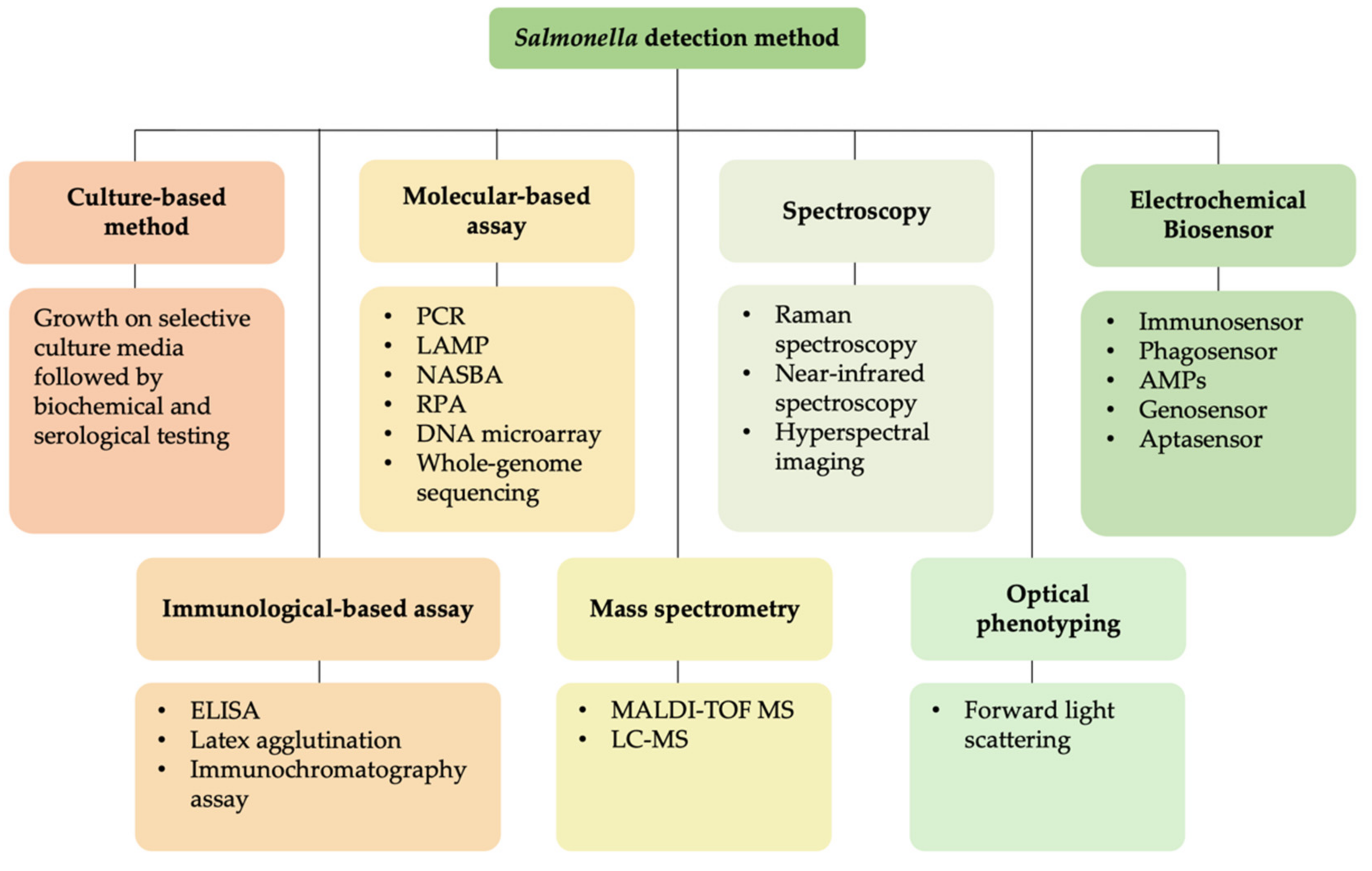
3. Detection of Salmonella
3.1. Conventional Detection of Salmonella
3.2. Rapid Salmonella Detection Methods
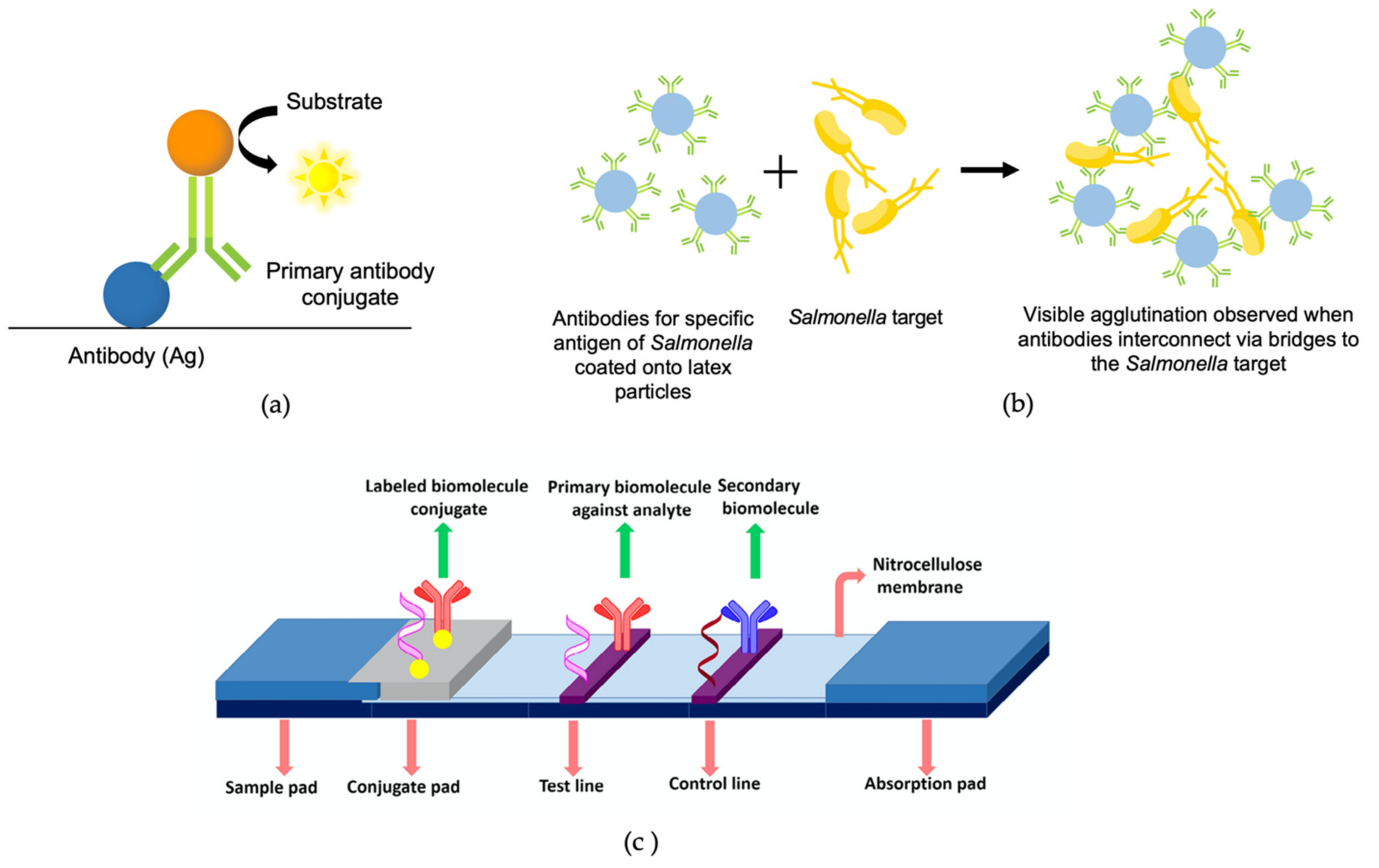
3.2.1. Immunological-Based Assay
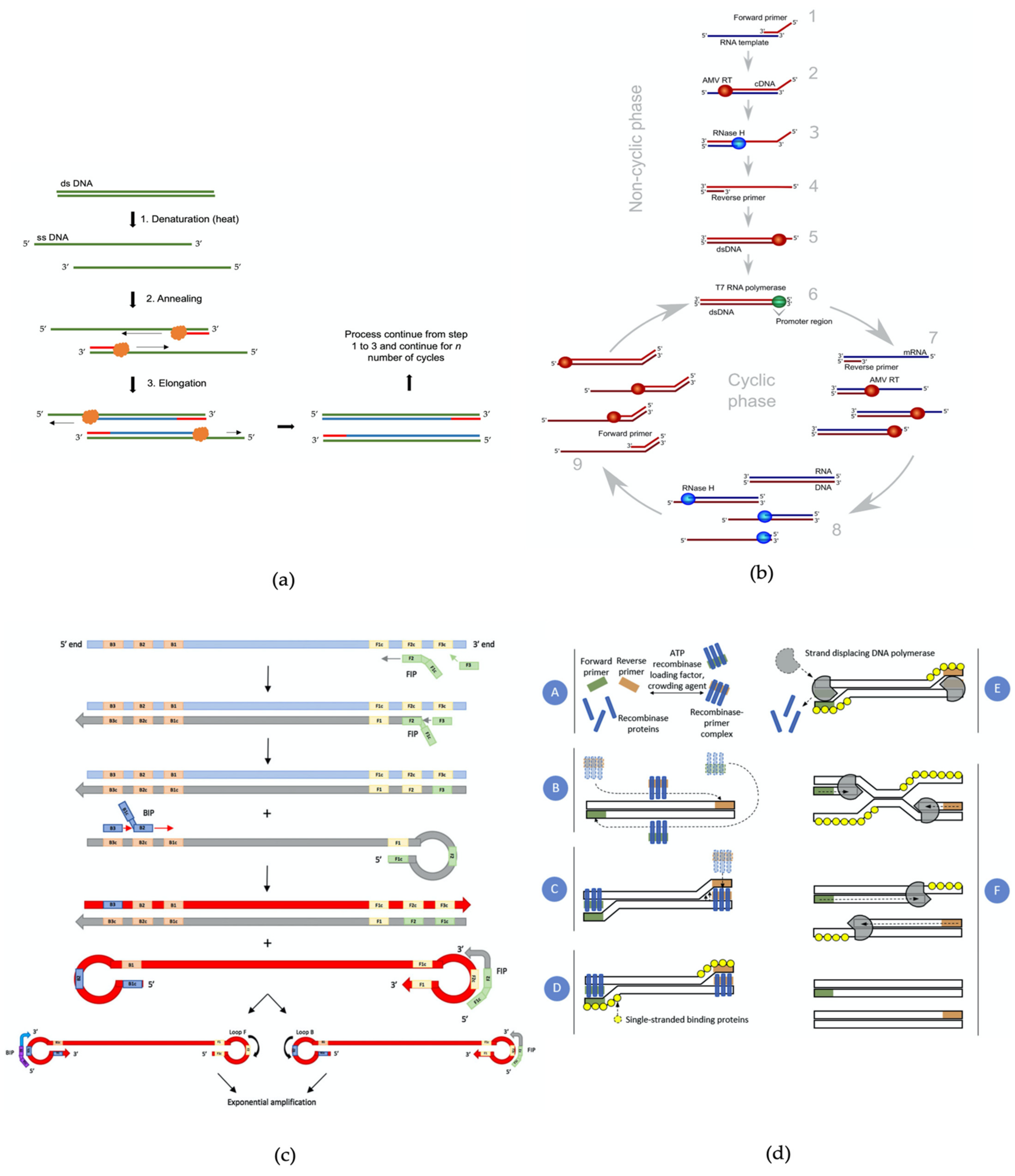
3.2.2. Molecular-Based Assay
3.2.3. Mass Spectrometry-Based Method
3.2.4. Spectroscopy Method
3.2.5. Optical Phenotyping Using Light Diffraction Technology Method
3.2.6. Electrochemical Biosensors
3.2.7. Advantages and Disadvantages of Salmonella Detection Methods
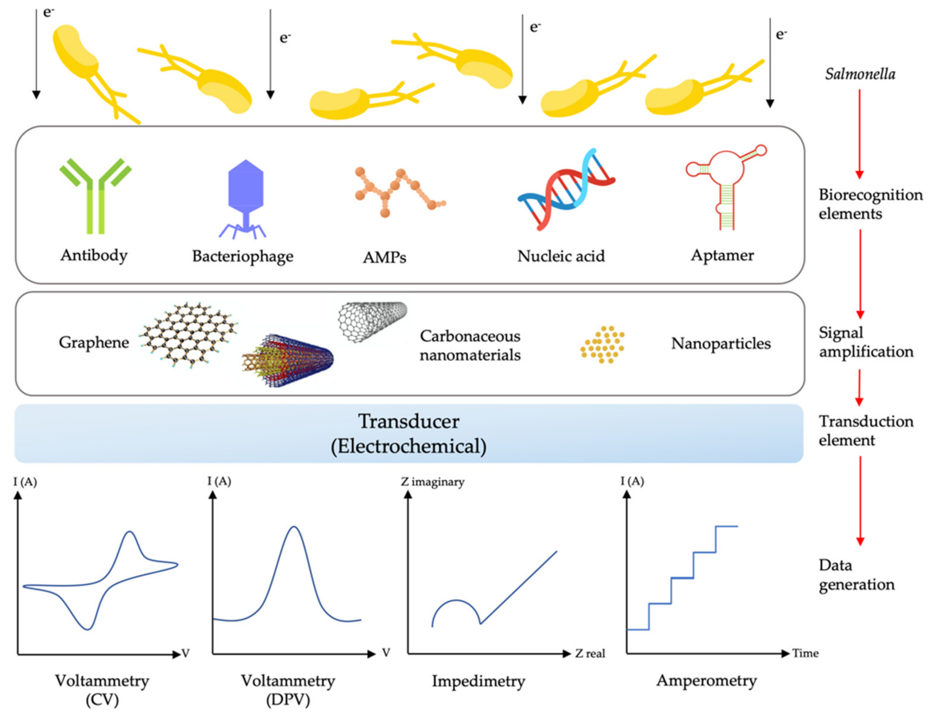
4. Biosensors Developed for Salmonella Detection
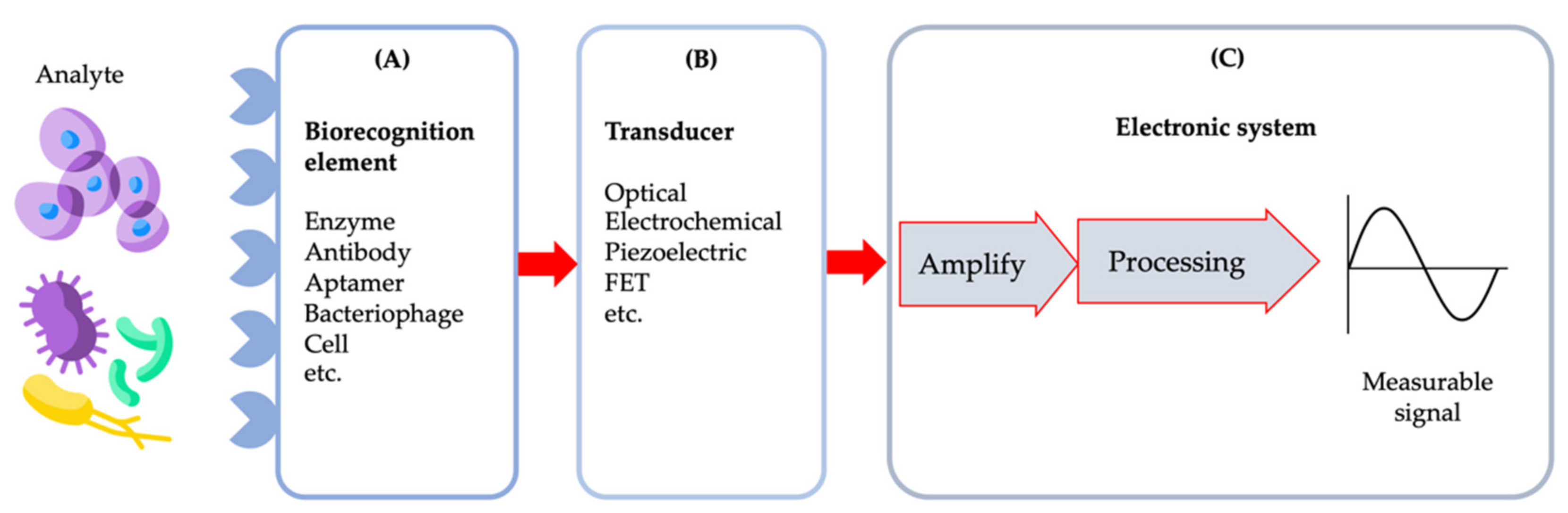
4.1. Biosensors
4.2. Electrochemical Biosensors
4.3. Bioreceptors Used in Salmonella Biosensors
4.3.1. Antibody–Antigen or Immunosensors
4.3.2. Bacteriophage-Based or Phagosensors
4.3.3. Antimicrobial Peptide-Based Biosensor (AMPs)
4.3.4. Nucleic Acid-Based Sensors or Genosensors
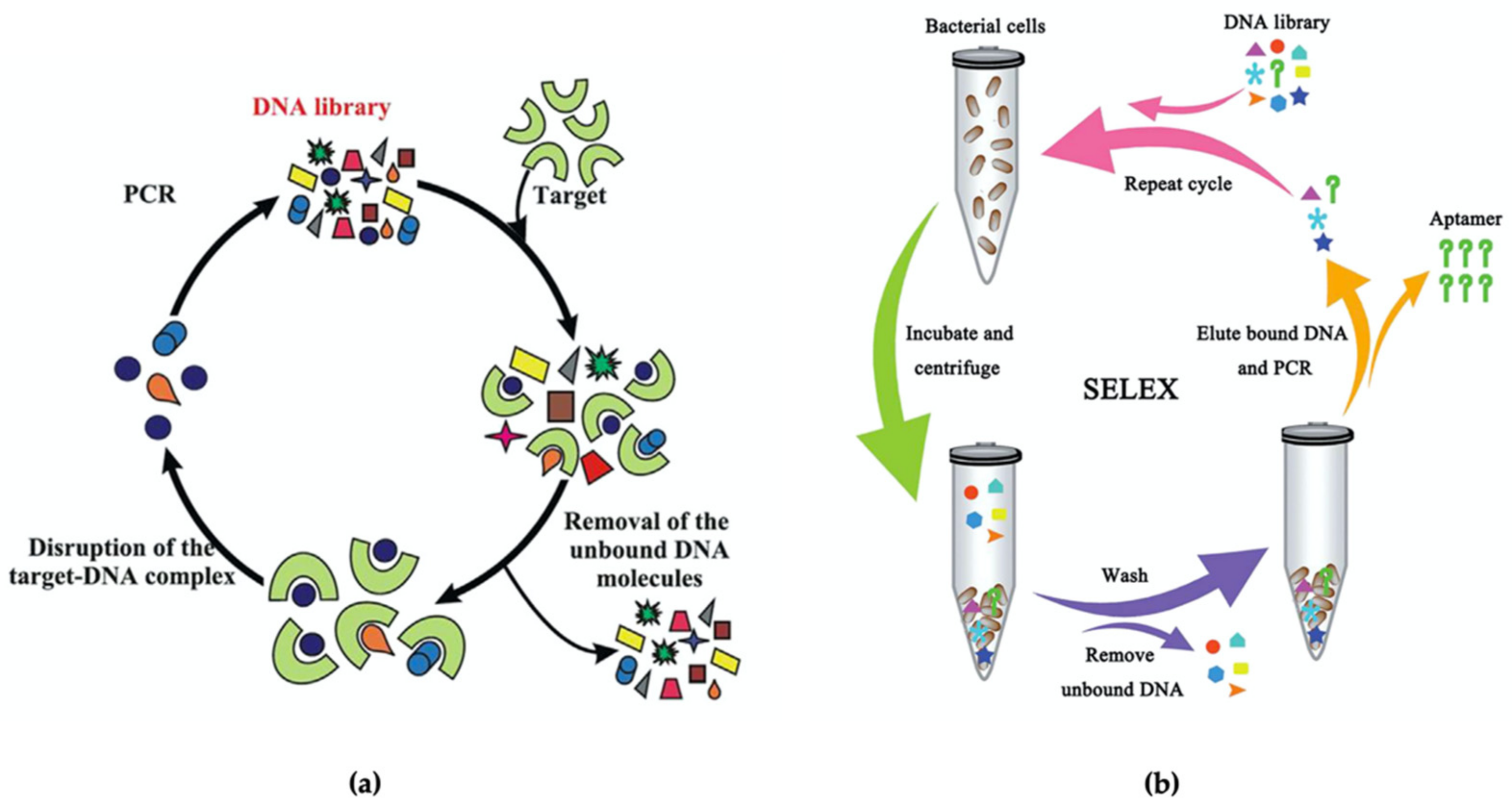
4.3.5. Aptamers as a New Bioreceptor for Salmonella Biosensors
5. Miniaturisation of Electrochemical Biosensors
5.1. Nanomaterials as the Surface Modifier of Electrochemical Biosensors
5.1.1. Carbon-Based Nanomaterials
5.1.2. Non-Carbon Nanomaterials
5.2. Lab-on-Chip Platforms for Rapid Detection of Salmonella
6. Conclusions and Future Impact of Salmonella Biosensors
Author Contributions
Funding
Institutional Review Board Statement
Informed Consent Statement
Data Availability Statement
Acknowledgments
Conflicts of Interest
References
- Kurtz, J.R.; Goggins, J.A.; McLachlan, J.B. Salmonella infection: Interplay between the bacteria and host immune system. Immunol. Lett. 2017, 190, 42–50. [Google Scholar] [CrossRef]
- Waddington, C.S.; Darton, T.C.; Pollard, A.J. The challenge of enteric fever. J. Infect. 2014, 68, S38–S50. [Google Scholar] [CrossRef] [PubMed]
- Harris, J.B.; Brooks, W.A. Typhoid and paratyphoid (enteric) fever. In Hunter’s Tropical Medicine and Emerging Infectious Diseases; Elsevier: Amsterdam, The Netherlands, 2020; pp. 608–616. [Google Scholar]
- Alhajj, M.; Farhana, A. Enzyme Linked Immunosorbent Assay [Updated 6 February 2021]. In StatPearls [Internet]; StatPearls Publishing: Treasure Island, FL, USA, 2020. Available online: https://0-www-ncbi-nlm-nih-gov.brum.beds.ac.uk/books/NBK555922/ (accessed on 8 September 2021).
- International Organization for Standardization (ISO). ISO 6579: 2002 Microbiology of Food and Animal Feeding Stuffs–Horizontal Method for the Detection of Salmonella spp., 2002; International Organization for Standardization IOS: Geneva, Switzerland, 2002; p. 27. [Google Scholar]
- Dilek, Ç.A.M. Lateral Flow Assay for Salmonella Detection and Potential Reagents. In New Insight into Brucella Infection and Foodborne Diseases; IntechOpen: London, UK, 2019. [Google Scholar]
- Fenollar, F.; Raoult, D. Molecular genetic methods for the diagnosis of fastidious microorganisms. APMIS 2004, 112, 785–807. [Google Scholar] [CrossRef] [PubMed]
- Notomi, T.; Okayama, H.; Masubuchi, H.; Yonekawa, T.; Watanabe, K.; Amino, N.; Hase, T. Loop-mediated isothermal amplification of DNA. Nucleic Acids Res. 2000, 28, E63. [Google Scholar] [CrossRef] [Green Version]
- Lobato, I.M.; O’Sullivan, C.K. Recombinase polymerase amplification: Basics, applications and recent advances. TrAC Trends Anal. Chem. 2018, 98, 19–35. [Google Scholar] [CrossRef] [PubMed]
- Fakruddin, M.D.; Mazumdar, R.M.; Chowdhury, A.; Mannan, K.S.B. Nucleic acid sequence based amplification (NASBA)-prospects and applications. Int. J. Life Sci. Pharma Res. 2012, 2, 106–121. [Google Scholar]
- Ehrenreich, A. DNA microarray technology for the microbiologist: An overview. Appl. Microbiol. Biotechnol. 2006, 73, 255–273. [Google Scholar] [CrossRef]
- Ibrahim, G.M.; Morin, P.M. Salmonella Serotyping Using Whole Genome Sequencing. Front. Microbiol. 2018, 9, 2993. [Google Scholar] [CrossRef] [Green Version]
- Mangmee, S.; Reamtong, O.; Kalambaheti, T.; Roytrakul, S.; Sonthayanon, P. MALDI-TOF mass spectrometry typing for predominant serovars of non-typhoidal Salmonella in a Thai broiler industry. Food Control 2020, 113, 107188. [Google Scholar] [CrossRef]
- Bell, R.L.; Jarvis, K.G.; Ottesen, A.R.; McFarland, M.A.; Brown, E.W. Recent and emerging innovations in Salmonella detection: A food and environmental perspective. Microb. Biotechnol. 2016, 9, 279–292. [Google Scholar] [CrossRef]
- Zhao, X.; Li, M.; Xu, Z. Detection of Foodborne Pathogens by Surface Enhanced Raman Spectroscopy. Front. Microbiol. 2018, 9, 1236. [Google Scholar] [CrossRef]
- Pereira, J.M.; Leme, L.M.; Perdoncini, M.R.F.G.; Valderrama, P.; Março, P.H. Fast Discrimination of Milk Contaminated with Salmonella sp. Via Near-Infrared Spectroscopy. Food Anal. Methods 2018, 11, 1878–1885. [Google Scholar] [CrossRef]
- Bonah, E.; Huang, X.; Aheto, J.H.; Osae, R. Application of hyperspectral imaging as a nondestructive technique for foodborne pathogen detection and characterization. Foodborne Pathog. Dis. 2019, 16, 712–722. [Google Scholar] [CrossRef]
- Abdelhaseib, M.U.; Singh, A.K.; Bhunia, A.K. Simultaneous detection of Salmonella enterica, Escherichia coli and Listeria monocytogenes in food using a light scattering sensor. J. Appl. Microbiol. 2019, 126, 1496–1507. [Google Scholar] [CrossRef] [PubMed]
- Liébana, S.; Brandão, D.; Alegret, S.; Pividori, M.I. Electrochemical immunosensors, genosensors and phagosensors for Salmonella detection. Anal. Methods 2014, 6, 8858–8873. [Google Scholar] [CrossRef]
- Lee, K.-M.; Runyon, M.; Herrman, T.J.; Phillips, R.; Hsieh, J. Review of Salmonella detection and identification methods: Aspects of rapid emergency response and food safety. Food Control 2015, 47, 264–276. [Google Scholar] [CrossRef]
- Lin, L.; Zheng, Q.; Lin, J.; Yuk, H.-G.; Guo, L. Immuno-and nucleic acid-based current technique for Salmonella detection in food. Eur. Food Res. Technol. 2020, 246, 373–395. [Google Scholar] [CrossRef]
- Silva, N.F.D.; Magalhães, J.M.C.S.; Freire, C.; Delerue-Matos, C. Electrochemical biosensors for Salmonella: State of the art and challenges in food safety assessment. Biosens. Bioelectron. 2018, 99, 667–682. [Google Scholar] [CrossRef] [PubMed] [Green Version]
- Ferone, M.; Gowen, A.; Fanning, S.; Scannell, A.G.M. Microbial detection and identification methods: Bench top assays to omics approaches. Compr. Rev. Food Sci. Food Saf. 2020, 19, 3106–3129. [Google Scholar] [CrossRef]
- Tiwari, H.; Kamat, R.S. Cross-reactions in cell-mediated immunity to Salmonella causing enteric fever. J. Med. Microbiol. 1986, 21, 233–237. [Google Scholar] [CrossRef] [PubMed]
- Dehghani, B.; Rasooli, I.; Gargari, S.L.M.; Nadooshan, M.R.J.; Owlia, P.; Nazarian, S. Immunogenicity of Salmonella enterica serovar Enteritidis virulence protein, InvH, and cross-reactivity of its antisera with Salmonella strains. Microbiol. Res. 2013, 168, 84–90. [Google Scholar] [CrossRef]
- Nga, T.V.T.; Karkey, A.; Dongol, S.; Thuy, H.N.; Dunstan, S.; Holt, K.; Tu, L.T.P.; Campbell, J.I.; Chau, T.T.; Chau, N.V.V.; et al. The sensitivity of real-time PCR amplification targeting invasive Salmonella serovars in biological specimens. BMC Infect. Dis. 2010, 10, 125. [Google Scholar] [CrossRef] [Green Version]
- Kostić, T.; Sessitsch, A. Microbial diagnostic microarrays for the detection and typing of food-and water-borne (bacterial) pathogens. Microarrays 2012, 1, 3–24. [Google Scholar] [CrossRef] [PubMed] [Green Version]
- Minoni, U.; Signoroni, A.; Nassini, G. On the application of optical forward-scattering to bacterial identification in an automated clinical analysis perspective. Biosens. Bioelectron. 2015, 68, 536–543. [Google Scholar] [CrossRef] [PubMed] [Green Version]
- Liu, D.; Wang, J.; Wu, L.; Huang, Y.; Zhang, Y.; Zhu, M.; Wang, Y.; Zhu, Z.; Yang, C. Trends in miniaturized biosensors for point-of-care testing. TrAC Trends Anal. Chem. 2020, 122, 115701. [Google Scholar] [CrossRef]
- Ansari, N.; Yazdian-Robati, R.; Shahdordizadeh, M.; Wang, Z.; Ghazvini, K. Aptasensors for quantitative detection of Salmonella Typhimurium. Anal. Biochem. 2017, 533, 18–25. [Google Scholar] [CrossRef] [PubMed]
- Naresh, V.; Lee, N. A Review on Biosensors and Recent Development of Nanostructured Materials-Enabled Biosensors. Sensors 2021, 21, 1109. [Google Scholar] [CrossRef] [PubMed]
- Eng, S.-K.; Pusparajah, P.; Ab Mutalib, N.-S.; Ser, H.-L.; Chan, K.-G.; Lee, L.-H. Salmonella: A review on pathogenesis, epidemiology and antibiotic resistance. Front. Life Sci. 2015, 8, 284–293. [Google Scholar] [CrossRef] [Green Version]
- Popoff, M.Y.; Bockemühl, J.; Gheesling, L.L. Supplement 2001 (no. 45) to the Kauffmann–White scheme. Res. Microbiol. 2003, 154, 173–174. [Google Scholar] [CrossRef]
- Reeves, M.W.; Evins, G.M.; Heiba, A.A.; Plikaytis, B.D.; Farmer, J.J. Clonal nature of Salmonella typhi and its genetic relatedness to other salmonellae as shown by multilocus enzyme electrophoresis, and proposal of Salmonella bongori comb. nov. J. Clin. Microbiol. 1989, 27, 313–320. [Google Scholar] [CrossRef] [Green Version]
- Hurley, D.; McCusker, M.P.; Fanning, S.; Martins, M. Salmonella–Host Interactions—Modulation of the Host Innate Immune System. Front. Immunol. 2014, 5, 481. [Google Scholar] [CrossRef] [PubMed] [Green Version]
- Giannella, R.A. Medical Microbiology. In Medical Microbiology, 4th ed.; University of Texas Medical Branch at Galveston: Texas, TX, USA, 1996. [Google Scholar]
- Luo, Y.; Yi, W.; Yao, Y.; Zhu, N.; Qin, P. Characteristic diversity and antimicrobial resistance of Salmonella from gastroenteritis. J. Infect. Chemother. 2018, 24, 251–255. [Google Scholar] [CrossRef] [PubMed]
- Jessica, M.H.; Beau, B.B. Chapter 4: Travel-related infectious diseases, Salmonellosis (Nontyphoidal). In CDC Yellow Book, Centers for Disease Control and Preventions (CDC); Oxford University Press: New York, NY, USA, 2019. [Google Scholar]
- Stanaway, J.D.; Reiner, R.C.; Blacker, B.F.; Goldberg, E.M.; Khalil, I.A.; Troeger, C.E.; Andrews, J.R.; Bhutta, Z.A.; Crump, J.A.; Im, J.; et al. The global burden of typhoid and paratyphoid fevers: A systematic analysis for the Global Burden of Disease Study 2017. Lancet Infect. Dis. 2019, 19, 369–381. [Google Scholar] [CrossRef] [Green Version]
- Vergese, A. The “typhoid state” revisited. Am. J. Med. 1985, 79, 370–372. [Google Scholar] [CrossRef]
- Bhutta, Z.A. Impact of age and drug resistance on mortality in typhoid fever. Arch. Dis. Child. 1996, 75, 214–217. [Google Scholar] [CrossRef]
- Qamar, F.N.; Azmatullah, A.; Bhutta, Z.A. Challenges in measuring complications and death due to invasive Salmonella infections. Vaccine 2015, 33, C16–C20. [Google Scholar] [CrossRef]
- Andoh, L.A.; Ahmed, S.; Olsen, J.E.; Obiri-Danso, K.; Newman, M.J.; Opintan, J.A.; Barco, L.; Dalsgaard, A. Prevalence and characterization of Salmonella among humans in Ghana. Trop. Med. Health 2017, 45, 1–11. [Google Scholar] [CrossRef] [Green Version]
- Muhammad, E.N.; Abdul Mutalip, M.H.; Hasim, M.H.; Paiwai, F.; Pan, S.; Mahmud, M.A.F.; Yeop, N.; Tee, G.H.; Senin, A.A.; Aris, T. The burden of typhoid fever in Klang Valley, Malaysia, 2011–2015. BMC Infect. Dis. 2020, 20, 843. [Google Scholar] [CrossRef]
- Chau, T.T.; Campbell, J.I.; Galindo, C.M.; Hoang, N.V.M.; Diep, T.S.; Nga, T.T.T.; Chau, N.V.V.; Tuan, P.Q.; Page, A.L.; Ochiai, R.L. Antimicrobial drug resistance of Salmonella enterica serovar Typhi in Asia and molecular mechanism of reduced susceptibility to the fluoroquinolones. Antimicrob. Agents Chemother. 2007, 51, 4315–4323. [Google Scholar] [CrossRef] [PubMed] [Green Version]
- Kumar, S.; Rizvi, M.; Berry, N. Rising prevalence of enteric fever due to multidrug-resistant Salmonella: An epidemiological study. J. Med. Microbiol. 2008, 57, 1247–1250. [Google Scholar] [CrossRef] [PubMed] [Green Version]
- Wang, X.; Biswas, S.; Paudyal, N.; Pan, H.; Li, X.; Fang, W.; Yue, M. Antibiotic resistance in Salmonella Typhimurium isolates recovered from the food chain through national antimicrobial resistance monitoring system between 1996 and 2016. Front. Microbiol. 2019, 10, 985. [Google Scholar] [CrossRef] [Green Version]
- Mather, A.E.; Reid, S.W.J.; Maskell, D.J.; Parkhill, J.; Fookes, M.C.; Harris, S.R.; Brown, D.J.; Coia, J.E.; Mulvey, M.R.; Gilmour, M.W. Distinguishable epidemics of multidrug-resistant Salmonella Typhimurium DT104 in different hosts. Science 2013, 341, 1514–1517. [Google Scholar] [CrossRef] [Green Version]
- Helms, M.; Ethelberg, S.; Mølbak, K.; Group, D.S. International Salmonella typhimurium DT104 infections, 1992–2001. Emerg. Infect. Dis. 2005, 11, 859. [Google Scholar] [CrossRef]
- Andrews, J.R.; Ryan, E.T. Diagnostics for invasive Salmonella infections: Current challenges and future directions. Vaccine 2015, 33, C8–C15. [Google Scholar] [CrossRef] [PubMed] [Green Version]
- Mogasale, V.; Ramani, E.; Mogasale, V.V.; Park, J. What proportion of Salmonella Typhi cases are detected by blood culture? A systematic literature review. Ann. Clin. Microbiol. Antimicrob. 2016, 15, 32. [Google Scholar] [CrossRef] [Green Version]
- Siala, M.; Barbana, A.; Smaoui, S.; Hachicha, S.; Marouane, C.; Kammoun, S.; Gdoura, R.; Messadi-Akrout, F. Screening and Detecting Salmonella in Different Food Matrices in Southern Tunisia Using a Combined Enrichment/Real-Time PCR Method: Correlation with Conventional Culture Method. Front. Microbiol. 2017, 8, 2416. [Google Scholar] [CrossRef] [PubMed]
- Gast, R.K.; Porter, R.E., Jr. Chapter 16: Salmonella Infections. In Diseases of Poultry, 14th ed.; John Wiley & Son Inc.: Hoboken, NJ, USA, 2020; pp. 717–753. [Google Scholar]
- Fung, D.Y.C. Rapid methods and automation in microbiology. In Proceedings of the Workshop MRAMA, Barcelona, Spain, 20–24 November 2002. [Google Scholar]
- Ge, B.; Meng, J. Advanced technologies for pathogen and toxin detection in foods: Current applications and future directions. JALA J. Assoc. Lab. Autom. 2009, 14, 235–241. [Google Scholar] [CrossRef] [Green Version]
- Klingler, J.M.; Stowe, R.P.; Obenhuber, D.C.; Groves, T.O.; Mishra, S.K.; Pierson, D.L. Evaluation of the Biolog automated microbial identification system. Appl. Environ. Microbiol. 1992, 58, 2089–2092. [Google Scholar] [CrossRef] [PubMed] [Green Version]
- El-Liethy, M.A.; Hemdan, B.A.; El-Taweel, G.E. Prevalence of E. coli, Salmonella, and Listeria spp. as potential pathogens: A comparative study for biofilm of sink drain environment. J. Food Saf. 2020, 40, e12816. [Google Scholar] [CrossRef]
- Sørensen, L.L.; Alban, L.; Nielsen, B.; Dahl, J. The correlation between Salmonella serology and isolation of Salmonella in Danish pigs at slaughter. Vet. Microbiol. 2004, 101, 131–141. [Google Scholar] [CrossRef]
- Alakomi, H.; Saarela, M. Salmonella importance and current status of detection and surveillance methods. Qual. Assur. Saf. Crop. Foods 2009, 1, 142–152. [Google Scholar] [CrossRef]
- Maciorowski, K.G.; Herrera, P.; Jones, F.T.; Pillai, S.D.; Ricke, S.C. Cultural and immunological detection methods for Salmonella spp. in animal feeds—A review. Vet. Res. Commun. 2006, 30, 127–137. [Google Scholar] [CrossRef]
- Manafi, M. New developments in chromogenic and fluorogenic culture media. Int. J. Food Microbiol. 2000, 60, 205–218. [Google Scholar] [CrossRef]
- Perry, J.D.; Freydière, A.M. The application of chromogenic media in clinical microbiology. J. Appl. Microbiol. 2007, 103, 2046–2055. [Google Scholar] [CrossRef] [PubMed]
- Lin, A. V Direct ELISA. In ELISA; Springer: Berlin/Heidelberg, Germany, 2015; pp. 61–67. [Google Scholar]
- Alhabbab, R.Y. Precipitation and Agglutination Reactions. In Basic Serological Testing; Springer: Berlin/Heidelberg, Germany, 2018; pp. 23–30. [Google Scholar]
- Bahadır, E.B.; Sezgintürk, M.K. Lateral flow assays: Principles, designs and labels. TrAC Trends Anal. Chem. 2016, 82, 286–306. [Google Scholar] [CrossRef]
- Blivet, D.; Soumet, C.; Ermel, G.; Colin, P. Rapid detection methods for pathogens. In Proceedings of the 3rd Karlsruhe Nutrition Symposium European Research towards Safer and Better Food, Review and Transfer Congress Centre, Karlsruhe, Germany, 18–20 October 1998; p. 3. [Google Scholar]
- Tietjen, M.; Fung, D.Y.C. Salmonellae and Food Safety. Crit. Rev. Microbiol. 1995, 21, 53–83. [Google Scholar] [CrossRef] [PubMed]
- Huang, Z.; Olson, N.A.; You, W.; Haugland, R.P. A sensitive competitive ELISA for 2, 4-dinitrophenol using 3, 6-fluorescein diphosphate as a fluorogenic substrate. J. Immunol. Methods 1992, 149, 261–266. [Google Scholar] [CrossRef]
- Sue, M.J.; Yeap, S.K.; Omar, A.R.; Tan, S.W. Application of PCR-ELISA in Molecular Diagnosis. Biomed Res. Int. 2014, 2014, 653014. [Google Scholar] [CrossRef]
- Peng, H.; Huang, Z.; Wu, W.; Liu, M.; Huang, K.; Yang, Y.; Deng, H.; Xia, X.; Chen, W. Versatile High-Performance Electrochemiluminescence ELISA Platform Based on a Gold Nanocluster Probe. ACS Appl. Mater. Interfaces 2019, 11, 24812–24819. [Google Scholar] [CrossRef]
- De La Rica, R.; Stevens, M.M. Plasmonic ELISA for the ultrasensitive detection of disease biomarkers with the naked eye. Nat. Nanotechnol. 2012, 7, 821–824. [Google Scholar] [CrossRef]
- Thorns, C.J.; McLaren, I.M.; Sojka, M.G. The use of latex particle agglutination to specifically detect Salmonella enteritidis. Int. J. Food Microbiol. 1994, 21, 47–53. [Google Scholar] [CrossRef]
- Eijkelkamp, J.M.; Aarts, H.J.M.; Van der Fels-Klerx, H.J. Suitability of rapid detection methods for Salmonella in poultry slaughterhouses. Food Anal. Methods 2009, 2, 1–13. [Google Scholar] [CrossRef] [Green Version]
- Love, D.C.; Sobsey, M.D. Simple and rapid F+ coliphage culture, latex agglutination, and typing assay to detect and source track fecal contamination. Appl. Environ. Microbiol. 2007, 73, 4110–4118. [Google Scholar] [CrossRef] [Green Version]
- Magwood, S.E.; Annau, E. The adsorption of somatic antigens of salmonella by polystyrene latex particles. Can. J. Comp. Med. Vet. Sci. 1961, 25, 69. [Google Scholar] [PubMed]
- Cheng-yu, Y. Using latex Agglutination Text Detecting Salmonella in Commercial Feed. Chin. Qinghai J. Anim. Vet. Sci. 2006, 2. [Google Scholar]
- Thompson, W.; Crabtree, D.; Bastin, B.; Koch, K.; Hahs, M.; Sohier, D. AOAC Validation Study of a Real-Time PCR Workflow for Salmonella Detection in Large Test Portions of Cocoa and Chocolate Products. IAFP 2021. Available online: https://assets.thermofisher.cn/TFS-Assets/MBD/posters/Poster-IAFP-2021-QLabs-AOAC-SureTect-Salm-LT2659A.pdf (accessed on 8 September 2021).
- Mao, X.; Wang, W.; Du, T.-E. Rapid quantitative immunochromatographic strip for multiple proteins test. Sens. Actuators B Chem. 2013, 186, 315–320. [Google Scholar] [CrossRef]
- Kang, J.; Kim, M.-G. Advancements in DNA-assisted Immunosensors. BioChip J. 2020, 14, 18–31. [Google Scholar] [CrossRef] [Green Version]
- Ma, K.-S.; Ma, Y.; Chiou, F. Nanotechnology Applications in Polymerase Chain Reaction (PCR) BT-Encyclopedia of Nanotechnology; Bhushan, B., Ed.; Springer: Dordrecht, The Netherlands, 2016; pp. 2869–2876. ISBN 978-94-017-9780-1. [Google Scholar]
- Hønsvall, B.K.; Robertson, L.J. From research lab to standard environmental analysis tool: Will NASBA make the leap? Water Res. 2017, 109, 389–397. [Google Scholar] [CrossRef]
- Salamin, O.; Kuuranne, T.; Saugy, M.; Leuenberger, N. Loop-mediated isothermal amplification (LAMP) as an alternative to PCR: A rapid on-site detection of gene doping. Drug Test. Anal. 2017, 9, 1731–1737. [Google Scholar] [CrossRef] [Green Version]
- Watson, J.D. The Polymerase Chain Reaction; Springer Science & Business Media: Berlin/Heidelberg, Germany, 2012; ISBN 1461202574. [Google Scholar]
- Chin, W.H.; Sun, Y.; Høgberg, J.; Quyen, T.L.; Engelsmann, P.; Wolff, A.; Bang, D.D. Direct PCR–A rapid method for multiplexed detection of different serotypes of Salmonella in enriched pork meat samples. Mol. Cell. Probes 2017, 32, 24–32. [Google Scholar] [CrossRef] [Green Version]
- Vinayaka, A.C.; Ngo, T.A.; Kant, K.; Engelsmann, P.; Dave, V.P.; Shahbazi, M.-A.; Wolff, A.; Bang, D.D. Rapid detection of Salmonella enterica in food samples by a novel approach with combination of sample concentration and direct PCR. Biosens. Bioelectron. 2019, 129, 224–230. [Google Scholar] [CrossRef]
- Park, S.H.; Aydin, M.; Khatiwara, A.; Dolan, M.C.; Gilmore, D.F.; Bouldin, J.L.; Ahn, S.; Ricke, S.C. Current and emerging technologies for rapid detection and characterization of Salmonella in poultry and poultry products. Food Microbiol. 2014, 38, 250–262. [Google Scholar] [CrossRef]
- De Medici, D.; Croci, L.; Delibato, E.; Di Pasquale, S.; Filetici, E.; Toti, L. Evaluation of DNA extraction methods for use in combination with SYBR green I real-time PCR to detect Salmonella enterica serotype enteritidis in poultry. Appl. Environ. Microbiol. 2003, 69, 3456–3461. [Google Scholar] [CrossRef] [Green Version]
- Malorny, B.; Huehn, S.; Dieckmann, R.; Krämer, N.; Helmuth, R. Polymerase chain reaction for the rapid detection and serovar identification of Salmonella in food and feeding stuff. Food Anal. Methods 2009, 2, 81–95. [Google Scholar] [CrossRef]
- Azinheiro, S.; Carvalho, J.; Prado, M.; Garrido-Maestu, A. Multiplex Detection of Salmonella spp., E. coli O157 and L. monocytogenes by qPCR Melt Curve Analysis in Spiked Infant Formula. Microorganisms 2020, 8, 1359. [Google Scholar] [CrossRef] [PubMed]
- Ruan, J.; Wang, W.; Zhang, T.; Zheng, T.; Zheng, J.; Yu, S.; Yu, D.; Huang, Y. Establishment of a duplex real-time qPCR method for detection of Salmonella spp. and Serratia fonticola in fishmeal. AMB Express 2020, 10, 1–8. [Google Scholar] [CrossRef]
- Parker, A.M.; Mohler, V.L.; Gunn, A.A.; House, J.K. Development of a qPCR for the detection and quantification of Salmonella spp. in sheep feces and tissues. J. Vet. Diagn. Investig. 2020, 32, 835–843. [Google Scholar] [CrossRef]
- Dmitric, M.; Vidanovic, D.; Matovic, K.; Sekler, M.; Saric, L.; Arsic, M.; Karabasil, N. In-house validation of real-time PCR methods for detecting the INV A and TTR genes of Salmonella spp. in food. J. Food Process. Preserv. 2018, 42, e13455. [Google Scholar] [CrossRef]
- Kulshreshtha, G.; Borza, T.; Rathgeber, B.; Stratton, G.S.; Thomas, N.A.; Critchley, A.; Hafting, J.; Prithiviraj, B. Red Seaweeds Sarcodiotheca gaudichaudii and Chondrus crispus down Regulate Virulence Factors of Salmonella Enteritidis and Induce Immune Responses in Caenorhabditis elegans. Front. Microbiol. 2016, 7, 421. [Google Scholar] [CrossRef] [PubMed]
- Tatavarthy, A.; Cannons, A. Real-time PCR detection of Salmonella species using a novel target: The outer membrane porin F gene (ompF). Lett. Appl. Microbiol. 2010, 50, 645–652. [Google Scholar] [CrossRef] [PubMed]
- McCabe, E.M.; Burgess, C.M.; O’Regan, E.; McGuinness, S.; Barry, T.; Fanning, S.; Duffy, G. Development and evaluation of DNA and RNA real-time assays for food analysis using the hilA gene of Salmonella enterica subspecies enterica. Food Microbiol. 2011, 28, 447–456. [Google Scholar] [CrossRef]
- Hassena, A.B.; Barkallah, M.; Fendri, I.; Grosset, N.; Neila, I.B.; Gautier, M.; Gdoura, R. Real time PCR gene profiling and detection of Salmonella using a novel target: The siiA gene. J. Microbiol. Methods 2015, 109, 9–15. [Google Scholar] [CrossRef]
- Ganz, K.; Gill, A. Inhibition of polymerase chain reaction for the detection of Escherichia coli O157: H7 and Salmonella enterica on walnut kernels. Food Microbiol. 2013, 35, 15–20. [Google Scholar] [CrossRef] [PubMed]
- Cossu, A.; Levin, R.E. Rapid conventional PCR and real-time-qPCR detection of low numbers of Salmonella enterica from ground beef without enrichment. Food Biotechnol. 2014, 28, 96–105. [Google Scholar] [CrossRef]
- Margot, H.; Stephan, R.; Guarino, S.; Jagadeesan, B.; Chilton, D.; O’Mahony, E.; Iversen, C. Inclusivity, exclusivity and limit of detection of commercially available real-time PCR assays for the detection of Salmonella. Int. J. Food Microbiol. 2013, 165, 221–226. [Google Scholar] [CrossRef] [PubMed] [Green Version]
- Xiao, L.; Zhang, Z.; Sun, X.; Pan, Y.; Zhao, Y. Development of a quantitative real-time PCR assay for viable Salmonella spp. without enrichment. Food Control 2015, 57, 185–189. [Google Scholar] [CrossRef]
- Li, Y.; Fan, P.; Zhou, S.; Zhang, L. Loop-mediated isothermal amplification (LAMP): A novel rapid detection platform for pathogens. Microb. Pathog. 2017, 107, 54–61. [Google Scholar] [CrossRef]
- Souii, A.; M’hadheb-Gharbi, M.B.; Gharbi, J. Nucleic acid-based biotechnologies for food-borne pathogen detection using routine time-intensive culture-based methods and fast molecular diagnostics. Food Sci. Biotechnol. 2016, 25, 11–20. [Google Scholar] [CrossRef]
- Yang, Q.; Wang, F.; Jones, K.L.; Meng, J.; Prinyawiwatkul, W.; Ge, B. Evaluation of loop-mediated isothermal amplification for the rapid, reliable, and robust detection of Salmonella in produce. Food Microbiol. 2015, 46, 485–493. [Google Scholar] [CrossRef]
- Kokkinos, P.A.; Ziros, P.G.; Bellou, M.; Vantarakis, A. Loop-mediated isothermal amplification (LAMP) for the detection of Salmonella in food. Food Anal. Methods 2014, 7, 512–526. [Google Scholar] [CrossRef]
- Techathuvanan, C.; Draughon, F.A.; D’Souza, D.H. Loop-mediated isothermal amplification (LAMP) for the rapid and sensitive detection of Salmonella Typhimurium from pork. J. Food Sci. 2010, 75, M165–M172. [Google Scholar] [CrossRef] [PubMed]
- Zhao, Y.; Jiang, X.; Qu, Y.; Pan, R.; Pang, X.; Jiang, Y.; Man, C. Salmonella detection in powdered dairy products using a novel molecular tool. J. Dairy Sci. 2017, 100, 3480–3496. [Google Scholar] [CrossRef] [PubMed] [Green Version]
- Liu, N.; Zou, D.; Dong, D.; Yang, Z.; Ao, D.; Liu, W.; Huang, L. Development of a multiplex loop-mediated isothermal amplification method for the simultaneous detection of Salmonella spp. and Vibrio parahaemolyticus. Sci. Rep. 2017, 7, 1–7. [Google Scholar] [CrossRef] [PubMed]
- Wu, G.P.; Chen, S.H.; Levin, R.E. Application of ethidium bromide monoazide for quantification of viable and dead cells of Salmonella enterica by real-time loop-mediated isothermal amplification. J. Microbiol. Methods 2015, 117, 41–48. [Google Scholar] [CrossRef]
- Deiman, B.; van Aarle, P.; Sillekens, P. Characteristics and applications of nucleic acid sequence-based amplification (NASBA). Mol. Biotechnol. 2002, 20, 163–179. [Google Scholar] [CrossRef]
- Zhai, L.; Liu, H.; Chen, Q.; Lu, Z.; Zhang, C.; Lv, F.; Bie, X. Development of a real-time nucleic acid sequence–based amplification assay for the rapid detection of Salmonella spp. from food. Braz. J. Microbiol. 2019, 50, 255–261. [Google Scholar] [CrossRef]
- D’souza, D.H.; Jaykus, L. Nucleic acid sequence based amplification for the rapid and sensitive detection of Salmonella enterica from foods. J. Appl. Microbiol. 2003, 95, 1343–1350. [Google Scholar] [CrossRef]
- Mollasalehi, H.; Yazdanparast, R. An improved non-crosslinking gold nanoprobe-NASBA based on 16S rRNA for rapid discriminative bio-sensing of major salmonellosis pathogens. Biosens. Bioelectron. 2013, 47, 231–236. [Google Scholar] [CrossRef]
- Chen, J.; Liu, X.; Chen, J.; Guo, Z.; Wang, Y.; Chen, G.; Chen, X.; Yan, Q.; Yang, P.; Li, R. Development of a rapid test method for Salmonella enterica detection based on fluorescence probe-based recombinase polymerase amplification. Food Anal. Methods 2019, 12, 1791–1798. [Google Scholar] [CrossRef]
- Rosser, A.; Rollinson, D.; Forrest, M.; Webster, B.L. Isothermal Recombinase Polymerase amplification (RPA) of Schistosoma haematobium DNA and oligochromatographic lateral flow detection. Parasit. Vectors 2015, 8, 1–5. [Google Scholar] [CrossRef] [Green Version]
- Dao, T.N.T.; Lee, E.Y.; Koo, B.; Jin, C.E.; Lee, T.Y.; Shin, Y. A microfluidic enrichment platform with a recombinase polymerase amplification sensor for pathogen diagnosis. Anal. Biochem. 2018, 544, 87–92. [Google Scholar] [CrossRef] [PubMed]
- Gao, W.; Huang, H.; Zhu, P.; Yan, X.; Fan, J.; Jiang, J.; Xu, J. Recombinase polymerase amplification combined with lateral flow dipstick for equipment-free detection of Salmonella in shellfish. Bioprocess Biosyst. Eng. 2018, 41, 603–611. [Google Scholar] [CrossRef] [PubMed]
- Ren, J.; Man, Y.; Li, A.; Liang, G.; Jin, X.; Pan, L. Detection of Salmonella enteritidis and Salmonella typhimurium in foods using a rapid, multiplex real-time recombinase polymerase amplification assay. J. Food Saf. 2020, 40, e12784. [Google Scholar] [CrossRef]
- Chen, Z.; Zhong, H.; Luo, H.; Zhang, R.; Huang, J. Recombinase polymerase amplification combined with unmodified gold nanoparticles for Salmonella detection in milk. Food Anal. Methods 2019, 12, 190–197. [Google Scholar] [CrossRef]
- Gao, W.; Huang, H.; Zhang, Y.; Zhu, P.; Yan, X.; Fan, J.; Chen, X. Recombinase polymerase amplification-based assay for rapid detection of Listeria monocytogenes in food samples. Food Anal. Methods 2017, 10, 1972–1981. [Google Scholar] [CrossRef]
- Schena, M.; Shalon, D.; Davis, R.W.; Brown, P.O. Quantitative monitoring of gene expression patterns with a complementary DNA microarray. Science 1995, 270, 467–470. [Google Scholar] [CrossRef] [Green Version]
- Raspoet, R.; Appia-Ayme, C.; Shearer, N.; Martel, A.; Pasmans, F.; Haesebrouck, F.; Ducatelle, R.; Thompson, A.; Van Immerseel, F. Microarray-based detection of Salmonella enterica serovar Enteritidis genes involved in chicken reproductive tract colonization. Appl. Environ. Microbiol. 2014, 80, 7710–7716. [Google Scholar] [CrossRef] [Green Version]
- Litrup, E.; Torpdahl, M.; Malorny, B.; Huehn, S.; Helms, M.; Christensen, H.; Nielsen, E.M. DNA microarray analysis of Salmonella serotype Typhimurium strains causing different symptoms of disease. BMC Microbiol. 2010, 10, 96. [Google Scholar] [CrossRef] [Green Version]
- Koyuncu, S.; Andersson, G.; Vos, P.; Häggblom, P. DNA microarray for tracing Salmonella in the feed chain. Int. J. Food Microbiol. 2011, 145, S18–S22. [Google Scholar] [CrossRef]
- Guo, D.; Liu, B.; Liu, F.; Cao, B.; Chen, M.; Hao, X.; Feng, L.; Wang, L. Development of a DNA microarray for molecular identification of all 46 Salmonella O serogroups. Appl. Environ. Microbiol. 2013, 79, 3392–3399. [Google Scholar] [CrossRef] [Green Version]
- Ronholm, J. Editorial: Game Changer—Next Generation Sequencing and Its Impact on Food Microbiology. Front. Microbiol. 2018, 9, 363. [Google Scholar] [CrossRef] [PubMed]
- Cheng, K.; Chui, H.; Domish, L.; Hernandez, D.; Wang, G. Recent development of mass spectrometry and proteomics applications in identification and typing of bacteria. Proteom.–Clin. Appl. 2016, 10, 346–357. [Google Scholar] [CrossRef] [PubMed]
- Yang, S.-M.; Kim, E.; Kim, D.; Baek, J.; Yoon, H.; Kim, H.-Y. Rapid Detection of Salmonella Enteritidis, Typhimurium, and Thompson by Specific Peak Analysis Using Matrix-Assisted Laser Desorption Ionization Time-of-Flight Mass Spectrometry. Foods 2021, 10, 933. [Google Scholar] [CrossRef] [PubMed]
- Callahan, J.H.; McFarland, M.A.; Musser, S.M.; Bell, R.; Andrzejewski, D. Anyl 282-intact protein liquid chromatography mass spectrometry for bacterial identification and differentiation. In Proceedings of the Abstracts of Papers of the American Chemical Society, Washington, DC, USA, 16 August 2009; Volume 238. [Google Scholar]
- McFarland, M.A.; Andrzejewski, D.; Musser, S.M.; Callahan, J.H. Platform for identification of Salmonella serovar differentiating bacterial proteins by top-down mass spectrometry: S. Typhimurium vs S. Heidelberg. Anal. Chem. 2014, 86, 6879–6886. [Google Scholar] [CrossRef] [PubMed]
- Smith, E.; Dent, G. Modern Raman Spectroscopy: A Practical Approach; John Wiley & Sons: Hoboken, NJ, USA, 2019; ISBN 1119440556. [Google Scholar]
- Duan, N.; Chang, B.; Zhang, H.; Wang, Z.; Wu, S. Salmonella typhimurium detection using a surface-enhanced Raman scattering-based aptasensor. Int. J. Food Microbiol. 2016, 218, 38–43. [Google Scholar] [CrossRef]
- Alexandrakis, D.; Downey, G.; Scannell, A.G.M. Detection and identification of bacteria in an isolated system with near-infrared spectroscopy and multivariate analysis. J. Agric. Food Chem. 2008, 56, 3431–3437. [Google Scholar] [CrossRef] [PubMed]
- Tian, Y.; Gao, X.; Qi, W.-L.; Wang, Y.; Wang, X.; Zhou, J.; Lu, D.; Chen, B. Advances in differentiation and identification of foodborne bacteria using near infrared spectroscopy. Anal. Methods 2021, 13, 2558–2566. [Google Scholar] [CrossRef]
- Gowen, A.A.; Feng, Y.; Gaston, E.; Valdramidis, V. Recent applications of hyperspectral imaging in microbiology. Talanta 2015, 137, 43–54. [Google Scholar] [CrossRef] [PubMed]
- Michael, M.; Phebus, R.K.; Amamcharla, J. Hyperspectral imaging of common foodborne pathogens for rapid identification and differentiation. Food Sci. Nutr. 2019, 7, 2716–2725. [Google Scholar] [CrossRef] [PubMed] [Green Version]
- Seo, Y.W.; Yoon, S.-C.; Park, B.; Windham, W.R.; Lawrence, K.C. Development of Hyperspectral Imaging Technique for Salmonella Enteritidis and Typhimurium on Agar Plates. Appl. Eng. Agric. 2014, 30, 495–506. [Google Scholar]
- Buzalewicz, I.; Suchwałko, A.; Trzciński, P.; Sas-Paszt, L.; Sumorok, B.; Kowal, K.; Kozera, R.; Wieliczko, A.; Podbielska, H. Integrated multi-channel optical system for bacteria characterization and its potential use for monitoring of environmental bacteria. Biomed. Opt. Express 2019, 10, 1165–1183. [Google Scholar] [CrossRef]
- Kim, H.; Singh, A.K.; Bhunia, A.K.; Bae, E. Laser-induced speckle scatter patterns in Bacillus colonies. Front. Microbiol. 2014, 5, 537. [Google Scholar] [CrossRef]
- Singh, A.K.; Bettasso, A.M.; Bae, E.; Rajwa, B.; Dundar, M.M.; Forster, M.D.; Liu, L.; Barrett, B.; Lovchik, J.; Robinson, J.P. Laser optical sensor, a label-free on-plate Salmonella enterica colony detection tool. MBio 2014, 5, e01019-13. [Google Scholar] [CrossRef] [Green Version]
- McNaught, A.D.; Wilkinson, A. Compendium of Chemical Terminology; Blackwell Science Oxford: Oxford, UK, 1997; Volume 1669. [Google Scholar]
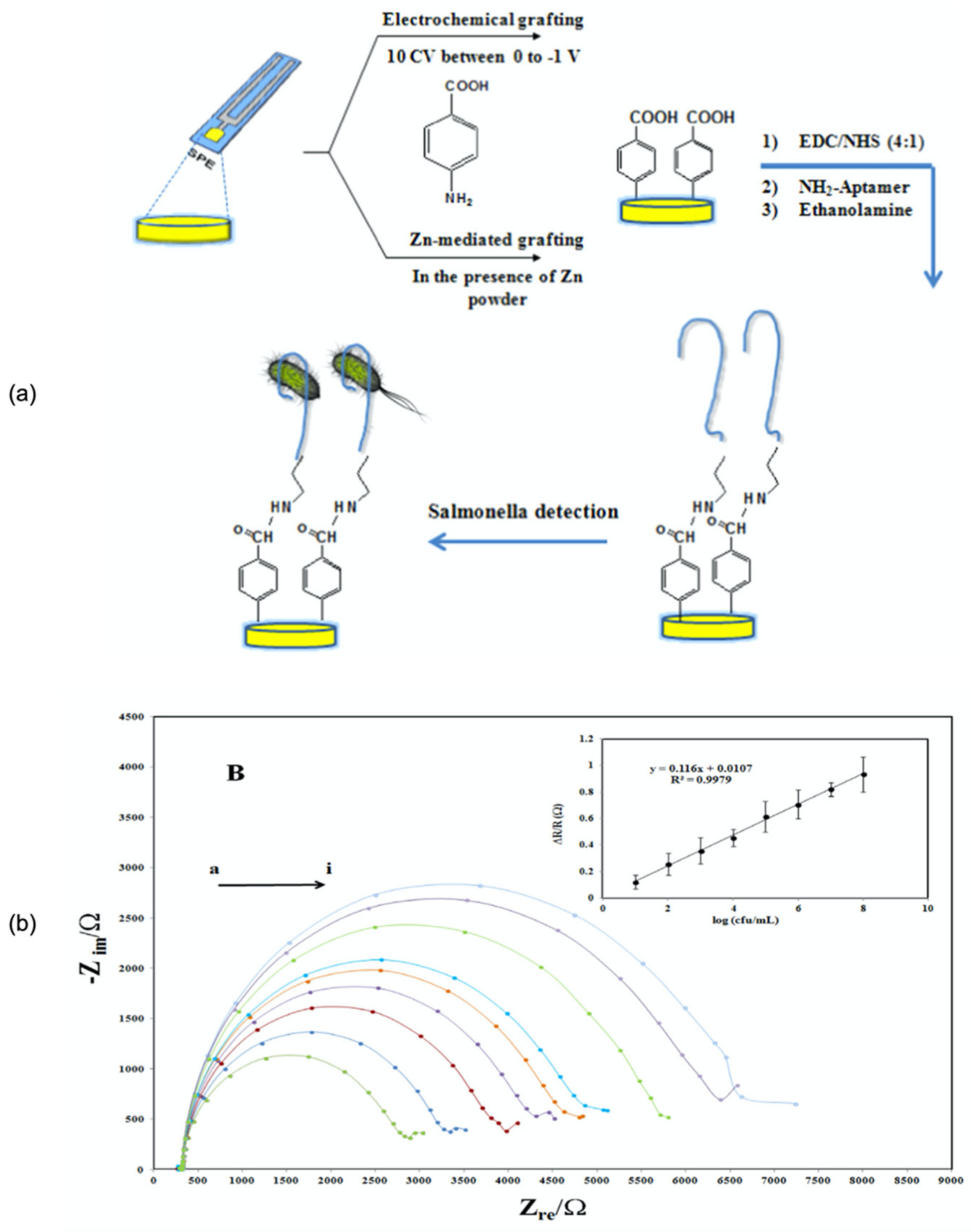
- Bagheryan, Z.; Raoof, J.-B.; Golabi, M.; Turner, A.P.F.; Beni, V. Diazonium-based impedimetric aptasensor for the rapid label-free detection of Salmonella typhimurium in food sample. Biosens. Bioelectron. 2016, 80, 566–573. [Google Scholar] [CrossRef] [Green Version]
- Malvano, F.; Pilloton, R.; Albanese, D. A novel impedimetric biosensor based on the antimicrobial activity of the peptide nisin for the detection of Salmonella spp. Food Chem. 2020, 325, 126868. [Google Scholar] [CrossRef] [PubMed]
- Melo, A.M.A.; Furtado, R.F.; de Fatima Borges, M.; Biswas, A.; Cheng, H.N.; Alves, C.R. Performance of an amperometric immunosensor assembled on carboxymethylated cashew gum for Salmonella detection. Microchem. J. 2021, 167, 106268. [Google Scholar] [CrossRef]
- Jia, F.; Duan, N.; Wu, S.; Dai, R.; Wang, Z.; Li, X. Impedimetric Salmonella aptasensor using a glassy carbon electrode modified with an electrodeposited composite consisting of reduced graphene oxide and carbon nanotubes. Microchim. Acta 2016, 183, 337–344. [Google Scholar] [CrossRef]
- Velusamy, V.; Arshak, K.; Korostynska, O.; Oliwa, K.; Adley, C. An overview of foodborne pathogen detection: In the perspective of biosensors. Biotechnol. Adv. 2010, 28, 232–254. [Google Scholar] [CrossRef]
- Cordova-Huaman, A.V.; Jauja-Ccana, V.R.; La Rosa-Toro, A. Low-cost smartphone-controlled potentiostat based on Arduino for teaching electrochemistry fundamentals and applications. Heliyon 2021, 7, e06259. [Google Scholar] [CrossRef]
- Dos Santos Schneid, A.; Rodrigues, K.L.; Chemello, D.; Tondo, E.C.; Ayub, M.A.Z.; Aleixo, J.A.G. Evaluation of an indirect ELISA for the detection of Salmonella in chicken meat. Braz. J. Microbiol. 2006, 37, 350–355. [Google Scholar] [CrossRef] [Green Version]
- Jesudason, M.V.; Sridharan, G.; Mukundan, S.; John, T.J. Vi-specific latex agglutination for early and rapid detection of Salmonella serotype typhi in blood cultures. Diagn. Microbiol. Infect. Dis. 1994, 18, 75–78. [Google Scholar] [CrossRef]
- Singh, J.; Sharma, S.; Nara, S. Nanogold based lateral flow assay for the detection of Salmonella typhi in environmental water samples. Anal. Methods 2015, 7, 9281–9288. [Google Scholar] [CrossRef]
- Kumar, R.; Surendran, P.K.; Thampuran, N. Evaluation of culture, ELISA and PCR assays for the detection of Salmonella in seafood. Lett. Appl. Microbiol. 2008, 46, 221–226. [Google Scholar] [CrossRef] [PubMed]
- Ogunremi, D.; Dupras, A.A.; Naushad, S.; Gao, R.; Duceppe, M.-O.; Omidi, K.; Márquez, I.G.; Huang, H.; Goodridge, L.; Lévesque, R.C.; et al. A New Whole Genome Culture-Independent Diagnostic Test (WG-CIDT) for Rapid Detection of Salmonella in Lettuce. Front. Microbiol. 2020, 11, 602. [Google Scholar] [CrossRef] [PubMed]
- Kuhns, M.; Zautner, A.E.; Rabsch, W.; Zimmermann, O.; Weig, M.; Bader, O.; Groß, U. Rapid discrimination of Salmonella enterica serovar Typhi from other serovars by MALDI-TOF mass spectrometry. PLoS ONE 2012, 7, e40004. [Google Scholar]
- Appaturi, J.N.; Pulingam, T.; Thong, K.L.; Muniandy, S.; Ahmad, N.; Leo, B.F. Rapid and sensitive detection of Salmonella with reduced graphene oxide-carbon nanotube based electrochemical aptasensor. Anal. Biochem. 2020, 589, 113489. [Google Scholar] [CrossRef]
- Bhalla, N.; Jolly, P.; Formisano, N.; Estrela, P. Introduction to biosensors. Essays Biochem. 2016, 60, 1–8. [Google Scholar] [CrossRef] [Green Version]
- Azizi, S.; Gholivand, M.-B.; Amiri, M.; Manouchehri, I. DNA biosensor based on surface modification of ITO by physical vapor deposition of gold and carbon quantum dots modified with neutral red as an electrochemical redox probe. Microchem. J. 2020, 159, 105523. [Google Scholar] [CrossRef]
- Ge, C.; Yuan, R.; Yi, L.; Yang, J.; Zhang, H.; Li, L.; Nian, W.; Yi, G. Target-induced aptamer displacement on gold nanoparticles and rolling circle amplification for ultrasensitive live Salmonella typhimurium electrochemical biosensing. J. Electroanal. Chem. 2018, 826, 174–180. [Google Scholar] [CrossRef]
- Purcell, R.V.; Pearson, J.; Frizelle, F.A.; Keenan, J.I. Comparison of standard, quantitative and digital PCR in the detection of enterotoxigenic Bacteroides fragilis. Sci. Rep. 2016, 6, 34554. [Google Scholar] [CrossRef] [PubMed] [Green Version]
- Hyeon, J.-Y.; Chon, J.-W.; Choi, I.-S.; Park, C.; Kim, D.-E.; Seo, K.-H. Development of RNA aptamers for detection of Salmonella Enteritidis. J. Microbiol. Methods 2012, 89, 79–82. [Google Scholar] [CrossRef] [PubMed]
- Sheikhzadeh, E.; Chamsaz, M.; Turner, A.P.F.; Jager, E.W.H.; Beni, V. Label-free impedimetric biosensor for Salmonella Typhimurium detection based on poly [pyrrole-co-3-carboxyl-pyrrole] copolymer supported aptamer. Biosens. Bioelectron. 2016, 80, 194–200. [Google Scholar] [CrossRef] [Green Version]
- Duan, Y.F.; Ning, Y.; Song, Y.; Deng, L. Fluorescent aptasensor for the determination of Salmonella typhimurium based on a graphene oxide platform. Microchim. Acta 2014, 181, 647–653. [Google Scholar] [CrossRef]
- Clark, L.C., Jr.; Lyons, C. Electrode systems for continuous monitoring in cardiovascular surgery. Ann. N. Y. Acad. Sci. 1962, 102, 29–45. [Google Scholar] [CrossRef]
- Yoo, E.-H.; Lee, S.-Y. Glucose Biosensors: An Overview of Use in Clinical Practice. Sensors 2010, 10, 4558–4576. [Google Scholar] [CrossRef] [Green Version]
- Jayanthi, V.S.A.; Das, A.B.; Saxena, U. Recent advances in biosensor development for the detection of cancer biomarkers. Biosens. Bioelectron. 2017, 91, 15–23. [Google Scholar] [CrossRef]
- Ameri, M.; Shabaninejad, Z.; Movahedpour, A.; Sahebkar, A.; Mohammadi, S.; Hosseindoost, S.; Ebrahimi, M.S.; Savardashtaki, A.; Karimipour, M.; Mirzaei, H. Biosensors for detection of Tau protein as an Alzheimer’s disease marker. Int. J. Biol. Macromol. 2020, 162, 1100–1108. [Google Scholar] [CrossRef]
- Lowe, C.R. Biosensors. Trends Biotechnol. 1984, 2, 59–65. [Google Scholar] [CrossRef]
- Grieshaber, D.; MacKenzie, R.; Vörös, J.; Reimhult, E. Electrochemical Biosensors—Sensor Principles and Architectures. Sensors 2008, 8, 1400–1458. [Google Scholar] [CrossRef]
- Kumar, A.; Malinee, M.; Dhiman, A.; Kumar, A.; Sharma, T.K. Chapter 2—Aptamer Technology for the Detection of Foodborne Pathogens and Toxins. In Advance Biosensors for Healthcare Application; Elsevier: Amsterdam, The Netherlands, 2019; pp. 45–69. ISBN 978-0-12-815743-5. [Google Scholar]
- Shen, Y.; Xu, L.; Li, Y. Biosensors for rapid detection of Salmonella in food: A review. Compr. Rev. Food Sci. Food Saf. 2021, 20, 149–197. [Google Scholar] [CrossRef]
- Nayak, M.; Kotian, A.; Marathe, S.; Chakravortty, D. Detection of microorganisms using biosensors—A smarter way towards detection techniques. Biosens. Bioelectron. 2009, 25, 661–667. [Google Scholar] [CrossRef] [PubMed]
- Evtugyn, G. Biosensors: Essentials; Springer: Berlin/Heidelberg, Germany, 2014; Volume 84, pp. 21–97. [Google Scholar]
- Singh, R.; Mukherjee, M.D.; Sumana, G.; Gupta, R.K.; Sood, S.; Malhotra, B.D. Biosensors for pathogen detection: A smart approach towards clinical diagnosis. Sens. Actuators B Chem. 2014, 197, 385–404. [Google Scholar] [CrossRef]
- Bergveld, P. A critical evaluation of direct electrical protein detection methods. Biosens. Bioelectron. 1991, 6, 55–72. [Google Scholar] [CrossRef] [Green Version]
- De Moraes, A.C.; Kubota, L.T. Recent Trends in Field-Effect Transistors-Based Immunosensors. Chemosens. 2016, 4, 20. [Google Scholar] [CrossRef] [Green Version]
- Atta, N.F.; Galal, A.; El-Ads, E.H. Graphene—a platform for sensor and biosensor applications. Biosens.-Micro Nanoscale Appl. 2015, 9, 38–84. [Google Scholar]
- Shanker, A.; Lee, K.; Kim, J. Chapter 12: Synthetic Hybrid Biosensors. In Nature Reviews Molecular Cell Biology; Wiley-VCH Verlag GmbH & Co. KgaA: Weinheim, Germany, 2014; pp. 359–386. [Google Scholar]
- Malhotra, B.D.; Ali, M.A. Nanomaterials in biosensors: Fundamentals and applications. Nanomater. Biosens. 2018, 1, 1–74. [Google Scholar]
- Zelada-Guillén, G.A.; Blondeau, P.; Rius, F.X.; Riu, J. Carbon nanotube-based aptasensors for the rapid and ultrasensitive detection of bacteria. Methods 2013, 63, 233–238. [Google Scholar] [CrossRef]
- Silva, N.F.D.; Magalhães, J.M.C.S.; Oliva-Teles, M.T.; Delerue-Matos, C. A potentiometric magnetic immunoassay for rapid detection of Salmonella typhimurium. Anal. Methods 2015, 7, 4008–4011. [Google Scholar] [CrossRef]
- Silva, N.F.D.; Almeida, C.M.R.; Magalhães, J.M.C.S.; Gonçalves, M.P.; Freire, C.; Delerue-Matos, C. Development of a disposable paper-based potentiometric immunosensor for real-time detection of a foodborne pathogen. Biosens. Bioelectron. 2019, 141, 111317. [Google Scholar] [CrossRef]
- Punbusayakul, N.; Talapatra, S.; Ajayan, P.M.; Surareungchai, W. Label-free as-grown double wall carbon nanotubes bundles for Salmonella typhimurium immunoassay. Chem. Cent. J. 2013, 7, 102. [Google Scholar] [CrossRef] [Green Version]
- Melo, A.M.A.; Alexandre, D.L.; Oliveira, M.R.F.; Furtado, R.F.; Borges, M.F.; Ribeiro, P.R.V.; Biswas, A.; Cheng, H.N.; Alves, C.R.; Figueiredo, E.A.T. Optimization and characterization of a biosensor assembly for detection of Salmonella Typhimurium. J. Solid State Electrochem. 2018, 22, 1321–1330. [Google Scholar] [CrossRef]
- Singh, C.; Ali, M.A.; Kumar, V.; Ahmad, R.; Sumana, G. Functionalized MoS2 nanosheets assembled microfluidic immunosensor for highly sensitive detection of food pathogen. Sens. Actuators B Chem. 2018, 259, 1090–1098. [Google Scholar] [CrossRef]
- Pal, N.; Sharma, S.; Gupta, S. Sensitive and rapid detection of pathogenic bacteria in small volumes using impedance spectroscopy technique. Biosens. Bioelectron. 2016, 77, 270–276. [Google Scholar] [CrossRef]
- Wang, L.; Huo, X.; Qi, W.; Xia, Z.; Li, Y.; Lin, J. Rapid and sensitive detection of Salmonella Typhimurium using nickel nanowire bridge for electrochemical impedance amplification. Talanta 2020, 211, 120715. [Google Scholar] [CrossRef]
- Afonso, A.S.; Pérez-López, B.; Faria, R.C.; Mattoso, L.H.C.; Hernández-Herrero, M.; Roig-Sagués, A.X.; Maltez-da Costa, M.; Merkoçi, A. Electrochemical detection of Salmonella using gold nanoparticles. Biosens. Bioelectron. 2013, 40, 121–126. [Google Scholar] [CrossRef]
- Fathi, S.; Saber, R.; Adabi, M.; Rasouli, R.; Douraghi, M.; Morshedi, M.; Farid-Majidi, R. Novel Competitive Voltammetric Aptasensor Based on Electrospun Carbon Nanofibers-Gold Nanoparticles Modified Graphite Electrode for Salmonella enterica serovar Detection. Biointerface Res. Appl. Chem. 2021, 11, 8702–8715. [Google Scholar]
- De Oliveira, T.R.; Martucci, D.H.; Faria, R.C. Simple disposable microfluidic device for Salmonella typhimurium detection by magneto-immunoassay. Sens. Actuators B Chem. 2018, 255, 684–691. [Google Scholar] [CrossRef]
- Dai, G.; Li, Z.; Luo, F.; Ai, S.; Chen, B.; Wang, Q. Electrochemical determination of Salmonella typhimurium by using aptamer-loaded gold nanoparticles and a composite prepared from a metal-organic framework (type UiO-67) and graphene. Microchim. Acta 2019, 186, 620. [Google Scholar] [CrossRef] [PubMed]
- Anany, H.; Chou, Y.; Cucic, S.; Derda, R.; Evoy, S.; Griffiths, M.W. From bits and pieces to whole phage to nanomachines: Pathogen detection using bacteriophages. Annu. Rev. Food Sci. Technol. 2017, 8, 305–329. [Google Scholar] [CrossRef]
- Morales, M.A.; Halpern, J.M. Guide to Selecting a Biorecognition Element for Biosensors. Bioconjug. Chem. 2018, 29, 3231–3239. [Google Scholar] [CrossRef] [PubMed]
- Melo, A.M.A.; Alexandre, D.L.; Furtado, R.F.; Borges, M.F.; Figueiredo, E.A.T.; Biswas, A.; Cheng, H.N.; Alves, C.R. Electrochemical immunosensors for Salmonella detection in food. Appl. Microbiol. Biotechnol. 2016, 100, 5301–5312. [Google Scholar] [CrossRef]
- Gizeli, E.; Lowe, C.R. Immunosensors. Curr. Opin. Biotechnol. 1996, 7, 66–71. [Google Scholar] [CrossRef]
- Alexandre, D.L.; Melo, A.M.A.; Furtado, R.F.; Borges, M.F.; Figueiredo, E.A.T.; Biswas, A.; Cheng, H.N.; Alves, C.R. A Rapid and Specific Biosensor for Salmonella Typhimurium Detection in Milk. Food Bioprocess Technol. 2018, 11, 748–756. [Google Scholar] [CrossRef]
- Sannigrahi, S.; Arumugasamy, S.K.; Mathiyarasu, J.; Suthindhiran, K. Magnetosome-anti-Salmonella antibody complex based biosensor for the detection of Salmonella typhimurium. Mater. Sci. Eng. C 2020, 114, 111071. [Google Scholar] [CrossRef] [PubMed]
- Bu, S.-J.; Wang, K.-Y.; Liu, X.; Ma, L.; Wei, H.-G.; Zhang, W.-G.; Liu, W.-S.; Wan, J.-Y. Ferrocene-functionalized nanocomposites as signal amplification probes for electrochemical immunoassay of Salmonella typhimurium. Microchim. Acta 2020, 187, 1–8. [Google Scholar] [CrossRef] [PubMed]
- Kasman, L.M.; Porter, L.D. Bacteriophages; [Updated 1 October 2020]. In StatPearls [Internet]; StatPearls Publishing: Treasure Island, FL, USA, 2020. Available online: https://0-www-ncbi-nlm-nih-gov.brum.beds.ac.uk/books/NBK493185/ (accessed on 8 September 2021).
- Vinay, M.; Franche, N.; Grégori, G.; Fantino, J.-R.; Pouillot, F.; Ansaldi, M. Phage-based fluorescent biosensor prototypes to specifically detect enteric bacteria such as E. coli and Salmonella enterica Typhimurium. PLoS ONE 2015, 10, e0131466. [Google Scholar] [CrossRef]
- Chai, Y.; Li, S.; Horikawa, S.; Park, M.-K.; Vodyanoy, V.; Chin, B.A. Rapid and Sensitive Detection of Salmonella Typhimurium on Eggshells by Using Wireless Biosensors. J. Food Prot. 2012, 75, 631–636. [Google Scholar] [CrossRef]
- Qiao, Z.; Lei, C.; Fu, Y.; Li, Y. Rapid and sensitive detection of E. coli O157: H7 based on antimicrobial peptide functionalized magnetic nanoparticles and urease-catalyzed signal amplification. Anal. Methods 2017, 9, 5204–5210. [Google Scholar] [CrossRef]
- Patel, S.; Akhtar, N. Antimicrobial peptides (AMPs): The quintessential ‘offense and defense’molecules are more than antimicrobials. Biomed. Pharmacother. 2017, 95, 1276–1283. [Google Scholar] [CrossRef]
- Brogden, K.A. Antimicrobial peptides: Pore formers or metabolic inhibitors in bacteria? Nat. Rev. Microbiol. 2005, 3, 238–250. [Google Scholar] [CrossRef]
- Hoyos-Nogués, M.; Gil, F.J.; Mas-Moruno, C. Antimicrobial Peptides: Powerful Biorecognition Elements to Detect Bacteria in Biosensing Technologies. Molecules 2018, 23, 1683. [Google Scholar] [CrossRef] [Green Version]
- Mannoor, M.S.; Zhang, S.; Link, A.J.; McAlpine, M.C. Electrical detection of pathogenic bacteria via immobilized antimicrobial peptides. Proc. Natl. Acad. Sci. USA 2010, 107, 19207–19212. [Google Scholar] [CrossRef] [Green Version]
- Datta, M.; Desai, D.; Kumar, A. Gene Specific DNA Sensors for Diagnosis of Pathogenic Infections. Indian J. Microbiol. 2017, 57, 139–147. [Google Scholar] [CrossRef] [PubMed]
- Vanegas, D.C.; Gomes, C.L.; Cavallaro, N.D.; Giraldo-Escobar, D.; McLamore, E.S. Emerging biorecognition and transduction schemes for rapid detection of pathogenic bacteria in food. Compr. Rev. Food Sci. Food Saf. 2017, 16, 1188–1205. [Google Scholar] [CrossRef] [Green Version]
- Das, R.; Sharma, M.K.; Rao, V.K.; Bhattacharya, B.K.; Garg, I.; Venkatesh, V.; Upadhyay, S. An electrochemical genosensor for Salmonella typhi on gold nanoparticles-mercaptosilane modified screen printed electrode. J. Biotechnol. 2014, 188, 9–16. [Google Scholar] [CrossRef]
- Park, K.S. Nucleic acid aptamer-based methods for diagnosis of infections. Biosens. Bioelectron. 2018, 102, 179–188. [Google Scholar] [CrossRef]
- Robati, R.Y.; Arab, A.; Ramezani, M.; Langroodi, F.A.; Abnous, K.; Taghdisi, S.M. Aptasensors for quantitative detection of kanamycin. Biosens. Bioelectron. 2016, 82, 162–172. [Google Scholar] [CrossRef]
- Davis, K.A.; Abrams, B.; Lin, Y.; Jayasena, S.D. Use of a high affinity DNA ligand in flow cytometry. Nucleic Acids Res. 1996, 24, 702–706. [Google Scholar] [CrossRef] [Green Version]
- Bayat, P.; Nosrati, R.; Alibolandi, M.; Rafatpanah, H.; Abnous, K.; Khedri, M.; Ramezani, M. SELEX methods on the road to protein targeting with nucleic acid aptamers. Biochimie 2018, 154, 132–155. [Google Scholar] [CrossRef]
- Wolter, A.C.; Weickhmann, A.K.; Nasiri, A.H.; Hantke, K.; Ohlenschläger, O.; Wunderlich, C.H.; Kreutz, C.; Duchardt-Ferner, E.; Wöhnert, J. A Stably Protonated Adenine Nucleotide with a Highly Shifted pKa Value Stabilizes the Tertiary Structure of a GTP-Binding RNA Aptamer. Angew. Chem. Int. Ed. 2017, 56, 401–404. [Google Scholar] [CrossRef]
- Zhou, J.; Satheesan, S.; Li, H.; Weinberg, M.S.; Morris, K.V.; Burnett, J.C.; Rossi, J.J. Cell-specific RNA aptamer against human CCR5 specifically targets HIV-1 susceptible cells and inhibits HIV-1 infectivity. Chem. Biol. 2015, 22, 379–390. [Google Scholar] [CrossRef] [PubMed] [Green Version]
- Wang, C.; Lin, B.; Chen, C. An aptamer targeting shared tumor-specific peptide antigen of MAGE-A3 in multiple cancers. Int. J. Cancer 2016, 138, 918–926. [Google Scholar] [CrossRef] [PubMed] [Green Version]
- Huang, Y.; Chen, X.; Duan, N.; Wu, S.; Wang, Z.; Wei, X.; Wang, Y. Selection and characterization of DNA aptamers against Staphylococcus aureus enterotoxin C1. Food Chem. 2015, 166, 623–629. [Google Scholar] [CrossRef] [PubMed]
- Mehlhorn, A.; Rahimi, P.; Joseph, Y. Aptamer-based biosensors for antibiotic detection: A review. Biosensors 2018, 8, 54. [Google Scholar] [CrossRef] [Green Version]
- Shrivastava, S.; Sohn, I.-Y.; Son, Y.-M.; Lee, W.-I.; Lee, N.-E. Real-time label-free quantitative fluorescence microscopy-based detection of ATP using a tunable fluorescent nano-aptasensor platform. Nanoscale 2015, 7, 19663–19672. [Google Scholar] [CrossRef] [PubMed]
- Lavu, P.S.R.; Mondal, B.; Ramlal, S.; Murali, H.S.; Batra, H.V. Selection and characterization of aptamers using a modified whole cell bacterium SELEX for the detection of Salmonella enterica serovar typhimurium. ACS Comb. Sci. 2016, 18, 292–301. [Google Scholar] [CrossRef]
- Park, H.-C.; Baig, I.A.; Lee, S.-C.; Moon, J.-Y.; Yoon, M.-Y. Development of ssDNA Aptamers for the Sensitive Detection of Salmonella typhimurium and Salmonella enteritidis. Appl. Biochem. Biotechnol. 2014, 174, 793–802. [Google Scholar] [CrossRef]
- Renuka, R.; Maroli, N.; Achuth, J.; Ponmalai, K.; Kadirvelu, K. Highly adaptable and sensitive FRET-based aptamer assay for the detection of Salmonella paratyphi A. Spectrochim. Acta Part A Mol. Biomol. Spectrosc. 2020, 243, 118662. [Google Scholar] [CrossRef]
- Dinshaw, I.J.; Muniandy, S.; Teh, S.J.; Ibrahim, F.; Leo, B.F.; Thong, K.L. Development of an aptasensor using reduced graphene oxide chitosan complex to detect Salmonella. J. Electroanal. Chem. 2017, 806, 88–96. [Google Scholar] [CrossRef] [Green Version]
- Ma, X.; Jiang, Y.; Jia, F.; Yu, Y.; Chen, J.; Wang, Z. An aptamer-based electrochemical biosensor for the detection of Salmonella. J. Microbiol. Methods 2014, 98, 94–98. [Google Scholar] [CrossRef]
- Fang, S.; Song, D.; Zhuo, Y.; Chen, Y.; Zhu, A.; Long, F. Simultaneous and sensitive determination of Escherichia coli O157:H7 and Salmonella Typhimurium using evanescent wave dual-color fluorescence aptasensor based on micro/nano size effect. Biosens. Bioelectron. 2021, 185, 113288. [Google Scholar] [CrossRef]
- Muniandy, S.; Teh, S.J.; Appaturi, J.N.; Thong, K.L.; Lai, C.W.; Ibrahim, F.; Leo, B.F. A reduced graphene oxide-titanium dioxide nanocomposite based electrochemical aptasensor for rapid and sensitive detection of Salmonella enterica. Bioelectrochemistry 2019, 127, 136–144. [Google Scholar] [CrossRef] [PubMed]
- Pathania, P.K.; Saini, J.K.; Vij, S.; Tewari, R.; Sabherwal, P.; Rishi, P.; Suri, C.R. Aptamer functionalized MoS2-rGO nanocomposite based biosensor for the detection of Vi antigen. Biosens. Bioelectron. 2018, 122, 121–126. [Google Scholar] [CrossRef] [PubMed]
- Hasan, M.R.; Pulingam, T.; Appaturi, J.N.; Zifruddin, A.N.; Teh, S.J.; Lim, T.W.; Ibrahim, F.; Leo, B.F.; Thong, K.L. Carbon nanotube-based aptasensor for sensitive electrochemical detection of whole-cell Salmonella. Anal. Biochem. 2018, 554, 34–43. [Google Scholar] [CrossRef] [PubMed]
- Jaiswal, N.; Tiwari, I. Recent build outs in electroanalytical biosensors based on carbon-nanomaterial modified screen printed electrode platforms. Anal. Methods 2017, 9, 3895–3907. [Google Scholar] [CrossRef]
- Barton, J.; García, M.B.G.; Santos, D.H.; Fanjul-Bolado, P.; Ribotti, A.; McCaul, M.; Diamond, D.; Magni, P. Screen-printed electrodes for environmental monitoring of heavy metal ions: A review. Microchim. Acta 2016, 183, 503–517. [Google Scholar] [CrossRef]
- Shi, Z.; Lu, Y.; Chen, Z.; Cheng, C.; Xu, J.; Zhang, Q.; Yan, Z.; Luo, Z.; Liu, Q. Electrochemical non-enzymatic sensing of glycoside toxins by boronic acid functionalized nano-composites on screen-printed electrode. Sens. Actuators B Chem. 2021, 329, 129197. [Google Scholar] [CrossRef]
- Munteanu, F.-D.; Titoiu, A.M.; Marty, J.-L.; Vasilescu, A. Detection of Antibiotics and Evaluation of Antibacterial Activity with Screen-Printed Electrodes. Sensors 2018, 18, 901. [Google Scholar] [CrossRef] [PubMed] [Green Version]
- Torre, R.; Costa-Rama, E.; Nouws, H.P.A.; Delerue-Matos, C. Screen-Printed Electrode-Based Sensors for Food Spoilage Control: Bacteria and Biogenic Amines Detection. Biosens. 2020, 10, 139. [Google Scholar] [CrossRef]
- Alonso-Lomillo, M.A.; Domínguez-Renedo, O.; Arcos-Martínez, M.J. Screen-printed biosensors in microbiology; a review. Talanta 2010, 82, 1629–1636. [Google Scholar] [CrossRef]
- Ferrari, A.G.-M.; Carrington, P.; Rowley-Neale, S.J.; Banks, C.E. Recent advances in portable heavy metal electrochemical sensing platforms. Environ. Sci. Water Res. Technol. 2020, 6, 2676–2690. [Google Scholar] [CrossRef]
- Choudhry, N.A.; Kampouris, D.K.; Kadara, R.O.; Jenkinson, N.; Banks, C.E. Next generation screen printed electrochemical platforms: Non-enzymatic sensing of carbohydrates using copper (II) oxide screen printed electrodes. Anal. Methods 2009, 1, 183–187. [Google Scholar] [CrossRef] [PubMed]
- Choudhry, N.A.; Kadara, R.O.; Jenkinson, N.; Banks, C.E. Screen printed electrodes provide micro-domain sites for fabricating disposable electro-catalytic ensembles. Electrochem. Commun. 2010, 12, 406–409. [Google Scholar] [CrossRef]
- Fei, J.; Dou, W.; Zhao, G. A sandwich electrochemical immunosensor for Salmonella pullorum and Salmonella gallinarum based on a screen-printed carbon electrode modified with an ionic liquid and electrodeposited gold nanoparticles. Microchim. Acta 2015, 182, 2267–2275. [Google Scholar] [CrossRef]
- Salam, F.; Tothill, I.E. Detection of Salmonella typhimurium using an electrochemical immunosensor. Biosens. Bioelectron. 2009, 24, 2630–2636. [Google Scholar] [CrossRef] [PubMed]
- Farka, Z.; Juřík, T.; Pastucha, M.; Kovář, D.; Lacina, K.; Skládal, P. Rapid Immunosensing of Salmonella Typhimurium Using Electrochemical Impedance Spectroscopy: The Effect of Sample Treatment. Electroanalysis 2016, 28, 1803–1809. [Google Scholar] [CrossRef]
- Fei, J.; Dou, W.; Zhao, G. A sandwich electrochemical immunoassay for Salmonella pullorum and Salmonella gallinarum based on a AuNPs/SiO2/Fe3O4 adsorbing antibody and 4 channel screen printed carbon electrode electrodeposited gold nanoparticles. RSC Adv. 2015, 5, 74548–74556. [Google Scholar] [CrossRef]
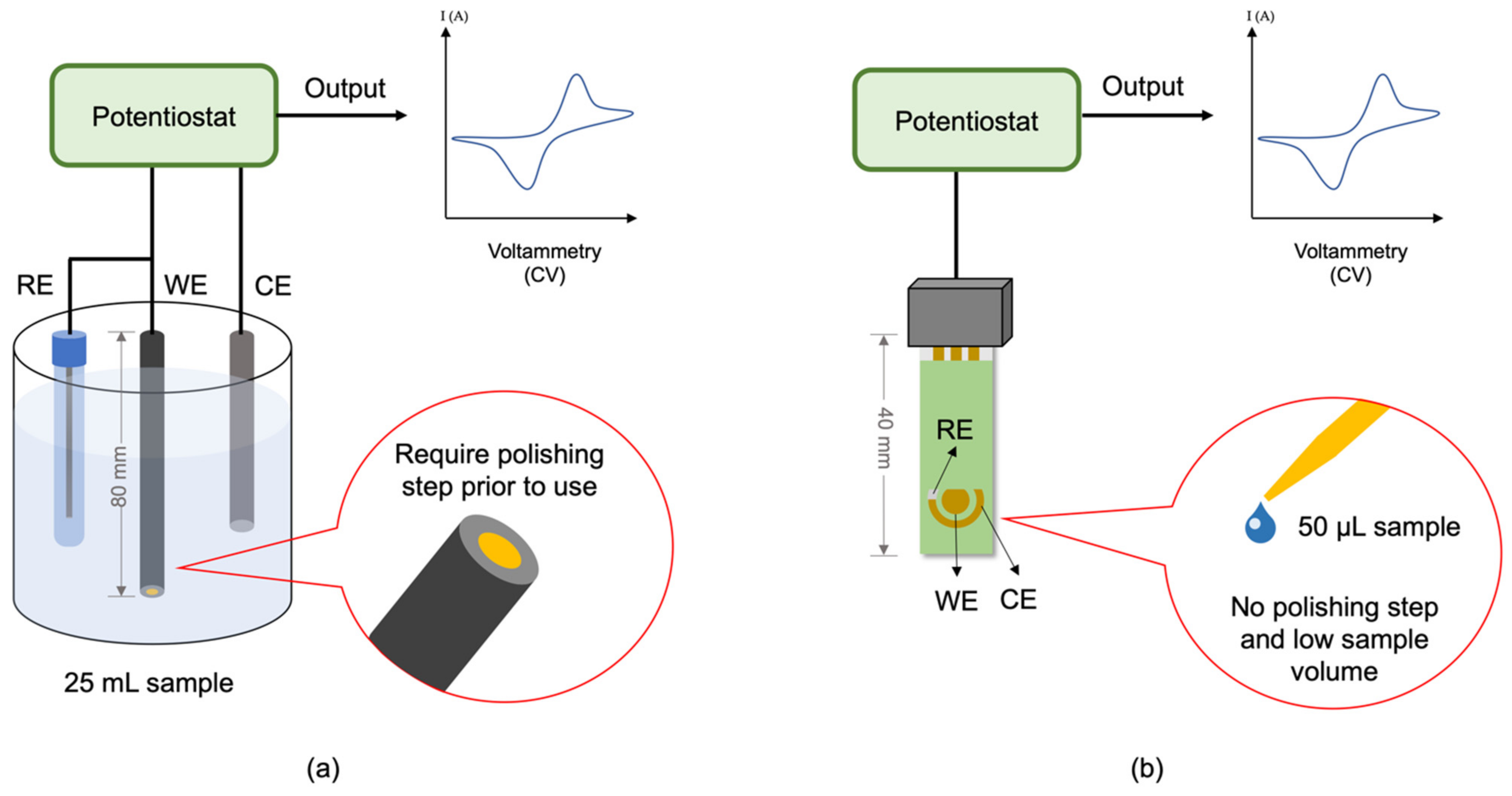
- Dahlin, A.B. Size Matters: Problems and Advantages Associated with Highly Miniaturized Sensors. Sensors 2012, 12, 3018–3036. [Google Scholar] [CrossRef] [PubMed] [Green Version]
- Sannomiya, T.; Vörös, J. Single plasmonic nanoparticles for biosensing. Trends Biotechnol. 2011, 29, 343–351. [Google Scholar] [CrossRef] [PubMed]
- Muniandy, S.; Teh, S.J.; Thong, K.L.; Thiha, A.; Dinshaw, I.J.; Lai, C.W.; Ibrahim, F.; Leo, B.F. Carbon nanomaterial-based electrochemical biosensors for foodborne bacterial detection. Crit. Rev. Anal. Chem. 2019, 49, 510–533. [Google Scholar] [CrossRef]
- Cho, I.-H.; Kim, D.H.; Park, S. Electrochemical biosensors: Perspective on functional nanomaterials for on-site analysis. Biomater. Res. 2020, 24, 1–12. [Google Scholar] [CrossRef] [PubMed] [Green Version]
- Sánchez-Tirado, E.; González-Cortés, A.; Yáñez-Sedeño, P.; Pingarrón, J.M. Carbon nanotubes functionalized by click chemistry as scaffolds for the preparation of electrochemical immunosensors. Application to the determination of TGF-beta 1 cytokine. Analyst 2016, 141, 5730–5737. [Google Scholar] [CrossRef] [PubMed] [Green Version]
- Eatemadi, A.; Daraee, H.; Karimkhanloo, H.; Kouhi, M.; Zarghami, N.; Akbarzadeh, A.; Abasi, M.; Hanifehpour, Y.; Joo, S.W. Carbon nanotubes: Properties, synthesis, purification, and medical applications. Nanoscale Res. Lett. 2014, 9, 393. [Google Scholar] [CrossRef] [PubMed] [Green Version]
- Bai, Y.; Xu, T.; Zhang, X. Graphene-Based Biosensors for Detection of Biomarkers. Micromachines 2020, 11, 60. [Google Scholar] [CrossRef] [PubMed] [Green Version]
- Novoselov, K.S.; Geim, A.K.; Morozov, S.V.; Jiang, D.; Zhang, Y.; Dubonos, S.V.; Grigorieva, I.V.; Firsov, A.A. Electric field effect in atomically thin carbon films. Science 2004, 306, 666–669. [Google Scholar] [CrossRef] [PubMed] [Green Version]
- Randviir, E.P.; Brownson, D.A.C.; Banks, C.E. A decade of graphene research: Production, applications and outlook. Mater. Today 2014, 17, 426–432. [Google Scholar] [CrossRef]
- Cho, I.-H.; Lee, J.; Kim, J.; Kang, M.; Paik, J.K.; Ku, S.; Cho, H.-M.; Irudayaraj, J.; Kim, D.-H. Current technologies of electrochemical immunosensors: Perspective on signal amplification. Sensors 2018, 18, 207. [Google Scholar] [CrossRef] [Green Version]
- Kuila, T.; Bose, S.; Khanra, P.; Mishra, A.K.; Kim, N.H.; Lee, J.H. Recent advances in graphene-based biosensors. Biosens. Bioelectron. 2011, 26, 4637–4648. [Google Scholar] [CrossRef] [PubMed]
- Zhu, Y.; Murali, S.; Cai, W.; Li, X.; Suk, J.W.; Potts, J.R.; Ruoff, R.S. Graphene and Graphene Oxide: Synthesis, Properties, and Applications. Adv. Mater. 2010, 22, 3906–3924. [Google Scholar] [CrossRef]
- Smith, A.T.; LaChance, A.M.; Zeng, S.; Liu, B.; Sun, L. Synthesis, properties, and applications of graphene oxide/reduced graphene oxide and their nanocomposites. Nano Mater. Sci. 2019, 1, 31–47. [Google Scholar] [CrossRef]
- Xue, Y. Chapter 11—Carbon Nanotubes for Biomedical Applications. In Micro and Nano Technologies; Elsevier: Amsterdam, The Netherlands, 2017; pp. 323–346. ISBN 978-0-323-41481-4. [Google Scholar]
- Zhang, X.; Li, C.-R.; Wang, W.-C.; Xue, J.; Huang, Y.-L.; Yang, X.-X.; Tan, B.; Zhou, X.-P.; Shao, C.; Ding, S.-J. A novel electrochemical immunosensor for highly sensitive detection of aflatoxin B1 in corn using single-walled carbon nanotubes/chitosan. Food Chem. 2016, 192, 197–202. [Google Scholar] [CrossRef] [PubMed]
- Soares, R.R.A.; Hjort, R.G.; Pola, C.C.; Parate, K.; Reis, E.L.; Soares, N.F.F.; McLamore, E.S.; Claussen, J.C.; Gomes, C.L. Laser-Induced Graphene Electrochemical Immunosensors for Rapid and Label-Free Monitoring of Salmonella enterica in Chicken Broth. ACS Sens. 2020, 5, 1900–1911. [Google Scholar] [CrossRef]
- Ye, Y.; Yan, W.; Liu, Y.; He, S.; Cao, X.; Xu, X.; Zheng, H.; Gunasekaran, S. Electrochemical detection of Salmonella using an invA genosensor on polypyrrole-reduced graphene oxide modified glassy carbon electrode and AuNPs-horseradish peroxidase-streptavidin as nanotag. Anal. Chim. Acta 2019, 1074, 80–88. [Google Scholar] [CrossRef] [PubMed]
- Xu, S.; Duo, H.; Zheng, C.; Zhao, S.; Song, S.; Simon, G. Novel approach to fabrication of DNA Biosensor Based on a Carboxylated Graphene Oxide Decorated with Fe3O4 NPs for the Detection of Typhoidal Salmonella. Int. J. Electrochem. Sci 2019, 14, 1248–1269. [Google Scholar] [CrossRef]
- Quiton, P.A.; Carreon, B.M.; Cruz-Papa, D.M.D.; Bergantin, J., Jr. Bacteriophage-modified graphene oxide screen-printed electrodes for the impedimetric biosensing of Salmonella enterica serovar Typhimurium. Sens. Transducers 2018, 28, 38–42. [Google Scholar]
- Weber, J.E.; Pillai, S.; Ram, M.K.; Kumar, A.; Singh, S.R. Electrochemical impedance-based DNA sensor using a modified single walled carbon nanotube electrode. Mater. Sci. Eng. C 2011, 31, 821–825. [Google Scholar] [CrossRef]
- Yang, M.; Peng, Z.; Ning, Y.; Chen, Y.; Zhou, Q.; Deng, L. Highly specific and cost-efficient detection of Salmonella Paratyphi A combining aptamers with single-walled carbon nanotubes. Sensors 2013, 13, 6865–6881. [Google Scholar] [CrossRef] [PubMed]
- Quintela, I.A.; de los Reyes, B.G.; Lin, C.-S.; Wu, V.C.H. Simultaneous Colorimetric Detection of a Variety of Salmonella spp. in Food and Environmental Samples by Optical Biosensing Using Oligonucleotide-Gold Nanoparticles. Front. Microbiol. 2019, 10, 1138. [Google Scholar] [CrossRef] [PubMed]
- Xiang, C.; Li, R.; Adhikari, B.; She, Z.; Li, Y.; Kraatz, H.-B. Sensitive electrochemical detection of Salmonella with chitosan–gold nanoparticles composite film. Talanta 2015, 140, 122–127. [Google Scholar] [CrossRef]
- Ye, Y.; Liu, Y.; He, S.; Xu, X.; Cao, X.; Ye, Y.; Zheng, H. Ultrasensitive electrochemical DNA sensor for virulence invA gene of Salmonella using silver nanoclusters as signal probe. Sens. Actuators B Chem. 2018, 272, 53–59. [Google Scholar] [CrossRef]
- Huang, F.; Guo, R.; Xue, L.; Cai, G.; Wang, S.; Li, Y.; Liao, M.; Wang, M.; Lin, J. An Acid-Responsive Microfluidic Salmonella Biosensor Using Curcumin as Signal Reporter and ZnO-Capped Mesoporous Silica Nanoparticles for Signal Amplification. Sens. Actuators B Chem. 2020, 312, 127958. [Google Scholar] [CrossRef]
- Appaturi, J.N.; Pulingam, T.; Muniandy, S.; Dinshaw, I.J.; Fen, L.B.; Johan, M.R. Supported cobalt nanoparticles on graphene oxide/mesoporous silica for oxidation of phenol and electrochemical detection of H2O2 and Salmonella spp. Mater. Chem. Phys. 2019, 232, 493–505. [Google Scholar] [CrossRef]
- Lin, D.; Harris, K.D.; Chan, N.W.C.; Jemere, A.B. Nanostructured indium tin oxide electrodes immobilized with toll-like receptor proteins for label-free electrochemical detection of pathogen markers. Sens. Actuators B Chem. 2018, 257, 324–330. [Google Scholar] [CrossRef]
- Lobo-Castañón, M.J.; Barreda-Garcia, S.; Miranda-Castro, R.; Estrela, P. Electrochemical Detection of Salmonella via On-surface Isothermal Amplification of its Genetic Material onto Highly Stable and Reproducible Indium Tin Oxide Platforms. In Proceedings of the 3rd International Electronic Conference on Medical Chemistry, Online. 1–30 November 2017. [Google Scholar]
- Thiha, A.; Ibrahim, F.; Muniandy, S.; Dinshaw, I.J.; Teh, S.J.; Thong, K.L.; Leo, B.F.; Madou, M. All-carbon suspended nanowire sensors as a rapid highly-sensitive label-free chemiresistive biosensing platform. Biosens. Bioelectron. 2018, 107, 145–152. [Google Scholar] [CrossRef]
- Mustafa, M.R.B.; Dhahi, T.S.; Ehfaed, N.A.K.H.; Adam, T.; Hashim, U.; Azizah, N.; Mohammed, M.; Noriman, N.Z. Specific and selective target detection of supra-genome 21 Mers Salmonella via silicon nanowires biosensor. In Proceedings of 2017 International Conference on Biotechnology and Bioengineering (ICBB-2017), Offenburg, Germany, 26–28 September 2017; AIP Publishing LLC: New York, NY, USA, 2017; Volume 1885, p. 20196. [Google Scholar]
- Cao, X.; Ye, Y.; Liu, S. Gold nanoparticle-based signal amplification for biosensing. Anal. Biochem. 2011, 417, 1–16. [Google Scholar] [CrossRef] [PubMed]
- Rasheed, P.A.; Sandhyarani, N. Electrochemical DNA sensors based on the use of gold nanoparticles: A review on recent developments. Microchim. Acta 2017, 184, 981–1000. [Google Scholar] [CrossRef]
- Pingarrón, J.M.; Yáñez-Sedeño, P.; González-Cortés, A. Gold nanoparticle-based electrochemical biosensors. Electrochim. Acta 2008, 53, 5848–5866. [Google Scholar] [CrossRef]
- Lepoitevin, M.; Lemouel, M.; Bechelany, M.; Janot, J.-M.; Balme, S. Gold nanoparticles for the bare-eye based and spectrophotometric detection of proteins, polynucleotides and DNA. Microchim. Acta 2015, 182, 1223–1229. [Google Scholar] [CrossRef]
- Elghanian, R.; Storhoff, J.J.; Mucic, R.C.; Letsinger, R.L.; Mirkin, C.A. Selective Colorimetric Detection of Polynucleotides Based on the Distance-Dependent Optical Properties of Gold Nanoparticles. Science 1997, 277, 1078–1081. [Google Scholar] [CrossRef] [Green Version]
- Yang, Z.; Chai, Y.; Yuan, R.; Zhuo, Y.; Li, Y.; Han, J.; Liao, N. Hollow platinum decorated Fe3O4 nanoparticles as peroxidase mimetic couple with glucose oxidase for pseudobienzyme electrochemical immunosensor. Sens. Actuators B Chem. 2014, 193, 461–466. [Google Scholar] [CrossRef]
- Wang, S.-F.; Tan, Y.-M. A novel amperometric immunosensor based on Fe 3 O 4 magnetic nanoparticles/chitosan composite film for determination of ferritin. Anal. Bioanal. Chem. 2007, 387, 703–708. [Google Scholar] [CrossRef]
- Aznar, E.; Oroval, M.; Pascual, L.; Murguía, J.R.; Martinez-Manez, R.; Sancenon, F. Gated materials for on-command release of guest molecules. Chem. Rev. 2016, 116, 561–718. [Google Scholar] [CrossRef] [PubMed]
- Cao, X.; Liu, S.; Feng, Q.; Wang, N. Silver nanowire-based electrochemical immunoassay for sensing immunoglobulin G with signal amplification using strawberry-like ZnO nanostructures as labels. Biosens. Bioelectron. 2013, 49, 256–262. [Google Scholar] [CrossRef]
- Jimenez-Falcao, S.; Parra-Nieto, J.; Pérez-Cuadrado, H.; Martínez-Máñez, R.; Martínez-Ruiz, P.; Villalonga, R. Avidin-gated mesoporous silica nanoparticles for signal amplification in electrochemical biosensor. Electrochem. Commun. 2019, 108, 106556. [Google Scholar] [CrossRef]
- Chopra, K.L.; Major, S.; Pandya, D.K. Transparent conductors—A status review. Thin Solid Films 1983, 102, 1–46. [Google Scholar] [CrossRef]
- Tran, D.-P.; Lu, H.-I.; Lin, C.-K. Conductive characteristics of indium tin oxide thin film on polymeric substrate under long-term static deformation. Coatings 2018, 8, 212. [Google Scholar] [CrossRef] [Green Version]
- Rabbani, M.; Hoque, M.E.; Mahbub, Z. Bin Nanosensors in biomedical and environmental applications: Perspectives and prospects. In Nanofabrication for Smart Nanosensor Applications; Elsevier: Amsterdam, The Netherlands, 2020; pp. 163–186. [Google Scholar]
- He, B.; Morrow, T.J.; Keating, C.D. Nanowire sensors for multiplexed detection of biomolecules. Curr. Opin. Chem. Biol. 2008, 12, 522–528. [Google Scholar] [CrossRef] [Green Version]
- Nambiar, S.; Yeow, J.T.W. Conductive polymer-based sensors for biomedical applications. Biosens. Bioelectron. 2011, 26, 1825–1832. [Google Scholar] [CrossRef]
- Liu, J.; Agarwal, M.; Varahramyan, K. Glucose sensor based on organic thin film transistor using glucose oxidase and conducting polymer. Sens. Actuators B Chem. 2008, 135, 195–199. [Google Scholar] [CrossRef]
- Gniadek, M.; Modzelewska, S.; Donten, M.; Stojek, Z. Modification of electrode surfaces: Deposition of thin layers of Polypyrrole− au nanoparticle materials using a combination of interphase synthesis and dip-in method. Anal. Chem. 2010, 82, 469–472. [Google Scholar] [CrossRef] [PubMed]
- Garcia-Cordero, J.L.; Ricco, A.J. Lab-on-a-Chip (General Philosophy) BT—Encyclopedia of Microfluidics and Nanofluidics; Springer: Boston, MA, USA, 2008; pp. 962–969. ISBN 978-0-387-48998-8. [Google Scholar]
- Mauk, M.; Song, J.; Bau, H.H.; Gross, R.; Bushman, F.D.; Collman, R.G.; Liu, C. Miniaturized devices for point of care molecular detection of HIV. Lab Chip 2017, 17, 382–394. [Google Scholar] [CrossRef] [Green Version]
- Xiao, J.; Liu, Y.; Su, L.; Zhao, D.; Zhao, L.; Zhang, X. Microfluidic chip-based wearable colorimetric sensor for simple and facile detection of sweat glucose. Anal. Chem. 2019, 91, 14803–14807. [Google Scholar] [CrossRef] [Green Version]
- Tsougeni, K.; Kaprou, G.; Loukas, C.M.; Papadakis, G.; Hamiot, A.; Eck, M.; Rabus, D.; Kokkoris, G.; Chatzandroulis, S.; Papadopoulos, V.; et al. Lab-on-Chip platform and protocol for rapid foodborne pathogen detection comprising on-chip cell capture, lysis, DNA amplification and surface-acoustic-wave detection. Sens. Actuators B Chem. 2020, 320, 128345. [Google Scholar] [CrossRef]
- Wang, S.; Liu, N.; Zheng, L.; Cai, G.; Lin, J. A lab-on-chip device for the sample-in-result-out detection of viable Salmonella using loop-mediated isothermal amplification and real-time turbidity monitoring. Lab Chip 2020, 20, 2296–2305. [Google Scholar] [CrossRef] [PubMed]
- Liu, D.Q.; Sato, M.; Tan, W.L.; Khoo, G.P.W.; Fisher, K.; Meibers, H.; Wei Foo, D.G.; Sen Phang, H.C.; Bird, P.; Joseph Benzinger, M. The Validation of the VereBeefTM Detection Kit. J. AOAC Int. 2019, 102, 508–524. [Google Scholar] [CrossRef] [PubMed]


| Detection Method | Advantages | Disadvantages | Example | a Time to Results | Detection Limit | Ref. |
|---|---|---|---|---|---|---|
| Input Sample | ||||||
| Conventional method | ||||||
| Microbiological culture method | Sensitivity, selectivity with chromogenic media, low cost | Labour intensive, lengthy analysis, requires a sterile environment, no on-site testing, VBNC | Food samples ** | Several days (3 to 7 days) | - | [52] |
| Immunological-based method | ||||||
| ELISA | High specificity, rapid, user friendly, high throughput, qualitative and quantitative | Batch to batch variation, low sensitivity, antigen cross-reaction, difficult synthesis of target antibody | Chicken meat ** | 2–3 days (detection in few hours) | - | [147] |
| Latex agglutination method | High specificity, user friendly, high throughput, low cost, rapid detection | Qualitative, susceptible to false negative results | Blood culture broth ** | 2–3 days (detection in few hours) | - | [148] |
| Immunochromatographic assay | Lightweight, disposable, on-site testing, low detection limit compared to other immunological method | Antibody instability | Water sample ** | 2–3 days (detection in 5–7 min) | 104 CFU mL−1 | [149] |
| Molecular-based method | ||||||
| PCR | High sensitivity and specificity, reliability, multiplex detection (mPCR), real-time detection (qPCR) | PCR inhibitor, unable to distinguish between live and dead cells, expensive equipment, requires well-trained personnel | Seafood sample ** | 2 days (detection in few hours) | - | [150] |
| LAMP | High specificity, real-time detection, requires less equipment due to isothermal properties, less sensitive to PCR inhibitor, low detection limit | Complex sample preparation, indirect detection method | Pork product and carcass ** | 2 days (detection in 90 min) | 101 CFU mL−1 | [105] |
| NASBA | High specificity, rapid, able to detect only viable cell, low detection limit | RNA is more labile than DNA | Beef, pork, and milk * | <24 h (detection in <2 h) | <101 CFU mL−1 | [110] |
| RPA | High specificity, rapid, minimal sample preparation, low detection limit | - | Milk * | <12 h (detection in 15 min) | 50 CFU mL−1 | [118] |
| DNA microarray | Easy to operate, online database for analysis and comparison | Expensive equipment, requires well-trained personnel for analysis | Spiked tomato sample ** | 2 days | - | [124] |
| Whole genome sequencing | Highly automated, wide variety of databases publicly available, able to detect genotypic | Expensive equipment, requires well-trained personnel | Lettuce * | 3–4 days | - | [151] |
| characteristics of bacteria such as antimicrobial susceptibility or virulent profile | ||||||
| Mass spectrometry method | ||||||
| MALDI-TOF MS | High sensitivity and selectivity, high throughput, rapid, low cost per testing, non-destructive, can be used in a complex sample | High early instrument cost, database limitations, no on-site testing, requires well-trained personnel | Blood culture ** | 1–2 days (detection in <5 min) | - | [152] |
| LC-MS | High sensitivity and selectivity, | Expensive equipment, requires well-trained personnel, | Pure culture ** | 2 days (detection in few hours) | - | [129] |
| Spectroscopy method | ||||||
| Raman spectroscopy | High sensitivity, high specificity, non-destructive, culture independent, multiplex detection, easy device handling | Fluorescent background, no on-site testing, expensive equipment, | Pure culture ** | 2 days (detection in few hours) | - | [131] |
| Near-infrared spectroscopy (NIR) | High sensitivity, non-destructive, real-time detection, real-time detection | Signal saturation due to water content, no on-site testing, expensive equipment, requires well-trained personnel, | Milk ** | 2 days (detection in few hours) | - | [16] |
| Hyperspectral imaging (HSI) | High specificity and selectivity, non-destructive, real-time detection | High detection limit, no on-site testing, expensive equipment, requires well-trained personnel, | Chicken carcass rinse ** | 2 days (detection in few hours) | - | [136] |
| Optical phenotyping method | ||||||
| Light diffraction/forward light scattering | High specificity and selectivity, non-destructive, real-time detection | database limitations, no on-site testing, expensive equipment, requires well-trained personnel, | Peanuts, spinach, chicken carcass, pork, and turkey samples ** | 2 days (detection in few hours) | - | [139] |
| Biosensor | ||||||
| Electrochemical biosensor | High sensitivity and specificity, rapid, high throughput, user friendly, low cost per testing, real-time detection, low detection limit, on-site testing | High early instrument cost, sample preparation depends on bioreceptor | Raw chicken sample * | 4 h (detection in 5 min.) | 101 CFUmL−1 | [153] |
| Types of Electrochemical Biosensor | Working Mechanism | Advantages | Disadvantages | Biosensor Developed | Ref. |
|---|---|---|---|---|---|
| Potentiometric | Measure the charge accumulation (potential) on the working electrode due to the interaction between the analyte and bioreceptor relative to the reference electrode under zero or negligible current flow. Usually, an ion-selective electrode and ion-sensitive field-effect transistors are used. | -Miniaturisation potential -Electrode surface area does not affect signal | Aptasensor for Salmonella detection using an ion-sensitive electrode (ISE) modified with single-walled carbon nanotubes (SWCNT). | [177] | |
| Immunosensor for S. Typhimurium detection using cadmium and sodium ion-selective electrodes as an indicator and pseudo-reference electrodes. | [178] | ||||
| Immunosensor for S. Typhimurium detection using a paper strip ion-selective electrode integrated with a filter paper pad as solution reservoir. | [179] | ||||
| Amperometric | Measure the current produced at the working electrode due to electrochemical oxidation or reduction of electroactive species when a constant potential is applied with respect to the reference electrode. Amperometric biosensor can operate in either two or three electrodes. The current produce is proportional to the analyte concentration present in the solution. | -Suitable for mass production -Sensitive, fast, precise, and provides a linear response compared to potentiometric biosensor | -Poor selectivity -Interference from other electroactive substances | Label-free immunosensor for S. Typhimurium detection using the as-grown double wall (DW) carbon nanotube bundles as an electrode and chronoamperometry as transducing method. | [180] |
| Immunosensor for S. Typhimurium detection using an enzymatic substrate and mediator for response detection | [181] | ||||
| Impedimetric | Measure the electrical impedance (change in electrical conductance or capacitance) produced at the electrode/electrolyte interface in a constant potential. | -Miniaturisation potential -Fast response | -Signal instability due to the electrode to electrode and probe variations | Aptasensor for S. Typhimurium detection using a diazonium-supporting layer SPE in spiked apple juice. | [141] |
| Immunosensor for S. Typhimurium detection using cetyltrimethyl ammonium bromide (CTAB) functionalised MoS2 nanosheets (CTAB-MoS2-NS) for protein conjugation on a microfluidics ITO-hydrolysed microelectrode. | [182] | ||||
| Immunosensor for S. Typhi detection using gold nanoparticle (AuNPs)-tagged bacteria via high-affinity antigen–antibody interactions in interdigitated microelectrodes. | [183] | ||||
| Impedimetric | Aptasensor for S. Typhimurium detection using an aptamer-coated gold interdigitated microelectrode for target capture and antibody modified nickel nanowires (NiNWs) for magnetic target separation in the spiked chicken sample. | [184] | |||
| Voltametric | Measure the changes in current during the controlled variation of applied potential. | -Highly sensitive measurement -Simultaneous detection of multiple analytes -Less prone to noise | Immunosensor for S. Typhimurium LT2(S) detection using magneto-immunoassay and gold nanoparticles (AuNPs) as a label in the skimmed milk sample. | [185] | |
| Aptasensor for S. enterica detection using a pencil graphite electrode decorated with chitosan (Chi)-electrospun carbon nanofibers (CNF)/gold nanoparticles (AuNPs). | [186] | ||||
| Immunosensor for S. Typhi detection using a disposable microfluidic device (DμFD) based on a carbon electrode array and magnetic gold nanoparticles (AuNPs) as a label. | [187] | ||||
| Aptasensor for S. Typhimurium detection using a metal-organic framework–graphene composite of type UiO-67/GR as a base substrate and aptamer–gold nanoparticles–horseradish peroxidase (Apt-AuNP-HRP) conjugate as the signal amplification probe. | [188] |
| Bioreceptor | Advantages | Disadvantages |
|---|---|---|
| Antibody | High affinity and specificity Possible for reusability | Low stability Possibility of batch variation High cost Laborious production |
| Bacteriophage | Can discriminate live and dead cells Phage structures can be engineered for better affinity, specificity, and robustness Low cost | Potential of bacterial lysis during detection Low capture efficiency when dry |
| AMPs | High affinity and stability Simple synthesis process Access to modification Low cost | Low specificity |
| Nucleic acid | High stability Simple synthesis process Access to modification Low detection limit | Laborious production Low specific binding Restricted to DNA target only |
| Aptamer | High affinity, stability and specify Simple synthesis process Access to modification Low cost Low detection limit | Sensitive to nuclease |
| No. | Serotype | Aptamer Target | Sample | Immobilisation Method | Detection Method | Linear Detection Range (CFU mL−1) | Limit of Detection (CFU mL−1) | Time to Results/Detection Time | Ref. |
|---|---|---|---|---|---|---|---|---|---|
| 1 | S. enterica | - | - | Chitosan (Chi)-electrospun carbon nanofibers (CNF)/gold nanoparticles (GNPs) | DPV | 10–105 | 1.223 | - | [186] |
| 2 | S. Typhimurium | Outer membrane proteins (OMPs) | Food/raw chicken sample | Reduced graphene oxide–carbon nanotubes (rGO-CNT) | DPV | 101–108 | 101 | 4 h/5 min | [153] |
| 3 | S. Typhimurium | Outer membrane proteins (OMPs) | Chicken meat | Reduced graphene oxide–titanium dioxide (rGO-TiO2) nanocomposite | DPV | 101–108 | 101 | 5 h/60 min | [223] |
| 4 | S. enterica | Vi polysaccharide antigen | sera and urine specimen | Molybdenum disulfide (MoS2) and reduced graphene oxide composite | CV, DPV and SWV | 0.1 ng mL−1–1000 ng mL−1 | 0.1 ng mL−1 | 3 h/45 min | [224] |
| 6 | S. Enteritidis S. Typhimurium | Outer membrane proteins (OMPs) | - | Multi-walled carbon nanotubes (MWCNTs) | CV and EIS | 5.5 × 101–5.5 × 10⁶ 6.7 × 101–6.7 × 10⁵ | 5.5 × 101 6.7 × 101 | -/10 min | [225] |
| 7 | S. Typhimurium | Outer membrane proteins (OMPs) | - | Reduced graphene oxide–chitosan (rGO-CHI) composite. Glutaraldehyde as a cross linker | CV and DPV | 102–106 | 101 | - | [220] |
| 8 | S. Typhimurium | Outer membrane proteins (OMPs) | Apple juice | Diazonium supporting layer | EIS | 101–108 | 101 | 2 h/30 min | [141] |
| 10 | Salmonella sp. ATCC 50761 | - | Spiked fresh chicken | Reduced graphene oxide (rGO) and carboxy-modified multi-walled carbon nanotubes (MWCNTs) | EIS | 75–7.5 × 10⁵ | 25 | Few hours/60 min | [144] |
| 11 | S. Typhimurium | - | Spiked chicken | Gold interdigitated microelectrode | EIS | 102–106 | 80 | -/120 min | [184] |
| 12 | S. Typhimurium | - | Spiked mineral water and milk | Gold nanoparticles (AuNPs) | CV and EIS | 20 to 2 × 10⁸ | 15 | - | [156] |
| 13 | S. Typhimurium | Outer membrane proteins (OMPs) | Spiked food sample | Poly [pyrrole-co-3-carboxyl-pyrrole] copolymer | EIS | 102–108 | 100 | 2 h/45 min | [159] |
| Working Electrode | Signal Monitoring | Surface Modification | Biomarker | Ref. |
|---|---|---|---|---|
| Carbon | DPV | Gold nanoparticle (AuNp) | S. Typhi Vi gene | [206] |
| Carbon | CV | Gold nanoparticle (AuNp) and ionic liquid | Antibody | [235] |
| Gold | CV | Cysteamine | Antibody | [193] |
| Gold | CV | Carboxymethyldextran | Antibody | [236] |
| Gold | EIS | Cysteamine | Antibody | [237] |
| Carbon | CV | Fe3O4/SiO2/AuNPs nanocomposites | Antibody | [238] |
| Carbon | EIS | Diazonium supporting layer | Aptamer | [141] |
| Class of Nanomaterials | Types of Nanomaterials | Biorecognition Elements | Transducer Type | Detection Technique | Input Sample/Sensitivity (LoD) | * Analysis Time | Ref |
|---|---|---|---|---|---|---|---|
| Carbon-based nanomaterials Graphene’s derivatives | Laser induced graphene electrode | Immunosensor | Electrochemical | EIS | Chicken broth/13 ± 7 CFU mL−1 | 48 h/22 min | [254] |
| rGO–MWCNTs nanocomposite | Aptasensor | Electrochemical | EIS | Spiked fresh chicken/25 CFU mL−1 | Few hours/60 min | [144] | |
| rGO–CNTs nanocomposite | Aptasensor | Electrochemical | DPV | Raw chicken sample/1 × 101 CFU mL−1 | 4 h/5 min | [153] | |
| rGO–chitosan complex | Aptasensor | Electrochemical | CV and DPV | 1 × 101 CFU mL−1 | - | [220] | |
| rGO–polypyrrole nanocomposite | Genosensor | Electrochemical | DPV | 8.07 × 101 CFU mL−1 | -/60 min | [255] | |
| Carboxylated GO decorated with Fe3O4 NPs | Genosensor | Electrochemical | DPV | 3.16 × 10⁻¹⁸ M | - | [256] | |
| Graphene oxide-modified SPE | Phagosensor | Electrochemical | EIS | 1 × 10⁻¹ CFU mL−1 | -/4 min | [257] | |
| Carbon nanotubes (CNTs) | MWCNTs | Aptasensor | Electrochemical | EIS | 5.5 × 101 CFU mL−1 for S. Enteritidis; 6.7 × 101 CFU mL−1 for S. Typhimurium | -/10 min | [225] |
| SWCNTs | Genosensor | Electrochemical | EIS | 1 × 10⁻⁹ mol L−1 | - | [258] | |
| SWCNTs | Aptasensor | Optical | Chemiluminescent | City water sample/1 × 103 CFU mL−1 | 48 h/- | [259] | |
| Non-carbon nanomaterials Metallic nanoparticles | AuNPs | Immunosensor | Electrochemical | DPV | Skimmed milk/143 cells mL−1 | -/90 min | [185] |
| AuNPs | Oligonucleotides | Optical | Colorimetric | Chicken meat and blueberry/<10 CFU mL−1 | 48 h/~35 min | [260] | |
| AuNPs–chitosan composite | Immunosensor | Electrochemical | DPV | Tap water and milk/5 CFU mL−1 | -/240 min | [261] | |
| silver nanoclusters (AgNCs) | Genosensor | Electrochemical | DPV | 0.162 fM | -/80 min | [262] | |
| Silica nanoparticle | Mesoporous silica nanoparticles capped with Zinc oxide (ZnO) | Immunosensor | Optical | Colorimetric Fluorescent | Chicken meat sample/63 CFU mL−1 40 CFU mL−1 | -/90 min | [263] |
| Silica nanoparticle | Mesoporous silica Nanoparticles–GO–cobalt nanocomposite | Aptasensor | Electrochemical | EIS | ~1 × 101 CFU mL−1 | - | [264] |
| Indium tin oxide (ITO) | 3D nanostructured indium–tin oxide (ITO) | Lipopolysaccharide (LPS) | Electrochemical | EIS | 2–3 ng mL−1 | - | [265] |
| Gold modified ITO electrode | Aptasensor | Electrochemical | DPV | 10 fM | - | [155] | |
| Indium–tin oxide (ITO) electrode | Genosensor | Electrochemical | DPV | Detection of Salmonella down to 10 genomes | -/60 min | [266] | |
| Nanowires | Nickel nanowire bridge | Immunosensor | Electrochemical | EIS | Poultry meats/80 CFU mL−1 | -/120 min | [184] |
| Highly suspended carbon nanowires | Apasensor | Electrochemical | Conductivity | 10 CFU mL−1 | -/5 min | [267] | |
| Silicon nanowire | Genosensor | Electrochemical | Amperometric | Hybridisation of the DNA probe gives a current of 1.05 × 10⁻¹⁰A at 1 V compared to 5.86 × 10⁻¹¹A at 1 V before hybridisation | - | [268] |
Publisher’s Note: MDPI stays neutral with regard to jurisdictional claims in published maps and institutional affiliations. |
© 2021 by the authors. Licensee MDPI, Basel, Switzerland. This article is an open access article distributed under the terms and conditions of the Creative Commons Attribution (CC BY) license (https://creativecommons.org/licenses/by/4.0/).
Share and Cite
Awang, M.S.; Bustami, Y.; Hamzah, H.H.; Zambry, N.S.; Najib, M.A.; Khalid, M.F.; Aziah, I.; Abd Manaf, A. Advancement in Salmonella Detection Methods: From Conventional to Electrochemical-Based Sensing Detection. Biosensors 2021, 11, 346. https://0-doi-org.brum.beds.ac.uk/10.3390/bios11090346
Awang MS, Bustami Y, Hamzah HH, Zambry NS, Najib MA, Khalid MF, Aziah I, Abd Manaf A. Advancement in Salmonella Detection Methods: From Conventional to Electrochemical-Based Sensing Detection. Biosensors. 2021; 11(9):346. https://0-doi-org.brum.beds.ac.uk/10.3390/bios11090346
Chicago/Turabian StyleAwang, Mohd Syafiq, Yazmin Bustami, Hairul Hisham Hamzah, Nor Syafirah Zambry, Mohamad Ahmad Najib, Muhammad Fazli Khalid, Ismail Aziah, and Asrulnizam Abd Manaf. 2021. "Advancement in Salmonella Detection Methods: From Conventional to Electrochemical-Based Sensing Detection" Biosensors 11, no. 9: 346. https://0-doi-org.brum.beds.ac.uk/10.3390/bios11090346