
Recent Advances in the Fabrication and Application of Screen-Printed Electrochemical (Bio)Sensors Based on Carbon Materials for Biomedical, Agri-Food and Environmental Analyses


Abstract
:1. Introduction
2. Screen-Printed Carbon Based Biosensors for the Determination of Glucose, Galactose, Glutamate, Lactate and Proteins
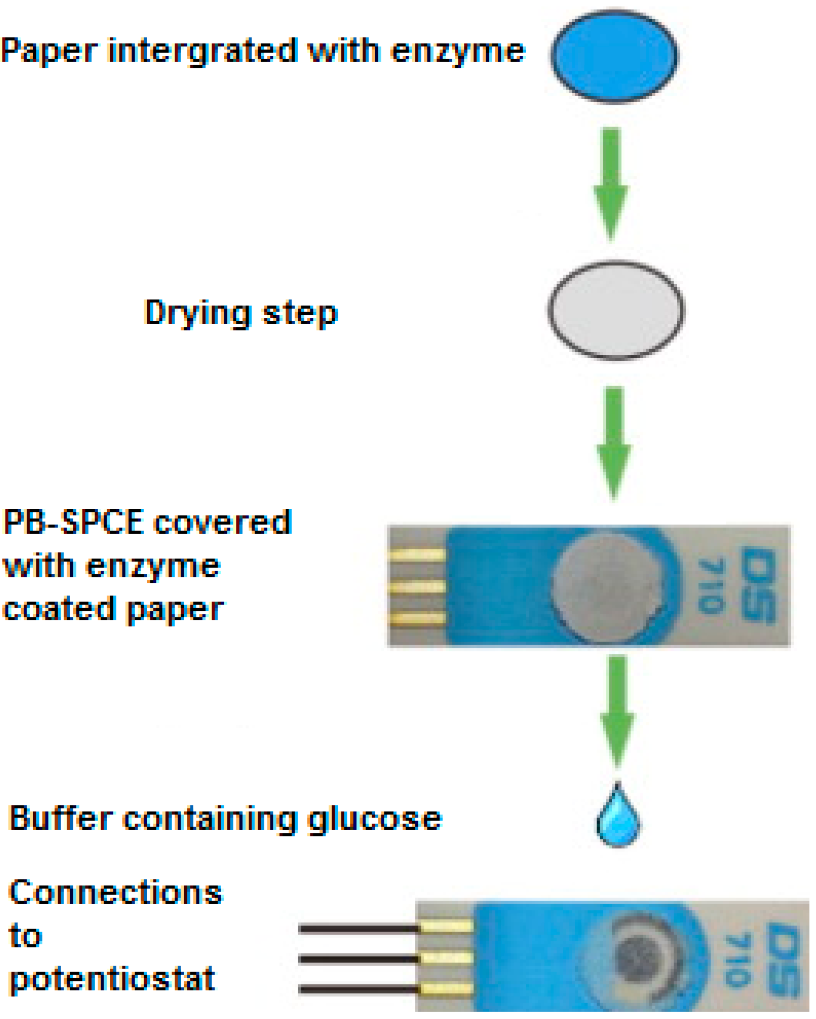
2.1. Glucose
2.2. Galactose
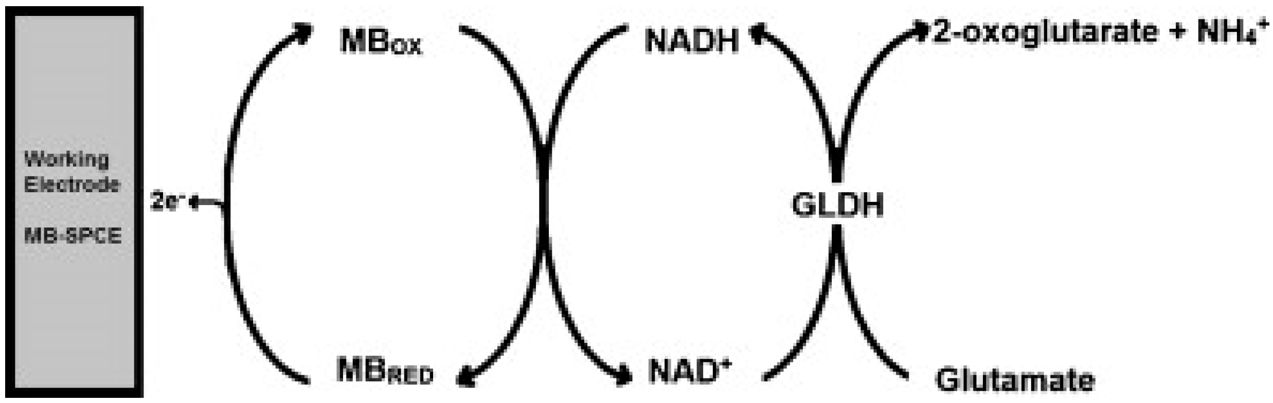
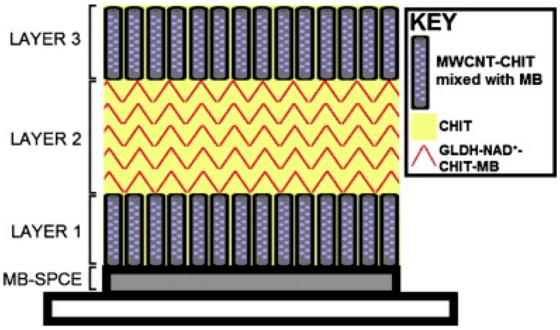
2.3. Glutamate
2.4. Lactate
2.5. Proteins
3. Screen-Printed Carbon Electrodes for Vitamin Analysis
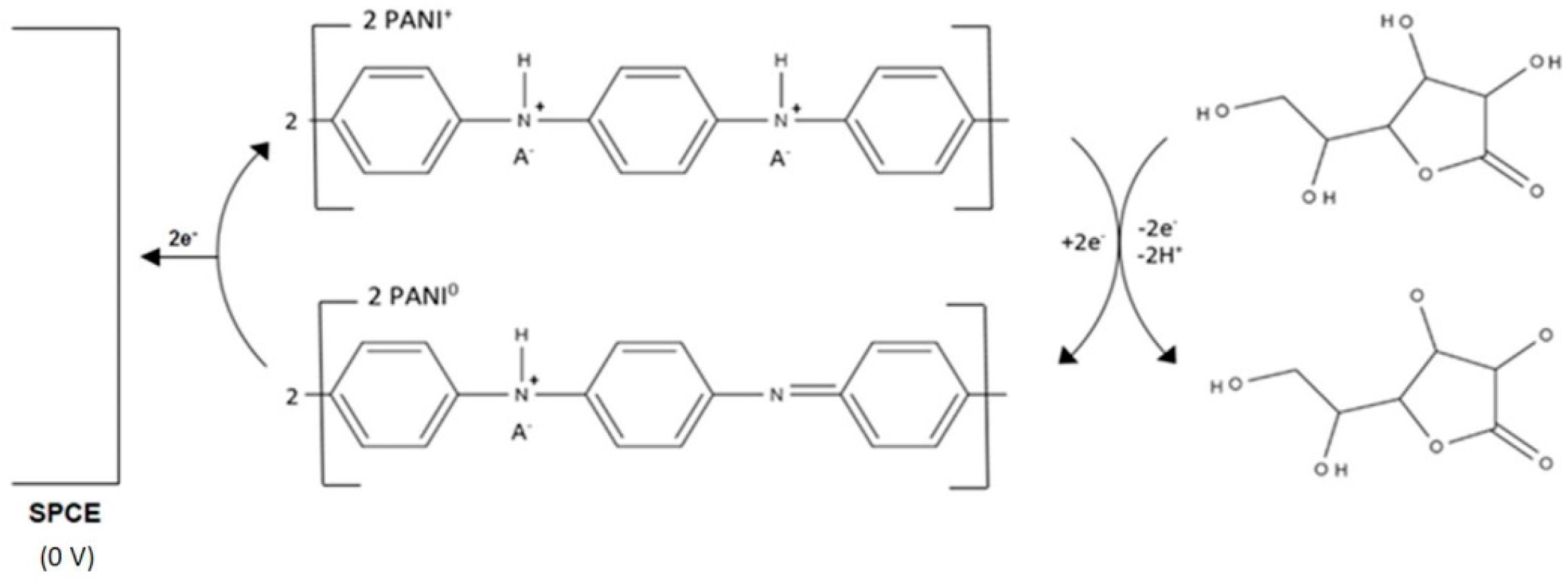
3.1. Vitamin C
3.1.1. Mediated Electron Transfer
3.1.2. Unmediated Electron Transfer
3.2. Vitamin B
3.2.1. Riboflavin
3.2.2. Pyridoxine
3.2.3. Biotin
3.2.4. Cyanocobalamin
4. Organophosphate (OP) Sensing
5. Screen-Printed Sensors for Metal Ion Determination
5.1. Unmodified Screen-Printed Carbon Electrodes
5.2. Mercury Modified Screen-Printed Carbon Electrodes
5.3. Bismuth Modified Screen-Printed Electrodes
5.4. Alternative Metal Based Screen-Printed Electrodes
5.5. Biosensor Based Screen-Printed Electrodes
6. Conclusions
Acknowledgments
Conflicts of Interest
References
- Hart, J.P.; Wring, S.A. Screen-printed voltammetric and amperometric electrochemical sensors for decentralised testing. Electroanalysis 1994, 6, 617–624. [Google Scholar] [CrossRef]
- Hart, J.P.; Wring, S.A. Recent developments in the design and application of screen-printed electrochemical sensors for biomedical, environmental and industrial analyses. Trend. Anal. Chem. 1997, 16, 89–103. [Google Scholar] [CrossRef]
- Wedge, R.; Pemberton, R.M.; Hart, J.P.; Luxton, R. Recent developments towards disposable screen-printed biosensors incorporating a carbon ink modified with the redox mediator, Meldola’s blue. Analysis 1999, 27, 570–577. [Google Scholar] [CrossRef]
- Cowell, D.C.; Abass, A.K.; Dowman, A.A.; Hart, J.P.; Pemberton, R.M.; Young, S.J. Screen-printed disposable biosensors for environmental pollution monitoring. In Biomonitors and Biomarkers as Indicators of Environmental Change 2; Butterworth, F.M., Gunatilaka, A.P., Gonsebatt, M.E., Eds.; Kluwer Academic Press/Plenum Publishers: New York, NY, USA, 2000; Volume 56, pp. 157–173. [Google Scholar]
- Hart, J.P.; Abass, A.K.; Honeychurch, K.C.; Pemberton, R.M.; Ryan, S.L.; Wedge, R. Sensors/biosensors, based on screen-printing technology for biomedical applications. Indian J. Chem. A 2003, 42, 709–718. [Google Scholar]
- Hart, J.P.; Crew, A.; Crouch, E.; Honeychurch, K.C.; Pemberton, R.M. Some Recent Designs and Developments of Screen-Printed Carbon Electrochemical Sensors/Biosensors for Biomedical, Environmental, and Industrial Analyses. Anal. Lett. 2004, 37, 789–830. [Google Scholar] [CrossRef]
- Hart, J.P.; Crew, A.; Crouch, E.; Honeychurch, K.C.; Pemberton, R.M. Chapter 23 Screen-printed electrochemical (bio)sensors in biomedical, environmental and industrial applications. In Comprehensive Analytical Chemistry; Alegret, S., Merkoçi, A., Eds.; Elsevier: Amsterdam, The Netherlands, 2007; Volume 49, pp. 497–557. [Google Scholar]
- Piermarini, S.; Volpe, G.; Esti, M.; Simonetti, M.; Palleschi, G. Real time monitoring of alcoholic fermentation with low-cost amperometric biosensors. Food Chem. 2011, 127, 749–754. [Google Scholar] [CrossRef] [PubMed] [Green Version]
- Jiang, L.; Liu, H.; Liu, J.; Yang, Q.; Cai, X. A sensitive biosensor based on Os-complex mediator and glucose oxidase for low concentration glucose determination. J. Electroanal. Chem. 2008, 619–620, 11–16. [Google Scholar] [CrossRef]
- Biscay, J.; Rama, E.C.; García, M.B.G.; Carrazón, J.M.P.; García, A.C. Enzymatic Sensor Using Mediator-Screen-Printed Carbon Electrodes. Electroanalysis 2011, 23, 209–214. [Google Scholar] [CrossRef]
- Gao, Q.; Guo, Y.; Zhang, W.; Qi, H.; Zhang, C. An amperometric glucose biosensor based on layer-by-layer GOx-SWCNT conjugate/redox polymer multilayer on a screen-printed carbon electrode. Sens. Actuators B Chem. 2011, 153, 219–225. [Google Scholar] [CrossRef]
- Gao, Q.; Guo, Y.; Liu, J.; Yuan, X.; Qi, H.; Zhang, C. A biosensor prepared by co-entrapment of a glucose oxidase and a carbon nanotube within an electrochemically deposited redox polymer multilayer. Bioelectrochemistry 2011, 81, 109–113. [Google Scholar] [CrossRef] [PubMed]
- Chandra Sekar, N.; Mousavi Shaegh, S.A.; Ng, S.H.; Ge, L.; Tan, S.N. A paper-based amperometric glucose biosensor developed with Prussian Blue-modified screen-printed electrodes. Sens. Actuators B Chem. 2014, 204, 414–420. [Google Scholar] [CrossRef]
- Pemberton, R.M.; Pittson, R.; Biddle, N.; Hart, J.P. Fabrication of microband glucose biosensors using a screen-printing water-based carbon ink and their application in serum analysis. Biosens. Bioelectron. 2009, 24, 1246–1252. [Google Scholar] [CrossRef] [PubMed]
- Pemberton, R.M.; Xu, J.; Pittson, R.; Biddle, N.; Drago, G.A.; Jackson, S.K.; Hart, J.P. Application of screen-printed microband biosensors to end-point measurements of glucose and cell numbers in HepG2 cell culture. Anal. Biochem. 2009, 385, 334–341. [Google Scholar] [CrossRef] [PubMed]
- Pemberton, R.M.; Xu, J.; Pittson, R.; Drago, G.A.; Griffiths, J.; Jackson, S.K.; Hart, J.P. A screen-printed microband glucose biosensor system for real-time monitoring of toxicity in cell culture. Biosens. Bioelectron. 2011, 26, 2448–2453. [Google Scholar] [CrossRef] [PubMed]
- Pemberton, R.M.; Cox, T.; Tuffin, R.; Sage, I.; Drago, G.A.; Biddle, N.; Griffiths, J.; Pittson, R.; Johnson, G.; Xu, J.; et al. Microfabricated glucose biosensor for culture well operation. Biosens. Bioelectron. 2013, 42, 668–677. [Google Scholar] [CrossRef] [PubMed]
- Chiu, J.-Y.; Yu, C.-M.; Yen, M.-J.; Chen, L.-C. Glucose sensing electrodes based on a poly(3,4-ethylenedioxythiophene)/Prussian blue bilayer and multi-walled carbon nanotubes. Biosens. Bioelectron. 2009, 24, 2015–2020. [Google Scholar] [CrossRef] [PubMed]
- Zuo, S.; Teng, Y.; Yuan, H.; Lan, M. Development of a Novel Silver Nanoparticles-Enhanced Screen-Printed Amperometric Glucose Biosensor. Anal. Lett. 2008, 41, 1158–1172. [Google Scholar] [CrossRef]
- Kanyong, P.; Pemberton, R.M.; Jackson, S.K.; Hart, J.P. Development of an amperometric screen-printed galactose biosensor for serum analysis. Anal. Biochem. 2013, 435, 114–119. [Google Scholar] [CrossRef] [PubMed]
- Kanyong, P.; Hughes, G.; Pemberton, R.M.; Jackson, S.K.; Hart, J.P. Amperometric Screen-Printed Galactose Biosensor for Cell Toxicity Applications. Anal. Lett. 2015, 49, 236–244. [Google Scholar] [CrossRef]
- Hughes, G.; Pemberton, R.M.; Fielden, P.R.; Hart, J.P. Development of a Disposable Screen Printed Amperometric Biosensor Based on Glutamate Dehydrogenase, for the Determination of Glutamate in Clinical and Food Applications. Anal. Bioanal. Electrochem. 2014, 6, 435–449. [Google Scholar]
- Krajewska, B. Application of chitin- and chitosan-based materials for enzyme immobilizations: A review. Enzyme Microb. Technol. 2004, 35, 126–139. [Google Scholar] [CrossRef]
- Hughes, G.; Pemberton, R.M.; Fielden, P.R.; Hart, J.P. Development of a novel reagentless, screen-printed amperometric biosensor based on glutamate dehydrogenase and NAD+, integrated with multi-walled carbon nanotubes for the determination of glutamate in food and clinical applications. Sens. Actuators B Chem. 2015, 216, 614–621. [Google Scholar] [CrossRef]
- Khan, R.; Gorski, W.; Garcia, C.D. Nanomolar Detection of Glutamate at a Biosensor Based on Screen-Printed Electrodes Modified with Carbon Nanotubes. Electroanalysis 2011, 23, 2357–2363. [Google Scholar] [CrossRef] [PubMed]
- Radoi, A.; Moscone, D.; Palleschi, G. Sensing the Lactic Acid in Probiotic Yogurts Using an L-Lactate Biosensor Coupled with a Microdialysis Fiber Inserted in a Flow Analysis System. Anal. Lett. 2010, 43, 1301–1309. [Google Scholar] [CrossRef]
- Piano, M.; Serban, S.; Pittson, R.; Drago, G.A.; Hart, J.P. Amperometric lactate biosensor for flow injection analysis based on a screen-printed carbon electrode containing Meldola’s Blue-Reinecke salt, coated with lactate dehydrogenase and NAD+. Talanta 2010, 82, 34–37. [Google Scholar] [CrossRef] [PubMed]
- Rawson, F.; Purcell, W.; Xu, J.; Pemberton, R.; Fielden, P.; Biddle, N.; Hart, J.P. A microband lactate biosensor fabricated using a water-based screen-printed carbon ink. Talanta 2009, 77, 1149–1154. [Google Scholar] [CrossRef] [PubMed]
- Pereira, A.C.; Aguiar, M.R.; Kisner, A.; Macedo, D.V.; Kubota, L.T. Amperometric biosensor for lactate based on lactate dehydrogenase and Meldola Blue coimmobilized on multi-wall carbon-nanotube. Sens. Actuators B Chem. 2007, 124, 269–276. [Google Scholar] [CrossRef]
- Hirst, N.A.; Hazelwood, L.D.; Jayne, D.G.; Millner, P.A. An amperometric lactate biosensor using H2O2 reduction via a Prussian Blue impregnated poly(ethyleneimine) surface on screen printed carbon electrodes to detect anastomotic leak and sepsis. Sens. Actuators B Chem. 2013, 186, 674–680. [Google Scholar] [CrossRef]
- Shimomura, T.; Sumiya, T.; Ono, M.; Ito, T.; Hanaoka, T. Amperometric L-lactate biosensor based on screen-printed carbon electrode containing cobalt phthalocyanine, coated with lactate oxidase-mesoporous silica conjugate layer. Anal. Chim. Acta 2012, 714, 114–120. [Google Scholar] [CrossRef] [PubMed]
- Prieto-Simón, B.; Fàbregas, E.; Hart, A. Evaluation of different strategies for the development of amperometric biosensors for l-lactate. Biosens. Bioelectron. 2007, 22, 2663–2668. [Google Scholar] [CrossRef] [PubMed]
- Kokkinos, C.; Prodromidis, M.; Economou, A.; Petrou, P.; Kakabakos, S. Disposable integrated bismuth citrate-modified screen-printed immunosensor for ultrasensitive quantum dot-based electrochemical assay of C-reactive protein in human serum. Anal. Chim. Acta 2015, 886, 29–36. [Google Scholar] [CrossRef] [PubMed]
- Kokkinos, C.; Prodromidis, M.; Economou, A.; Petrou, P.; Kakabakos, S. Quantum dot-based electrochemical DNA biosensor using a screen-printed graphite surface with embedded bismuth precursor. Electrochem. Commun. 2015, 60, 47–51. [Google Scholar] [CrossRef]
- Kokkinos, C.; Angelopoulou, M.; Economou, A.; Prodromidis, M.; Florou, A.; Haasnoot, W.; Petrou, P.; Kakabakos, S. Lab-on-a-Membrane Foldable Devices for Duplex Drop-Volume Electrochemical Biosensing Using Quantum Dot Tags. Anal. Chem. 2016, 88, 6897–6904. [Google Scholar] [CrossRef] [PubMed]
- Xu, Q.; Yan, F.; Lei, J.; Leng, C.; Ju, H. Disposable Electrochemical Immunosensor by Using Carbon Sphere/Gold Nanoparticle Composites as Labels for Signal Amplification. Chem. A Eur. J. 2012, 18, 4994–4998. [Google Scholar] [CrossRef] [PubMed]
- Qi, H.; Ling, C.; Ma, Q.; Gao, Q.; Zhang, C. Sensitive electrochemical immunosensor array for the simultaneous detection of multiple tumor markers. Analyst 2012, 137, 393–399. [Google Scholar] [CrossRef] [PubMed]
- Viswanathan, S.; Rani, C.; Vijay Anand, A.; Ho, J.A. Disposable electrochemical immunosensor for carcinoembryonic antigen using ferrocene liposomes and MWCNT screen-printed electrode. Biosens. Bioelectron. 2009, 24, 1984–1989. [Google Scholar] [CrossRef] [PubMed]
- Elyashevich, G.K.; Sidorovich, A.V.; Smirnov, M.A.; Kuryndin, I.S.; Bobrova, N.V.; Trchova, M.; Stejskal, J. Thermal and structural stability of composite systems based onpolyaniline deposited on porous polyethylene films. Polym. Degrad. Stab. 2006, 91, 2786–2792. [Google Scholar] [CrossRef]
- Ambrosi, A.; Morrin, A.; Smyth, M.R.; Killard, A.J. The application of conducting polymer nanoparticle electrodes to the sensing of ascorbic acid. Anal. Chim. Acta 2006, 609, 37–43. [Google Scholar] [CrossRef] [PubMed]
- Milakin, K.A.; Korovin, A.N.; Moroz, E.V.; Levon, K.; Guiseppi-Elie, A.; Sergeyev, V.G. Polyaniline-Based Sensor Material for Potentiometric Determination of Ascorbic Acid. Electroanalysis 2013, 25, 1323–1330. [Google Scholar] [CrossRef]
- Kit-Anan, W.; Olarnmanich, A.; Sriprachuabwong, C.; Karuman, C.; Tuantranont, A.; Wisitsoraat, A.; Srituravanch, W.; Pimpin, A. Disposable paper-based electrochemical sensor utilizing inkjet-printing Polyaniline modified screen-printed carbon electrode for ascorbic acid detection. J. Electroanal. Chem. 2012, 685, 72–78. [Google Scholar] [CrossRef]
- Nassef, H.M.; Civit, L.; Fragoso, A.; O’Sullivan, C.K. Amperometric sensing of ascorbic acid using a disposable screen-printed electrode modified with electrografted o-aminophenol film. Analyst 2008, 133, 1736–1741. [Google Scholar] [CrossRef] [PubMed]
- Civit, L.; Nassef, H.M.; Fragoso, A.; O’Sullivan, C.K. Amperometric determination of ascorbic acid in real samples using a disposable screen-printed electrode modified with electrografted o-aminophenol film. J. Agric. Food Chem. 2008, 56, 10452–10455. [Google Scholar] [CrossRef] [PubMed]
- Wonsawat, W. Determination of Vitamin C (Ascorbic Acid) in Orange Juices Product. Int. J. Biol. Biomol. Agric. Food Biotechnol. Eng. 2006, 8, 623–625. [Google Scholar]
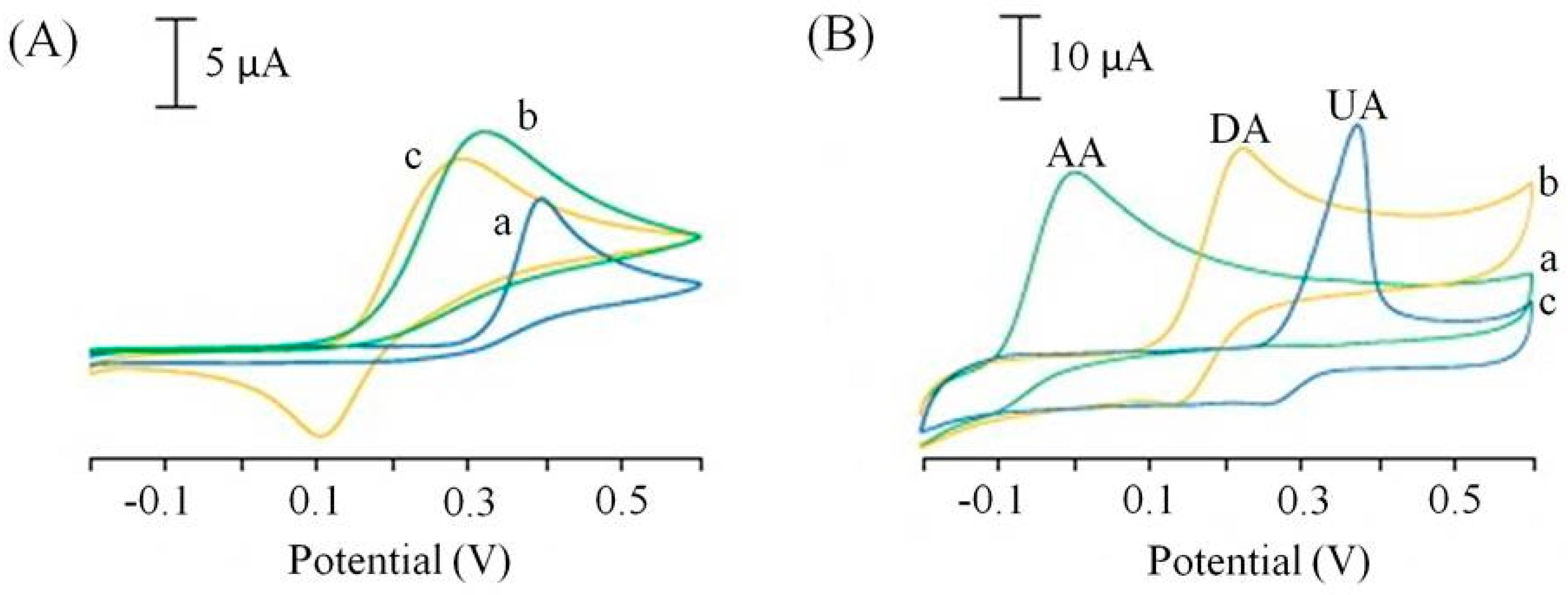
- Ping, J.; Wu, J.; Wang, Y.; Ying, Y. Simultaneous determination of ascorbic acid, dopamine and uric acid using high-performance screen-printed graphene electrode. Biosens. Bioelectron. 2012, 34, 70–76. [Google Scholar] [CrossRef] [PubMed]
- Wang, J. Carbon-Nanotube based Electrochemical Biosensors: A Review. Electroanalysis 2005, 17, 7–14. [Google Scholar] [CrossRef]
- Crevillén, A.G.; Pumera, M.; González, M.C.; Escarpa, A. Carbon nanotube disposable detectors in microchip capillary electrophoresis for water-soluble vitamin determination: Analytical possibilities in pharmaceutical quality control. Electrophoresis 2008, 29, 2997–3004. [Google Scholar] [CrossRef] [PubMed]
- Crevillén, A.G.; Avila, M.; Pumera, M.; González, M.C.; Escarpa, A. Food Analysis on Microfluidic Devices using Ultrasensitive Carbon Nanotube Detectors. Anal. Chem. 2007, 79, 7408–7415. [Google Scholar] [CrossRef] [PubMed]
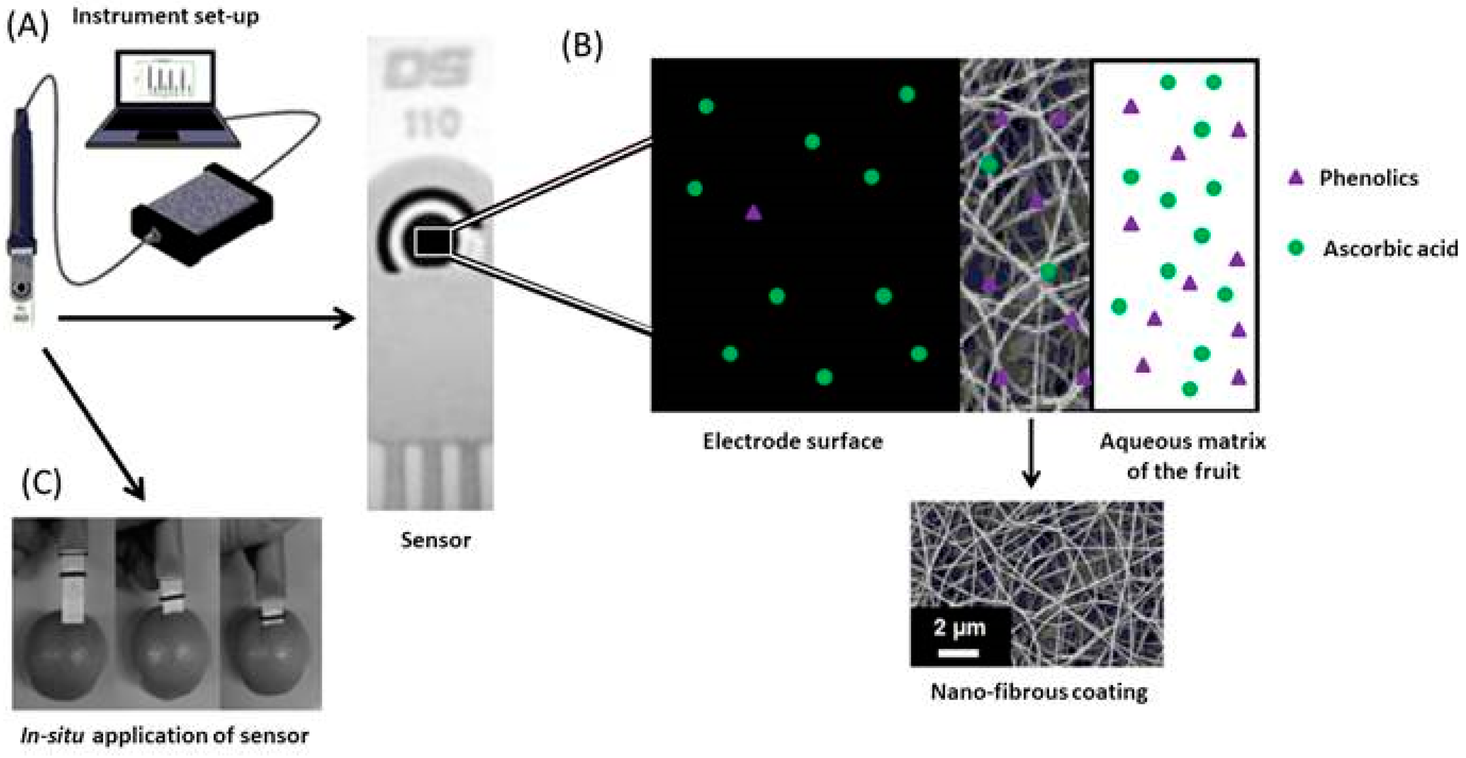
- Fuenmayor, C.A.; Benedetti, S.; Pellicanὸ, A.; Cosio, M.S.; Mannino, S. Direct In Situ Determination of Ascorbic Acid in Fruits by Screen-Printed Carbon Electrodes Modified with Nylon-6 Nanofibers. Electroanalysis 2014, 26, 704–710. [Google Scholar] [CrossRef]
- Kadara, R.O.; Haggett, B.G.D.; Birch, B.J. Disposable Sensor for Measurement of Vitamin B2 in Nutritional Premix, Cereal, and Milk Powder. J. Agric. Food Chem. 2006, 54, 4921–4924. [Google Scholar] [CrossRef] [PubMed]
- Kadara, R.O.; Fogg, A.G.; Haggett, B.D.G.; Birch, B.J. Enhancement by Copper(II) of the Voltammetric Signal of Vitamin B2 Applied to Its Determination in Breakfast Cereals. J. Agric. Food Chem. 2009, 57, 804–806. [Google Scholar] [CrossRef] [PubMed]
- Riman, D.; Avgeropoulos, A.; Hrbac, J.; Prodromidis, M.I. Sparked-bismuth oxide screen-printed electrodes for the determination of riboflavin at sub-nanomolar range in non-deoxygenated solutions. Electrochim. Acta 2015, 165, 410–415. [Google Scholar] [CrossRef]
- Riman, D.; Jirovsky, D.; Hrbac, J.; Prodromidis, M.I. Green and facile electrode modification by spark discharge: Bismuth oxide-screen printed electrodes for the screening of ultra-trace Cd(II) and Pb(II). Electrochem. Commun. 2015, 50, 20–23. [Google Scholar] [CrossRef]
- Brunetti, B.; Desimoni, E. Voltammetric determination of vitamin B6 in food samples and dietary supplements. J. Food Compos. Anal. 2014, 33, 155–160. [Google Scholar] [CrossRef]
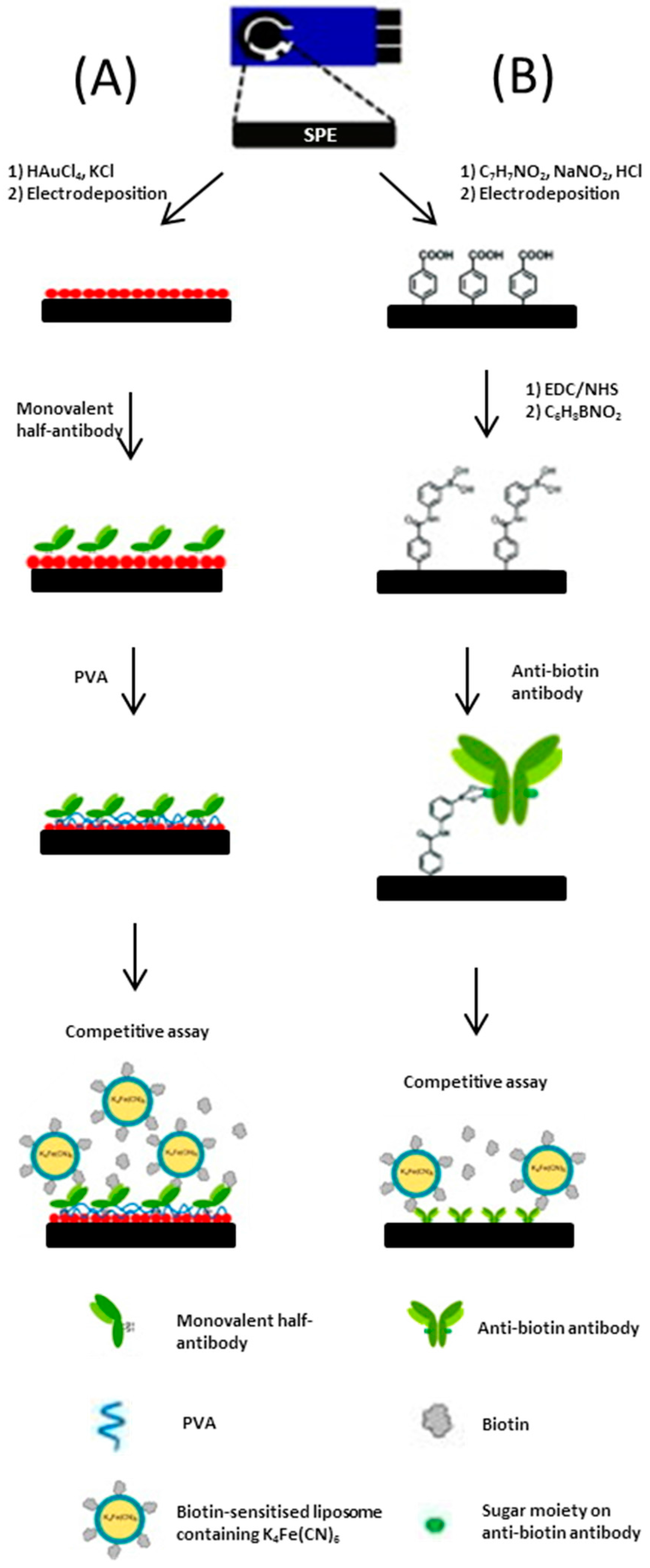
- Ho, J.A.; Chiu, J.K.; Hong, J.C.; Lin, C.C.; Hwang, K.C.; Hwu, J.R. Gold-nanostructured immunosensor for the electrochemical sensing of biotin based on liposomal competitive assay. J. Nanosci. Nanotechnol. 2009, 9, 2324–2329. [Google Scholar] [CrossRef] [PubMed]
- Ho, J.A.; Hsu, W.-L.; Liao, W.-C.; Chiu, J.-K.; Chen, M.-L.; Chang, H.-C.; Li, C.-C. Ultrasensitive electrochemical detection of biotin using electrically addressable site-oriented antibody immobilization approach via aminophenyl boronic acid. Biosens. Bioelectron. 2010, 26, 1021–1027. [Google Scholar] [CrossRef] [PubMed]
- Biscay, J.; González-García, M.B.; Costa-García, A. Electrochemical biotin determination based on a screen printed carbon electrode array and magnetic beads. Sens. Actuators B Chem. 2014, 205, 426–432. [Google Scholar] [CrossRef]
- Biscay, J.; González-García, M.B.; Costa-García, A. Electrochemical biotin detection based on magnetic beads and a new magnetic flow cell for screen printed electrode. Talanta 2015, 131, 706–711. [Google Scholar] [CrossRef] [PubMed]
- Michopoulos, A.; Florou, A.B.; Prodromidis, M.I. Ultrasensitive Determination of Vitamin B12 Using Disposable Graphite Screen-Printed Electrodes and Anodic Adsorptive Voltammetry. Electroanalysis 2015, 27, 1876–1882. [Google Scholar] [CrossRef]
- Kostaki, V.T.; Florou, A.B.; Prodromidis, M.I. Electrochemically induced chemical sensor properties in graphite screen-printed electrodes: The case of a chemical sensor for uranium. Electrochim. Acta 2011, 56, 8857–8860. [Google Scholar] [CrossRef]
- Domínguez Renedo, O.; Alonso-Lomillo, M.A.; Arcos Martínez, M.J. Recent developments in the field of screen-printed electrodes and their related applications. Talanta 2007, 73, 202–219. [Google Scholar] [CrossRef] [PubMed]
- Li, H.; Li, J.; Yang, Z.; Xu, Q.; Hu, X. A novel photoelectrochemical sensor for the organophosphorus pesticide dichlofenthion based on nanometer-sized titania coupled with a screen-printed electrode. Anal. Chem. 2011, 83, 5290–5295. [Google Scholar] [CrossRef] [PubMed]
- Songa, E.A.; Okonkwo, J.O. Recent approaches to improving selectivity and sensitivity of enzyme-based biosensors for organophosphorus pesticides: A review. Talanta 2016, 155, 289–304. [Google Scholar] [CrossRef] [PubMed]
- Pundir, C.S.; Chauhan, N. Acetylcholinesterase inhibition-based biosensors for pesticide determination: A review. Anal. Biochem. 2012, 429, 19–31. [Google Scholar] [CrossRef] [PubMed]
- Grosmanová, Z.; Krejčí, J.; Týnek, J.; Cuhra, P.; Baršová, S. Comparison of biosensoric and chromatographic methods for the detection of pesticides. Int. J. Environ. Anal. Chem. 2005, 85, 885–893. [Google Scholar] [CrossRef]
- Chen, D.; Jiao, Y.; Jia, H.; Guo, Y.; Sun, X.; Wang, X.; Xu, J. Acetylcholinesterase Biosensor for Chlorpyrifos Detection Based on Multi-Walled Carbon Nanotubes-SnO2-chitosan Nanocomposite Modified Screen-Printed Electrode. Int. J. Electrochem. Sci. 2015, 10, 10491–10501. [Google Scholar]
- Istamboulie, G.; Sikora, T.; Jubete, E.; Ochoteco, E.; Marty, J.-L.; Noguer, T. Screen-printed poly(3,4-ethylenedioxythiophene) (PEDOT): A new electrochemical mediator for acetylcholinesterase-based biosensors. Talanta 2010, 82, 957–961. [Google Scholar] [CrossRef] [PubMed]
- Mishra, R.K.; Alonso, G.A.; Istamboulie, G.; Bhand, S.; Marty, J.-L. Automated flow based biosensor for quantification of binary organophosphates mixture in milk using artificial neural network. Sens. Actuators B 2015, 208, 228–237. [Google Scholar] [CrossRef]
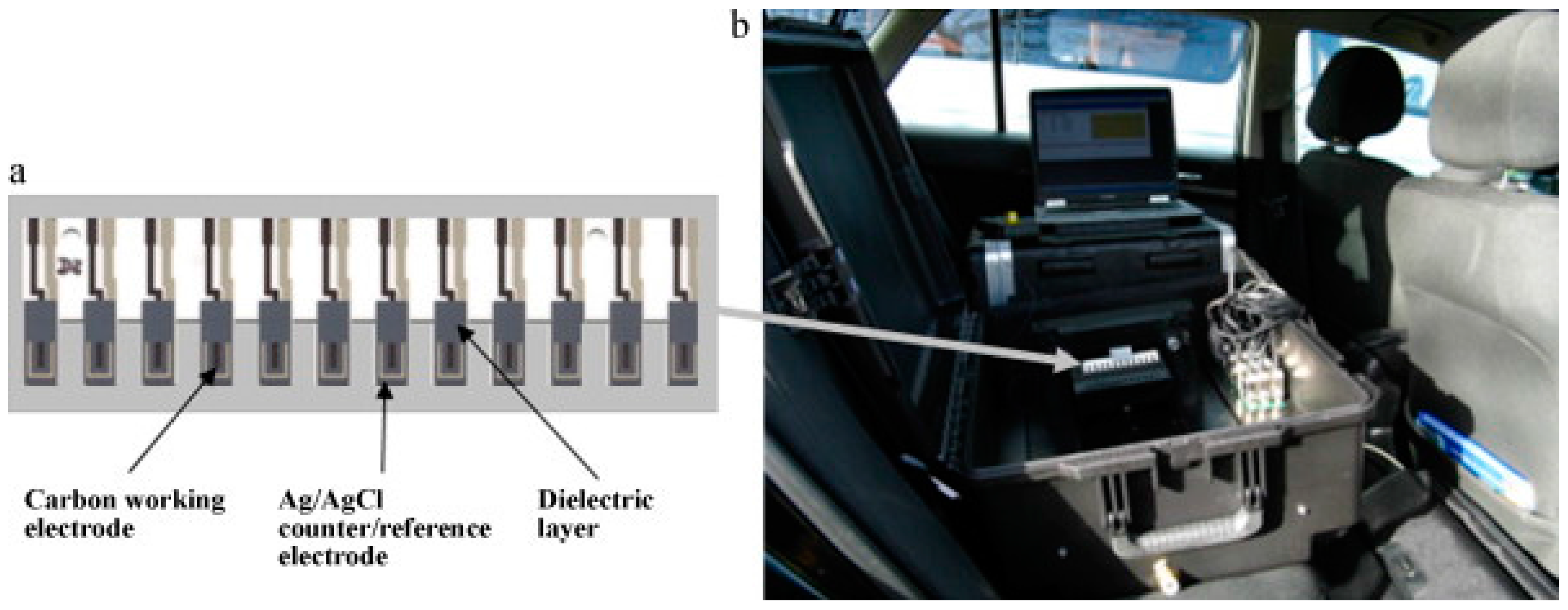
- Crew, A.P.; Lonsdale, D.; Byrd, N.; Pittson, R.; Hart, J.P. A screen-printed, amperometric biosensor array incorporated into a novel automated system for the simultaneous determination of organophosphate pesticides. Biosens. Bioelectron. 2011, 26, 2847–2851. [Google Scholar] [CrossRef] [PubMed]
- Pohanka, M.; Drtinova, L.; Kuca, K. Acetylcholinesterase based assay of eleven organophosphorus pesticides: finding of assay limitations. Int. J. Environ. Anal. Chem. 2012, 92, 125–132. [Google Scholar] [CrossRef]
- Arduini, F.; Guidone, S.; Amine, A.; Palleschi, G.; Moscone, D. Acetylcholinesterase biosensor based on self-assembled monolayer-modified gold-screen printed electrodes for organophosphorus insecticide detection. Sens. Actuators B 2013, 179, 201–208. [Google Scholar] [CrossRef]
- Upadhyay, S.; Sharma, M.K.; Rao, G.R.; Bhattacharya, B.K.; Rao, V.K.; Vijayaraghavan, R. Application of bimetallic nanoparticles modified screen printed electrode for the detection of organophosphate compounds using an enzyme inhibition approach. Anal. Methods 2011, 3, 2246–2253. [Google Scholar] [CrossRef]
- Cinti, S.; Neagu, D.; Carbone, M.; Cacciotti, I.; Moscone, D.; Arduini, F. Novel carbon black-cobalt phthalocyanine nanocomposite as sensing platform to detect organophosphorus pollutants at screen-printed electrode. Electrochim. Acta 2016, 188, 574–581. [Google Scholar] [CrossRef]
- Pohanka, M.; Drobik, O.; Krenkova, Z.; Zdarova-Karasova, J.; Pikula, J.; Cabal, J.; Kuca, K. Voltammetric biosensor based on acetylcholinesterase and different immobilization protocols: A simple tool for toxic organophosphate assay. Anal. Lett. 2011, 44, 1254–1264. [Google Scholar] [CrossRef]
- Kumar, A.; Arora, S.; Mogha, N.; Al-Deyab, S.S.; Ansari, Z.A.; Ansari, S.G. Glutathione coated zinc oxide nanoparticles: A promising material for pesticide detection. Energy Environ. Focus 2013, 2, 101–107. [Google Scholar] [CrossRef]
- Eremenko, V.; Dontsova, E.A.; Nazarov, A.P.; Evtushenko, E.G.; Amitonov, S.V.; Savilov, S.V.; Martynova, L.F.; Lunin, V.V.; Kurochkin, I.N. Manganese dioxide nanostructures as a novel electrochemical mediator for thiol sensors. Electroanalysis 2012, 24, 573–580. [Google Scholar] [CrossRef]
- Ivanov, A.N.; Younusov, R.R.; Evtugyn, G.A.; Arduini, F.; Moscone, D.; Palleschi, G. Acetylcholinesterase biosensor based on single-walled carbon nanotubes–Co phtalocyanine for organophosphorus pesticides detection. Talanta 2011, 85, 216–221. [Google Scholar] [CrossRef] [PubMed]
- Gan, N.; Yang, X.; Xie, D.; Wu, Y.; Wen, W. A disposable organophosphorus pesticides enzyme biosensor based on magnetic composite nano-particles modified screen printed carbon electrode. Sensors 2010, 10, 625–638. [Google Scholar] [CrossRef] [PubMed]
- Alonso, G.A.; Istamboulie, G.; Ramírez-García, A.; Noguer, T.; Marty, J.-L.; Muñoza, R. Artificial neural network implementation in single low-cost chip for the detection of insecticides by modelling of screen-printed enzymatic sensors response. Comput. Electron. Agric. 2010, 74, 223–229. [Google Scholar] [CrossRef]
- Crew, A.P.; Hart, J.P.; Wedge, R.; Marty, J.-L.; Fournier, D. A screen-printed, amperometric, biosensor array for the detection of organophosphate pesticides based on inhibition of wild type, and mutant acetylcholinesterases, from Drosophila melanogaster. Anal. Lett. 2004, 37, 1601–1610. [Google Scholar] [CrossRef]
- Florence, T.M. The speciation of trace elements in waters. Talanta 1982, 29, 345–364. [Google Scholar] [CrossRef]
- Stotyk, W.; Weiss, D.; Appleby, P.G.; Cheburkin, A.K.; Gloor, F.M.; Kramers, J.D.; van der Knaap, W.O. History of atmospheric lead deposition since 12,370 C-14 yr BP from a peat bog, Jura Mountains, Switzerland. Science 1998, 281, 1635–1640. [Google Scholar] [CrossRef]
- MacDonald, R.W.; Barrie, L.A.; Bidleman, T.F.; Diamond, M.L.; Gregor, D.J.; Semkin, R.G.; Strachan, W.M.J.; Li, Y.F.; Wania, F.; Alaee, M.; et al. Contaminants in the Canadian Arctic: 5 years of progress in understanding sources, occurrence and pathways. Sci. Total Environ. 2000, 254, 93–234. [Google Scholar] [CrossRef]
- Hong, S.; Candelone, J.-P.; Patterson, C.C.; Boutron, C.F. Greenland ice evidence of hemispheric lead pollution two millennia ago by Greek and roman civilizations. Science 1994, 265, 1841–1843. [Google Scholar] [CrossRef] [PubMed]
- Warwick, R.M. Evidence for the effects of metal contamination on the intertidal macrobenthic assemblages of the Fal Estuary. Marine Poll. Bull. 2001, 42, 145–148. [Google Scholar] [CrossRef]
- Hamilton, E.I. Environmental variables in a holistic evaluation of land contaminated by historic mine wastes: A study of multi-element mine wastes in West Devon, England using arsenic as an element of potential concern to human health. Sci. Total Environ. 2000, 249, 171–221. [Google Scholar] [CrossRef]
- Wang, J. Analytical Electrochemistry, 3rd ed.; John Wiley & Sons Inc.: Hoboken, NJ, USA, 2006. [Google Scholar]
- Rajeshwar, K.; Ibanez, J.G. Environmental Electrochemistry, Fundamentals and Applications in Pollution Abatement; Academic Press: London, UK, 1997; p. 276. [Google Scholar]
- Wang, J. Stripping Analysis: Principles, Instrumentation and Applications; VCH: Weinheim, Germany, 1985. [Google Scholar]
- Vydra, F.; Stulik, K.; Julakova, E. Electrochemical Stripping Analysis; Ellis Horwood: Chichester, UK, 1976. [Google Scholar]
- Barek, J.; Fogg, A.G.; Muck, A.; Zima, J. Polarography and Voltammetry at Mercury Electrodes. Crit. Rev. Anal. Chem. 2001, 31, 291–309. [Google Scholar] [CrossRef]
- Švancara, I.; Walcarius, A.; Kalcher, K.; Vytřas, K. Carbon paste electrodes in the new millennium. Cent. Eur. J. Chem. 2009, 7, 598–656. [Google Scholar] [CrossRef]
- Stozhko, N.Y.; Malakhova, N.A.; Fyodorov, M.V.; Brainina, K.Z. Modified carbon-containing electrodes in stripping voltammetry of metals. Part II. Composite and microelectrodes. J. Solid State Electrochem. 2008, 12, 1219–1230. [Google Scholar] [CrossRef]
- Honeychurch, K.C. Screen-printed electrochemical sensors and biosensors for monitoring metal pollutants. Insci. J. 2012, 2, 1–51. [Google Scholar] [CrossRef]
- Li, M.; Li, Y.T.; Long, D.W.L.; Long, Y.T. Recent developments and applications of screen-printed electrodes in environmental assays–A review. Anal. Chim. Acta 2012, 734, 31–44. [Google Scholar] [CrossRef] [PubMed]
- Niu, X.H.; Lan, M.B.; Zhao, H.L.; Chen, C.; Li, Y.X.; Zhu, X. Review: Electrochemical Stripping Analysis of Trace Heavy Metals Using Screen-Printed Electrodes. Anal. Lett. 2013, 46, 2479–2502. [Google Scholar] [CrossRef]
- Hayat, A.; Marty, J.L. Disposable Screen Printed Electrochemical Sensors: Tools for Environmental Monitoring. Sensors 2014, 14, 10432–10453. [Google Scholar] [CrossRef] [PubMed]
- Barton, J.; García, M.B.G.; Santos, D.H.; Fanjul-Bolado, P.; Ribotti, A.; McCaul, M.; Diamond, D.; Magn, P. Screen-printed electrodes for environmental monitoring of heavy metal ions: A review. Microchim. Acta 2016, 183, 503–517. [Google Scholar] [CrossRef]
- Serrano, N.; Díaz-Cruz, J.M.; Ariño, C.; Esteban, M. Antimony–based electrodes for analytical determinations. TrAC 2016, 77, 203–213. [Google Scholar] [CrossRef]
- Amine, A.; Arduini, F.; Moscone, D.; Palleschi, G. Recent advances in biosensors based on enzyme inhibition. Biosens. Bioelectron. 2016, 76, 180–194. [Google Scholar] [CrossRef] [PubMed]
- Duarte, K.; Justino, C.I.L.; Freitas, A.C.; Gomes, A.M.P.; Duarte, A.C.; Rocha-Santos, T.A.P. Disposable sensors for environmental monitoring of lead, cadmium and mercury. TrAC 2015, 64, 183–190. [Google Scholar] [CrossRef]
- Crew, A.; Cowell, D.C.; Hart, J.P. Development of an anodic stripping voltammetric assay, using a disposable mercury-free screen-printed carbon electrode, for the determination of zinc in human sweat. Talanta 2008, 75, 1221–1226. [Google Scholar] [CrossRef] [PubMed]
- Honeychurch, K.C.; Al-Berezanchi, S.; Hart, J.P. The voltammetric behaviour of lead at a microband screen-printed carbon electrode and its determination in acetate leachates from glazed ceramic plates. Talanta 2011, 84, 717–723. [Google Scholar] [CrossRef] [PubMed]
- Bergamini, M.F.; Santos, D.P.; Zanoni, M.V.B. Screen-Printed Carbon Electrode Modified with Poly-l-histidine Applied to Gold(III) Determination. J. Braz. Chem. Soc. 2009, 20, 100–106. [Google Scholar] [CrossRef]
- Arduini, F.; Majorani, C.; Amine, A.; Moscone, D.; Palleschi, G. Hg2+ detection by measuring thiol groups with a highly sensitive screen-printed electrode modified with a nanostructured carbon black film. Electrochim. Acta 2011, 56, 4209–4215. [Google Scholar] [CrossRef]
- Aragay, G.; Puig-Font, A.; Cadevall, M.; Merkoc, A. Surface Characterizations of Mercury-Based Electrodes with the Resulting Micro and Nano Amalgam Wires and Spheres Formations May Reveal Both Gained Sensitivity and Faced Nonstability in Heavy Metal Detection. J. Phys. Chem. C 2010, 114, 9049–9055. [Google Scholar] [CrossRef]
- Hallam, P.M.; Kampouris, D.K.; Kadara, R.O.; Banks, C.E. Graphite screen printed electrodes for the electrochemical sensing of chromium(VI). Analyst 2010, 135, 1947–1952. [Google Scholar] [CrossRef] [PubMed]
- Renedo, O.D.; Martınez, M.J.A. A novel method for the anodic stripping voltammetry determination of Sb(III) using silver nanoparticle-modified screen-printed electrodes. Electrochem. Commun. 2007, 9, 820–826. [Google Scholar] [CrossRef]
- Renedo, O.D.; Martınez, M.J.A. Anodic stripping voltammetry of antimony using gold nanoparticle-modified carbon screen-printed electrodes. Anal. Chim. Acta 2007, 589, 255–260. [Google Scholar] [CrossRef] [PubMed]
- Renedo, O.D.; González, M.J.G.; Martínez, M.J.A. Determination of Antimony (III) in Real Samples by Anodic Stripping Voltammetry Using a Mercury Film Screen-Printed Electrode. Sensors 2009, 9, 219–231. [Google Scholar] [CrossRef] [PubMed]
- Betelu, S.; Vautrin-Ula, C.; Lyb, J.; Chaussé, A. Screen-printed electrografted electrode for trace uranium analysis. Talanta 2009, 80, 372–376. [Google Scholar] [CrossRef] [PubMed]
- Choi, H.S.; Kim, H.D. Development of a Portable Heavy Metal Ion Analyzer Using Disposable Screen-Printed Electrodes. Bull. Korean Chem. Soc. 2009, 30, 1881–1883. [Google Scholar]
- Sánchez, A.; Morante-Zarcero, S.; Pérez-Quintanilla, D.; Sierra, I.; del Hierro, I. Development of screen-printed carbon electrodes modified with functionalized mesoporous silica nanoparticles: Application to voltammetric stripping determination of Pb(II) in non-pretreated natural waters. Electrochim. Acta 2010, 55, 6983–6990. [Google Scholar] [CrossRef]
- Sanllorente-Méndez, S.; Dominguez-Renedo, O.; Arcos-Martinez, M.J. Determination of Arsenic(III) Using Platinum Modified Screen-Printed Carbon-Based Electrodes. Electroanalysis 2009, 21, 635–639. [Google Scholar] [CrossRef]
- Somerset, V.; Iwuoha, E.; Hernandez, L. Stripping Voltammetric Measurement of Trace Metal Ions at Screen-printed Carbon and Carbon Paste Electrodes. Procedia Chem. 2009, 1, 1279–1282. [Google Scholar] [CrossRef]
- Cugnet, C.; Zaouak, O.; René, A.; Pécheyran, C.; Potin-Gautier, M.; Authier, L. A novel microelectrode array combining screen-printing and femtosecond laser ablation technologies: Development, characterization and application to cadmium detection. Sens. Actuators B Chem. 2009, 143, 158–163. [Google Scholar] [CrossRef]
- Zaouak, O.; Authier, L.; Cugnet, C.; Castetbon, A.; Potin-Gautier, M. Electroanalytical Device for Cadmium Speciation in Waters. Part 1: Development and Characterization of a Reliable Screen-Printed Sensor. Electroanalysis 2010, 22, 1151–1158. [Google Scholar] [CrossRef]
- Christidis, K.; Robertson, P.; Gow, K.; Pollard, P. Voltammetric in situ measurements of heavy, metals in soil using a portable electrochemical instrument. Measurement 2007, 40, 960–967. [Google Scholar] [CrossRef]
- Pollard, P.; Adams, M.; Robertson, P.J.; Christidis, K.; Officer, S.; Prabhu, G.R.; Gow, K.; Morrisson, A.R. Environmental Forensic Investigations: The Potential Use of a Novel Heavy Metal Sensor and Novel Tangents. In Criminal and Environmental Soil Forensics IV 2009; Ritz, K., Dawson, L., Miller, D., Eds.; Springer: Berlin, Germany, 2009; pp. 477–490. [Google Scholar]
- Krystofova, O.; Trnkova, L.; Adam, V.; Zehnalek, J.; Hubalek, J.; Babula, P.; Kizek, R. Electrochemical Microsensors for the Detection of Cadmium(II) and Lead(II) Ions in Plants. Sensors 2010, 10, 5308–5328. [Google Scholar] [CrossRef] [PubMed]
- Güell, R.; Aragay, G.; Fontàs, C.; Anticó, E.; Merkoci, A. Sensitive and stable monitoring of lead and cadmium in seawater using screen-printed electrode and electrochemical stripping analysis. Anal. Chim. Acta 2008, 627, 219–224. [Google Scholar] [CrossRef] [PubMed]
- Somerset, V.; Leaner, J.; Mason, R.; Iwuoha, E.; Morrin, A. Determination of inorganic mercury using a polyaniline and polyaniline-methylene blue coated screen-printed carbon electrode. Int. J. Environ. Anal. Chem. 2010, 90, 671–685. [Google Scholar] [CrossRef]
- Somerset, V.; Leaner, J.; Mason, R.; Iwuoha, E.; Morrin, A. Development and application of a poly(2,2’-dithiodianiline) (PDTDA)-coated screen-printed carbon electrode in inorganic mercury determination. Electrochim. Acta 2010, 55, 4240–4246. [Google Scholar] [CrossRef]
- Zen, J.-M.; Kumar, A.S.; Lee, S.-C.; Shih, Y. Microliter Volume Determination of Cosmetic Mercury with a Partially Crosslinked Poly(4-vinylpyridine) Modified Screen-Printed Three-Electrode Portable Assembly. Electroanalysis 2007, 19, 2369–2374. [Google Scholar] [CrossRef]
- Mandil, A.; Idrissi, L.; Amine, A. Stripping voltammetric determination of mercury(II) and lead(II) using screen-printed electrodes modified with gold films, and metal ion preconcentration with thiol-modified magnetic particles. Microchim. Acta 2010, 170, 299–305. [Google Scholar] [CrossRef]
- Meucci, V.; Laschi, S.; Minunni, M.; Pretti, C.; Intorre, L.; Soldani, G.; Mascini, M. An optimized digestion method coupled to electrochemical sensor for the determination of Cd, Cu, Pb and Hg in fish by square wave anodic stripping voltammetry. Talanta 2009, 77, 1143–1148. [Google Scholar] [CrossRef] [PubMed]
- Meucci, V.; Intorre, L.; Pretti, C.; Laschi, S.; Minunni, M.; Mascini, M. Disposable electrochemical sensor for rapid measurement of heavy metals in fish by square wave anodic stripping voltammetry (SWASV). Vet. Res. Commun. 2009, 33, 249–252. [Google Scholar] [CrossRef] [PubMed]
- Redha, Z.M.; Baldock, S.J.; Fielden, P.R.; Goddard, N.J.; Brown, B.J.T.; Haggett, B.G.D.; Andres, R.; Birch, B.J. Hybrid Microfluidic Sensors Fabricated by Screen Printing and Injection Molding for Electrochemical and Electrochemiluminescence Detection. Electroanalysis 2009, 21, 422–430. [Google Scholar] [CrossRef]
- Khaled, E.; Hassan, H.N.A.; Habib, I.H.I.; Metelka, R. Chitosan Modified Screen-Printed Carbon Electrode for Sensitive Analysis of Heavy Metals. Int. J. Electrochem. Sci. 2010, 5, 158–167. [Google Scholar]
- Chailapakul, O.; Korsrisakul, S.; Siangproh, W.; Grudpan, K. Fast and simultaneous detection of heavy metals using a simple and reliable microchip-electrochemistry route: An alternative approach to food analysis. Talanta 2008, 74, 683–689. [Google Scholar] [CrossRef] [PubMed]
- Choudhry, N.A.; Kadara, R.O.; Banks, C.E. “Cosmetic electrochemistry”: The facile production of graphite microelectrode ensembles. Phys. Chem. Chem. Phys. 2010, 12, 2285–2287. [Google Scholar] [CrossRef] [PubMed]
- Choudhry, N.A.; Khairy, M.; Kadara, R.O.; Jenkinson, N.; Banks, C.E. Cosmetic Electrochemistry II: Rapid and Facile Production of Metallic Electrocatalytic Ensembles. Electroanalysis 2010, 22, 1831–1836. [Google Scholar] [CrossRef]
- ŠVancara, I.; Vytřas, K. Elektroanalýza S Bismutovými Elektrodami. Chem. Listy 2006, 100, 90–113. [Google Scholar]
- Arduini, F.; Calvo, J.Q.; Amine, A.; Palleschi, G.; Moscone, D. Bismuth-modified electrodes for lead detection. TrAC 2010, 29, 1295–1304. [Google Scholar] [CrossRef]
- Svancara, I.; Prior, C.; Hocevar, S.B.; Wang, J. A Decade with Bismuth-Based Electrodes in Electroanalysis. Electroanalysis 2010, 22, 1405–1420. [Google Scholar] [CrossRef]
- Kokkinos, C.; Economou, A. Stripping at Bismuth-Based Electrodes. Curr. Anal. Chem. 2008, 4, 183–190. [Google Scholar] [CrossRef]
- Kadara, R.O.; Tothill, I.E. Development of disposable bulk-modified screen-printed electrode based on bismuth oxide for stripping chronopotentiometric analysis of lead (II) and cadmium (II) in soil and water samples. Anal. Chim. Acta 2008, 623, 76–81. [Google Scholar] [CrossRef] [PubMed]
- Khairy, M.; Kadara, R.O.; Kampouris, D.K.; Banks, C.E. Disposable Bismuth Oxide Screen Printed Electrodes for the Sensing of Zinc in Seawater. Electroanalysis 2010, 22, 1455–1459. [Google Scholar] [CrossRef]
- Malakhova, N.A.; Stojko, N.Y.; Brainina, K.Z. Novel approach to bismuth modifying procedure for voltammetric thick film carbon containing electrodes. Electrochem. Commun. 2007, 9, 221–227. [Google Scholar] [CrossRef]
- Yong, L.; Armstrong, K.C.; Dansby-Sparks, R.N.; Carrington, N.A.; Chambers, J.Q.; Xue, Z.L. Quantitative analysis of trace chromium in blood samples. Combination of the advanced oxidation process with catalytic adsorptive stripping voltammetry. Anal. Chem. 2006, 78, 7582–7587. [Google Scholar] [CrossRef] [PubMed]
- Kruusma, J.; Banks, C.E.; Compton, R.G. Mercury-free sono-electroanalytical detection of lead in human blood by use of bismuth-film-modified boron-doped diamond electrodes. Anal. Bioanal. Chem. 2004, 379, 700–706. [Google Scholar] [CrossRef] [PubMed]
- Hutton, E.A.; van Elteren, J.T.; Ogorevc, B.; Smyth, M.R. Validation of bismuth film electrode for determination of cobalt and cadmium in soil extracts using ICP–MS. Talanta 2004, 63, 849–855. [Google Scholar] [CrossRef] [PubMed]
- Khairy, M.; Kadara, R.O.; Kampouris, D.K.; Banks, C.E. In situ bismuth film modified screen printed electrodes for the bio-monitoring of cadmium in oral (saliva) fluid. Anal. Methods 2010, 2, 645–649. [Google Scholar] [CrossRef]
- Serrano, N.; Díaz-Cruz, J.M.; Ariño, C.; Esteban, M. Ex situ Deposited Bismuth Film on Screen-Printed Carbon Electrode: A Disposable Device for Stripping Voltammetry of Heavy Metal Ions. Electroanalysis 2010, 22, 1460–1467. [Google Scholar] [CrossRef]
- Li, M.; Li, D.-W.; Li, Y.-T.; Xu, D.-K.; Long, Y.-T. Highly Selective In Situ Metal Ion Determination by Hybrid Electrochemical “Adsorption-Desorption” and Colorimetric Methods. Anal. Chim. Acta 2011, 701, 157–163. [Google Scholar] [CrossRef] [PubMed]
- Mandil, A.; Amine, A. Screen-Printed Electrodes Modified by Bismuth Film for the Determination of Released Lead in Moroccan Ceramics. Anal. Lett. 2009, 42, 1245–1257. [Google Scholar] [CrossRef]
- Lu, D.; Belle, J.L.; Ninivin, C.L.; Mabic, S.; Dimitrakopoulos, T. In situ electrochemical detection of trace metal vapors at bismuth doped carbon screen printed electrodes. J. Electroanal. Chem. 2010, 642, 157–159. [Google Scholar] [CrossRef]
- Injang, U.; Noyrod, P.; Siangproh, W.; Dungchai, W.; Motomizu, S.; Chailapakul, O. Determination of trace heavy metals in herbs by sequential injection analysis-anodic stripping voltammetry using screen-printed carbon nanotubes electrodes. Anal. Chim. Acta 2010, 668, 54–60. [Google Scholar] [CrossRef] [PubMed]
- Chuanuwatanakul, S.; Dungchai, W.; Chailapakul, O.; Motomizu, S. Determination of trace heavy Metals by Sequential Injection- anodic Stripping Voltammetry using Bismuth Film Screen printed Carbon Electrode. Anal. Sci. 2008, 24, 589–594. [Google Scholar] [CrossRef] [PubMed]
- Siriangkhawut, W.; Pencharee, S.; Grudpan, K.; Jakmunee, J. Sequential injection monosegmented flow voltammetric determination of cadmium and lead using a bismuth film working electrode. Talanta 2009, 79, 1118–1124. [Google Scholar] [CrossRef] [PubMed]
- Rico, M.A.G.; Olivares-Marin, M.; Gil, E.P. A Novel Cell Design for the Improved Stripping Voltammetric Detection of Zn(II), Cd(II), and Pb(II) on Commercial Screen-Printed Strips by Bismuth Codeposition in Stirred Solutions. Electroanalysis 2008, 20, 2608–2613. [Google Scholar] [CrossRef]
- Hwang, G.-H.; Han, W.-K.; Park, J.-S.; Kang, S.-G. An electrochemical sensor based on the reduction of screen-oxide for the determination of trace lead and cadmium. Sensor. Actuators B Chem. 2008, 135, 309–316. [Google Scholar] [CrossRef]
- Nie, Z.; Nijhuis, C.A.; Gong, J.; Chen, X.; Kumachev, A.; Martinez, A.W.; Narovlyansky, M.; Whitesides, G.M. Electrochemical sensing in paper-based microfluidic devices. Lab Chip 2010, 10, 477–483. [Google Scholar] [CrossRef] [PubMed]
- Tan, S.N.; Ge, L.; Wang, W. Paper Disk on Screen Printed Electrode for One-Step Sensing with an Internal Standard. Anal. Chem. 2010, 82, 8844–8847. [Google Scholar] [CrossRef] [PubMed]
- Amine, A.; Mohammadi, H. Electrochemical biosensors for heavy metal based on enzyme inhibition. In Electrochemical Sensor Analysis; Alegret, S., Merkoçi, A., Eds.; Elsevier: Amsterdam, The Netherlands, 2007; Volume 49, pp. 299–310. [Google Scholar]
- Verma, N.; Singh, M. Biosensors for Metals. BioMetals 2005, 18, 121–129. [Google Scholar] [CrossRef] [PubMed]
- Gooding, J.J.; Chow, E.; Finlayson, R. Biosensors for Detecting Metal Ions: New Trends. Aust. J. Chem. 2003, 56, 159–162. [Google Scholar] [CrossRef]
- Sanllorente-Méndez, S.; Domínguez-Renedo, O.; Arcos-Martínez, M.J. Immobilization of Acetylcholinesterase on Screen-Printed Electrodes. Appl. Determ. Arsenic(III) Sens. 2010, 10, 2119–2128. [Google Scholar]
- Guascito, M.R.; Malitesta, C.; Mazzotta, E.; Turco, A. Screen-Printed Glucose Oxidase-Based Biosensor for Inhibitive Detection of Heavy Metal Ions in a Flow Injection System. Sens. Lett. 2009, 7, 153–159. [Google Scholar] [CrossRef]
- Maczuga, M.; Economou, A.; Bobrowski, A.; Prodromidis, M.I. Novel screen-printed antimony and tin voltammetric sensors for anodic stripping detection of Pb(II) and Cd(II). Electrochim. Acta 2013, 114, 758–765. [Google Scholar] [CrossRef]
- Punrat, E.; Chuanuwatanakul, S.; Kaneta, T.; Motomizu, S.; Chailapakul, O. Method development for the determination of arsenic by sequential injection/anodic stripping voltammetry using long-lasting gold-modified screen-printed carbon electrode. Talanta 2013, 116, 1018–1025. [Google Scholar] [CrossRef] [PubMed]
- Hassan, S.S.; Solangi, A.R.; Kazi, T.G.; Kalhoro, M.S.; Junejo, Y.; Tagar, Z.A.; Kalwar, N.H. Nafion stabilized ibuprofen-gold nanostructures modified screen printed electrode as arsenic(III) sensor. J. Electroanal. Chem. 2012, 682, 77–82. [Google Scholar] [CrossRef]
- Gamboa, J.C.M.; Cornejo, L.; Squella, J.A. Vibrating screen printed electrode of gold nanoparticle-modified carbon nanotubes for the determination of arsenic(III). J. Appl. Electrochem. 2014, 44, 1255–1260. [Google Scholar] [CrossRef]
- Cinti, S.; Politi, S.; Moscone, D.; Palleschi, G.; Arduini, F. Stripping Analysis of As(III) by Means of Screen-Printed Electrodes Modified with Gold Nanoparticles and Carbon Black Nanocomposite. Electroanalysis 2014, 26, 931–939. [Google Scholar] [CrossRef]
- Sarkar, P.; Banerjee, S.; Bhattacharyay, D.; Turner, A.P.F. Electrochemical sensing systems for arsenate estimation by oxidation of L-cysteine, Ecotox. Ecotoxicol. Environ. Saf. 2010, 73, 1495–1501. [Google Scholar] [CrossRef] [PubMed]
- Domínguez-Renedo, O.; Alonso-Lomillo, M.A.; Ferreira-Goncalves, L.; Arcos-Martínez, M.J. Development of urease based amperometric biosensors for the inhibitive determination of Hg (II). Talanta 2009, 79, 1306–1310. [Google Scholar] [CrossRef] [PubMed]
- Prasad, K.S.; Arun, A.B.; Rekha, P.D.; Young, C.C.; Chang, J.L.; Zen, J.M. A Microbial Sensor Based on Direct Electron Transfer at Shewanella Sp. Drop-Coated Screen-Printed Carbon Electrodes. Electroanalysis 2009, 21, 1646–1650. [Google Scholar] [CrossRef]
- Viguier, B.; Zor, K.; Kasotakis, E.; Mitraki, A.; Clausen, C.H.; Svendsen, W.E.; Castillo-Leon, J. Development of an Electrochemical Metal-Ion Biosensor Using Self-Assembled Peptide Nanofibrils. ACS Appl. Mater. Interfaces 2011, 3, 1594–1600. [Google Scholar] [CrossRef] [PubMed]
- Niu, X.; Ding, Y.; Chen, C.; Zhao, H.; Lan, M. A novel electrochemical biosensor for Hg2+ determination based on Hg2+-induced DNA hybridization. Sens. Actuators B Chem. 2011, 158, 383–387. [Google Scholar] [CrossRef]
- Ilangovan, R.; Daniel, D.; Krastanov, A.; Zachariah, C.; Elizabeth, R. Enzyme Based Biosensor for Heavy Metal Ions Determination. Biotechnol. Biotechnol. Equip. 2006, 20, 184–189. [Google Scholar] [CrossRef]
- Florescu, M.; Badea, M.; Coman, G.; Marty, J.-L.; Mitrica, M. Screen Printed Electrodes Used for Detection of Ionic Heavy Metals. Bull. Transilv. Univ. Brasov. Ser. VI Med. Sci. 2009, 2, 49–54. [Google Scholar]
- Grand View Research Glucose Biosensor Market Size, Industry Report, 2022. Available online: http://www.grandviewresearch.com/industry-analysis/glucose-biosensors-market (accessed on 4 August 2016).
- Imani, S.; Bandodkar, A.J.; Mohan, A.M.V.; Kumar, R.; Yu, S.; Wang, J.; Mercier, P.P. A wearable chemical–electrophysiological hybrid biosensing system for real-time health and fitness monitoring. Nat. Commun. 2016, 7, 11650. [Google Scholar] [CrossRef] [PubMed]
- Steinacker, J.; Zügel, M.; Schumann, U.; Machus, K.; Schneider, M.; Harald, H.; Sareban, M.; Treff, G. Monitoring rowers to determine under-performance. BMC Sports Sci. Med. Rehabil. 2015, 7, 1. [Google Scholar] [CrossRef]
- Pinnacle Technology. Available online: http://www.pinnaclet.com/glutamate.html (accessed on 4 August 2016).
- Sarissa Biomedical. Available online: http://www.sarissa-biomedical.com/products/sarissaprobes/glutamate.aspx (accessed on 4 August 2016).
- Gwent Electronic Materials. Available online: http://www.gwent.org/gem_biosensors.html (accessed on 4 August 2016).
- Liébana, S.; Jones, L.; Drago, G.; Pittson, R.; Liu, D.; Perrie, W.; Hart, J.P. Design and development of novel screen-printed microelectrode and microbiosensor arrays fabricated using ultrafast pulsed laser ablation. Sens. Actuators B Chem. 2016, 231, 384–392. [Google Scholar] [CrossRef]


| Immobilization Technique | Mediator | Assay Time (s) | Lower Linear Range (µM) | Upper Linear Range (µM) | Sensitivity | Applied Potential (mV) | Storage Stability (Weeks) | Reference |
|---|---|---|---|---|---|---|---|---|
| Crosslinking with glutaraldehyde & Nafion | Prussian Blue | N/A | 20 | 700 | N/A | 200 | 90% activity after 6 months | [8] |
| Crosslinking with glutaraldehyde & BSA | Osmium-polyvinyl pyridine wired HRP | 60 | 0 | 700 | 28.24 nA/μM/cm | 0 | 90% activity after 15 months | [9] |
| Drop coating | Ferrocene | N/A | 50 | 1000 | 2.12 µA/mM | −100 | 100% activity after 3 months | [10] |
| Use of SWCNT | PVI | 5 | 500 | 800 | 32 μA/mM/cm | 300 | 90% activity after 1 month | [11] |
| Use of SWCNT | Osmium bipyridine-complexed PVI | 5 | 200 | 6000 | 16.4 μA/mM/cm | 300 | 90% activity after 1 month | [12] |
| Immobilization on paper disk | Prussian Blue | N/A | 250 | 2000 | 2.13 µA/mM | −300 | 72% activity after 45 days | [13] |
| Enzyme contained within water-based ink | CoPC | 20 | 270 | 2000 | 16.4 nA/mM | 400 | N/A | [14] |
| Enzyme contained within water-based ink | CoPC | 400 s | Buffer: 450 | 9000 | Buffer: 26 nA/mM | 400 | N/A | [16] |
| Culture Medium: 2000 | 13,000 | Culture Medium: 13 nA/mM | ||||||
| Enzyme contained within water-based ink | CoPC | 30 | 0 | 2000 | 7 nA/mM | 400 | N/A | [17] |
| Enzyme entrapped by electro-polymerization of PEDOT | Prussian Blue | N/A | 1000 | 10,000 | 2.67 μA/cm/mM | −100 | 82% activity after 1 month | [18] |
| Drop coating | Prussian Blue | 5 | 12.5 | 2560 | 20.09 mA/M/cm2 | −50 | 91% activity after 30 days | [19] |
| Immobilization Technique | Mediator | Assay Time (s) | Lower Linear Range (µM) | Upper Linear Range (µM) | Sensitivity | Applied Potential (mV) | Storage Stability | Reference |
|---|---|---|---|---|---|---|---|---|
| Cellulose acetate | CoPC | 10 | 100 | 25,000 | 7.00 µA/mM/cm | 500 | 100% activity after two weeks | [20] |
| Cellulose acetate | CoPC | 10 | 1980 | 9520 | 7.27 µA/mM/cm | 500 | N/A | [21] |
| Immobilization Technique | Mediator | Assay Time (s) | Lower Linear Range (µM) | Upper Linear Range (µM) | Sensitivity | Applied Potential (mV) | Storage Stability | Reference |
|---|---|---|---|---|---|---|---|---|
| Entrapment with chitosan | Meldola’s Blue | 2 | 12.5 | 150 | 0.44 nA/µM | 100 | N/A | [22] |
| Entrapment with chitosan & MWCNTs | Meldola’s Blue | 20 | 7 | 105 | 0.39 nA/µM | 100 | 100% after two weeks | [24] |
| Drop coated onto surface of CNTs | None | <5 | 0.01 | 10 | 0.72 ± 0.05 μA/μM | 950 | 92% after 24 days | [25] |
| Immobilization Technique | Mediator | Assay Time (s) | Lower Linear Range (µM) | Upper Linear Range (µM) | Sensitivity | Applied Potential (mV) | Storage Stability (Weeks) | Reference |
|---|---|---|---|---|---|---|---|---|
| Nafion | Variamine Blue | N/A | 200 | 1000 | 0.46 nA/mM | 200 | N/A | [26] |
| Cellulose acetate | Meldola’s Blue | 10 | 550 | 10,000 | 0.53 nA/mM | 50 | 100% activity for 17 days | [27] |
| Enzyme contained within water-based ink | CoPC | 100 | 1000 | 6000 | 3.63 nA/mM | 400 | N/A | [28] |
| Crosslinking with glutaraldehyde | Meldola’s Blue | 5 | 100 | 10,000 | 3.46 μA cm/mM | 0 | N/A | [29] |
| Dropcoating onto a polyethyleneimine surface | Prussian Blue | 5 | 200 | 800 | 3 µA/mM | 0 | N/A | [30] |
| Polyvinyl alcohol | CoPC | 90 | 18.3 | 1500 | 4.54 μA/cm/mM | 450 | 98% activity after 9 months | [31] |
| Polysulfone precipitation | Meldola’s Blue | 30 | 1 | 125 | 80 mA/M | −100 | 75% activity after one week | [32] |
| CoPC: Cobalt pthalocyanine |
| Electrode Components | Supporting Electrolyte | Measurement Technique | Detection Limit (µM) | Linear Range (µM) | Sample/s | Modification Method | Reference |
|---|---|---|---|---|---|---|---|
| W: Nano-PANI SPCE R: Ag/AgCl C: Pt mesh | PBS pH 6.8 | Amperometry 0 V | 8.3 | 500–8000 | Tablet pharmaceutical | Drop coating | [40] |
| W: PANI-SPCE R: Ag/AgCl C: GCE | 0.05 M Phosphate buffer pH 7.0 & 0.5 M NaCl | Cyclic Voltammetry | 0.1 | 1.00–80.00 | Grapefruit juice | Oxidative chemical polymerisation | [41] |
| W: PANI-SPCE R: Carbon C: Carbon | 0.1 M Acetate buffer pH 5.0 | Chronoamperometry 0.4 V | 30 | 30.00–270.00 | None reported | Inkjet printed Paper based design | [42] |
| W: o-AP-SPCE R: Ag C: Carbon | 0.1 M Phosphate buffer pH 7.2 | Amperometry 0.2 V | 0.86 | 2.00–20.00 | Apple, Kiwi, Lemon, Orange, Pineapple, Strawberry, Tomato | Electrografted film | [43,44] |
| W: SPCE R: Ag/AgCl C: Carbon | 0.1 M Phosphate buffer pH 2.0 | DPV 0.0 V > −1.2 V | 1360 | 1000–10,000 | Orange juice | Unmodified | [45] |
| W: SPGNE R: Ag/AgCl C: Pt | 0.1 M Phosphate buffer pH 7.0 | DPV −0.2 V > +0.6 V Ep = −0.5 V | 0.95 | 4.00–4500.00 | Injection formula | Graphene ink formulation | [46] |
| W: MWCNT-SPCE R: Ag/AgCl wire C: Pt wire | 0.01 M Phosphate buffer pH 7.0 | Amperometry −1.2 V | 11 | 50.00–400.00 | Tablet pharmaceutical Capsule pharmaceutical | Drop coating | [48,49] |
| W: N6-NFM-SPCE R: Ag C: Graphite | Buffer citrate pH 4 | Amperometry 0.35 V | Not reported | 56.78–7381.33 | Tangerine, Apple, Pear, Kiwi, Lemon, Strawberry | Electrospun membrane | [50] |
| Analyte | Electrode Components | Supporting Electrolyte | Measurement Technique | Detection Limit | Linear Range | Sample/s | Reference |
|---|---|---|---|---|---|---|---|
| Vitamin B2 (Riboflavin) | W: Carbon R: Ag/AgCl C: Carbon | 0.05 M Acetate-phosphate /KCl buffer pH 6.0 | DPV −0.6 V > −0.2 V Ep = −0.42 V | 2.39 µM | 2.66–61.11 µM | Vitamin B premix, Dietetic milk powder, Corn flake cereal | [51] |
| Vitamin B2 (Riboflavin) | W: Carbon R: Ag/AgCl C: Carbon | 0.10 M Acetate-phosphate /KCl buffer pH 8.0 | LSV −0.1 V > 1.0 V Ep = −0.65 V | 0.13 µM | 0.016–0.399 µM | Variety of breakfast cereals | [52] |
| Vitamin B2 (Riboflavin) | W: Sparked Bi-SPCE R: Ag/AgCl KCl C: Pt wire | 0.1 M Acetate buffer pH 4.5 | SWV 0 V > +1.0 V Ep = +0.3 V | 0.7 nM | 0.001–0.01 µM | Tablet pharmaceutical | [53] |
| Vitamin B6 (Pyridoxine) | W: MWCNT-SPCE R: Ag/AgCl wire C: Pt wire | 0.01 M Phosphate buffer pH 7.0 | Amperometry +1.2 V | 8.00 µM | 25.00–300.00 µM | Tablet pharmaceutical Capsule pharmaceutical | [48,49] |
| Vitamin B6 (Pyridoxine) | W: MWCNT-SPCE R: Ag C: Carbon | Acetate buffer pH 5.0 | DPV 0 V > +1.0 V Ep = +0.75 V | 1.50 µM | 2.00–72.00 µM | Tablet pharmaceutical Energy drink Cereal | [55] |
| Vitamin B7 (Biotin) | W: PAH/nanoAu/SPCE R: Ag/AgCl C: Pt | 0.1M PBS pH 7.2 | SWV +0.6 V > −0.3 V Ep = +0.2 V | 8.30 nM | 0.01 nM–0.01 M | None reported | [56] |
| Vitamin B7 (Biotin) | W: Ab/APBA/SPGrE R: Ag C: Carbon | Phosphate buffer pH 7.2 | Amperometry −0.2 V | 0.16 nM | 0.1 nM–1.0 mM | None reported | [57] |
| Vitamin B7 (Biotin) | W: MonoAb/nanoAu/SPGnE R: Ag C: Carbon | Phosphate buffer pH 7.2 | Amperometry −0.2 V | 14.00 nM | 1.0 nM–1.0 µM | None reported | [57] |
| Vitamin B7 (Biotin) | W: Carbon R: Ag C: Carbon | Phosphate buffer pH 7.2 | Amperometry −0.2 V | Not reported | 0.10–250.00 nM | None reported | [58] |
| Vitamin B7 (Biotin) | W: Carbon R: Ag C: Carbon | Phosphate buffer pH 7.2 | Amperometry −0.2 V | Not reported | 0.01–1.00 nM | Tablet pharmaceutical Liquid pharmaceutical | [59] |
| Vitamin B9 (Folic) | W: MWCNT-SPCE R: Ag/AgCl wire C: Pt wire | 0.01 M Phosphate buffer pH 7.0 | Amperometry +1.2 V | 8.00 µM | 50.00–400.00 µM | Tablet pharmaceutical Capsule pharmaceutical | [48,49] |
| Vitamin B12 (Cyanocobalomin) | W: SPGrE R: Ag/AgCl /3M KCl C: Pt | 0.1 M Phosphate buffer, 0.1 M KCl, 10 mM/L EDTA pH 3 | SWV −1.2 V > −0.3 V Ep = −0.73 V | 0.07 nM | 0.10–0.80 nM | Tablet pharmaceutical Liquid pharmaceutical | [60] |
| SPE Material | SPE Modification | Immobilization Method | Enzyme | Limit of Detection | Real Sample Analysis | Analytical Technique | Incubation Time | Reference |
|---|---|---|---|---|---|---|---|---|
| Gold | Cysteamine | Cross-linking | EE AChE | 2 ppb paraoxon | Drinking water | CV | 15 | [67] |
| Carbon | MWCNT, SnO2, chitosan | Entrapment | EE AChE | 0.05 µg/L chlorpyrifos | Vegetable extract | CV | 14 | [62] |
| Carbon | Carbon black, CoPC | Entrapment | BChE | 18 nM paraoxon | Industrial waste water | Chronoamp | 20 | [69] |
| Carbon | MnO2 | n/a | BChE | 0.6 nM diazinon | n/a | Chronoamp | 15 | [71] |
| Carbon | Magnetic composite nano-particles, prussian blue | Entrapment | DmAChE | 0.56 ng/L dimethoate | Vegetable extract | DPV | 5 | [72] |
| Carbon | n/a | Not declared | Not declared | n/a | Food extracts | CV/Chronoamp | n/a | [61] |
| Carbon | PEDOT, PSS | Entrapment | EE AChE | 4 nM chlorpyrifos | n/a | Chronoamp | 10 | [63] |
| Carbon | SWCNT, CoPC | Cross-linking | EE AChE | 5 ppb paraoxon, 2 ppb malaoxon | Water | Chronoamp | 15 | [73] |
| Gold | Glutathione, ZnO nanoparticles | Adsorption | EE AChE | 10 ppb chlorpyrifos | n/a | CV | n/a | [70] |
| Carbon | Titania nanoparticles | n/a | n/a | 2 nM dichlofenthion | Vegetable extract | DPV/Photoelec | n/a | [58] |
| Platinum | n/a | Entrapment | Human AChE | n/a | n/a | SWV/CV | 5 | [66] |
| Carbon | CoPC | Entrapment | DmAChE/PTE/EE AChE | Chronoamp | 10 | [74] | ||
| Carbon | CoPC | Cross-linking | DmAChE | n/a | Lake water | Chronoamp | 10 | [64] |
| Carbon | Ag/Pt bimetallic nanoparticles | Cross-linking | EE AChE/ChO | 0.2 µM paraoxon/carbofuran | n/a | Chronoamp | 10 | [68] |
| Carbon | CoPC | Cross-linking | DmAChE | <1 nM pirimiphos/chlorpyrifos/ malaoxon/omethoate/dichlorvos | Food extracts, waste water, drinking water, river/lake water | Chronoamp | 3 | [65] |
| Analyte | Modifier | Medium Exchange | Accumulation Media | Measurement Technique | Linear Range | Detection Limit (Time) | Sample/s | Reference |
|---|---|---|---|---|---|---|---|---|
| Pb2+, Cd2+, Cu2+ | Hg thin film | No | Sample acidified with HCl pH 2 | SWASV, −1.1 V, 120 s | 0–500 ng/mL in acidified seawater | Cd2+ 7.0 ng/mL, Pb2+ 0.31 ng/mL, Cu2+ 0.53 ng/mL | Seawater | [107] |
| Cr6+ | Unmodified carbon | No | 0.1 M H2SO4 | LSCSV | 100–1000 ng/mL | 19 ng/mL | Canal water | [108] |
| Sb3+ | Electrochemical generated silver nanoparticles | No | pH 2 Britton–Robinson buffer | DPASV, −0.6 V, (200 s) | 9.90 × 10⁻⁸–9.09 × 10⁻⁷ M | 6.79 × 10⁻¹⁰ M | Seawater, pharmaceutical preparations | [109] |
| Sb3+ | Electrochemical generated gold nanoparticles | No | pH 2 Britton-Robinson buffer | DPASV, −0.55 V (200 s) | 9.90 × 10⁻⁸–9.09 × 10⁻⁷ M | 9.44 × 10⁻¹⁰ M | Seawater, pharmaceutical preparations | [110] |
| Sb3+ | Mercury film | No | HCl 3 M | DPASV, −0.9 V (600 s) | 0.99 × 10⁻⁸–8.26 × 10⁻⁸ M | 1.27 × 10⁻⁸ M | Glucantime and seawater | [111] |
| U | 4-Carboxyphenyl | No | Ammonium acetate | 15 min | 8.5 × 10⁻¹⁰–10−7 M | 2 × 10⁻⁹ M | Estuarine water | [112] |
| Pb2+ | No | 0.1 M KCl | DPASV, −1.1 V (400 s) | 10–60 μg/dL | 2 μg/dL | - | [113] | |
| Pb2+ | Functionalized mesoporous silica | No | 0.2 M HCl | SWASV, −1.2 V | 1–30 ng/mL | 0.1 ng/mL, 5 min accumulation, 120 s electrolysis | Drinking water, river water, groundwater | [114] |
| As3+ | Platinum nanoparticle | No | 1 M H2SO4 | CV, −0.2 V to +1.3 V, 100 mV/s | 1.6 × 10⁻⁷–1.3 × 10⁻⁶ M | 5.68 ± 1.18 mg/L | Certificated water sample | [115] |
| Hg2+, Pb2+, Ni2+, Cd2+ | PANI, or PANI-poly(DTDA) | No | 0.1 M H2SO4; 0.5M HCl | DPASV, −0.4 V (120 s) | 1 × 10⁻⁹–1 × 10⁻⁶ M | - | - | [116] |
| Cd2+ | Hg modified microelectrode array formed by femtosecond laser ablation | No | acetate buffer 0.2 M, pH 4.5 | SWASV | 1–10 ng/mL | 1.3 ng/mL (300 s) | River water | [117] |
| Cd2+ | Ex-situ Hg plated thin film | No | acetate buffer 0.2 M pH 4.5 | SWASV, −1.0 V | 0.2–40 ng/mL | 0.2 ng/mL, (60 s) | River water | [118] |
| Hg2+, Pb2+, Ni2+, Cd2+, Cu2+ | Unmodified carbon | No | 0.1 M NaCl, pH 1.35 | DPASV, −1.4 V | - | - | Soil | [119] |
| Pb2+, Ni2+, Cd2+, Cu2+ | Unmodified carbon | No | 0.1 M NaCl, pH 1.35 | DPASV, −1.4 V | - | - | Forensic soil analysis | [120] |
| Cd2+, Pb2+ | Unmodified carbon | No | 0.2 M acetic acid & 0.2 M sodium acetate | DPASV, −1.0 V | Cd2+ 2–100 µM, Pb2+ 5–100 µM | Cd2+ 500 nM, Pb2+ 800 nM (120 s) | Rainwater, flour, maize & seedlings | [121] |
| Pb2+, Cd2+ | Thin-film Hg | No | 0.6 M NaCl, pH 8 | SWASV, −1.1 V | 10–2000 ng/mL | Pb2+ 1.8 ng/mL, Cd2+ 2.9 ng/mL (120 s) | Seawater | [122] |
| Hg2+ | PANI-methylene blue coated | No | 0.5 M HCl | DPASV, −0.3 V | 1 × 10⁻⁸−1 × 10⁻⁵ M | 54.27 ng/mL (120 s) | Ultra-pure water | [123] |
| Hg2+ | Electrochemically coated PANI-poly(DTDA) | No | 0.5 M HCl | DPASV, −0.3 V | 1 × 10⁻⁸–1 × 10⁻⁵ M | 56 ng/mL (120 s) | - | [124] |
| Hg2+ | poly(4-vinlylpyridine) | No | pH 4 acetate buffer + 2 M KCl | SWASV | 100–1000 ppb | 69.5 ppb | Skin-lightening cosmetics | [125] |
| Hg2+, Pb2+ | Au film | Yes | 0.05 M HCl | SWASV, −1.0 V | Hg2+ 2–16 ng/mL, Pb2+ 4–16 ng/mL | Hg2+ 1.5 ng/mL, Pb2+ 0.5 ng/mL, (120 s) | Drinking water | [126] |
| Cd2+, Cu2+, Pb2+, Hg2+ | Cd2+, Cu2+, Pb2+ by thin Hg film, Hg2+ Au screen-printed electrode | No | 0.1 M HCl | SWASV, Hg2+ +0.2 V, Cd2+, Cu2+, Pb2+, −1.1 V | 1 ng/mL–1 µg/mL for all | Hg2+ 0.9 ng/mL, (120 s), Cd2+, 1.0 ng/mL, Cu2+ 0.5 ng/mL, Pb2+ 0.3 ng/mL (300 s) | Dogfish muscle, Mussel tissue, Atlantic hake fillets | [127,128] |
| Cd2+, Cu2+, Pb2+ | Injection modelled flow cell containing screen-printed sensor | No | Cu2+ 0.1 M HNO3, Cd2+ 0.1 M pH 9 ammonium citrate buffer, Pb2+ 0.1 M pH 9 glycine buffer | Cu2+ & Cd2+ DPASV, Pb2+ SWASV | Pb2+ 30–70 ng/mL, Cu2+ 9 ng/mL–26 ng/mL | Cu2+ 4.4 ng/mL (300 s), Pb2+ 5.9 ng/mL (500 s), Cd2+ | Lake water, industrial waste water | [129] |
| Cd2+, Cu2+, Pb2+, Hg2+ | Chitosan | No | 0.1 M HCl/KCl | DPASV, −1.0 V | 10–200 ng/mL | Pb2+ 3.4 ng/mL, Cu2+ 5 ng/mL, Cd2+ 5 ng/mL Hg2+ 2 ng/mL (30 s) | Tap water | [130] |
| Cd2+, Cu2+, Pb2+ | Microchip capillary electrophoresis | No | MES buffer (pH 7.0, 25 mM) | −0.8 V | 100–1000 µM | Pb2+ 1.74 µM, Cd2+ 0.73 µM, 0.13 µM | Green vegetable, Tomato and pine apple juices | [131] |
| Pb2+ | Random micro-array formed by spraying screen-printed working with a commercial deodorant (200 mm for 12 s). | No | 0.1 M HNO3 | SWASV, −0.5 V | 20–50 µM and 75–200 µM | 9.5 µM | - | [132] |
| As3+ | Au array for ASV, Pt array for direct oxidation, formed by spraying screen-printed working with a commercial deodorant (200 mm for 6 s). | No | 1 M H2SO4 | LSASV, −1.2 V | 1–5 µM | 4.8 × 10⁻⁷ M | - | [133] |
| Analyte | Modifier | Medium Exchange | Accumulation Media | Measurement Technique | Linear Range | Detection Limit (Time) | Sample/s | Reference |
|---|---|---|---|---|---|---|---|---|
| Zn2+, Cd2+ Pb2+ | Chemically synthesized Bi nanoparticles | No | pH 4.5 0.1 M acetate buffer | SWASV, −1.4 V, flow cell & convective cell | - | 0.52 ng/mL Zn2+, 0.45 ng/mL Cd2+, 0.41 ng/mL Pb2+, (120 s) | Waste water CRM, drinking water | [147] |
| Zn2+, Cd2+ Pb2+ | bismuth oxide modified ink | No | 0.1 M NaOAc solution containing 0.05 M HCl or 0.1 M HCl | SWASV, −1.2 V | Cd2+ 10–150 ng/mL, Pb2+ 10–150 ng/mL, Zn2+ 40–150 ng/mL | 5, 10 and 30 ng/mL | River water | [148] |
| Cd2+ | Microband ex-situ Bi plated | No | pH 4.5, acetate buffer 0.2M | SWASV, −1.0 V | 5.6 ng/mL–45 ng/mL | 1.3 ng/mL | River water (mining area) | [149] |
| Cd2+, Pb2+ | Bismuth oxide modified ink | No | 0.5 M ammonium acetate + 0.1 M HCl pH 4.6 | Chrono-potentiometric | 20–300 ng/mL | Pb2+ 8.0 ng/mL, Cd2+ 16 ng/mL | Soil, water | [150] |
| Zn2+, Pb2+ | Ex-situ deposited bismuth | No | 0.01 M KNO3 | DPASV, −1.5 V, 60 s. stripping chrono-potentiometry | Up to: Zn2+ 250 ng/mL, Pb2+ 50 ng/mL, Cd2+ 600 ng/mL | Zn2+ 3.5 ng/mL Pb2+ 0.5 ng/mL, Cd2+ 3.9 ng/mL | Tap water (Barcelona) | [151] |
| Pb2+ | Bi, 0.5% Nafion | No | 10.0 mM acetate 50 mM KCl buffer + 500 mg/L Bi | SWASV, −1.0 V, 120 s | 5 ng/mL–80 ng/mL | 4 ng/mL | Leachates from cooking vessels | [152] |
| Zn2+, Cd2+ Pb2+ | Dip coated hydrogel modified Bi doped ink | Yes | Volatile metal species generated at room temperature by the addition of sodium tetrahydroborate (III) to an acidified solution. | SWASV, −1.2 V | 10–80 ng | 1 ng (120 s) | Metal vapours | [153] |
| Zn2+, Cd2+ Pb2+ | In situ plated Bi | No | 1 M HCl | SIA-ASV, −1.4 V | 2–100 ng/mL Pb2+ and Cd2+, 12–100 ng/mL Zn2+ | 0.2 ng/mL Pb2+, 0.8 ng/mL Cd2+, 11 ng/mL Zn2+ | Herbs | [154] |
| Zn2+, Cd2+ Pb2+ | In situ plated Bi | No | 1 M HCl | SIA-ASV, −1.4 V | 0–70 ng/mL Pb2+ and Cd2+, 75–200 ng/mL Zn2+. | 0.89 ng/mL Pb2+, 0.69 ng/mL Cd2+ | Drinking water | [155] |
| Cd2+ Pb2+ | In situ plated Bi | No | 0.2 M, pH 4.6 acetate buffer | SI-MSFA, −1.1 V | 10 ng/mL–100 ng/mL | Cd2+ 1.4 ng/mL, Pb2+ 6.9 ng/mL | Water from a zinc mining draining pond | [156] |
| Zn2+, Cd2+ Pb2+ | In situ plated Bi | No | 0.1 M pH 4.5 acetate buffer, 10−2 M KCl | SWASV | 10 ng/mL–100 ng/mL | Zn2+ 8.2 ng/mL, Cd2+ 3.6 ng/mL, Pb2+ 2.5 ng/mL | Tap water, waste water | [157] |
| Cd2+ Pb2+ | Bismuth oxide modified ink | No | 0.1 M, pH 4.5 acetate buffer | SWASV, −1.2 V | 20 ng/mL–100 ng/mL | 2.3 ng/mL Pb2+, 1.5 ng/mL Cd2+ | River water | [158] |
| Pb2+ | In-situ plated Bi Lab on a chip | No | 0.1 M acetate buffer pH 4.5 | SWASV, −1.2 V | 2.5 ng/mL–100 ng/mL | 1.0 ng/mL (120 s) | - | [159] |
| Pb2+ | SPCE modified with filter paper containing electrolyte, Bi and internal standard (Zn) salts. | No | 0.1 M pH 4.5 acetate buffer, containing Zn (60 ng/mL) as internal standard | SWASV, −1.4 V | 10 ng/mL–100 ng/mL | 2.0 ng/mL (120 s) | - | [160] |
| Analyte | Modifier | Medium Exchange | Accumulation Media | Measurement Technique | Linear Range | Detection Limit (Time) | Sample/s | Reference |
|---|---|---|---|---|---|---|---|---|
| Cu2+, Cd2+ and Pb2+ | Urease | sol-Gel | pH 7.0 0.02 mM phosphate buffer | Conductometric | 0.1–10 | - | - | [171] |
| Ni2+, Cu2+ and Cd2+ | Acetylcholinesterase | - | - | Amperometric, TCNQ as mediator | Cu2+ 0.001–0.1 | - | - | [172] |
© 2016 by the authors; licensee MDPI, Basel, Switzerland. This article is an open access article distributed under the terms and conditions of the Creative Commons Attribution (CC-BY) license (http://creativecommons.org/licenses/by/4.0/).
Share and Cite
Hughes, G.; Westmacott, K.; Honeychurch, K.C.; Crew, A.; Pemberton, R.M.; Hart, J.P. Recent Advances in the Fabrication and Application of Screen-Printed Electrochemical (Bio)Sensors Based on Carbon Materials for Biomedical, Agri-Food and Environmental Analyses. Biosensors 2016, 6, 50. https://0-doi-org.brum.beds.ac.uk/10.3390/bios6040050
Hughes G, Westmacott K, Honeychurch KC, Crew A, Pemberton RM, Hart JP. Recent Advances in the Fabrication and Application of Screen-Printed Electrochemical (Bio)Sensors Based on Carbon Materials for Biomedical, Agri-Food and Environmental Analyses. Biosensors. 2016; 6(4):50. https://0-doi-org.brum.beds.ac.uk/10.3390/bios6040050
Chicago/Turabian StyleHughes, Gareth, Kelly Westmacott, Kevin C. Honeychurch, Adrian Crew, Roy M. Pemberton, and John P. Hart. 2016. "Recent Advances in the Fabrication and Application of Screen-Printed Electrochemical (Bio)Sensors Based on Carbon Materials for Biomedical, Agri-Food and Environmental Analyses" Biosensors 6, no. 4: 50. https://0-doi-org.brum.beds.ac.uk/10.3390/bios6040050