Enzyme-Based Electrochemical Biosensors for Microfluidic Platforms to Detect Pharmaceutical Residues in Wastewater

 , , ,
, , ,  and
and 
Abstract
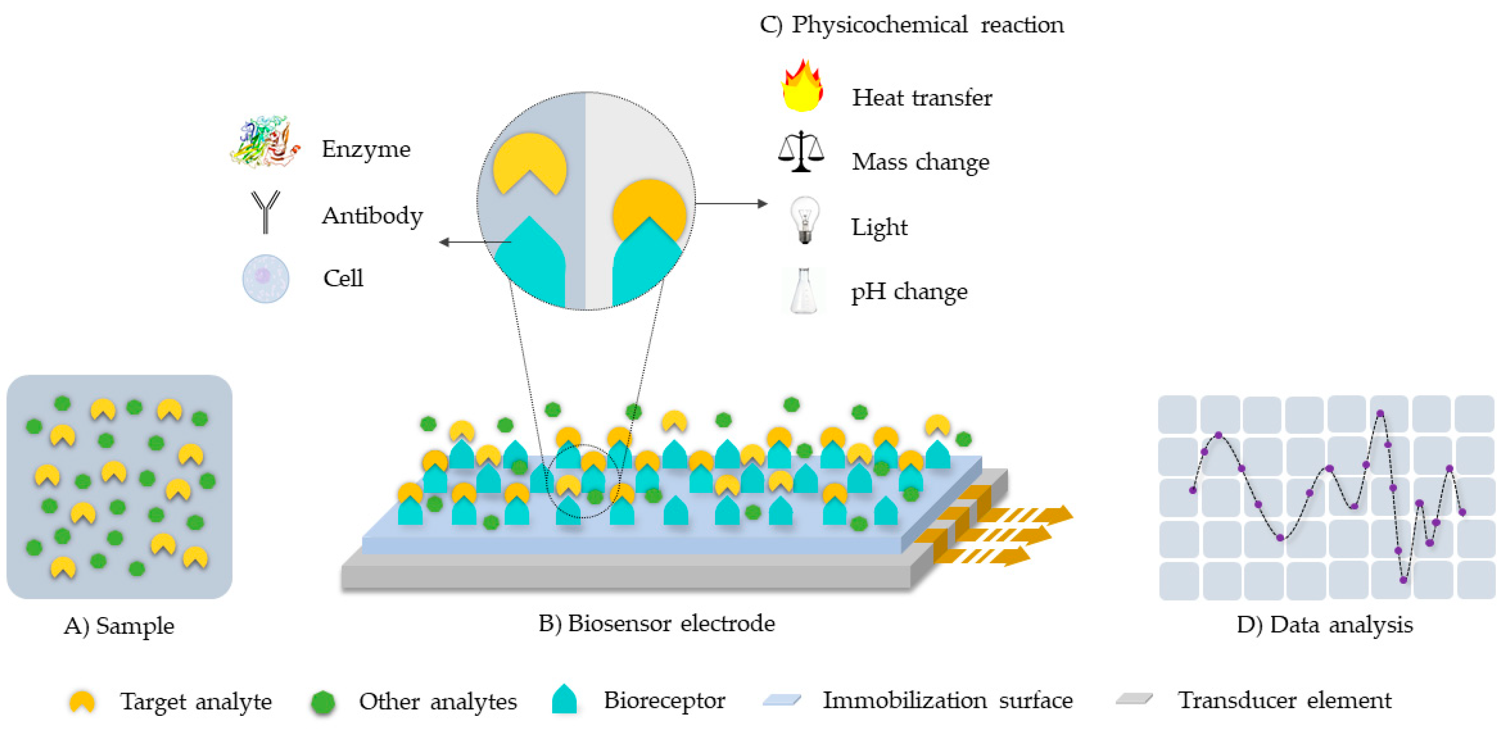
:1. Introduction
2. Enzymatic Biosensors Applied to Microfluidic Systems
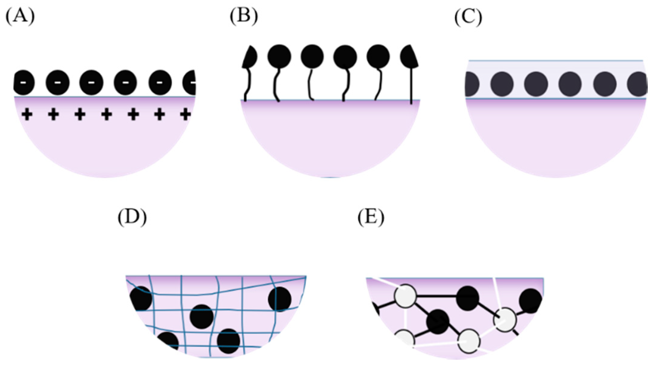
2.1. Immobilization Methods
2.2. Device Prototyping and Testing
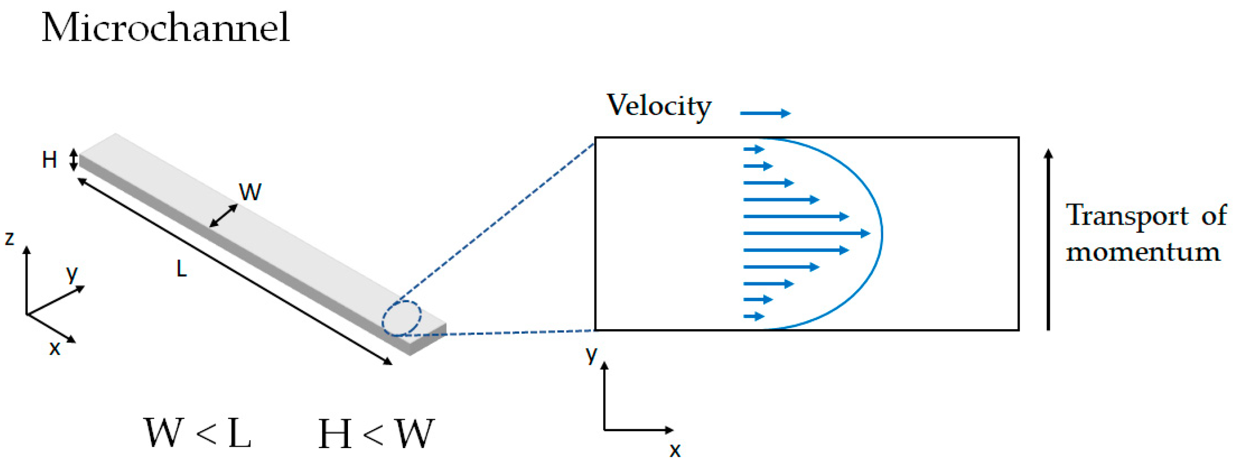
2.2.1. Microfluidics Fundamentals
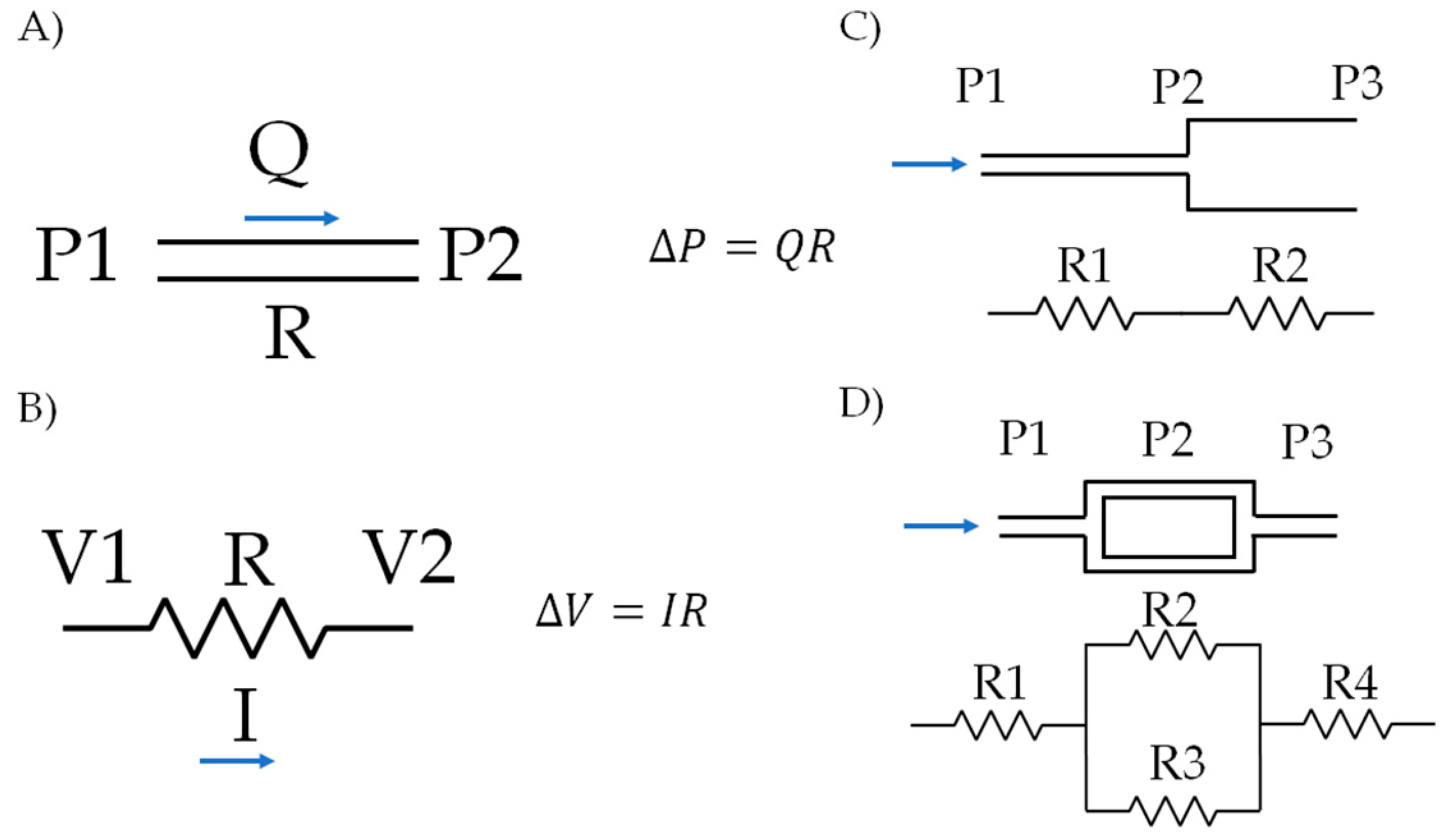
2.2.2. Prototypes Circuit Approximation
2.2.3. Mixing and Separation
2.2.4. Simulations
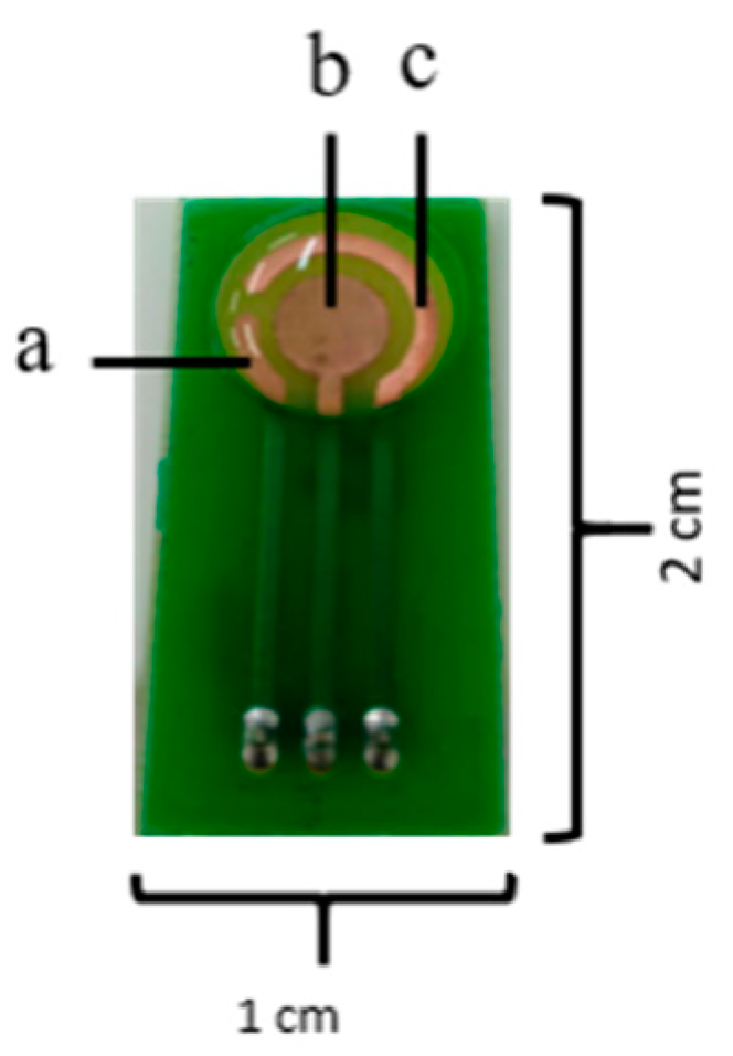
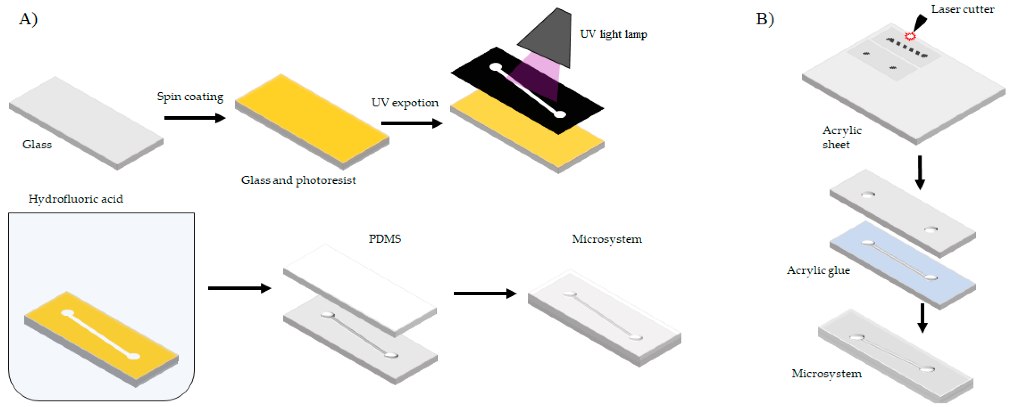
2.2.5. Fabrication of Prototypes
3. Oxidase/Peroxidase Based-Biosensors
4. Laccase Based-Biosensors
5. Concluding Remarks and Future Perspectives
Author Contributions
Funding
Acknowledgments
Conflicts of Interest
References
- Ejeian, F.; Etedali, P.; Mansouri-Tehrani, H.A.; Soozanipour, A.; Low, Z.X.; Asadnia, M.; Taheri-Kafrani, A.; Razmjou, A. Biosensors for wastewater monitoring: A review. Biosens. Bioelectron. 2018, 118, 66–79. [Google Scholar] [CrossRef] [PubMed]
- Morais, R.L.; Garcia, L.F.; Moreno, E.K.; Thomaz, D.V.; Rodrigues, M.F.; Vaz, B.G.; de Souza Gi, E. Electrochemical remediation of industrial pharmaceutical wastewater containing hormones in a pilot scale treatment system. Eclética Quím. J. 2019, 44, 40–52. [Google Scholar] [CrossRef]
- Coleman, C.; Huang, C.-H.; Lee, W.-N.; Nowack, K.; Carter, J.; Fu, J. Removal of pharmaceuticals and personal care products by two-stage biofiltration for drinking water treatment. Sci. Total Environ. 2019, 664, 240–248. [Google Scholar]
- Paucar, N.E.; Kim, I.; Tanaka, H.; Sato, C. Ozone treatment process for the removal of pharmaceuticals and personal care products in wastewater. Ozone Sci. Eng. 2019, 41, 3–16. [Google Scholar] [CrossRef]
- Gaudin, V. Advances in biosensor development for the screening of antibiotic residues in food products of animal origin—A comprehensive review. Biosens. Bioelectron. 2017, 90, 363–377. [Google Scholar] [CrossRef] [PubMed]
- Wang, Q.; Zhao, W.-M. Optical methods of antibiotic residues detections: A comprehensive review. Sens. Actuators B Chem. 2018, 269, 238–256. [Google Scholar] [CrossRef]
- Wang, S.; Xu, L.-P.; Zhang, X. Ultrasensitive Electrochemical Biosensor Based on Noble Metal Nanomaterials. Sci. Adv. Mater. 2015, 7, 2084–2102. [Google Scholar] [CrossRef]
- Lan, L.; Yao, Y.; Ping, J.; Ying, Y. Recent advances in nanomaterial-based biosensors for antibiotics detection. Biosens. Bioelectron. 2017, 91, 504–514. [Google Scholar] [CrossRef] [PubMed]
- Nikoleli, G.P.; Nikolelis, D.P.; Siontorou, C.G.; Karapetis, S.; Nikolelis, M.T. Application of biosensors based on lipid membranes for the rapid detection of toxins. Biosensors 2018, 8, 61. [Google Scholar] [CrossRef] [PubMed]
- Rackus, D.G.; Shamsi, M.H.; Wheeler, A.R. Electrochemistry, biosensors and microfluidics: A convergence of fields. Chem. Soc. Rev. 2015, 44, 5320–5340. [Google Scholar] [CrossRef] [PubMed]
- Bahadır, E.B.; Sezgintürk, M.K. Applications of commercial biosensors in clinical, food, environmental, and biothreat/biowarfare analyses. Anal. Biochem. 2015, 478, 107–120. [Google Scholar] [CrossRef] [PubMed]
- Huet, A.-C.; Delahaut, P.; Fodey, T.; Haughey, S.A.; Elliott, C.; Weigel, S. Advances in biosensor-based analysis for antimicrobial residues in foods. TrAC Trends Anal. Chem. 2010, 29, 1281–1294. [Google Scholar] [CrossRef]
- Pullano, S.A.; Critello, C.D.; Mahbub, I.; Tasneem, N.T.; Shamsir, S.; Islam, S.K.; Greco, M.; Fiorillo, A.S. EGFET-based sensors for bioanalytical applications: A review. Sensors 2018, 18, 4042. [Google Scholar] [CrossRef] [PubMed]
- Monteiro, T.; Almeida, M.G. Electrochemical Enzyme Biosensors Revisited: Old Solutions for New Problems. Crit. Rev. Anal. Chem. 2018, 1–23. [Google Scholar] [CrossRef] [PubMed]
- Zhu, C.; Yang, G.; Li, H.; Du, D.; Lin, Y. Electrochemical Sensors and Biosensors Based on Nanomaterials and Nanostructures. Anal. Chem. 2015, 87, 230–249. [Google Scholar] [CrossRef] [PubMed]
- Kurbanoglu, S.; Ozkan, S.A.; Merkoçi, A. Nanomaterials-based enzyme electrochemical biosensors operating through inhibition for biosensing applications. Biosens. Bioelectron. 2017, 89, 886–898. [Google Scholar] [CrossRef] [PubMed]
- Rocchitta, G.; Spanu, A.; Babudieri, S.; Latte, G.; Madeddu, G.; Galleri, G.; Nuvoli, S.; Bagella, P.; Demartis, M.; Fiore, V.; et al. Enzyme Biosensors for Biomedical Applications: Strategies for Safeguarding Analytical Performances in Biological Fluids. Sensors 2016, 16, 780. [Google Scholar] [CrossRef] [PubMed]
- Gonzalez-Rivera, J.C.; Osma, J.F. Fabrication of an Amperometric Flow-Injection Microfluidic Biosensor Based on Laccase for In Situ Determination of Phenolic Compounds. BioMed Res. Int. 2015, 2015, 845261. [Google Scholar] [CrossRef] [PubMed]
- Pohanka, M.; Skládal, P. Electrochemical Biosensors—Principles and Applications—A Review. J. Appl. Biomed. 2008, 6, 57–64. [Google Scholar]
- García-Morales, R.; García-García, A.; Orona-Navar, C.; Osma, J.F.; Nigam, K.D.P.; Ornelas-Soto, N. Biotransformation of emerging pollutants in groundwater by laccase from P. sanguineus CS43 immobilized onto titania nanoparticles. J. Environ. Chem. Eng. 2018, 6, 710–717. [Google Scholar] [CrossRef]
- Reyes-De-Corcuera, J.I.; Olstad, H.E.; García-Torres, R. Stability and Stabilization of Enzyme Biosensors: The Key to Successful Application and Commercialization. Annu. Rev. Food Sci. Technol. 2018, 9, 293–322. [Google Scholar] [CrossRef] [PubMed]
- Rodríguez-Delgado, M.M.; Alemán-Nava, G.S.; Rodríguez-Delgado, J.M.; Dieck-Assad, G.; Martínez-Chapa, S.O.; Barceló, D.; Parra, R. Laccase-based biosensors for detection of phenolic compounds. TrAC Trends Anal. Chem. 2015, 74, 21–45. [Google Scholar] [CrossRef] [Green Version]
- Yashas, S.R.; Shivakumara, B.R.; Udayashankara, T.H.; Krishna, B.M. Laccase biosensor: Green technique for quantification of phenols in wastewater (a review). Orient. J. Chem. 2018, 34, 631–637. [Google Scholar]
- Gul, I.; Ahmad, M.S.; Naqvi, S.S.; Hussain, A.; Wali, R.; Farooqi, A.A.; Ahmed, I. Polyphenol oxidase (PPO) based biosensors for detection of phenolic compounds: A Review. J. Appl. Biol. Biotechnol. 2017, 5, 72–85. [Google Scholar]
- Rodríguez-Delgado, M.; Ornelas-Soto, N. Laccases: A blue enzyme for greener alternative technologies in the detection and treatment of emerging pollutants. In Green Technologies and Environmental Sustainability; Springer International Publishing: Cham, Switzerland, 2017; pp. 45–65. [Google Scholar]
- Nazari, M.; Kashanian, S.; Rafipour, R. Laccase immobilization on the electrode surface to design a biosensor for the detection of phenolic compound such as catechol. Spectrochim. Acta Part A Mol. Biomol. Spectrosc. 2015, 145, 130–138. [Google Scholar] [CrossRef] [PubMed]
- Morozova, O.V.; Shumakovich, G.P.; Gorbacheva, M.A.; Shleev, S.V.; Yaropolov, A.I. ‘Blue’ laccases. Biochemistry 2007, 72, 1136–1150. [Google Scholar] [CrossRef] [PubMed]
- Riva, S. Laccases: Blue enzymes for green chemistry. Trends Biotechnol. 2006, 24, 219–226. [Google Scholar] [CrossRef] [PubMed]
- Bai, X.; Gu, H.; Chen, W.; Shi, H.; Yang, B.; Huang, X.; Zhang, Q. Immobilized laccase on activated poly(vinyl alcohol) microspheres for enzyme thermistor application. Appl. Biochem. Biotechnol. 2014, 173, 1097–1107. [Google Scholar] [CrossRef] [PubMed]
- Chinnadayyala, S.R.; Kakoti, A.; Santhosh, M.; Goswami, P. A novel amperometric alcohol biosensor developed in a 3rd generation bioelectrode platform using peroxidase coupled ferrocene activated alcohol oxidase as biorecognition system. Biosens. Bioelectron. 2014, 55, 120–126. [Google Scholar] [CrossRef] [PubMed]
- Kuswandi, B.; Irmawati, T.; Hidayat, M.A.; Jayus; Ahmad, M. A simple visual ethanol biosensor based on alcohol oxidase immobilized onto polyaniline film for halal verification of fermented beverage samples. Sensors 2014, 14, 2135–2149. [Google Scholar] [CrossRef] [PubMed]
- Mazlan, S.Z.; Lee, Y.H.; Hanifah, S.A. A new Laccase based biosensor for tartrazine. Sensors 2017, 17, 2859. [Google Scholar] [CrossRef] [PubMed]
- Reverté, L.; Prieto-Simón, B.; Campàs, M. New advances in electrochemical biosensors for the detection of toxins: Nanomaterials, magnetic beads and microfluidics systems. A review. Anal. Chim. Acta 2016, 908, 8–21. [Google Scholar] [CrossRef] [PubMed]
- Thakur, S.; Rao, S.N. Theoretical Microfluidics; Oxford University Press: Oxford, UK, 2014. [Google Scholar]
- Kirby, B.J. Micro- and Nanoscale Fluid Mechanics: Transport in Microfluidic Devices: Species and Charge Transport; Cambridge University Press: Cambridge, MA, USA, 2010. [Google Scholar]
- Fox, A.T.; McDonald, W.R.; Pritchard, P.J. Fox and McDonald’s Introduction to Fluid Mechanics, 8th ed.; Wiley: Hoboken, NJ, USA, 1999. [Google Scholar]
- Prakash, S.; Yeom, J. Nanofluidics and Microfluidics: Systems and Applications; Elsevier: Amsterdam, The Netherlands, 2014. [Google Scholar]
- Martel, J.M.; Toner, M. Inertial focusing dynamics in spiral microchannels. Phys. Fluids 2012, 24, 371–396. [Google Scholar] [CrossRef] [PubMed]
- Salafi, T.; Zeming, K.K.; Zhang, Y. Advancements in microfluidics for nanoparticle separation. Lab Chip 2017, 17, 11–33. [Google Scholar] [CrossRef] [PubMed] [Green Version]
- SooHoo, J.R.; Walker, G.M. Microfluidic aqueous two phase system for leukocyte concentration from whole blood. Biomed. Microdevices 2009, 11, 323–329. [Google Scholar] [CrossRef] [PubMed]
- Tsukamoto, M.; Taira, S.; Yamamura, S.; Morita, Y.; Nagatani, N.; Takamura, Y.; Tamiya, E. Cell separation by an aqueous two-phase system in a microfluidic device. Analyst 2009, 134, 1994–1998. [Google Scholar] [CrossRef] [PubMed]
- Yung, C.W.; Fiering, J.; Mueller, A.J.; Ingber, D.E. Micromagnetic-microfluidic blood cleansing device. Lab Chip 2009, 9, 1171–1177. [Google Scholar] [CrossRef] [PubMed]
- Wu, D.; Qin, J.; Lin, B. Electrophoretic separations on microfluidic chips. J. Chromatogr. A 2008, 1184, 542–559. [Google Scholar] [CrossRef] [PubMed]
- Lee, C.Y.; Chang, C.L.; Wang, Y.N.; Fu, L.M. Microfluidic mixing: A review. Int. J. Mol. Sci. 2011, 12, 3263–3287. [Google Scholar] [CrossRef] [PubMed]
- Hessel, V.; Löwe, H.; Schönfeld, F. Micromixers—A review on passive and active mixing principles. Chem. Eng. Sci. 2005, 60, 2479–2501. [Google Scholar] [CrossRef]
- Meijer, H.E.H.; Singh, M.K.; Kang, T.G.; den Toonder, J.M.J.; Anderson, P.D. Passive and active mixing in microfluidic devices. Macromol. Symp. 2009, 279, 201–209. [Google Scholar] [CrossRef]
- Dickinson, E.J.F.; Ekström, H.; Fontes, E. COMSOL Multiphysics®: Finite element software for electrochemical analysis. A mini-review. Electrochem. Commun. 2014, 40, 71–74. [Google Scholar] [CrossRef]
- Kaffash, A.; Rostami, K.; Zare, H.R. Modeling of an electrochemical nanobiosensor in COMSOL Multiphysics to determine phenol in the presence of horseradish peroxidase enzyme. Enzym. Microb. Technol. 2019, 121, 23–28. [Google Scholar] [CrossRef] [PubMed]
- Hossen, M.N.; Ferdous, M.; Khalek, M.A.; Chakma, S.; Paul, B.K.; Ahmed, K. Design and analysis of biosensor based on surface plasmon resonance. Sens. Bio-Sensing Res. 2018, 21, 1–6. [Google Scholar] [CrossRef]
- Wu, G.; Meyyappan, M.; Lai, K.W.C. Simulation of graphene field-effect transistor biosensors for bacterial detection. Sensors 2018, 18, 1715. [Google Scholar] [CrossRef] [PubMed]
- Berkowski, K.L.; Plunkett, K.N.; Yu, Q.; Moore, J.S. Introduction to Photolithography: Preparation of Microscale Polymer Silhouettes. J. Chem. Educ. 2005, 82, 1365. [Google Scholar] [CrossRef]
- Sun, S.; Leggett, G.J. Generation of Nanostructures by Scanning Near-Field Photolithography of Self-Assembled Monolayers and Wet Chemical Etching. Nano Lett. 2002, 2, 1223–1227. [Google Scholar] [CrossRef]
- Brower, K.; White, A.K.; Fordyce, P.M. Multi-step Variable Height Photolithography for Valved Multilayer Microfluidic Devices. J. Vis. Exp. 2017, 55276. [Google Scholar] [CrossRef] [PubMed]
- Szilasi, S.Z.; Cserháti, C. Selective etching of PDMS: Etching technique for application as a positive tone resist. Appl. Surf. Sci. 2018, 457, 662–669. [Google Scholar] [CrossRef]
- Huang, Y.; Liu, S.; Yang, W.; Yu, C. Surface roughness analysis and improvement of PMMA-based microfluidic chip chambers by CO2laser cutting. Appl. Surf. Sci. 2010, 256, 1675–1678. [Google Scholar] [CrossRef]
- Carbaugh, D.J.; Wright, J.T.; Rahman, F. Negative tone photolithography with photo-sensitised polymethyl methacrylate (PMMA). Microelectron. Eng. 2017, 171, 53–59. [Google Scholar] [CrossRef]
- Trantidou, T.; Friddin, M.S.; Gan, K.B.; Han, L.; Bolognesi, G.; Brooks, N.J.; Ces, O. Mask-free laser lithography for rapid and low-cost microfluidic device fabrication. Anal. Chem. 2018, 90, 13915–13921. [Google Scholar] [CrossRef] [PubMed]
- Stasyuk, N.; Gayda, G.; Zakalskiy, A.; Zakalska, O.; Serkiz, R.; Gonchar, M. Amperometric biosensors based on oxidases and PtRu nanoparticles as artificial peroxidase. Food Chem. 2019, 285, 213–220. [Google Scholar] [CrossRef] [PubMed]
- Soylemez, S.; Kaya, H.Z.; Udum, Y.A.; Toppare, L. A multipurpose conjugated polymer: Electrochromic device and biosensor construction for glucose detection. Org. Electron. Phys. Mater. Appl. 2019, 65, 327–333. [Google Scholar] [CrossRef]
- Monti, D.; Ottolina, G.; Carrea, G.; Riva, S. Redox Reactions Catalyzed by Isolated Enzymes. Chem. Rev. 2011, 111, 4111–4140. [Google Scholar] [CrossRef] [PubMed]
- Colmati, F.; Sgobbi, L.F.; Teixeira, G.F.; Vilela, R.S.; Martins, T.D.; Figueiredo, G.O. Electrochemical Biosensors Containing Pure Enzymes or Crude Extracts as Enzyme Sources for Pesticides and Phenolic Compounds with Pharmacological Property Detection and Quantification. In Environmental Biosensors; IntechOpen: Goiânia, Brazil, 2019. [Google Scholar]
- Gayda, G.Z.; Demkiv, O.M.; Stasyuk, N.Y.; Serkiz, R.Y.; Lootsik, M.D.; Errachid, A.; Gonchar, M.V.; Nisnevitch, M. Metallic Nanoparticles Obtained via ‘Green’ Synthesis as a Platform for Biosensor Construction. Appl. Sci. 2019, 9, 720. [Google Scholar] [CrossRef]
- Xiao, F.; Wang, L.; Duan, H. Nanomaterial based electrochemical sensors for in vitro detection of small molecule metabolites. Biotechnol. Adv. 2016, 34, 234–249. [Google Scholar] [CrossRef] [PubMed]
- Pitschmann, V.; Urban, M.; Dědič, J.; Matějovský, L.; Dymák, M.; Lobotka, M. Modified Biosensor for Cholinesterase Inhibitors with Guinea Green B as the Color Indicator. Biosensors 2018, 8, 81. [Google Scholar] [CrossRef] [PubMed]
- Antunes, R.; Ferraz, D.; Garcia, L.; Thomaz, D.; Luque, R.; Lobón, G.; Gil, E.; Lopes, F. Development of a Polyphenol Oxidase Biosensor from Jenipapo Fruit Extract (Genipa americana L.) and Determination of Phenolic Compounds in Textile Industrial Effluents. Biosensors 2018, 8, 47. [Google Scholar] [CrossRef] [PubMed]
- Sassolas, A.; Blum, L.J.; Leca-Bouvier, B.D. Immobilization strategies to develop enzymatic biosensors. Biotechnol. Adv. 2012, 30, 489–511. [Google Scholar] [CrossRef] [PubMed]
- Khan, M.; Park, S. Glucose biosensor based on GOx/HRP bienzyme at liquid–crystal/aqueous interface. J. Colloid Interface Sci. 2015, 457, 281–288. [Google Scholar] [CrossRef] [PubMed]
- Xia, H.Q.; Kitazumi, Y.; Shirai, O.; Ohta, H.; Kurihara, S.; Kano, K. Putrescine oxidase/peroxidase-co-immobilized and mediator-less mesoporous microelectrode for diffusion-controlled steady-state amperometric detection of putrescine. J. Electroanal. Chem. 2017, 804, 128–132. [Google Scholar] [CrossRef]
- Pérez, J.P.H.; López-Ruiz, B.; López-Cabarcos, E. Synthesis and characterization of microparticles based on poly-methacrylic acid with glucose oxidase for biosensor applications. Talanta 2016, 149, 310–318. [Google Scholar] [CrossRef] [PubMed]
- Bottoni, P.; Caroli, S. Detection and quantification of residues and metabolites of medicinal products in environmental compartments, food commodities and workplaces. A review. J. Pharm. Biomed. Anal. 2015, 106, 3–24. [Google Scholar] [CrossRef] [PubMed]
- Ahmed, M.J.; Hameed, B.H. Removal of emerging pharmaceutical contaminants by adsorption in a fixed-bed column: A review. Ecotoxicol. Environ. Saf. 2018, 149, 257–266. [Google Scholar] [CrossRef] [PubMed]
- Rebollar-Pérez, G.; Campos-Terán, J.; Ornelas-Soto, N.; Méndez-Albores, A.; Torres, E. Biosensors based on oxidative enzymes for detection of environmental pollutants. Biocatalysis 2016, 1, 118–129. [Google Scholar] [CrossRef]
- Narang, J.; Malhotra, N.; Singh, S.; Singh, G.; Pundir, C.S. Monitoring analgesic drug using sensing method based on nanocomposite. RSC Adv. 2015, 5, 2396–2404. [Google Scholar] [CrossRef]
- Jaouani, A.; Guillén, F.; Penninckx, M.J.; Martínez, A.T.; Martínez, M.J. Role of Pycnoporus coccineus laccase in the degradation of aromatic compounds in olive oil mill wastewater. Enzym. Microb. Technol. 2005, 36, 478–486. [Google Scholar] [CrossRef]
- Sheikhi, F.; Ardakani, M.R.; Enayatizamir, N.; Rodriguez-Couto, S. The Determination of Assay for Laccase of Bacillus subtilis WPI with Two Classes of Chemical Compounds as Substrates. Indian J. Microbiol. 2012, 52, 701–707. [Google Scholar] [CrossRef] [PubMed] [Green Version]
- Viswanath, B.; Rajesh, B.; Janardhan, A.; Kumar, A.P.; Narasimha, G. Fungal laccases and their applications in bioremediation. Enzym. Res. 2014, 2014, 163242. [Google Scholar] [CrossRef] [PubMed]
- Bhalla, N.; Jolly, P.; Formisano, N.; Estrela, P. Introduction to biosensors. Essays Biochem. 2016, 60, 1–8. [Google Scholar] [CrossRef] [PubMed] [Green Version]
- Leonardo, S.; Toldrà, A. Trends and Prospects on Electrochemical Biosensors for the Detection of Marine Toxins. Compr. Anal. Chem. 2017, 78, 303–341. [Google Scholar]
- Brondani, D.; Scheeren, C.W.; Dupont, J.; Vieira, I.C. Biosensor based on platinum nanoparticles dispersed in ionic liquid and laccase for determination of adrenaline. Sens. Actuators B Chem. 2009, 140, 252–259. [Google Scholar] [CrossRef]
- Baluta, S.; Lesiak, A.; Graphene, J.C. Quantum Dots-based Electrochemical Biosensor for Catecholamine Neurotransmitters Detection. Electroanalysis 2018, 30, 1773–1782. [Google Scholar] [CrossRef]
- Santhiago, M.; Vieira, I.C. l-Cysteine determination in pharmaceutical formulations using a biosensor based on laccase from Aspergillus oryzae. Sens. Actuators B Chem. 2007, 128, 279–285. [Google Scholar] [CrossRef]
- Zhao, W.; Wang, K.; Wei, Y.; Ma, Y.; Liu, L.; Huang, X. Laccase biosensor based on phytic acid modification of nanostructured SiO2 surface for sensitive detection of dopamine. Langmuir 2014, 30, 11131–11137. [Google Scholar] [CrossRef] [PubMed]
- Leite, O.D.; Lupetti, K.O.; Fatibello-Filho, O.; Vieira, I.C.; Barbosa, A.D. Synergic effect studies of the bi-enzymatic system laccaseperoxidase in a voltammetric biosensor for catecholamines. Talanta 2003, 59, 889–896. [Google Scholar] [CrossRef]
- Das, P.; Barbora, L.; Das, M.; Goswami, P. Highly sensitive and stable laccase based amperometric biosensor developed on nano-composite matrix for detecting pyrocatechol in environmental samples. Sens. Actuators B Chem. 2014, 192, 737–744. [Google Scholar] [CrossRef]
- Rahman, M.A.; Noh, H.B.; Shim, Y.B. Direct electrochemistry of laccase immobilized on Au nanoparticles encapsulated-dendrimer bonded conducting polymer: Application for a catechin sensor. Anal. Chem. 2008, 80, 8020–8027. [Google Scholar] [CrossRef] [PubMed]
- Chen, T.; Xu, Y.; Wei, S.; Li, A.; Huang, L.; Liu, J. A signal amplification system constructed by bi-enzymes and bi-nanospheres for sensitive detection of norepinephrine and miRNA. Biosens. Bioelectron. 2019, 124–125, 224–232. [Google Scholar] [CrossRef] [PubMed]
- Portaccio, M.; Di Martino, S.; Maiuri, P.; Durante, D.; De Luca, P.; Lepore, M.; Bencivenga, U.; Rossi, S.; De Maio, A.; Mita, D.G. Biosensors for phenolic compounds: The catechol as a substrate model. J. Mol. Catal. B Enzym. 2006, 41, 97–102. [Google Scholar] [CrossRef]
- Kulys, J.; Vidziunaite, R. Amperometric biosensors based on recombinant laccases for phenols determination. Biosens. Bioelectron. 2002, 18, 319–325. [Google Scholar] [CrossRef]
- Roy, J.J.; Abraham, T.E.; Abhijith, K.S.; Kumar, P.V.S.; Thakur, M.S. Biosensor for the determination of phenols based on Cross-Linked Enzyme Crystals (CLEC) of laccase. Biosens. Bioelectron. 2005, 21, 206–211. [Google Scholar] [CrossRef] [PubMed]
- Odaci, D.; Timur, S.; Pazarlioğlu, N.; Kirgöz, Ü.A.; Telefoncu, A. Effects of mediators on the laccase biosensor response in paracetamol detection. Biotechnol. Appl. Biochem. 2006, 45, 23–28. [Google Scholar] [PubMed]
- Li, Y.; Zhang, L.; Li, M.; Pan, Z.; Li, D. A disposable biosensor based on immobilization of laccase with silica spheres on the MWCNTs-doped screen-printed electrode. Chem. Cent. J. 2012, 6, 103. [Google Scholar] [CrossRef] [PubMed]
- Chen, X.; Li, D.; Li, G.; Luo, L.; Ullah, N.; Wei, Q.; Huang, F. Facile fabrication of gold nanoparticle on zein ultrafine fibers and their application for catechol biosensor. Appl. Surf. Sci. 2015, 328, 444–452. [Google Scholar] [CrossRef]
- Bauer, C.G.; Kühn, A.; Gajovic, N.; Skorobogatko, O.; Holt, P.J.; Bruce, N.C.; Makower, A.; Lowe, C.R.; Scheller, F.W. New enzyme sensors for morphine and codeine based on morphine dehydrogenase and laccase. Fresenius J. Anal. Chem. 1999, 364, 179–183. [Google Scholar] [CrossRef]
- Moraes, J.T.; Salamanca-Neto, C.A.; Švorc, Ĺ.; Schirmann, J.G.; Barbosa-Dekker, A.M.; Dekker, R.F.; Sartori, E.R. Laccase from Botryosphaeria rhodina MAMB-05 as a biological component in electrochemical biosensing devices. Anal. Methods 2019, 11, 717–720. [Google Scholar] [CrossRef]
- Liu, Y.; Qu, X.; Guo, H.; Chen, H.; Liu, B.; Dong, S. Facile preparation of amperometric laccase biosensor with multifunction based on the matrix of carbon nanotubes-chitosan composite. Biosens. Bioelectron. 2006, 21, 2195–2201. [Google Scholar] [CrossRef] [PubMed]
- Ferry, Y.; Leech, D. Amperometric Detection of Catecholamine Neurotransmitters Using Electrocatalytic Substrate Recycling at a Laccase Electrode. Electroanalysis 2005, 17, 113–119. [Google Scholar] [CrossRef]
- Sanz, J.; de Marcos, S.; Galbán, J. Autoindicating optical properties of laccase as the base of an optical biosensor film for phenol determination. Anal. Bioanal. Chem. 2012, 404, 351–359. [Google Scholar] [CrossRef] [PubMed]
- Huang, J.; Fang, H.; Liu, C.; Gu, E.; Jiang, D. A novel fiber optic biosensor for the determination of adrenaline based on immobilized laccase catalysis. Anal. Lett. 2008, 41, 1430–1442. [Google Scholar] [CrossRef]
- Silva, L.I.B.; Ferreira, F.D.P.; Freitas, A.C.; Rocha-Santos, T.A.P.; Duarte, A.C. Optical fiber biosensor coupled to chromatographic separation for screening of dopamine, norepinephrine and epinephrine in human urine and plasma. Talanta 2009, 80, 853–857. [Google Scholar] [CrossRef] [PubMed]
- Abdullah, J.; Ahmad, M.; Lee, Y.H.; Karuppiah, N.; Sidek, H. An optical biosensor based on immobilization of lacease and MBTH in stacked films for the detection of catechol. Sensors 2007, 7, 2238–2250. [Google Scholar] [CrossRef] [PubMed]
- Karami, C.; Taher, M.A. A catechol biosensor based on immobilizing laccase to Fe3O4@Au core-shell nanoparticles. Int. J. Biol. Macromol. 2019, 129, 84–90. [Google Scholar] [CrossRef] [PubMed]
- Ferreira, F.D.P.; Silva, L.I.B.; Freitas, A.C.; Rocha-Santos, T.A.P.; Duarte, A.C. High performance liquid chromatography coupled to an optical fiber detector coated with laccase for screening catecholamines in plasma and urine. J. Chromatogr. A 2009, 1216, 7049–7054. [Google Scholar] [CrossRef] [PubMed]
- Mohapatra, S.; Padhye, L.P.; Mukherji, S. Challenges in Detection of Antibiotics in Wastewater Matrix; Springer: Singapore, 2018; pp. 3–20. [Google Scholar]
- Anku, W.W.; Mamo, M.A.; Govender, P.P. Phenolic Compounds in Water: Sources, Reactivity, Toxicity and Treatment Methods. In Phenolic Compounds—Natural Sources, Importance and Applications; InTech: Johannesburg, South Africa, 2017. [Google Scholar] [Green Version]
- Zilly, A.; da Silva Coelho-Moreira, J.; Bracht, A.; De Souza, C.G.; Carvajal, A.E.; Koehnlein, E.A.; Peralta, R.M. Influence of NaCl and Na2SO4 on the kinetics and dye decolorization ability of crude laccase from Ganoderma lucidum. Int. Biodeterior. Biodegrad. 2011, 65, 340–344. [Google Scholar] [CrossRef] [Green Version]
- Margot, J.; Copin, P.J.; von Gunten, U.; Barry, D.A.; Holliger, C. Sulfamethoxazole and isoproturon degradation and detoxification by a laccase-mediator system: Influence of treatment conditions and mechanistic aspects. Biochem. Eng. J. 2015, 103, 47–59. [Google Scholar] [CrossRef] [Green Version]
- Mogharabi, M.; Rezaei, S.; Faramarzi, M.A. Trends in Peptide and Protein Sciences; Shahid Beheshti University of Medical Sciences, Laser Application in Medical Sciences Research Center: Tehran, Iran, 2017; Volume 1. [Google Scholar]
- Denard, C.A.; Ren, H.; Zhao, H. Improving and repurposing biocatalysts via directed evolution. Curr. Opin. Chem. Biol. 2015, 25, 55–64. [Google Scholar] [CrossRef] [PubMed]


| Transduction Method | Immobilization Method | Target Analyte | Measurement | Detection (Range| Limit| Time) | Characteristics (Selectivity | Stability) | Ref. |
|---|---|---|---|---|---|---|
| Electrochemical Biosensor | Adsorption | Adrenaline | Pt-BMI.PF6-Laccase Ag/AgCl reference electrode Pt wire as counter electrode | M M NS | Suitable (Untested) 20% loss of response after 90 days | [79] |
| Epinephrine | Glassy carbon-GQDs-Laccase electrode Ag/AgCl reference electrode Pt wire as counter electrode | µM 83 ηM NS | High (Against ascorbic acid, uric acid, cysteine, glutathione, tryptophan and a mix of all) NS | [80] | ||
| L-Cysteine | Carbon-paste electrode Ag/AgCl reference electrode Pt wire as counter electrode | M NS NS | High (Against hydroquinone and other inhibitors) Lifetime of 9 months (950 measurements) Maximum response at pH 7.0 | [81] | ||
| Dopamine | Laccase/(h-SiO2—PA)/Glassy-carbon electrode Saturated calomel reference electrode | µM µM s | Good anti-interference ability 11% loss of response after 20 days | [82] | ||
| Dopamine Adrenaline L-dopa Isoprenaline | Nujol/Graphite powder Laccase/Peroxidase as working electrode Ag/AgCl reference electrode Pt wire as counter electrode | D: M A: M L: M I: M D: M A: M L: M I: M 60 s | NS Lifetime of 2 months (500 measurements) D: Maximum response at pH 6.0 and 35 °C A: Maximum response at pH 7.0 and 35 °C L: Maximum response at pH 6.5 and 35 °C I: Maximum response at pH 6.0 and 35 °C | [83] | ||
| Covalent binding | Pyrocatechol | Glassy-carbon electrode Ag/AgCl reference electrode Pt wire as counter electrode | ηM 2.82 ηM NS | ≤5% change in the response by environmental interferents 19% loss of response after 21 days | [84] | |
| Catechin | PDATT/Den(AuNPs) on glassy-carbon electrode Ag/AgCl electrode | µM µM <10 s | NS 8% loss of response after 60 days Maximum response at pH 6.5 and 30 °C | [85] | ||
| Norepinephrine | PDA-Laccase/Au-glucose dehydrogenase | 0.5 ηM0.5 µM 0.07 ηM NS | NS 8.57% loss of response after 30 days | [86] | ||
| Tartrazine | Laccase -AuNPs coated on a carbon-paste screen-printed electrode | µM 0.04 µM 2 min | ±10% change in the response by common coexisting substances 48.9% loss of response after 90 days Maximum response at pH 5.0 | [32] | ||
| Catechol | Graphite electrode Ag/AgCl reference electrode Pt wire as counter electrode | A: up to—2 mM B-C: up to—0.1 mM NS | NS A: 10 days, B: 30 days and C: 60 days of stability Maximum response at pH 5.0 | [87] | ||
| Catechol | Glassy carbon as working electrode Ag/AgCl reference electrode Pt wire as counter electrode | M M NS | NS | [26] | ||
| Cross-linking | Acetaminophen Diclofenac | TiO2-Lac nanoparticles | NS | NS High stability at low pH values of 2–3 and 50–60 °C | [20] | |
| Pyrocatechol 1-naphthol o-phenylenediamine | Graphite electrode SCE as reference electrode Pt wire as counter electrode | mM NS NS | NS Lifetime of 9 days (decreased response at longer times) | [88] | ||
| 2-amino phenol Catechol Pyrogallol Guaiacol | Clarke-type electrode (Au cathode and Ag/AgCl reference electrode) | mM NS 60 s | NS 40% loss of response after 30 measurements Maximum response at pH 5.5–6.0 | [89] | ||
| Paracetamol: With/Without HBT | Dissolve oxygen electrode | HBT: µM W-HBT: µM 10 min | NS 0% loss of response after 7 h Maximum response at pH 4.5 and 35 °C | [90] | ||
| Entrapment | Dopamine | Lac/Si/MWCNTs/ SPE electrode Ag/AgCl reference electrode | µM 0.42 µM NS | High (Against ascorbic acid [AA]) 14% loss of response after 30 days | [91] | |
| Catechol | Nafion/Laccase-glassy carbon as working electrode Ag/AgCl reference electrode Pt wire for counter electrode | µM 0.166 µM NS | ≤3% change in the response by phenolic interferents 12.9% loss of response after 30 days | [92] | ||
| Morphine | Clark oxygen electrode | µM A: 32 ηM N-A: 10 µM 1 min | High (Against codeine) NS | [93] | ||
| Epinephrine | Laccase-carbon paste working electrode Ag/AgCl reference electrode Pt wire as counter electrode | µM 1.84 µM NS | High (Against dopamine and phenol) 7.0% loss of response after 7 days | [94] | ||
| Catechol | CNTs–CS/GC electrode Ag/AgCl reference electrode Pt wire as counter electrode | µM 0.66 µM NS | NS <1% loss of response after 15 days | [95] | ||
| Epinephrine Norepinephrine Dopamine | Os(PVI) 10-Laccase electrode Glassy carbon working electrode Ag/AgCl reference electrode Pt wire as counter electrode | NS E: 11 ηM N: 8 ηM D: 4 ηM 5 s | No selectivity between the catecholamines Lifetime of at least 1 month | [96] | ||
| Optical Biosensor | Adsorption | Catechol | Lac-polyacrylamide sensor film | L: M H: M NS 450 s | NS Lifetime of 30 measurements Maximum response at pH 5.0–6.0 | [97] |
| Adrenaline | Laccase-CuTAPc-Fe3O4-NPs | M M NS 30 s | NS 16% loss of response after 10 measurements | [98] | ||
| Dopamine Norepinephrine Epinephrine | LacOF biosensor | NS D: 2.1 pg / mL N: 2.6 pg / mL E: 3.4 pg / mL 3 min | High (Against urine and plasma) <5% loss of response after 60 days | [99] | ||
| Covalent binding | Adrenaline | Laccase-CuTAPc-Fe3O4 NPs | M M NS 30 s | NS 16% loss of response after 10 measurements | [98] | |
| Cross-linking | Catechol | Laccase-Hybrid Nafion/sol-gel silicate-MBTH film | mM 0.33 mM 10 min | Suitable selectivity against Nafion/sol-gel silicate Lifetime of at least 2 months | [100] | |
| Catechol | Laccase-Au-Fe3O4 NPs | µM 2 µM 40 min | NS Maximum response at pH 5.0 | [101] | ||
| Entrapment | Epinephrine Dopamine Norepinephrine | Liquid chromatography (HPLC) and detection by optical fiber (OF) coated with Laccase | pg / mL E: 3.5 pg / mL D: 2.9 pg / mL N: 3.3 pg / mL 7 min | NS Lifetime of at least 2 months | [102] | |
| Thermal Biosensor | Cross-linking | Phenol | Lac/PVA Microspheres | mM NS NS | NS 13.7% loss of response after 100 days | [29] |
© 2019 by the authors. Licensee MDPI, Basel, Switzerland. This article is an open access article distributed under the terms and conditions of the Creative Commons Attribution (CC BY) license (http://creativecommons.org/licenses/by/4.0/).
Share and Cite
Campaña, A.L.; Florez, S.L.; Noguera, M.J.; Fuentes, O.P.; Ruiz Puentes, P.; Cruz, J.C.; Osma, J.F. Enzyme-Based Electrochemical Biosensors for Microfluidic Platforms to Detect Pharmaceutical Residues in Wastewater. Biosensors 2019, 9, 41. https://0-doi-org.brum.beds.ac.uk/10.3390/bios9010041
Campaña AL, Florez SL, Noguera MJ, Fuentes OP, Ruiz Puentes P, Cruz JC, Osma JF. Enzyme-Based Electrochemical Biosensors for Microfluidic Platforms to Detect Pharmaceutical Residues in Wastewater. Biosensors. 2019; 9(1):41. https://0-doi-org.brum.beds.ac.uk/10.3390/bios9010041
Chicago/Turabian StyleCampaña, Ana Lucia, Sergio Leonardo Florez, Mabel Juliana Noguera, Olga P. Fuentes, Paola Ruiz Puentes, Juan C. Cruz, and Johann F. Osma. 2019. "Enzyme-Based Electrochemical Biosensors for Microfluidic Platforms to Detect Pharmaceutical Residues in Wastewater" Biosensors 9, no. 1: 41. https://0-doi-org.brum.beds.ac.uk/10.3390/bios9010041