Article

Effect of Citrus aurantium L. Essential Oil on Streptococcus mutans Growth, Biofilm Formation and Virulent Genes Expression

1
Oral Ecology Research Group, Faculty of Dentistry, Laval University, 2420, Rue de la Terrasse, Quebec City, QC G1V 0A6, Canada
2
Groupe de Recherche sur les Biofilms et la Biocontamination des Matériaux, Faculté des Sciences, Université d’Annaba, Annaba 23000, Algeria
3
Laboratoire de Bio Surveillance Environnementale, Université Badji Mokhtar, Annaba 23220, Algeria
*
Author to whom correspondence should be addressed.
Submission received: 21 December 2020 / Revised: 4 January 2021 / Accepted: 5 January 2021 / Published: 8 January 2021

Abstract

:
In an oral cavity, dental caries, periodontal disease, and endodontic lesions are caused by well-known bacterial and fungal pathogens. Essential oils (EOs) have demonstrated antimicrobial activity suggesting their use for oral hygiene. The goal of this study was to evaluate the interaction of bitter orange flower (Citrus aurantium L.) essential oil with cariogenic bacteria Streptococcus mutans and human gingival epithelial cells. After extraction, the chemical composition of the essential oil was analyzed by gas chromatography, and its antimicrobial activity was evaluated against the growth and the expression of virulent genes in S. mutans. Finally, the effects of this essential oil on human gingival epithelial cell adhesion and growth were assessed using cell adhesion and proliferation assays. We showed that the major constituents of the tested essential oil were limonene, linalool, and β-ocimene. The essential oil reduced the growth of S. mutans, and decreased expression of comC, comD, comE, gtfB, gtfC, and gbpB genes. It should, however, be noted that essential oil at high concentration was toxic to gingival epithelial cells. Overall, this study suggests that C. aurantium L. essential oil could be used to prevent/control oral infections.

1. Introduction

Essential oils (EOs) are natural products empirically used to treat various illnesses [1,2]. In the literature on the subject, EOs have been suggested for various applications to treat sleep quality and anxiety as well as for antimicrobial products to synergize the antimicrobial activity of conventional antimicrobial agents [3,4]. In the oral cavity, the antimicrobial properties of EOs have shown promising health benefits, including the reduction of gingival inflammation and halitosis and the control of biofilm formation [5,6].
Biofilms refer to bacteria embedded with extracellular polysaccharides, lipoprotein, and fibrinogen. Biofilm formation is promoted by microorganism adherence to a favorable support and by their capacity producing extracellular matrix [7,8]. During biofilm formation, microorganisms modify their gene expression patterns. As an example, in Streptococcus mutans, genes associated with glucan (gtfB) and fructan synthesis (ftf) were regulate differentially in biofilm [9]. In Candida albicans biofilm formation involves different genes, including inositol phosphoryl transferase1 (IPT1) and ECM33 genes [10,11]. It has been shown that biofilms confer a high bacterial resistance to antibiotics [12] leading to biofilm-related infections such as in oral cavities.
Oral microbiota is a normal component of an oral cavity and has an important function to protect against the colonization of extrinsic bacteria that could affect systemic health [13,14]. However, under certain conditions, including poor oral hygiene and the use of immunosuppressive drugs, commensal oral microbes can cause oral diseases such as dental caries and periodontal diseases [15], hence the emergence of multiple initiatives based on a modulation of the oral microbiome to maintain or reestablish a healthy oral ecosystem [16].
Oral hygiene is among the most efficient methods to prevent oral diseases [17]. This includes the use of mouthwash [18]. Mouthwash has become the ideal vehicle in which to incorporate antimicrobial molecules, as this easy-to-use product contributes to reducing plaque biofilm and preventing bad breath, to name a few [19]. Aside from antimicrobial peptides, mouthwash can also contain chlorhexidine, cetylpyridinium chloride, and essential oils [6]. These types of mouthwash can be considered therapeutic because they contain active ingredients intended to help control or reduce such conditions as halitosis, gingivitis, dental plaque, periodontitis, and tooth decay [19,20]. In the EOs used in mouthwash can be found thymol, menthol, and eucalyptol [21].
The EO extracted from Citrus aurantium L. (CA) has been reported to be efficient against microbial growth [22] and can serve as a food-flavoring substance [23,24] because it contains bioactive chemicals, such as phenolics and flavonoids [24,25]. With these reported beneficial effects, we sought to investigate the effects of the EO of CA on the oral bacteria S. mutans and to evaluate the safety/toxicity of this EO when in contact with gingival epithelial cells.

2. Results

2.1. Chemical Composition and Antioxidant Activities of C. aurantium L. Essential Oil

Chromatographic analyses resulted in the identification of 24 chemicals in the EO of CA (Table 1). The high-level chemicals in the EO were limonene (50.5%), linalool (9.89%), β-ocimene (7.82%), and sabinene (5.1%).

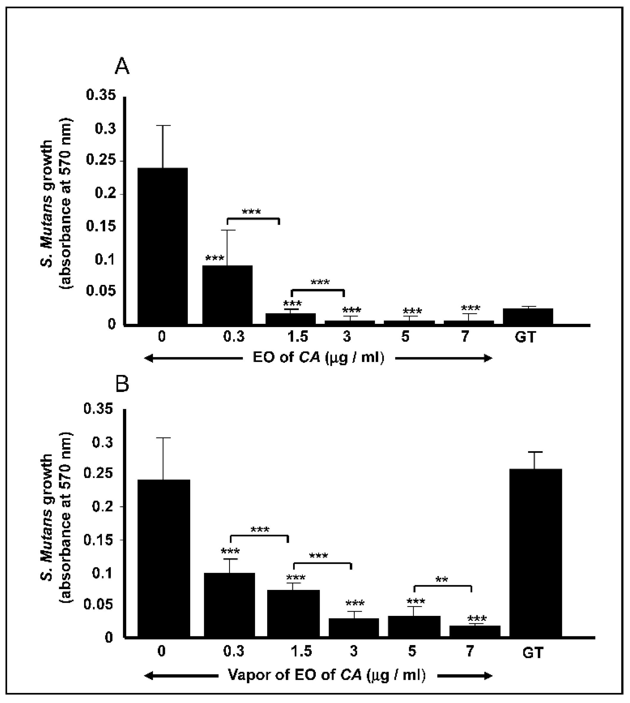
2.2. C. aurantium L. Essential Oil decreased S. mutans Cell Growth

Direct contact of S. mutans cells with the EO led to a significant (p < 0.01) inhibition of the bacteria growth (Figure 1A). After 24 h of culture, the inhibition of S. mutans cell growth was observed even at a low EO concentration. At 0.3 μg/mL, the OD600 decreased to 0.09 ± 0.05, compared to that recorded by the control, which was OD600 of 0.24 ± 0.067. At the highest concentration (7 μg/mL) of EO, the OD600 dramatically decreased to 0.009 ± 0.008, confirming the antimicrobial effect of the EO. It is interesting to note that starting from 1.5 μg/mL, the effect of the EO was greater than that of gentamicin at 5 μg/mL (Figure 1A). EO vapor was also found to be effective against S. mutans cell growth (Figure 1B). Indeed, with the lowest concentration (0.3 μg/mL), the EO vapor was capable of reducing S. mutans cell growth from 0.24 ± 0.067 (control) to 0.1 ± 0.02, which were comparable to the values obtained in the direct contact tests. At the highest concentration (7 μg/mL) of EO, the vapor produced an OD600 of 0.018 ± 0.005. Overall findings indicate that both direct contact and vapor contributed to significantly reducing the growth of the S. mutans cells.

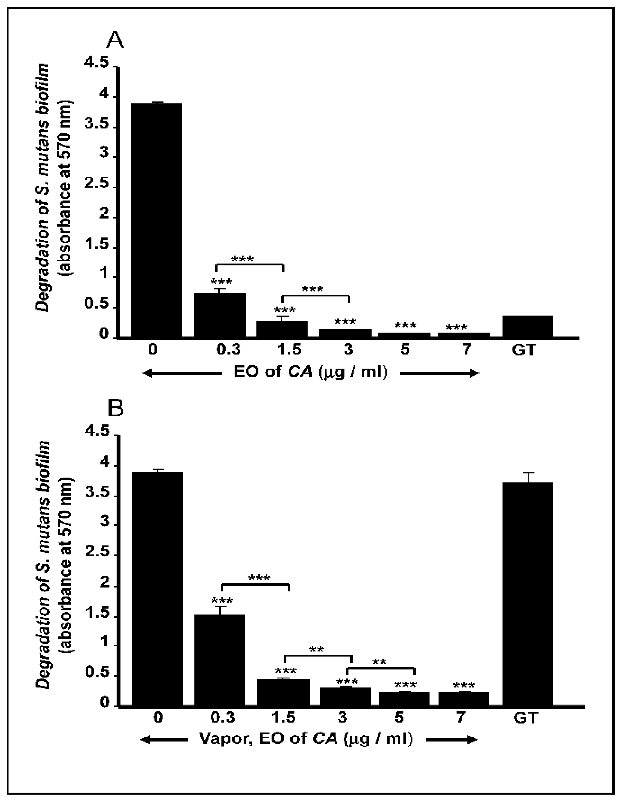
2.3. C. aurantium L. Essential Oil Disrupted Mature S. mutans Biofilms

The mature biofilms showed a significant reduction in OD570 values following treatment with the EO (Figure 2). The presence of direct-contact EO was shown to degrade the biofilms, regardless of the concentration used (Figure 2A). The EO vapor also decreased S. mutans viability in the biofilms (Figure 2B). It is worth to be noted that both the direct and indirect contact with the EO resulted in a degradation of mature S. mutans biofilm in a dose-dependent manner (Figure 2). The effect of the EO on biofilm degradation was either equivalent to or greater (with 3 μg/mL of EO) than the effect obtained with gentamicin.

2.4. C. aurantium L. Essential Oil Decreased the mRNA Expression of Various Virulent Genes by S. mutans

As shown in Figure 3, the mRNA expression of the comC, comD, comE, gtfB, gtfC, and gbpB genes significantly (p < 0.05) decreased following direct contact with the EO. Similar findings were observed with the EO vapor. However, the mRNA expression of gtfB and gtfC showed no significant change following the exposure of the S. mutans cells to the EO. Furthermore, the EO in both direct and indirect contact had no effect on the mRNA expression of atpH gene (Figure 3). It should be noted that the effect of gentamicin on all of the tested genes was higher than that obtained with the EO in either direct or indirect contact, although the decrease by the EO on gene expression remained significant.

2.5. C. aurantium L. Essential Oil Reduced Gingival Epithelial Cell Attachment

As shown in Figure 4 the addition of EO to gingival epithelial cell cultures had an important effect on the adhesion of these cells to the surface of the culture plates. Indeed, after 24 h of incubation, we observed the attachment of a high number of cells in the control groups and those supplemented with 0.3 μg/mL of the EO. In contrast, those fed with 1.5 μg/mL of the EO in direct contact resulted in total inhibition of cell adhesion. The EO vapor also had a significant reduction effect on gingival epithelial cell adhesion. While the cell cultures supplemented with a low concentration (0.3 μg/mL) of the EO showed good cell adhesion, at higher concentrations, cell adhesion was significantly inhibited (Figure 5). This suggests the use of low concentrations of the EO to prevent cell damage.

2.6. C. aurantium L. Essential Oil Decreased Gingival Epithelial Cell Viability

After 48 h of incubation, gingival epithelial cell cultures were supplemented or not with various concentrations of the EO through direct or indirect contact. The results presented in Figure 5A show that direct and indirect contact with the EO reduced cell viability. The MTT 3-(4,5-dimethylthiazol-2-yl)-2,5-monotetrazolium bromide assay (MTT) results reveal that with direct contact, the control cultures had a mean OD550 of 2.38 ± 0.14, and with the lowest concentration (0.3 μg/mL) of the EO, the OD550 dropped slightly, but significantly (p < 0.05) reaching to 1.9 ± 0.13. With the highest concentration (7 μg/mL) of the EO, epithelial cell viability dropped to an OD550 of 0.18 ± 0.01. Similar results were obtained with the EO vapor. As shown in Figure 5B, the viability of the epithelial cells went from 2.19 ± 0.05 with the control to 2.01 ± 0.18 with the lowest concentration (0.3 μg/mL) of the EO. This decrease was not significant as compared to the control (non-exposed cells). At high concentration (7 μg/mL), the OD550 dropped significantly (p < 0.01) reaching an OD550 of 0.43 ± 0.05. Overall results indicate that the use of EO vapor is less toxic to gingival epithelial cells.

3. Discussion

Combating the growth and virulence of oral pathogens, such as C. albicans, Porphyromonas gingivalis, S. mutans, etc., is important to ensure oral health. Frequent brushing with toothpaste reportedly reduces periodontitis and dental caries [26]. Furthermore, the use of cleaning solutions/mouthwash contributes to maintaining good oral hygiene by removing oral pathogens in dental plaque [27].
Mouthwashes contain various chemicals such as chlorhexidine, fluoride, delmopinol chloride, cetylpyridinium chloride, and alcohol, to name a few [28]. Although these ingredients are effective in reducing oral diseases, patients are concerned by the frequent use of these chemicals and are, therefore, looking for a mouthwash with natural products [29]. In this respect, essential oils may represent an interesting approach in the design of effective mouthwashes.
We extracted the essential oil from C. aurantium L. and analyzed its biological properties. The extracted EO had an elevated amount of limonene, followed by linalool. Both of these products display several attributes, including anti-inflammatory, antioxidant, anti-stress, and possibly disease-preventing properties [30,31]. This antioxidant activity is known to prevent tissue damage due to oxidation involving reactive oxygen species [32]. The antioxidant activity of the EO may be attributed to the presence of cineol, pinene, and limonene [33,34]. This activity was also in the range of what has been previously reported [22,33].
Limonene and linalool were shown to display antimicrobial properties [34], which may explain our results showing a significant decrease of S. mutans growth after exposure to the C. aurantium L. EO. These observations support those reported previously against Listeria monocytogenes [35] and aspergillus flavors [36], confirming the possible use of C. aurantium L. EO to control the growth of various bacterial strains. Growth inhibition may translate to reducing biofilm formation and disruption [37]. With this study, we are the first to show that C. aurantium L. EO can disrupt mature S. mutans biofilms.
The effects of the EO on S. mutans cell growth and biofilm degradation may involve certain virulent genes. Our results show that the EO is capable of decreasing comC, comD, comE, gftB, gftC, and gbpB mRNA expression in S. mutans. Activation of the gtf gene is known to lead to the production of GTFase synthesized glucans, which contribute to bacterial adherence and biofilm formation, while activation of the gtfB gene leads to the synthetization of polysaccharide containing α-1,3-linked glucans, facilitating cell aggregation in stable biofilms. The gtfC gene encodes the GTFC protein, which is involved in the synthesis of α-1,3- and α-1,6-linked glucans required for the formation and stabilization of biofilms [38]. The gbpB gene is involved in oral bacterial cell–cell aggregation, which facilitates the adhesion of the bacterium and the formation of mature biofilms [39].
S. mutans cell–cell interactions to form biofilm involve different gene products encoded by comAB [40] and comCDE [41]. The comC, comD, and comE genes, respectively, encode a competence-stimulating peptide (CSP) precursor contributing to biofilm formation [42]. In our study, the observed decrease in the mRNA expression of comC, comD, comE, gftB, gftC, and gbpB by the EO may explain the observed growth reduction and biofilm disruption.
The use of essential oil to prevent and/or control oral pathogens brings it into contact with other constituents in the oral cavity, such as the oral mucosa. For this reason, EO must be non-toxic to the host’s oral tissues. Our findings demonstrate that C. aurantium L. EO at 1.5 μg/mL and more is indeed toxic to gingival epithelial cells by inhibiting their adhesion and viability/growth. Of interest is that a low concentration (0.3 μg/mL) of EO showed reduced toxicity to the gingival epithelial cells, while it decreased significantly the growth of S. mutans, which supports the results of previous studies using various cell types [42].
The contribution of C. aurantium L. EO as a mouthwash supplement thus shows enormous potential, as its contact with human oral mucosa will only be for a short period (less than 30 s), and the oral mucosa tissue is more resistant (than are monolayer cell cultures). Furthermore, as shown in this study, even at a low concentration, C. aurantium L. essential oil is effective in reducing bacterial growth and degrading mature biofilms.

4. Materials and Methods

4.1. Plant Material, Extraction of C. aurantium L. Essential Oil, and Chemical Analyses

Bitter orange flowers (C. aurantium L.) (CA) were sampled during April 2016 from the Chiffa area in the central north region of Algeria. Botanical identification of the plant was determined in the Botany Laboratory at the Department of Biology at Badji Mokhtar—Annaba University. The extraction of the EO was carried out on fresh flowers using water vapor on an industrial scale by the Extral-Bio® company located in Chiffa (Blida, Algeria). The extracted oil was subjected to gas chromatography (GC) analyses. Its components were characterized by comparing the retention indices on polar and non-polar columns using mass spectra compared with standard solutions of n-alkanes (C8–C28) as the control [43]. The GC conditions involved an injector with a temperature of 250 °C and the following injection mode: Split 1/50, volume injected: 0.2 mL. The column used was an HP-5 (L 30 m, internal diameter 0.25 mm; film thickness 0.25 mm) (Société Chromopic, Villejust, France). The stationary phase consisted of 5% phenyl and 95% dimethylpolysiloxane. The oven temperature was 60 °C for 8 min, followed by 2 °C/min up to 240 °C, and finally isothermal for 10 min. The duration of the analyses was 108 min and the carrier gas was nitrogen, with a flow of 0.5 mL/min.

4.2. Effect of C. aurantium L. EO on S. mutans Growth

S. mutans cells (ATCC 25175) were seeded (105 CFU) in brain heart infusion (BHI) medium in the presence or absence of the EO at concentrations of either 0.3, 1.5, 3, 5, or 7 μg/mL. The EO concentrations were selected to identify the lowest concentration with antimicrobial activity and low toxicity for human cells. The EO was used in direct contact or as a vapor. For the vapor experiments, the EO was put into a sterile cotton swab and suspended over the surface of the culture medium at a distance of 3 cm (indirect contact). All the cultures were incubated aerobically at 37 °C in a 5% CO2 humid atmosphere. Negative (no EO) and positive (gentamicin (GT) at 5 μg/mL) controls were included in each experiment. Following incubation for 24 h, each culture was washed twice with Phosphate-buffered saline (PBS), after which time the pellets were suspended in 200 µL of a 1% crystal violet solution and incubated thereafter for 15 min under shaking conditions at 22 °C. Each cell suspension was then repeatedly suspended in distilled water then centrifuged until no visible blue color leached from the cell pellets. At this time, each pellet was air-dried for 24 h at 40 °C, suspended in 500 μL of a 30% acetic acid solution, and incubated for 15 min at 22 °C under agitation. The supernatants were distributed into a 96-well plate at 100 μL per well to measure the absorbance at 570 nm. Results were reported as the means ± SD, n = 6.

4.3. Effect of C. aurantium L. EO on the Disruption of Mature Biofilm

To produce mature biofilms, S. mutans (106 CFU) cells were seeded into a 3D porous collagen matrix (5 × 5 mm) [44], after which time the samples were cultured for four days in BHI medium containing 1% glucose. Following this culture period, the biofilms were treated or not with the EO (0.3, 1.5, 3, or 7 μg/mL) under either direct contact or indirect vapor. Gentamicin-treated biofilms (5 μg/mL) were used as positive controls. The biofilms with the EO were treated for 24 h and subjected to crystal violet staining, and the OD570 was measured.

4.4. Effect of C. aurantium L. EO on the Activation/Repression of Various S. mutans Genes

S. mutans cells were cultured in the presence or absence of a chosen concentration (3 μg/ml) of the EO (direct contact or vapor) at 37 °C for 12 h. Following the incubation period, the RNA was extracted from each sample, as we previously reported [44] and was used to perform quantitative reverse transcription-polymerase chain reactions (RT-PCR). The RNA (500 ng of each sample) was reverse transcribed into cDNA by means of the iScript cDNA synthesis kit (Bio-Rad Laboratoties Ltd., Saint-Laurant, Quebec, Canada) and was subsequently used for quantitative PCR (qPCR). Reactions were performed using a PCR supermix (Bio-Rad, Laboratoties Ltd., Saint-Laurant, Quebec, Canada; iQ SYBR Green supermix). Specific primers for comC, comD, comE, gtfB, gtfC, gbpB, and atpH (Table 2) were added to the reaction mix at a final concentration of 250 nmol/l. Reactions were performed in triplicate, as we previously described [44]. The specificity of each primer pair was determined by the presence of a single melting temperature peak. The 16S rRNA gene was included as a reference for this study. The results were analyzed using the 2−ΔΔCt (Livak) relative expression method. The selected time (12 h) and concentration (3 μg/mL) were to enable us to collect enough cells to extract useful amounts of RNA. Indeed, a higher concentration of C. aurantium L. EO will reduce considerably the growth of C. ablicans, thus making it impossible to extract enough RNA for our gene expression analyses.

4.5. Effect of C. aurantium L. EO on Gingival Epithelial Cell Adhesion and Morphology

A gingival epithelial cell line (Ca9-22) purchased from the Health Science Research Resources Bank (Osaka, Japan) was seeded (5 × 10⁵) in sterile tissue culture Petri dishes in RPMI-1640 medium supplemented with 10% fetal calf serum. The EO at concentrations of 0, 0.3, 1.5, 3, 5, or 7 μg/mL was introduced to the cell cultures either in direct contact or as vapor. The cultures were then incubated at 37 °C for 24 h. The cells were then fixed with 4% paraformaldehyde for 60 min, stained with crystal violet dye, examined under an optical microscope, and photographed (n = 5).

4.6. Effect of C. aurantium L. EO on Gingival Epithelial Cell Growth and Proliferation

Ca9-22 cells (105) were cultured in RPMI-1640 medium supplemented with 10% fetal calf serum for 48 h prior to contact with the EO. Following incubation, the culture medium was refreshed and the cells were exposed to the EO (direct contact or vapor) and subsequently incubated for 24 h in a 5% CO2 humid atmosphere at 37 °C. An MTT assay was then performed on each culture, as we previously reported [45]. The results were presented as the means ± SD; (n = 5).

4.7. Statistical Analysis

Each experiment was performed at least four times, with experimental values expressed as means ± SD. The statistical significance of the differences between the control (absence of the EO) and test (presence of the EO or gentamicin) values was determined by means of a one-way ANOVA. Posteriori comparisons were conducted using Tukey’s method. P values were declared significant at ≤ 0.05. The data were analyzed using a statistical version 8.2 (SAS Institute Inc., Cary, NC, USA).

5. Conclusions

In this study, we demonstrate that Citrus aurantium L. essential oil could decrease the growth of Streptococcus mutans and degrading its mature biofilms. Our findings also show a significant decrease in certain virulent genes (comC, comD, comE, gtfB, gtfC, and gbpB) expressed by S. mutans. While at mid and high concentrations, the essential oil (EO) was toxic to gingival epithelial cells, this toxicity was lower with a low concentration of the EO, which was effective in reducing S. mutans growth. Overall results suggest the possible use of C. aurantium L. essential oil to reduce the pathogenesis of S. mutans.

Author Contributions

Conceptualization, project administration, funding acquisition, supervision, M.R.; methodology, data analysis, validation, C.B., A.B. and M.R.; data validation, R.D., and L.T.; writing first draft, C.B. and M.R.; review of the manuscript and editing, A.B., R.D., and L.T. All authors have read and agreed to the published version of the manuscript

Funding

This study was supported by funding from the “Fonds Émile Beaulieu” Laval University Foundation, grant number FO117430.

Institutional Review Board Statement

Not applicable.

Informed Consent Statement

Not applicable.

Data Availability Statement

All data were included in the manuscript.

Conflicts of Interest

The authors declare no conflict of interest. The funders had no role in the design of the study; in the collection, analyses, or interpretation of data; in the writing of the manuscript, or in the decision to publish the results.

References

  1. Ferrentino, G.; Morozova, K.; Horn, C.; Scampicchio, M. Extraction of essential oils from medical plants and their utilization as food antioxidants. Curr. Pharm. Des. 2020, 26, 519–541. [Google Scholar] [CrossRef]
  2. Bighetti, E.J.; Hiruma-Lima, C.A.; Gracioso, J.S.; Brito, A.R. Anti-inflammatory and antinociceptive effects in rodents of the essential oil of Croton cajucara Benth. J. Pharm. Pharmacol. 1999, 51, 1447–1453. [Google Scholar] [CrossRef] [PubMed]
  3. Nasiri Lari, Z.; Hajimonfarednejad, M.; Riasatian, M.; Abolhassanzadeh, Z.; Iraji, A.; Vojoud, M.; Heydari, M.; Shams, M. Efficacy of inhaled Lavandula angustifolia Mill. Essential oil on sleep quality, quality of life and metabolic control in patients with diabetes mellitus type II and insomnia. J. Ethnopharmacol. 2020, 251, 112560. [Google Scholar] [CrossRef]
  4. Khan, M.; Alkhathlan, H.Z.; Khan, S.T. Antibiotic and Antibiofilm Activities of Salvadora persica L. Essential Oils against Streptococcus mutans: A Detailed Comparative Study with Chlorhexidine Digluconate. Pathogens 2020, 9, 66. [Google Scholar] [CrossRef] [PubMed] [Green Version]
  5. Jünger, H.; Jaun-Ventrice, A.; Guldener, K.; Ramseier, C.A.; Reissmann, D.R.; Schimmel, M. Anti-inflammatory potential of an essential oil-containing mouthwash in elderly subjects enrolled in supportive periodontal therapy: A 6-week randomised controlled clinical trial. Clin. Oral Investig. 2020, 24, 3203–3211. [Google Scholar] [CrossRef] [PubMed] [Green Version]
  6. Richards, D. Effect of essential oil mouthwashes on plaque and gingivitis. Evid. Based Dent. 2017, 18, 39–40. [Google Scholar] [CrossRef] [PubMed]
  7. Fanning, S.; Mitchell, A.P. Fungal biofilms. PLoS Pathog. 2012, 8, e1002585. [Google Scholar] [CrossRef] [PubMed] [Green Version]
  8. Pantanella, F.; Valenti, P.; Natalizi, T.; Passeri, D.; Berlutti, F. Analytical techniques to study microbial biofilm on abiotic surfaces: Pros and cons of the main techniques currently in use. Ann Ig. 2013, 25, 31–42. [Google Scholar] [PubMed]
  9. Li, Y.; Burne, R.A. Regulation of the gtfBC and ftf genes of Streptococcus mutans in biofilms in response to pH and carbohydrate. Microbiology 2001, 147, 2841–2848. [Google Scholar] [CrossRef] [Green Version]
  10. Rouabhia, M.; Mukherjee, P.K.; Lattif, A.A.; Curt, S.; Chandra, J.; Ghannoum, M.A. Disruption of sphingolipid biosynthetic gene IPT1 reduces Candida albicans adhesion and prevents activation of human gingival epithelial cell innate immune defense. Med. Mycol. 2011, 49, 458–466. [Google Scholar]
  11. Rouabhia, M.; Semlali, A.; Chandra, J.; Mukherjee, P.; Chmielewski, W.; Ghannoum, M.A. Disruption of the ECM33 gene in Candida albicans prevents biofilm formation, engineered human oral mucosa tissue damage and gingival cell necrosis/apoptosis. Mediat. Inflamm. 2012, 2012, 398207. [Google Scholar] [CrossRef] [PubMed] [Green Version]
  12. Høiby, N.; Bjarnsholt, T.; Givskov, M.; Molin, S.; Ciofu, O. Antibiotic resistance of bacterial biofilms. Int. J. Antimicrob. Agents. 2010, 35, 322–332. [Google Scholar] [CrossRef] [PubMed] [Green Version]
  13. Samaranayake, L.; Matsubara, V.H. Normal Oral Flora and the Oral Ecosystem. Dent. Clin. North Am. 2017, 61, 199–215. [Google Scholar] [CrossRef] [PubMed]
  14. Belstrøm, D. The salivary microbiota in health and disease. J. Oral 2020, 12, 1723975. [Google Scholar] [CrossRef] [Green Version]
  15. Peacock, M.E.; Arce, R.M.; Cutler, C.W. Periodontal and other oral manifestations of immunodeficiency diseases. Oral Dis. 2017, 23, 866–888. [Google Scholar] [CrossRef]
  16. Kilian, M.; Chapple, I.L.; Hannig, M.; Marsh, P.D.; Meuric, V.; Pedersen, A.M.; Tonetti, M.S.; Wade, W.G.; Zaura, E. The oral microbiome—An update for oral healthcare professionals. Br. Dent. J. 2016, 221, 657–666. [Google Scholar] [CrossRef]
  17. Lauritano, D.; Moreo, G.; Della Vella, F.; Di Stasio, D.; Carinci, F.; Lucchese, A.; Petruzzi, M. Oral Health Status and Need for Oral Care in an Aging Population: A Systematic Review. Int. J. Environ. Res. Public Health. 2019, 16, 4558. [Google Scholar] [CrossRef] [Green Version]
  18. Takenaka, S.; Ohsumi, T.; Noiri, Y. Evidence-based strategy for dental biofilms: Current evidence of mouthwashes on dental biofilm and gingivitis. Jpn. Dent. Sci. Rev. 2019, 55, 33–40. [Google Scholar] [CrossRef]
  19. Cantarelli, R.; Negrini, T.C.; Muniz, F.W.; Oballe, H.J.; Arthur, R.A.; Rösing, C.K. Antimicrobial potential and gustatory perception of chlorhexidine gluconate mouthwashes with or without alcohol after a single rinse—A randomized controlled crossover clinical trial. Int. J. Dent. Hyg. 2017, 15, 280–286. [Google Scholar] [CrossRef]
  20. Araujo, M.W.B.; Charles, C.A.; Weinstein, R.B.; McGuire, J.A.; Parikh-Das, A.M.; Du, Q.; Zhang, J.; Berlin, J.A.; Gunsolley, J.C. Meta-analysis of the effect of an essential oil-containing mouthrinse on gingivitis and plaque. J. Am. Dent. Assoc. 2015, 146, 610–622. [Google Scholar] [CrossRef] [Green Version]
  21. Lynch, M.C.; Cortelli, S.C.; McGuire, J.A.; Zhang, J.; Ricci-Nittel, D.; Mordas, C.J.; Aquino, D.R.; Cortelli, J.R. The effects of essential oil mouthrinses with or without alcohol on plaque and gingivitis: A randomized controlled clinical study. BMC Oral Health 2018, 18, 6. [Google Scholar] [CrossRef] [PubMed] [Green Version]
  22. Değirmenci, H.; Erkurt, H. Relationship between volatile components, antimicrobial and antioxidant properties of the essential oil, hydrosol and extracts of Citrus aurantium L. flowers. J. Infect. Public Health. 2020, 13, 58–67. [Google Scholar] [CrossRef] [PubMed]
  23. Salma, M.; Abdellah, F.; Houssine, A.; Kawtar, B.; Dalila, B. Comparison of the chemical composition and the bioactivity of the essential oils of three medicinal and aromatic plants from Jacky Garden of Morocco. Int. J. Pharm. Phytochem. Res. 2016, 8, 537–545. [Google Scholar]
  24. Zhao, H.Y.; Yang, L.; Wei, J.; Huang, M.; Jiang, J.G. Bioactivity evaluations of ingredients extracted from the flowers of Citrus aurantium L. var. amara Engl. Food Chem. 2012, 135, 2175–2181. [Google Scholar] [CrossRef] [PubMed]
  25. Gironés-Vilaplana, A.; Valentão, P.; Andrade, P.B.; Ferreres, F.; Moreno, D.A.; García-Viguera, C. Beverages of lemon juice and exotic noni and papaya with potential for anticholinergic effects. Food Chem. 2015, 170, 16–21. [Google Scholar] [CrossRef]
  26. Pedersen, A.M.L.; Darwish, M.; Nicholson, J.; Edwards, M.I.; Gupta, A.K.; Belstrøm, D. Gingival health status in individuals using different types of toothpaste. J. Dent. 2019, 80 (Suppl. 1), S13–S18. [Google Scholar] [CrossRef]
  27. James, P.; Worthington, H.V.; Parnell, C.; Harding, M.; Lamont, T.; Cheung, A.; Whelton, H.; Riley, P. Chlorhexidine mouthrinse as an adjunctive treatment for gingival health. Cochrane Database Syst. Rev. 2017, 3, CD008676. [Google Scholar] [CrossRef]
  28. Guerra, F.; Pasqualotto, D.; Rinaldo, F.; Mazur, M.; Corridore, D.; Nofroni, I.; Ottolenghi, L.; Nardi, G.M. Therapeutic efficacy of chlorhexidine-based mouthwashes and its adverse events: Performance-related evaluation of mouthwashes added with Anti-Discoloration System and cetylpyridinium chloride. Int. J. Dent. Hyg. 2019, 17, 229–236. [Google Scholar] [CrossRef]
  29. Ekor, M. The growing use of herbal medicines: Issues relating to adverse reactions and challenges in monitoring safety. Front. Pharmacol. 2014, 4, 177. [Google Scholar] [CrossRef] [Green Version]
  30. Suntar, I.; Khan, H.; Patel, S.; Celano, R.; Rastrelli, L. An Overview on Citrus aurantium L.: Its Functions as Food Ingredient and Therapeutic Agent. Oxid. Med. Cell. Longev. 2018, 2018, 7864269. [Google Scholar] [CrossRef] [Green Version]
  31. Ellouze, I.; Abderrabba, M.; Sabaou, N.; Mathieu, F.; Lebrihi, A.; Bouajila, J. Season’s variation impact on Citrus aurantium leaves essential oil: Chemical composition and biological activities. J. Food Sci. 2012, 77, T173–T180. [Google Scholar] [CrossRef] [PubMed]
  32. López-Alarcón, C.; Denicola, A. Evaluating the antioxidant capacity of natural products: A review on chemical and cellular-based assays. Anal. Chim. Acta. 2013, 763, 1–10. [Google Scholar] [CrossRef] [PubMed]
  33. Ben Hsouna, A.; Ben Halima, N.; Smaoui, S.; Hamdi, N. Citrus lemon essential oil: Chemical composition, antioxidant and antimicrobial activities with its preservative effect against Listeria monocytogenes inoculated in minced beef meat. Lipids Health Dis. 2017, 16, 146. [Google Scholar] [CrossRef] [PubMed] [Green Version]
  34. Maté, J.; Periago, P.M.; Palop, A. Combined effect of a nanoemulsion of d-limonene and nisin on Listeria monocytogenes growth and viability in culture media and foods. Food Sci. Technol. Int. 2016, 22, 146–152. [Google Scholar] [CrossRef]
  35. Fancello, F.; Petretto, G.L.; Marceddu, S.; Venditti, T.; Pintore, G.; Zara, G.; Mannazzu, I.; Budroni, M.; Zara, S. Antimicrobial activity of gaseous Citrus limon var pompia leaf essential oil against Listeria monocytogenes on ricotta salata cheese. Food Microbiol. 2020, 87, 103386. [Google Scholar] [CrossRef] [PubMed]
  36. Restuccia, C.; Oliveri Conti, G.; Zuccarello, P.; Parafati, L.; Cristaldi, A.; Ferrante, M. Efficacy of different citrus essential oils to inhibit the growth and B1 aflatoxin biosynthesis of Aspergillus flavus. Environ. Sci. Pollut. Res. Int. 2019, 26, 31263–31272. [Google Scholar] [CrossRef]
  37. Sahal, G.; Woerdenbag, H.J.; Hinrichs, W.L.J.; Visser, A.; Tepper, P.G.; Quax, W.J.; van der Mei, H.C.; Bilkay, I.S. Antifungal and biofilm inhibitory effect of Cymbopogon citratus (lemongrass) essential oil on biofilm forming by Candida tropicalis isolates; an in vitro study. J. Ethnopharmacol. 2020, 246, 112188. [Google Scholar] [CrossRef]
  38. Veloz, J.J.; Saavedra, N.; Alvear, M.; Zambrano, T.; Barrientos, L.; Salazar, L.A. Polyphenol-Rich Extract from Propolis Reduces the Expression and Activity of Streptococcus mutans Glucosyltransferases at Subinhibitory Concentrations. Biomed. Res. Int. 2016, 2016, 4302706. [Google Scholar] [CrossRef] [Green Version]
  39. Duque, C.; Stipp, R.N.; Wang, B.; Smith, D.J.; Höfling, J.F.; Kuramitsu, H.K.; Duncan, M.J.; Mattos-Graner, R.O. Downregulation of GbpB, a component of the VicRK regulon, affects biofilm formation and cell surface characteristics of Streptococcus mutans. Infect. Immun. 2011, 79, 786–796. [Google Scholar] [CrossRef] [Green Version]
  40. Petersen, F.C.; Scheie, A.A. Genetic transformation in Streptococcus mutans requires a peptide secretion-like apparatus. Oral Microbiol. Immunol. 2000, 15, 329–334. [Google Scholar] [CrossRef]
  41. Li, Y.H.; Tang, N.; Aspiras, M.B.; Lau, P.C.; Lee, J.H.; Ellen, R.P.; Cvitkovitch, D.G. A quorum-sensing signaling system essential for genetic competence in Streptococcus mutans is involved in biofilm formation. J. Bacteriol. 2002, 184, 2699–2708. [Google Scholar] [CrossRef] [PubMed] [Green Version]
  42. Lorençoni, M.F.; Figueira, M.M.; Toledo, E.; Silva, M.V.; Pimentel Schmitt, E.F.; Endringer, D.C.; Scherer, R.; Barth, T.; Vilela Bertolucci, S.K.; Fronza, M. Chemical composition and anti-inflammatory activity of essential oil and ethanolic extract of Campomanesia phaea (O. Berg.) Landrum leaves. J. Ethnopharmacol. 2020, 252, 112562. [Google Scholar] [CrossRef] [PubMed]
  43. Dosoky, N.S.; Satyal, P.; Pokharel, S.; Setzer, W.N. Chemical Composition, Enantiomeric Distribution, and Biological Activities of Rhododendron anthopogon Leaf Essential Oil from Nepal. Nat. Prod. Commun. 2016, 11, 1895–1898. [Google Scholar] [CrossRef] [Green Version]
  44. Belmadani, A.; Semlali, A.; Rouabhia, M. Dermaseptin-S1 decreases Candida albicans growth, biofilm formation and the expression of hyphal wall protein 1 and aspartic protease genes. J. Appl. Microbiol. 2018, 125, 72–83. [Google Scholar] [CrossRef] [PubMed]
  45. Rouabhia, M.; Rouabhia, D.; Park, H.J.; Giasson, L.; Zhang, Z. Effect of soft foods on primary human gingival epithelial cell growth and the wound healing process. Food Res. Int. 2017, 100, 433–441. [Google Scholar] [CrossRef] [PubMed]
Figure 1. Citrus aurantium L. essential oil decreased Streptococcus mutans cell growth. Cell growth was evaluated after 24 h of either direct contact with the essential oil (EO) (A) or its vapor (B). Cell growth was assessed using crystal violet staining. GT = gentamicin. ** p < 0.01, *** p < 0.001.
Antibiotics 10 00054 g001
Figure 2. C. aurantium L. essential oil disrupted mature S. mutans biofilms. Bacteria were seeded into a 3D porous matrix for four days and then exposed to different concentrations of EO either in direct contact (A) or as vapor (B). Biofilm disruption was assessed by crystal violet staining. GT = gentamicin. ** p < 0.01; *** p < 0.001.
Antibiotics 10 00054 g002
Figure 3. C. aurantium L. essential oil inhibited the expression of virulent genes by S. mutans cells. After exposure or not to the EO for 12 h, the total RNA was extracted and subjected to qPCR analyses. V-EO = essential oil vapor; GT = gentamicin; Ctrl = control. * p < 0.05; *** p < 0.001.
Antibiotics 10 00054 g003
Figure 4. High concentrations of C. aurantium L. essential oil inhibited human gingival epithelial cell adhesion. Cells were seeded and exposed immediately to the EO for cell adhesion assessment. After 24 h incubation, the adherent cells were stained with crystal violet, cell adhesion was evaluated under an optical microscope and photographed.
Antibiotics 10 00054 g004
Figure 5. High concentrations of C. aurantium L. essential oil reduced human gingival epithelial cell viability. Cells were seeded and grow for 48 h prior to their exposure to the EO (Panel A) or the EO vapor (Panel B). After 48 h, the culture medium was refreshed and supplemented with various concentrations of the EO, and the cells were incubated for 24 h. Cell viability was assessed by MTT assay. * p < 0.05; ** p < 0.01, *** p < 0.001.
Antibiotics 10 00054 g005
Table 1. Identification of the different chemicals in the Citrus aurantium L. Essential oil by means of gas chromatography (GC) analyses.
Compound Retention Time (min) Retention Index Aire%
α-Pinene3.229360.63
Camphene3.599550.02
β-pinene4.309903.00
Sabinene4.5910015.01
β-Myrcene 5.1710262.17
δ-3- carene5.5410480.86
Limonene5.62105350.56
(E)-β—Ocimene5.8910637.82
Z- β—Ocimene6.3810753.54
γ—Terpinene6.5910880.64
Terpinolene6.7711020.48
Citronellal7.5411390.27
Linalool7.7111529.89
Terpinen-4-ol8.0311750.32
Linalylacetate8.3912811.61
α—Terpineol8.8311850.12
Neral12.16312680.08
Geraniol13.8212801.53
Geranial14.3412910.26
Nerylacetate19.7313641.52
Geranylacetate20.0913852.26
Methyl N-methylanthranilate22.1714022.35
Betacaryophyllene22.6014161.21
Total 96.15%
Table 2. Primer sequences used for the quantitative polymerase chain reaction (qPCR).
GenePrimer Sequence (5′ à 3′)Annealing Temperature (°C)Amp Size (bp)
16S rRNAForward: CTTACCAGGTCTTGACATCCCG
Reverse: ACCCAACATCTCACGACACGAG
63.3113
gbpBForward: AGCAACAGAAGCACAACCATCAG
Reverse: CCACCATTACCCCAGTAGTTTCC
65.6150
gtfBForward: ACACTTTCGGGTGGCTTG
Reverse: GCTTAGATGTCACTTCGGTTG
62127
gtfCForward: CCAAAATGGTATTATGGCTGTCG
Reverse: GAGTCTCTATCAAAGTAACGCAGT
63135
comCForward: GACTTTAAAGAAATTAAGACTG
Reverse: AAGCTTGTGTAAAACTTCTGT
54103
comDForward: CTCTGATTGACCATTCTTCTGG
Reverse: CATTCTGAGTTTATGCCCCTC
62150
comEForward: CCTGAAAAGGGCAATCACCAG
Reverse: GGGGCATAAACTCAGAATGTGTCG
65148
atpHForward: ACCATACATTTCAGGCTG
Reverse: TTTTAGCACTTGGGATTG
56101
Publisher’s Note: MDPI stays neutral with regard to jurisdictional claims in published maps and institutional affiliations.

