
Antibacterial Activity of Kaolin–Silver Nanomaterials: Alternative Approach to the Use of Antibiotics in Animal Production

 ,
,
Abstract
:1. Introduction
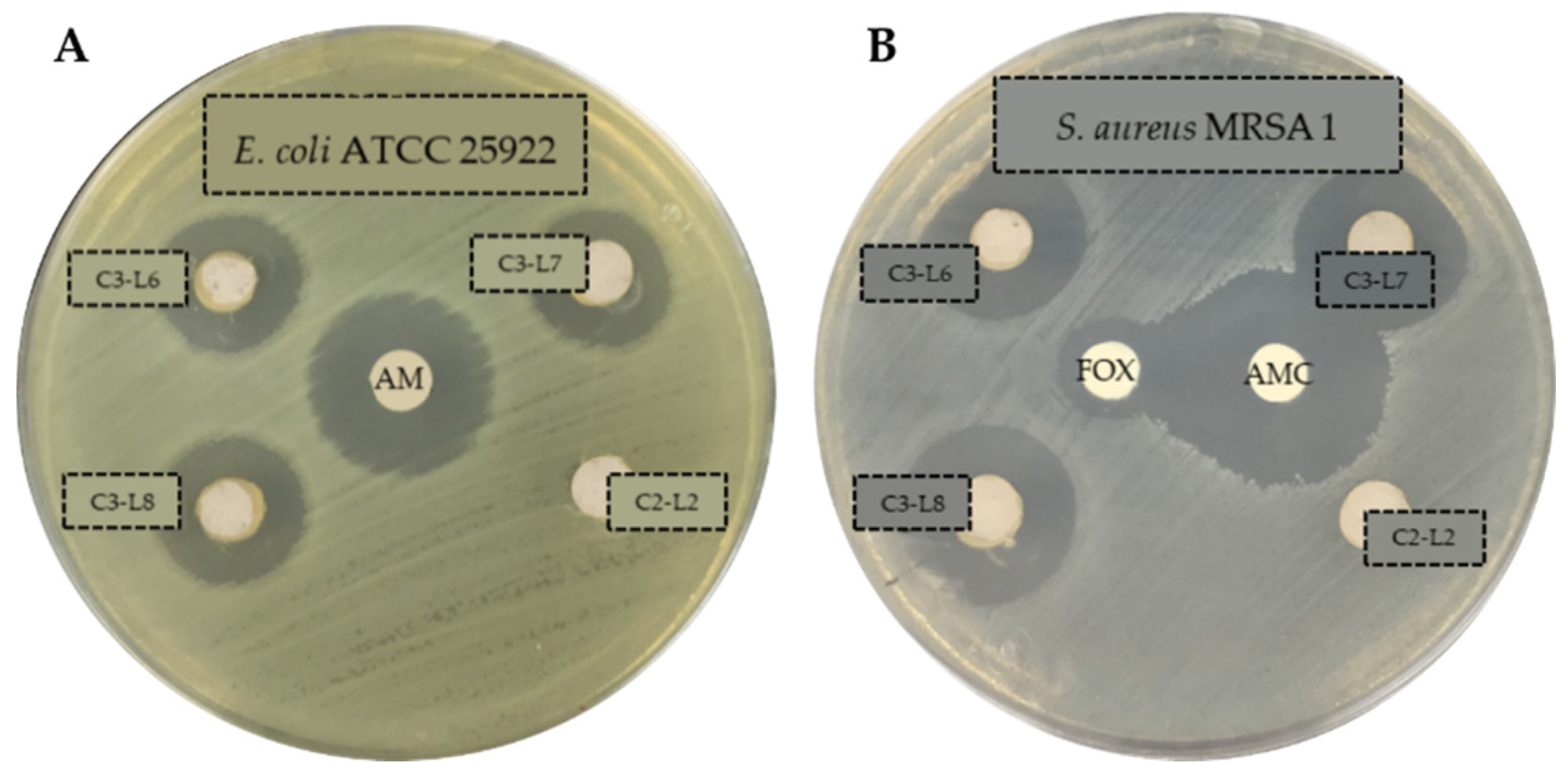
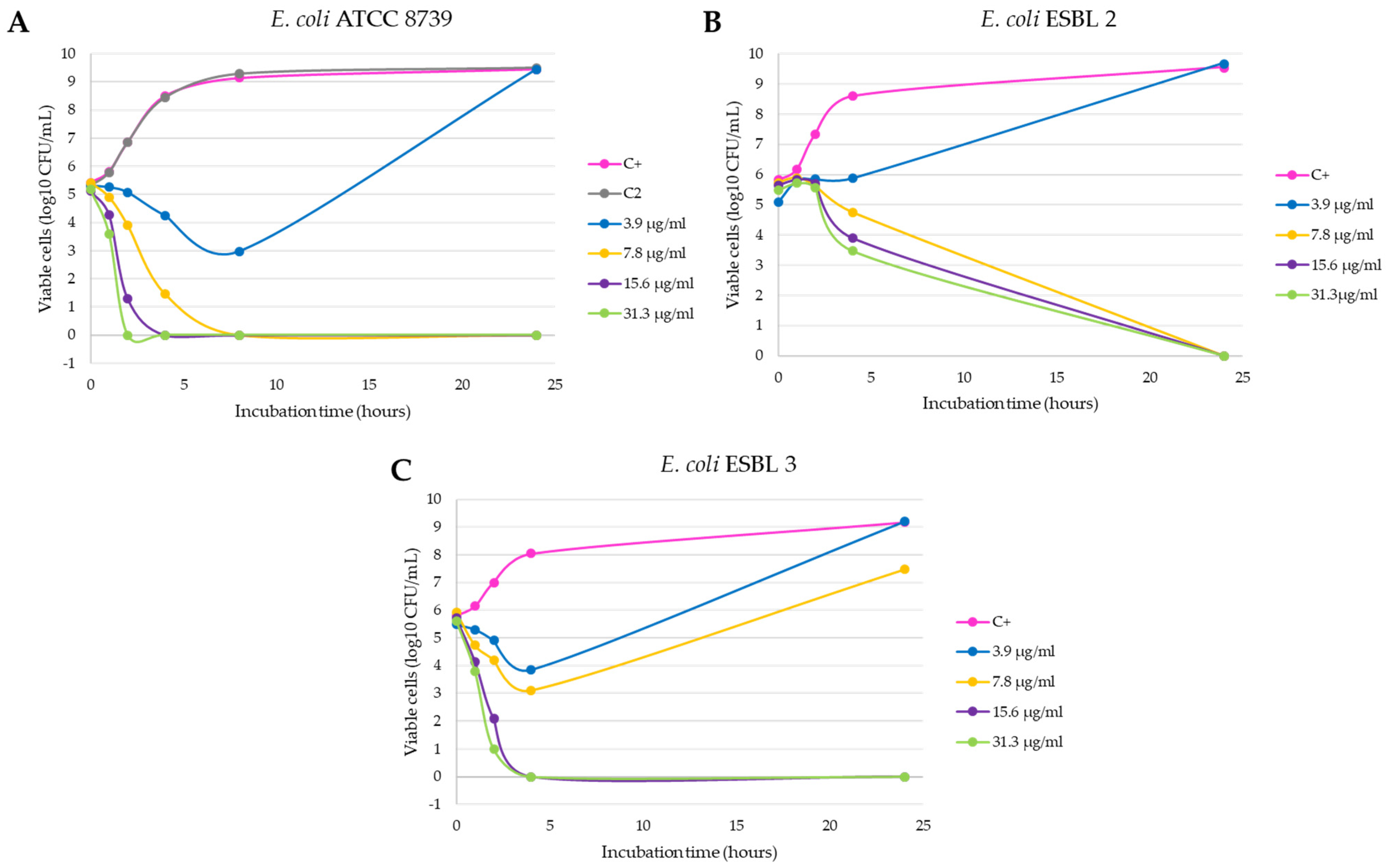
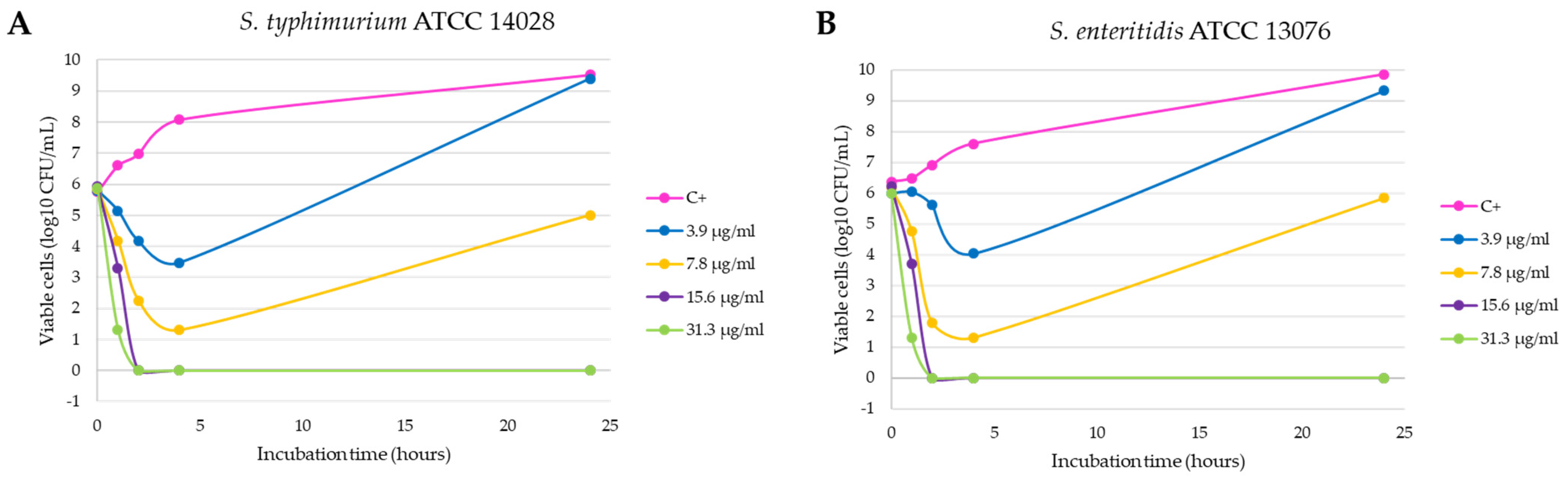
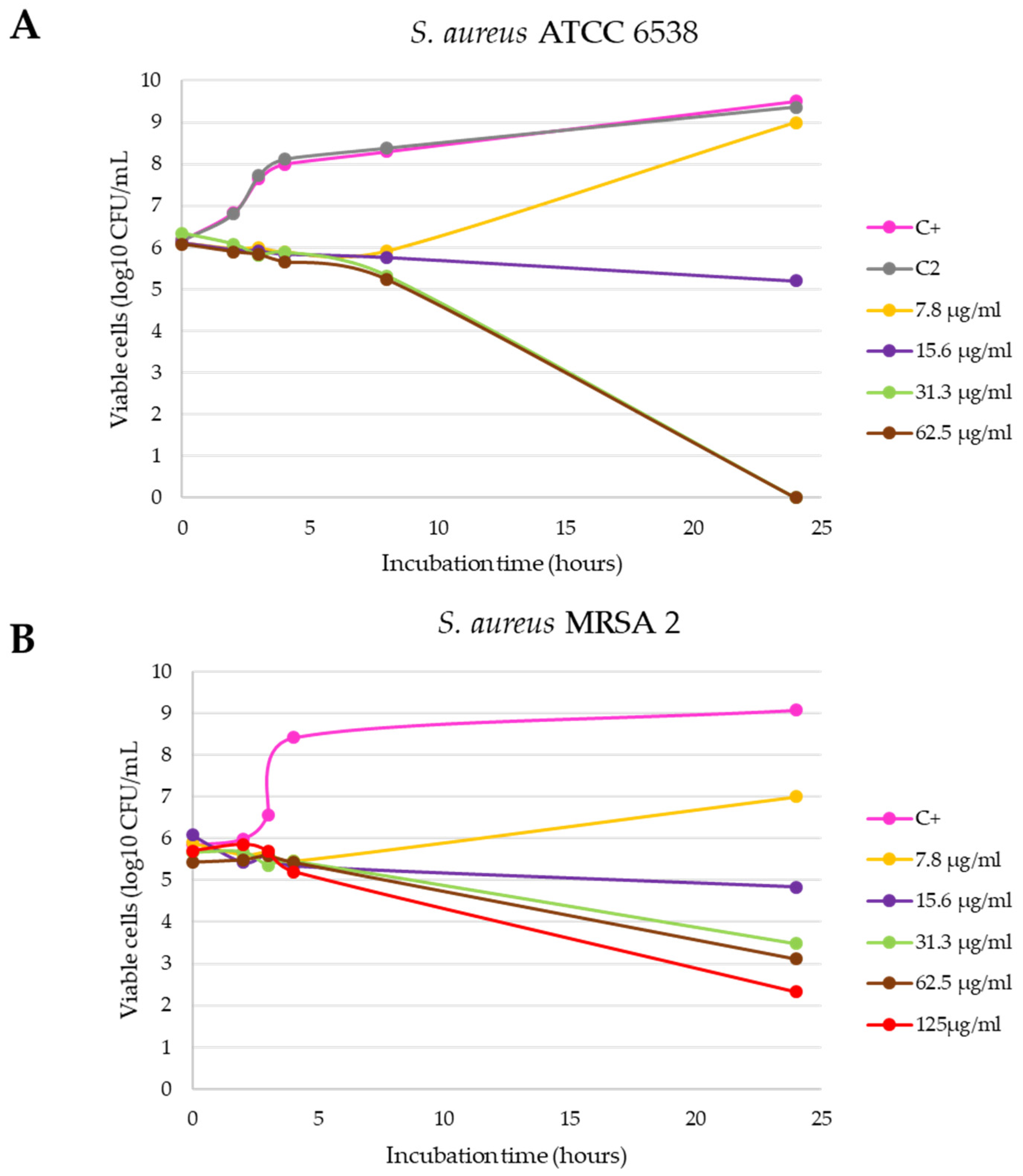
2. Results
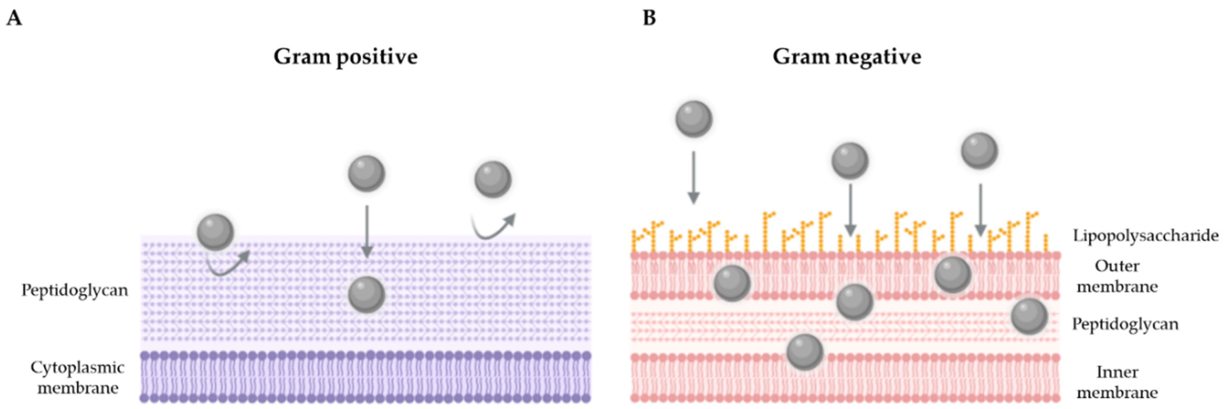
3. Discussion
4. Materials and Methods
4.1. Products
4.2. Agar Well-Diffusion Antibacterial Assay
4.3. Minimal Inhibitory Concentration (MIC) and Minimal Bactericidal Concentration (MBC) Determination
4.4. Growth Inhibition Curves in Liquid Medium
4.5. Statistical Analysis
5. Conclusions
6. Patents
Author Contributions
Funding
Institutional Review Board Statement
Informed Consent Statement
Data Availability Statement
Conflicts of Interest
References
- Holmer, I.; Salomonsen, C.M.; Jorsal, S.E.; Astrup, L.B.; Jensen, V.F.; Høg, B.B.; Pedersen, K. Antibiotic resistance in porcine pathogenic bacteria and relation to antibiotic usage. BMC Vet. Res. 2019, 15, 1–13. [Google Scholar] [CrossRef] [PubMed] [Green Version]
- Brown, E.E.F.; Cooper, A.; Carrillo, C.; Blais, B. Selection of multidrug-resistant bacteria in medicated animal feeds. Front. Microbiol. 2019, 10, 1–8. [Google Scholar] [CrossRef] [PubMed] [Green Version]
- Roth, N.; Käsbohrer, A.; Mayrhofer, S.; Zitz, U.; Hofacre, C.; Domig, K.J. The application of antibiotics in broiler production and the resulting antibiotic resistance in Escherichia coli: A global overview. Poult. Sci. 2019, 98, 1791–1804. [Google Scholar] [CrossRef] [PubMed]
- Calbo, E.; Freixas, N.; Xercavins, M.; Riera, M.; Nicolás, C.; Monistrol, O.; Solé, M.D.M.; Sala, M.R.; Vila, J.; Garau, J. Foodborne nosocomial outbreak of SHV1 and CTX-M-15-producing Klebsiella pneumoniae: Epidemiology and control. Clin. Infect. Dis. 2011, 52, 743–749. [Google Scholar] [CrossRef]
- Gieraltowski, L.; Higa, J.; Peralta, V.; Green, A.; Schwensohn, C.; Rosen, H.; Libby, T.; Kissler, B.; Marsden-Haug, N.; Booth, H.; et al. National outbreak of multidrug resistant Salmonella Heidelberg infections linked to a single poultry company. PLoS ONE 2016, 11, 1–13. [Google Scholar] [CrossRef] [PubMed] [Green Version]
- Yang, Y.; Ashworth, A.J.; Willett, C.; Cook, K.; Upadhyay, A.; Owens, P.R.; Ricke, S.C.; DeBruyn, J.M.; Moore, P.A. Review of Antibiotic Resistance, Ecology, Dissemination, and Mitigation in U.S. Broiler Poultry Systems. Front. Microbiol. 2019, 10, 1–10. [Google Scholar] [CrossRef]
- EFSA. The European Union Summary Report on Antimicrobial Resistance in Zoonotic and Indicator Bacteria from Humans, Animals and Food in 2017/2018; EFSA: Parma, Italy, 2020; Volume 18.
- EU. Ban on Antibiotics as Growth Promoters in Animal Feed Enters into Effect; Regulation 1831/2003/EC on Additives for Use in Animal Nutrition; European Union: Brussels, Belgium, 2006. [Google Scholar]
- EMA. Sales of veterinary antimicrobial agents in 30 European countries in 2015. EMA/184855/2017. In Seventh ESVAC Report; European Union: London, UK, 2017; p. 184. [Google Scholar]
- AEMPS Consumo Total de Antibióticos; Agencia Española Medicam. y Prod. Sanit.: Madrid, Spain, 2019; Volume 8, p. 4.
- Griggs, J.P. Alternatives to antibiotics for organic poultry production. J. Appl. Poult. Res. 2005, 14, 750–756. [Google Scholar] [CrossRef]
- Yazdankhah, S.; Rudi, K.; Bernhoft, A. Zinc and copper in animal feed-development of resistance and co-resistance to antimicrobial agents in bacteria of animal origin. Microb. Ecol. Health Dis. 2014, 25, 1–7. [Google Scholar] [CrossRef] [Green Version]
- Zhang, B.; Guo, Y. Beneficial effects of tetrabasic zinc chloride for weanling piglets and the bioavailability of zinc in tetrabasic form relative to ZnO. Anim. Feed Sci. Technol. 2007, 135, 75–85. [Google Scholar] [CrossRef]
- Fondevila, M. Potential Use of Silver Nanoparticles as an Additive in Animal Feeding. Silver Nanopart. 2010, 17, 10. [Google Scholar]
- Franci, G.; Falanga, A.; Galdiero, S.; Palomba, L.; Rai, M.; Morelli, G.; Galdiero, M. Silver nanoparticles as potential antibacterial agents. Molecules 2015, 20, 8856–8874. [Google Scholar] [CrossRef] [PubMed] [Green Version]
- Garipov, I.T.; Khaydarov, R.R.; Gapurova, O.U.; Khaydarov, R.A.; Lutfi, F.M.; Efimova, I.L.; Evgrafova, S.Y. Silver Nanoparticles as a New Generation of Antimicrobial Prophylaxis. J. Sib. Fed. Univ. Biol. 2019, 12, 266–276. [Google Scholar] [CrossRef]
- Mohanta, Y.K.; Biswas, K.; Jena, S.K.; Hashem, A.; Abd_Allah, E.F.; Mohanta, T.K. Anti-biofilm and Antibacterial Activities of Silver Nanoparticles Synthesized by the Reducing Activity of Phytoconstituents Present in the Indian Medicinal Plants. Front. Microbiol. 2020, 11, 1–15. [Google Scholar]
- Fondevila, M.; Herrer, R.; Casallas, M.C.; Abecia, L.; Ducha, J.J. Silver nanoparticles as a potential antimicrobial additive for weaned pigs. Anim. Feed Sci. Technol. 2009, 150, 259–269. [Google Scholar] [CrossRef]
- Taraszkiewicz, A.; Fila, G.; Grinholc, M.; Nakonieczna, J. Innovative strategies to overcome biofilm resistance. Biomed Res. Int. 2013, 2013, 1–13. [Google Scholar] [CrossRef] [Green Version]
- Rai, M.; Kon, K.; Ingle, A.; Duran, N.; Galdiero, S.; Galdiero, M. Broad-spectrum bioactivities of silver nanoparticles: The emerging trends and future prospects. Appl. Microbiol. Biotechnol. 2014, 98, 1951–1961. [Google Scholar] [CrossRef]
- Silver, S. Bacterial silver resistance: Molecular biology and uses and misuses of silver compounds. FEMS Microbiol. Rev. 2003, 27, 341–353. [Google Scholar] [CrossRef] [Green Version]
- Salas-Orozco, M.; Niño-Martínez, N.; Martínez-Castañón, G.A.; Méndez, F.T.; Jasso, M.E.C.; Ruiz, F. Mechanisms of resistance to silver nanoparticles in endodontic bacteria: A literature review. J. Nanomater. 2019, 2019, 1–11. [Google Scholar] [CrossRef]
- Marambio-Jones, C.; Hoek, E.M.V. A review of the antibacterial effects of silver nanomaterials and potential implications for human health and the environment. J. Nanopart. Res. 2010, 12, 1531–1551. [Google Scholar] [CrossRef]
- Abad-Álvaro, I.; Trujillo, C.; Bolea, E.; Laborda, F.; Fondevila, M.; Latorre, M.A.; Castillo, J.R. Silver nanoparticles-clays nanocomposites as feed additives: Characterization of silver species released during in vitro digestions. Effects on silver retention in pigs. Microchem. J. 2019, 149, 104040. [Google Scholar] [CrossRef]
- Martynková, G.S.; Valášková, M. Antimicrobial nanocomposites based on natural modified materials: A review of carbons and clays. J. Nanosci. Nanotechnol. 2014, 14, 673–693. [Google Scholar] [CrossRef]
- EFA183/16/OUTBIOTICS. Innovative Technologies for Diagnosis, Prevention and Elimination of Emerging Pollutants (Antibiotics) from the Waters of the POCTEFA Territory; Project Co-Financed 65% by the European Regional Development Fund (FEDER) through the Interreg V-A Spain-Franc (POCTEFA 2014-2020). Available online: https://www.fresh-thoughts.eu/userfiles/file/Pharma-2-Vidal.pdf (accessed on 26 September 2021).
- UE REGLAMENTO (CE) No. 1831/2003 DEL PARLAMENTO EUROPEO Y DEL CONSEJO de 22 de septiembre de 2003 sobre los aditivos en la alimentación animal. D. Of. Unión Eur. 2003, 4, 24–28.
- Clinical and Laboratory Standards Institute (CLSI). Performance Standards for Antimicrobial Susceptibility Testing, 28th ed.; CLSI: Wayne, PA, USA, 2018. [Google Scholar]
- Lara, H.H.; Ayala-Núñez, N.V.; del Turrent, L.C.I.; Padilla, C.R. Bactericidal effect of silver nanoparticles against multidrug-resistant bacteria. World J. Microbiol. Biotechnol. 2010, 26, 615–621. [Google Scholar] [CrossRef]
- Dakal, T.C.; Kumar, A.; Majumdar, R.S.; Yadav, V. Mechanistic basis of antimicrobial actions of silver nanoparticles. Front. Microbiol. 2016, 7, 1–17. [Google Scholar] [CrossRef] [PubMed] [Green Version]
- Doudi, M.; Naghsh, N.; Setorki, M. Comparison of the effects of silver nanoparticles on pathogenic bacteria resistant to beta-lactam antibiotics (ESBLs) as a prokaryote model and Wistar rats as a eukaryote model. Med. Sci. Monit. Basic Res. 2013, 19, 103–110. [Google Scholar] [CrossRef] [Green Version]
- Kar, D.; Bandyopadhyay, S.; Dimri, U.; Mondal, D.B.; Nanda, P.K.; Das, A.K.; Batabyal, S.; Dandapat, P.; Bandyopadhyay, S. Antibacterial effect of silver nanoparticles and capsaicin against MDR-ESBL producing Escherichia coli: An in vitro study. Asian Pacific J. Trop. Dis. 2016, 6, 807–810. [Google Scholar] [CrossRef]
- Yuan, Y.G.; Peng, Q.L.; Gurunathan, S. Effects of silver nanoparticles on multiple drug-resistant strains of Staphylococcus aureus and Pseudomonas aeruginosa from mastitis-infected goats: An alternative approach for antimicrobial therapy. Int. J. Mol. Sci. 2017, 18, 569. [Google Scholar] [CrossRef] [Green Version]
- Subashini, J.; Gopiesh Khanna, V.; Kannabiran, K. Anti-ESBL activity of silver nanoparticles biosynthesized using soil Streptomyces species. Bioprocess Biosyst. Eng. 2014, 37, 999–1006. [Google Scholar] [CrossRef]
- Kim, J.S.; Kuk, E.; Yu, K.N.; Kim, J.H.; Park, S.J.; Lee, H.J.; Kim, S.H.; Park, Y.K.; Park, Y.H.; Hwang, C.Y.; et al. Antimicrobial effects of silver nanoparticles. Nanomed. Nanotechnol. Biol. Med. 2007, 3, 95–101. [Google Scholar] [CrossRef]
- Loo, Y.Y.; Rukayadi, Y.; Nor-Khaizura, M.A.R.; Kuan, C.H.; Chieng, B.W.; Nishibuchi, M.; Radu, S. In vitro antimicrobial activity of green synthesized silver nanoparticles against selected Gram-negative foodborne pathogens. Front. Microbiol. 2018, 9, 1–7. [Google Scholar] [CrossRef]
- Zarei, M.; Jamnejad, A.; Khajehali, E. Antibacterial effect of silver nanoparticles against four foodborne pathogens. Jundishapur J. Microbiol. 2014, 7, 1–4. [Google Scholar] [CrossRef] [Green Version]
- Flores, C.Y.; Diaz, C.; Rubert, A.; Benítez, G.A.; Moreno, M.S.; Fernández Lorenzo de Mele, M.A.; Salvarezza, R.C.; Schilardi, P.L.; Vericat, C. Spontaneous adsorption of silver nanoparticles on Ti/TiO2 surfaces. Antibacterial effect on Pseudomonas aeruginosa. J. Colloid Interface Sci. 2010, 350, 402–408. [Google Scholar] [CrossRef] [PubMed]
- WHO. Global Priority List of Antibiotic-Resistant Bacteria to Guide Research, Discovery, and Development of New Antibiotics; WHO: Geneva, Switzerland, 2017. [Google Scholar]
- Saleh, A.A.; El-Magd, M.A. Beneficial effects of dietary silver nanoparticles and silver nitrate on broiler nutrition. Environ. Sci. Pollut. Res. 2018, 25, 27031–27038. [Google Scholar] [CrossRef] [PubMed]
- Pérez-Etayo, L.; González, D.; Leiva, J.; Vitas, A.I. Multidrug-resistant bacteria isolated from different aquatic environments in the North of Spain and South of France. Microorganisms 2020, 8, 1425. [Google Scholar] [CrossRef] [PubMed]


| Strain | C2 1 | C3 1 | Control Antibiotics 2 | ||||
|---|---|---|---|---|---|---|---|
| L2 | L6 | L7 | L8 | X ± sd | |||
| E. faecium ATCC 19434 | 6 | 19 | 20 | 21 | 20 ± 1 | AMC | 37 |
| S. aureus ATCC 25923 | 6 | 20 | 20 | 20 | 20 ± 0 | GM | 24 |
| K. pneumoniae ATCC 700603 | 6 | 15 | 15 | 15 | 15 ± 0 | AMC | 19 |
| A. baumannii ATCC 19606 | 6 | 19 | 20 | 20 | 19.67 ± 0.58 | GM | 17 |
| P. aeruginosa ATCC 27853 | 6 | 17 | 18 | 17 | 17.33 ± 0.58 | GM | 17 |
| E. cloacae ATCC 13047 | 6 | 12 | 11 | 12 | 11.67 ± 0.58 | GM | 20 |
| E. coli ATCC 25922 | 6 | 17 | 16 | 16 | 16.33 ± 0.58 | AM | 20 |
| Salmonella enteritidis ATCC 13076 | 6 | 15 | 15 | 15 | 15 ± 0 | AM | 27 |
| Salmonella typhimurium ATCC 14028 | 6 | 15 | 16 | 15 | 15.33 ± 0.58 | AM | 26 |
| E. coli ESBL 1 | 6 | 15 | 15 | 14 | 14.67 ± 0.58 | AMC | 22 |
| E. coli ESBL 2 | 6 | 16 | 16 | 16 | 16 ± 0 | AMC | 19 |
| E. coli ESBL 3 | 6 | 18 | 18 | 18 | 18 ± 0 | AMC | 20 |
| S. fonticola ESBL | 6 | 15 | 16 | 16 | 15.67 ± 0.58 | AMC | 19 |
| K. pneumoniae ESBL | 6 | 14 | 14 | 15 | 14.33 ± 0.58 | AMC | 19 |
| E. coli CARBA | 6 | 15 | 15 | 15 | 15 ± 0 | GM | 21 |
| C. freundii CARBA | 6 | 17 | 17 | 17 | 17 ± 0 | GM | 14 |
| P. aeruginosa CARBA 1 | 6 | 21 | 21 | 21 | 21 ± 0 | GM | 21 |
| P. aeruginosa CARBA 2 | 6 | 20 | 20 | 21 | 20.33 ± 0.58 | GM | 19 |
| E. coli COL | 6 | 16 | 16 | 16 | 16 ± 0 | AMC | 22 |
| K. oxytoca COL | 6 | 17 | 18 | 17 | 17.33 ± 0.58 | AMC | 30 |
| S. aureus MRSA 1 | 6 | 20 | 20 | 19 | 19.67 ± 0.58 | AMC | 25 |
| S. aureus MRSA 2 | 6 | 18 | 19 | 18 | 18.33 ± 0.58 | GM | 25 |
| E. faecium VANCO 2 | 6 | 20 | 20 | 21 | 20.33 ± 0.58 | AMC | 40 |
| Strain | MIC (µg/mL) | MBC (µg/mL) |
|---|---|---|
| E. coli ATCC 8739 | 7.8 | 15.6 |
| E. coli ESBL 1 | 7.8 | 15.6 |
| E. coli ESBL 2 | 7.8 | 15.6 |
| E. coli ESBL 3 | 7.8 | 15.6 |
| E. coli CARBA | 7.8 | 15.6 |
| E. coli COL | 7.8 | 15.6 |
| C. freundii CARBA | 7.8 | 15.6 |
| Salmonella. enteritidis ATCC 13076 | 7.8 | 15.6 |
| Salmonella typhimurium ATCC 14028 | 7.8 | 15.6 |
| P. aeruginosa ATTC 9027 | 3.9 | 7.8 |
| P. aeruginosa CARBA 2 | 3.9 | 7.8 |
| S. aureus ATCC 6538 | 15.6 | 31.3 |
| S. aureus MRSA 2 | 15.6 | 125 |
| E. faecium ATCC 19434 | 7.8 | 250 |
| E. faecium VANCO 1 | 7.8 | 250 |
| Strain | Origin 1 | Resistance | Resistance Gene |
|---|---|---|---|
| Enterococcus faecium ATCC 19434 | CECT (410) | - | - |
| Staphylococcus aureus ATCC 25923 | CECT (435) | - | - |
| Klebsiella pneumoniae ATCC 700603 | CECT (7787) | Penicillins | blaSHV-18 |
| Acinetobacter baumannii ATCC 19606 | CECT (9111) | - | - |
| Pseudomonas aeruginosa ATCC 27853 | CECT (108) | - | - |
| Enterobacter cloacae ATCC 13047 | CECT (194) | - | - |
| Escherichia coli ATCC 25922 | CECT (434) | - | - |
| Salmonella enteritidis ATCC 13076 | CECT (4300) | - | - |
| Salmonella typhimurium ATCC 14028 | CECT (4594) | - | - |
| Escherichia coli ESBL 1 | River | Penicillins/cephalosporins | blaCTX-M14, blaTEML-278 |
| Escherichia coli ESBL 2 | River | Penicillins/cephalosporins | blaSHV-12, blaTEML-278 |
| Escherichia coli ESBL 3 | Pig farm | Penicillins/cephalosporins | ND |
| Serratia fonticola ESBL | River | Penicillins/cephalosporins | blaCTX-M1 |
| Klebsiella pneumoniae ESBL | WWTP | Penicillins/cephalosporins | blaCTX-M14 |
| Escherichia coli CARBA | WWTP | Penicillins/cephalosporins/ carbapenems | blaTEML-278, KPC |
| Citrobacter freundii CARBA | WWTP | Penicillins/cephalosporins/ carbapenems | blaTEML-278, KPC |
| Pseudomonas aeruginosa CARBA 1 | WWTP | Penicillins/cephalosporins/ carbapenems | blaTEML-278 |
| Pseudomonas aeruginosa CARBA 2 | WWTP | Penicillins/cephalosporins/ carbapenems | ND |
| Escherichia coli COL | Rabbit farm collector | Colistin | mcr-1 |
| Klebsiella oxytoca COL | River | Colistin | ND |
| Staphylococcus aureus MRSA 1 | River | Methicillin | ND |
| Staphylococcus aureus MRSA 2 | Clinical sample | Methicillin | mecA |
| Enterococcus faecium VANCO 2 | Rabbit farm collector | Vancomycin | vanB |
Publisher’s Note: MDPI stays neutral with regard to jurisdictional claims in published maps and institutional affiliations. |
© 2021 by the authors. Licensee MDPI, Basel, Switzerland. This article is an open access article distributed under the terms and conditions of the Creative Commons Attribution (CC BY) license (https://creativecommons.org/licenses/by/4.0/).
Share and Cite
Pérez-Etayo, L.; González, D.; Leiva, J.; Díez-Leturia, M.; Ezquerra, A.; Lostao, L.; Vitas, A.I. Antibacterial Activity of Kaolin–Silver Nanomaterials: Alternative Approach to the Use of Antibiotics in Animal Production. Antibiotics 2021, 10, 1276. https://0-doi-org.brum.beds.ac.uk/10.3390/antibiotics10111276
Pérez-Etayo L, González D, Leiva J, Díez-Leturia M, Ezquerra A, Lostao L, Vitas AI. Antibacterial Activity of Kaolin–Silver Nanomaterials: Alternative Approach to the Use of Antibiotics in Animal Production. Antibiotics. 2021; 10(11):1276. https://0-doi-org.brum.beds.ac.uk/10.3390/antibiotics10111276
Chicago/Turabian StylePérez-Etayo, Lara, David González, José Leiva, María Díez-Leturia, Alba Ezquerra, Luis Lostao, and Ana Isabel Vitas. 2021. "Antibacterial Activity of Kaolin–Silver Nanomaterials: Alternative Approach to the Use of Antibiotics in Animal Production" Antibiotics 10, no. 11: 1276. https://0-doi-org.brum.beds.ac.uk/10.3390/antibiotics10111276