Review

The Role of Staphylococcus aureus YycFG in Gene Regulation, Biofilm Organization and Drug Resistance

1
Department of Orthopedics, West China Hospital, Sichuan University, Chengdu 610041, China
2
West China School of Public Health, Sichuan University, Chengdu 610041, China
3
West China Hospital of Stomatology, Sichuan University, Chengdu 610041, China
*
Authors to whom correspondence should be addressed.
Submission received: 10 November 2021 / Revised: 9 December 2021 / Accepted: 16 December 2021 / Published: 19 December 2021
(This article belongs to the Special Issue Design and Preparation of Antimicrobial Agents)

Abstract

:
Antibiotic resistance is a serious global health concern that may have significant social and financial consequences. Methicillin-resistant Staphylococcus aureus (MRSA) infection is responsible for substantial morbidity and leads to the death of 21.8% of infected patients annually. A lack of novel antibiotics has prompted the exploration of therapies targeting bacterial virulence mechanisms. The two-component signal transduction system (TCS) enables microbial cells to regulate gene expression and the subsequent metabolic processes that occur due to environmental changes. The YycFG TCS in S. aureus is essential for bacterial viability, the regulation of cell membrane metabolism, cell wall synthesis and biofilm formation. However, the role of YycFG-associated biofilm organization in S. aureus antimicrobial drug resistance and gene regulation has not been discussed in detail. We reviewed the main molecules involved in YycFG-associated cell wall biosynthesis, biofilm development and polysaccharide intercellular adhesin (PIA) accumulation. Two YycFG-associated regulatory mechanisms, accessory gene regulator (agr) and staphylococcal accessory regulator (SarA), were also discussed. We highlighted the importance of biofilm formation in the development of antimicrobial drug resistance in S. aureus infections. Data revealed that inhibition of the YycFG pathway reduced PIA production, biofilm formation and bacterial pathogenicity, which provides a potential target for the management of MRSA-induced infections.

1. Introduction

Staphylococcus aureus (S. aureus) is a life-threatening, opportunistic pathogen [1]. The social and financial burdens caused by S. aureus-related infections continue to increase globally [2]. S. aureus may infect host sites via implantable medical devices, including tubes and orthopedic or cardiac prostheses [3,4]. Evidence indicates that bacteria growing in biofilms better tolerate the action of antimicrobial drugs than planktonic cells, because biofilms facilitate cell–cell contact and concentrate nutrients, such as carbon and nitrogen [5,6]. Bacterial cells adjust their metabolism in response to environmental stress, such as exposure to antibiotics or extremes of temperature and pH. The two-component signal transduction system (TCS) enables microbial cells to regulate gene expression and the subsequent metabolic process associated with environmental changes [7].
YycFG, also designated as WalRK or VicRK, is a highly conserved TCS in Gram-positive bacteria with a low G-C content [8]. The YycFG system was first identified as part of a system that is essential for the survival of temperature-sensitive mutants of Bacillus subtilis [9] and S. aureus [10]. The YycFG TCS in S. aureus is essential for bacterial viability, the regulation of cell wall synthesis and physiological metabolic processes, but attempts to construct viable YycFG deletion mutants were not successful [11]. The histidine kinase YycG/WalK/VicK is anchored by a cytoplasmic membrane, and it monitors environmental stimuli. YycG responds to extracellular changes by transferring the phosphoryl group to activate the response regulator YycF/WalR/VicR, which results in modification of the expression of downstream target genes to adapt to environmental changes [12].
Biofilms are primarily protected by the extracellular matrix (ECM), which is composed of lipids, proteins, exocellular DNA (eDNA) and polysaccharides (EPS) [13]. Staphylococcal biofilm formation is mediated by polysaccharide intercellular adhesin (PIA), which is synthesized by the ica operon [14]. The icaADBC locus contains icaA, icaD, icaB and icaC genes, which are arranged into the operon [15]. Notably, enzymes that degrade PIAs were not found in staphylococci [16]. Recent data suggested a role for the S. aureus YycFG TCS in PIA matrix-associated drug resistance [17]. The ica-independent biofilms are more commonly observed in methicillin-sensitive S. aureus (MRSA) [18], whereas SarA-regulated PIA are more commonly observed in methicillin-resistant S. aureus (MSSA) biofilms. Previous studies investigated the mecA gene, which is responsible for methicillin resistance [19], but the potential mechanisms of the role of the mecA gene in PIA biosynthesis and biofilm formation in MRSA remain largely undetermined. Our previous data showed that YycF directly bound to the promoter regions of icaA genes and may regulate icaA expression, which suggests that biofilm polysaccharides and the subsequent antimicrobial drug resistance of S. aureus are targeted via the YycFG pathway [20]. We reviewed the bacterial factors involved in YycFG-dependent biofilm development, the impact of YycFG two-component systems in antibacterial agent resistance and the strategies for targeting S. aureus two-component systems in the management of this human pathogen.

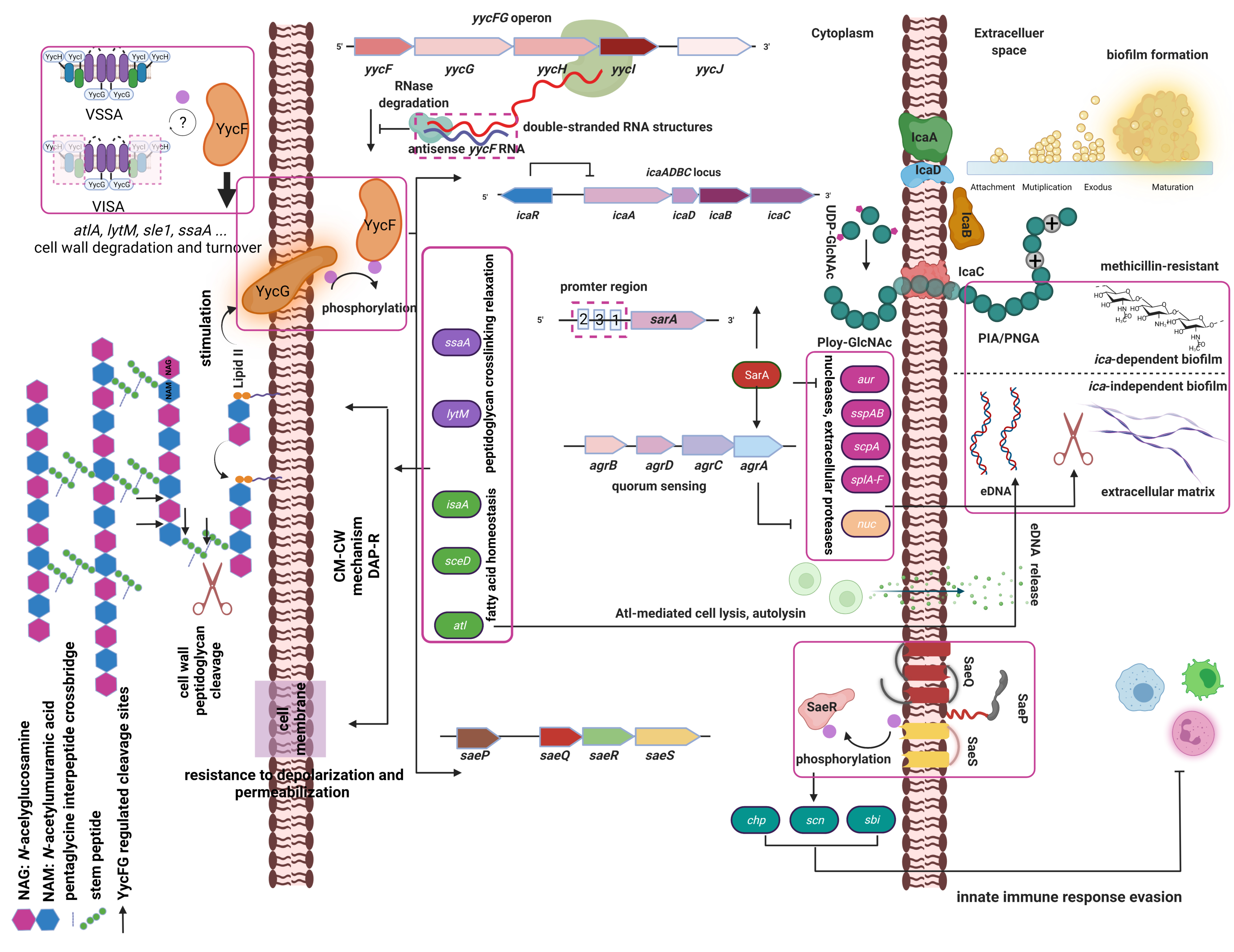
2. Regulation of S. aureus YycFG Two-Component Systems

2.1. S. aureus Two-Component Systems

Fifteen TCSs in the whole genome of S. aureus are involved in the regulation of bacterial physiological metabolism [21,22]. The regulation of the S. aureus TCS inextricably affects bacterial antimicrobial resistance [23]. The TCS system is comprised of two components: (i) histidine protein kinase (HPK) receptor proteins, which are anchored to the cell membrane and sense external environmental stimuli, and (ii) response regulators (RRs), which regulate downstream target gene expression. After physical or chemical stimulation by the external environment, HPKs undergo phosphorylation. The phosphate group is transferred to the response regulator (RR). Phosphorylated response regulators directly bind to the promoter regions of downstream target genes and enhance the adaptive viability of bacteria [24,25].

2.2. Regulatory Roles of the YycFG TCS in Cell Wall Biosynthesis and Biofilm Formation

The yyc operon in S. aureus is comprised of four genes, yycF, yycG, yycH, yycI and yycJ. The membrane-associated regulator YycHI is an activator of YycG function in S. aureus [26]. Despite its essential role in bacterial viability, the physiological or mechanical signals sensed by YycG are not well understood. Recent structural analysis of the YycG PASCYT domain revealed a metal-binding site that binds zinc ions (Zn2+). The abrogation of metal binding increased YycG kinase activity and YycF phosphorylation, which indicates that Zn2+ binding negatively regulates YycFG [27]. The response regulator YycF participates in the regulation of cell wall synthesis and binds promoter regions that contain a conserved motif sequence [5′-TGT(A/T) A(A/T/C)-N5-TGT(A/T)A(A/T/C)-3′] of target genes via the helix-turn-helix domain of YycF [12,20].
The specific binding of YycF to promoter regions, including icaA, agr, sarA and sarX, modifies biofilms in an ica-dependent manner [8]. The ArlRS regulon is a global regulator of relevant genes, including cell wall-anchored adhesins, polysaccharide synthesis genes, cell wall remodeling genes, the urease operon and a large number of virulence factors [28,29]. The accessory gene regulator (agr) system greatly contributes to the formation of S. aureus biofilms [30], and the staphylococcal accessory regulator (SarA) drives biofilm organization by altering ica transcription and producing PIA [31,32]. The sarA gene in Staphylococcus epidermidis is an essential regulator of ica operon activation in biofilm formation [33]. The sarA gene in S. aureus is associated with bacterial oxidation sensing and virulence factors [34,35]. Notably, our recent study revealed that YycF directly regulated the predicted promoter regions of sarA, and YycFG TCS sensitized S. aureus biofilm formation to H2O2 exposure via the sarA pathway [20].

2.3. Regulatory Roles of the YycFG TCS in Response to Host Immunity

S. aureus is a major opportunistic human pathogen. S. aureus interacts with its human host as an innocuous member of the microbiota, or it breaks immune barriers to become an invasive pathogen [36]. Because YycFG TCS positively regulates certain virulence genes, including genes associated with host–matrix interactions (efb, emp, fnbA, and fnbB), cytolysis (hlgACB, hla, and hlb) and innate immune defense evasion (scn, chp, and sbi), its activity is closely linked to the host inflammatory response that is induced during infection [37]. However, virulence gene modulation is achieved via the coordination of another TCS, SaeSR (short for S. aureus exoprotein expression). YycF positively activates SaeSR TCS. SaeSR is a virulence factor regulation system that promotes lysis of polymorphonuclear leukocytes (PMNs) after phagocytosis, and it plays an essential role in S. aureus evasion of innate immunity [38]. During host cell–pathogen interactions, the innate and adaptive immune systems respond to S. aureus. Adaptive immunity amplifies the activity of innate immune cells and influences host susceptibility to S. aureus, and it is associated with chronic persistent infections [39]. S. aureus also developed evasion mechanisms from the adaptive immune response using virulence strategies. A second immunoglobulin binding protein (Sbi) is a cell wall-anchored surface protein that binds with the Fcγ portion of IgG, or it is secreted as a virulence factor that interferes with soluble complement factor C3, which manipulates adaptive immune responses to S. aureus [40]. Aurélia et al. [41] found that YycFG triggered cell wall turnover and degradation. Degradation of the cell wall via the NF-κB system resulted in the clearance of bacterial cells by the host immune system. Activated YycF stimulated the SaeSR TCS to increase the virulence gene expression involved in human–pathogen interactions and innate immune system evasion. Therefore, the fine tuning of YycFG plays an important role in determining the conditions of S. aureus infection.

3. The Impact of YycFG Two-Component Systems on Antibiotic Resistance

3.1. S. aureus Biofilms and Antimicrobial Drug Resistance

After biofilm formation due to intercellular aggregation, bacterial cell detachment caused by the action of bacterial products is critical for subsequent bacterial dissemination [42]. Staphylococcus biofilms are mediated by polysaccharide intercellular adhesin (PIA), which is synthesized by the ica operon [14]. PIA contributes to the facilitation of initial biofilm adherence [43]. PIA is a major component of the extracellular matrix that fixes staphylococcal cells within the biofilm mass, which increases resistance to mechanical force [44]. The classical and predominant adaptive modules from the TCS systems [45] modulate mechanisms associated with antibiotic resistance in most bacteria, including increased drug efflux, upregulation of antibiotic-degrading enzymes, biofilm production and enhanced cell permeability, which depend on the expression of corresponding downstream effectors [25]. Therefore, an understanding of the YycFG two-component system contributed to developments in our ability to combat S. aureus infections.
An animal study using co-infection models demonstrated that ica-positive S. aureus showed better in vivo survival than their corresponding ica mutants in wild-type mice [46]. The significant contribution of the ica genes toward S. epidermidis infection was confirmed using a C. elegans infection model, which indicated that ica genes were required for a lethal infection [47]. PIA, produced by S. aureus in vivo, significantly affected S. aureus systemic infections in mice [48]. S. aureus could grow synergistically with Candida albicans within biofilms [49]. These studies suggest that PIA production is important for infection and/or co-infection in vivo, especially S. aureus. Biofilm organization decreases the susceptibility to antimicrobial agents and/or antibiotics [25]. The potential mechanisms include persister cell formation, altered metabolic conditions and a decreased penetration into the biofilm extracellular matrix [50]. S. aureus persister cells were first observed in 1942 and demonstrated that non-growing dormant cells were resistant to penicillin [51]. Biofilms exhibit characteristics similar to persisted cells, and the biofilm matrix contributes to persistent infection by protecting bacterial cells from the immune system and antibiotics [52,53]. PIA, which accounts for most of the extracellular matrix, affects susceptibility to antibiotics by impairing penetration through the biofilm matrix. PIA likely enhances the horizontal transfer of drug resistance genes via its effect on cell-to-cell contact in biofilms [50] because S. aureus within biofilms was 1000 times more resistant than the bacteria in a planktonic state. Previous studies showed that ica-positive S. aureus strains had increased resistance to a variety of antibiotics, such as oxacillin, gentamicin, ciprofloxacin, levofloxacin, erythromycin and vancomycin, compared to ica-negative strains [54,55].

3.2. YycFG TCS-Associated Cell Membrane and Cell Wall Biogenesis Involvement in Drug Resistance

Antibiotic-resistant strains, particularly MRSA, are increasing in prevalence in hospital- and community-acquired infections and pose a significant threat to public health [56,57]. MRSA infections are responsible for substantial morbidity and lead to the death of 21.8% of infected patients annually [58]. To understand the mechanisms of resistance in MRSA strains, recent genomic studies demonstrated that antibiotic resistance in MRSA was primarily due to extensive modification of bacterial cell wall biogenesis [59,60]. These studies used a clinical MRSA strain to demonstrate that exposure to certain antibiotic combinations was associated with the development of mutations in specific genes, including yycFG [61]. Wu et al. isolated clinical MRSA strains from chronic osteomyelitis tissues. These MRSA strains demonstrated an accelerated growth rate compared to the MSSA strains and an accumulation of PIA matrix in the biofilms with the increased expression of the yycF/G/H and icaA/D genes [17]. The YycFG system is essential for S. aureus viability. Therefore, a recombinant plasmid shuttle vector was used to overexpress an antisense RNA and inhibit target gene expression, which led to the construction of antisense yycG RNA (ASyycG)-overexpressing MRSA strains. The ASyycG strains showed a reduction in biofilm formation and an increased antibiotic sensitivity to cefoxitin compared to MRSA strains, which may be attributed to altered PIA production [62]. To further investigate the regulatory roles of ASyycG in the pathogenicity of MRSA strains in vivo, a rat model of tibial osteomyelitis was developed and infected with MRSA- or ASyycG-overexpressing strains. The ASyycG strains exhibited suppressed invasive ability and pathogenicity in vivo, and the production of pro-inflammatory cytokines was reduced compared to MRSA strains [63].
To explain the potential mechanisms of ASyycG in regulating S. aureus biofilms, transcriptome analyses showed that ASyycG overexpression influenced the pathways associated with biofilm metabolism, virulence and glycolysis/gluconeogenesis utilization in S. aureus, including the sarA and icaA genes [20]. For the potential role of the response regulator YycF in biofilm formation and pathogenicity, endogenous antisense yycF RNA (ASyycF) was detected using a 5′ RACE assay. The over-production of ASyycF reduced YycF production and biofilm formation. Antibiotic sensitivity to vancomycin was significantly improved in ASyycF-overexpressing strains compared to MRSA strains. ASyycF-overexpressing MRSA strains exhibited suppressed invasion in a rat tibial infection model [63], which supports ASyycF as a supplementary strategy for the management of S. aureus and MRSA infections. Taken together, these data indicate that inhibition of the YycFG pathway reduced PIA production, biofilm formation and bacterial pathogenicity, which provides a potential target for the management of MRSA infections. The YycFG TCS was identified in the process of cell wall biosynthesis. Cell wall thickening and an aberrant division of septa are closely associated with YycFG [64]. LytM and SsaA play crucial roles in cell wall peptidoglycan crosslinking relaxation during the cell division process and are regulated by the YycFG system, which is required for cell viability. A previous study revealed lipid II as an essential component of the cell wall and a signal that is sensed by YycG kinase. Antibiotics, such as β-lactams, which characteristically target lipid II, activate YycFG TCS [65]. However, the efficacies of last resort agents, such as vancomycin, linezolid and daptomycin, in the treatment of serious MRSA infections are controversial.
Vancomycin is a glycopeptide antibiotic that inhibits cell wall synthesis via binding to the D-alanyl-D-alanine residue on the bacterial cell wall [66]. Ten types of vancomycin-intermediate resistance in S. aureus (VISA) were analyzed, and three types of heterogeneous vancomycin-intermediate S. aureus (hVISA) strains were sequenced using high-throughput techniques. Site mutations in the yycFG gene were detected in eight types of VISA and two types of hVISA strains, which exhibited the highest mutation frequency [67]. These results suggest that the yycFG gene plays an important role in the generation of VISA and hVISA strains. Jansen et al. reported that the insertion of an enhanced promoter sequence in the promoter region of the yycFG gene of the S. aureus VISA strain increased the expression of its downstream target genes and up-regulated bacterial cell wall biosynthesis, which increased antimicrobial drug resistance [68]. Domains of the yycFG gene of the VISA strain are affected by the mutation of a single nucleotide, such as the A96T mutation in the yycF gene, which is a mutation from base G to base A at site 24673 of the S. aureus genome. This mutated base is located in a conserved region of the yycF gene, and it is associated with a conformational change to protein phosphorylation regulation. These mutations decrease the activity of the YycFG protein and down-regulate the expression of bacterial autolytic enzymes, which inhibits the bacterial autolytic process [67]. The yycHI genes are downstream of yycFG and bind to the YycG histidine kinase receptor to interact with the YycFG pathway [26]. Auxiliary YycH and YycI are ‘connector’ proteins that physically interact with the YycG sensor kinase to form a ternary protein complex that activates the YycFG TCS. Mutation of these auxiliary proteins disrupted the integration of YycFG two-component networks and reduced vancomycin susceptibility in clinical VISA strains. The mutation rate of the yycHI gene in these strains was significantly higher than the vancomycin-sensitive S. aureus strain. Mutation of yycHI may lead to the enhancement of cell wall synthesis and enhance antimicrobial resistance [67], but further investigation is required.
Membrane-bound receptors and cognate cytosolic response regulators, such as the YycG receptor and the YycF regulator, are closely associated with a phospho-relay mechanism on initiation. Upon phosphorylated, the YycF response regulator plays a role in transcription factor binding to DNA and modulates associated gene expression to orchestrate several physiological functions involved virulence, cell wall metabolism and biofilm formation [37,69]. Daptomycin (Dap) is a cell membrane-targeting lipopeptide antibiotic that exhibits excellent antibacterial activities against susceptible Gram-positive pathogens [70]. The combination of Dap with calcium significantly reduced cell viability via cell membrane depolarization and permeabilization [71]. Because the YycFG TCS regulates the cell envelope and lipid metabolism-associated genes, including atl, lytM, sceD, isaA, and ssaA, it plays a fundamental role in cell membrane metabolism [8,65]. Because S. aureus extracellular genomic DNA (eDNA) is released from bacteria via cell lysis, the role of S. aureus autolysin, Atl, may be implicated in biofilm development, especially in initial attachment [72]. YycFG TCS maintains cell membrane fatty acid homeostasis [73], confers resistance to depolarization and/or permeabilization and contributes to daptomycin resistance (DAP-R). The development of DAP-R in S. aureus was observed clinically during therapy, and it is often associated with treatment failure [74]. DAP-R strains acquire a progressive accumulation of single nucleotide polymorphisms in the YycFG TCS of the yycFGHI operon, which is involved in key cell membrane events, and the yyc operon is involved in the generalized response to antimicrobials [71]. Clinical MRSA strains that emerged with daptomycin non-susceptibility were isolated to examine the influence of certain antibiotic combinations, including daptomycin with or without adjunctive clarithromycin, linezolid, or oxacillin, on the development of mutations in specific genes, including the multi-peptide resistance factor gene (mprF) and yycFG. Daptomycin alone or combined with other antibiotics resulted in mutations in mprF and yycFG, which suggests that combining daptomycin with different antimicrobials affects the mutational space required for daptomycin nonsusceptibility development [61]. These results indicate that the use of adjunctive antibiotic therapy in a clinical setting alters the mutational space permitted for drug resistance development, which warrants the exploration of novel targeted molecular treatments.

4. Targeting the S. aureus Two-Component Systems

4.1. Molecular Targets

Two-component systems integrate with other signaling molecules in bacteria via cross-activation with other transcription factors [75]. The staphylococcal accessory regulator (SarA) is a global regulator that controls the transcription of a wide range of virulence genes [76]. A novel inhibitor of SarA was designed to prevent S. aureus biofilm formation, and it was developed as a potential antimicrobial strategy in prosthetic joint infections [77]. Global SarA expression is linked to the YycFG TCS, which binds to the promotor region of the sarA gene [20]. This activation affects biofilm formation and the pathogenic and antimicrobial resistance potential of S. aureus, which offers a supplementary therapeutic target.

4.2. Non-Coding RNA Regulation

Two-component systems are integrated with other signaling molecules in bacteria, such as regulatory RNAs. RNA III is a “small” regulatory RNA (sRNA) in S. aureus, which is controlled by the AgrAC TCS, and participates in quorum sensing and pathogenicity “antisense regulation” via spa, rot or hla at the post-transcriptional level [78]. The formation of double-stranded RNA structures via base pairing of the 5’ or 3′ terminal sequences of target mRNAs with corresponding antisense RNAs provides positive translation initiation and mRNA stability and stimulates RNA degradation by RNase for transcription interference and attenuation [79,80]. Endonuclease RNase III plays a critical role in the latter effect via the cleavage of double-stranded RNA structures [81]. Our previous study showed that promoter regions of the RNase III–encoding gene (rnc) bound and were directly regulated by the YycF ortholog gene VicR in S. mutans [82]. Antisense regulation mechanisms were used to inhibit antibiotic resistance in bacterial infections using antisense oligonucleotides [83]. Notably, an endogenous antisense RNA base paired with yycF mRNA was identified in S. aureus [84], which belongs to a trans-encoded sRNA [79]. The length of the ASyycF operon is approximately 400 bp, and because endogenous ASyycF significantly downregulated the expression of YycFG TCS, it may restrict biofilm formation and reduce antibiotic resistance and pathogenesis. Therefore, this pathway should be considered as a supplementary strategy for the management of S. aureus and MRSA infections.

5. Concluding Remarks

YycFG TCS is the only essential TCS for S. aureus to adapt to a wide variety of environments. Many other resistance mechanisms, including cell wall peptidoglycan metabolism, cell membrane lipid metabolism and innate immune system evasion, are directly or indirectly regulated by YycFG (Figure 1). S. aureus acquired a collection of virulence factors that enabled the bacterial cells to colonize biotic and abiotic surfaces and form a biofilm. This biofilm structure allows S. aureus to sense and resist harsh environmental conditions, physical and chemical stimuli and antimicrobial drugs, thereby enabling S. aureus to contribute to chronic and recalcitrant infections. The S. aureus YycFG TCS and several global gene regulators coordinate important functions during the establishment and maturation of the biofilm. Despite recent advances in this field, the available data are generally limited to in vitro studies using laboratory strains. Most of the studies on S. aureus biofilms were performed under static conditions and do not account for environmental signals that may variably affect biofilms. The development of new models that mimic the processes during biofilm growth in human infections is critical for the study of the mechanisms that drive S. aureus biofilm production. Future novel and effective anti-infection therapies will likely include antimicrobial agents that exhibit antibiofilm properties. Exploration of the molecular targets present in S. aureus two-component systems and their gene regulators, such as the ica operon, sarA, atl, lytM, sceD, isaA, and ssaA, is worthwhile. Non-coding RNAs elucidated the post-transcription regulation of biofilm growth and bacterial resistance to antimicrobials. The designing of a nucleotide delivery system with high transfection efficiencies, favorable biocompatibility and safety are needed to target RNA interference and as a novel strategy to treat infections and tackle drug resistance.

Author Contributions

Conceptualization, S.W., L.L. and H.Z.; software, S.W., J.Z. and Q.P.; formal analysis, S.W., Y.L. and J.Z.; investigation, L.L. and H.Z.; resources, S.W., Q.P. and J.Z.; data curation, S.W., Y.L. and J.Z.; writing—original draft preparation, S.W. and L.L.; writing—review and editing, S.W., L.L. and H.Z.; supervision, L.L. and H.Z.; funding acquisition, S.W. and H.Z. All authors have read and agreed to the published version of the manuscript.

Funding

This work was supported in part by the Sichuan Provincial Natural Science Foundation of China (Grant No. 2021YJ0455; 2019YFS0270) and Post-Doctor Research Project, West China Hospital, Sichuan University (Grant No. 2020HXBH134).

Conflicts of Interest

The authors declare no conflict of interest.

References

  1. Bhattacharya, M.; Wozniak, D.J.; Stoodley, P.; Hall-Stoodley, L. Prevention and treatment of Staphylococcus aureus biofilms. Expert Rev. Anti. Infect. Ther. 2015, 13, 1499–1516. [Google Scholar] [CrossRef] [Green Version]
  2. Tong, S.Y.C.; Davis, J.S.; Eichenberger, E.; Holland, T.L.; Fowler, V.G., Jr. Staphylococcus aureus Infections: Epidemiology, Pathophysiology, Clinical Manifestations, and Management. Clin. Microbiol. Rev. 2015, 28, 603–661. [Google Scholar] [CrossRef] [PubMed] [Green Version]
  3. Maiti, P.; Chatterjee, S.; Dey, R.; Kundu, A. Biofilms on indwelling urologic devices: Microbes and antimicrobial management prospect. Ann. Med. Heal. Sci. Res. 2014, 4, 100–104. [Google Scholar] [CrossRef] [Green Version]
  4. Francischetto, O.; Da Silva, L.A.P.; E Senna, K.M.S.; Vasques, M.R.; Barbosa, G.F.; Weksler, C.; Ramos, R.G.; Golebiovski, W.F.; Lamas, C. Healthcare-Associated Infective Endocarditis: Case Series in Referral Hospital from 2006 to 2011. Arq. Bras. Cardiol. 2014, 103, 292–298. [Google Scholar] [CrossRef] [PubMed]
  5. Cha, J.-O.; Yoo, J.I.; Yoo, J.S.; Chung, H.-S.; Park, S.-H.; Kim, H.S.; Lee, Y.S.; Chung, G.T. Investigation of Biofilm Formation and its Association with the Molecular and Clinical Characteristics of Methicillin-resistant Staphylococcus aureus. Osong Public Heal Res. Perspect. 2013, 4, 225–232. [Google Scholar] [CrossRef] [PubMed] [Green Version]
  6. Savage, V.J.; Chopra, I.; O’Neill, A.J. Staphylococcus aureus Biofilms Promote Horizontal Transfer of Antibiotic Resistance. Antimicrob. Agents Chemother. 2013, 57, 1968–1970. [Google Scholar] [CrossRef] [PubMed] [Green Version]
  7. Mattos-Graner, R.O.; Duncan, M.J. Two-component signal transduction systems in oral bacteria. J. Oral Microbiol. 2017, 9, 1400858. [Google Scholar] [CrossRef] [PubMed] [Green Version]
  8. Xu, T.; Wu, Y.; Lin, Z.; Bertram, R.; Götz, F.; Zhang, Y.; Qu, D. Identification of Genes Controlled by the Essential YycFG Two-Component System Reveals a Role for Biofilm Modulation in Staphylococcus epidermidis. Front. Microbiol. 2017, 8, 724. [Google Scholar] [CrossRef] [PubMed] [Green Version]
  9. Fabret, C.; Hoch, J.A. A two-component signal transduction system essential for growth of Bacillus subtilis: Implications for anti-infective therapy. J. Bacteriol. 1998, 180, 6375–6383. [Google Scholar] [CrossRef]
  10. Martin, P.K.; Li, T.; Sun, D.; Biek, D.P.; Schmid, M.B. Role in Cell Permeability of an Essential Two-Component System in Staphylococcus aureus. J. Bacteriol. 1999, 181, 3666–3673. [Google Scholar] [CrossRef] [Green Version]
  11. Villanueva, M.; García, B.; Valle, J.; Rapún, B.; Mozos, I.R.D.L.; Solano, C.; Martí, M.; Penadés, J.R.; Toledo-Arana, A.; Lasa, I. Sensory deprivation in Staphylococcus aureus. Nat. Commun. 2018, 9, 1–12. [Google Scholar] [CrossRef] [PubMed]
  12. Dubrac, S.; Msadek, T. Identification of Genes Controlled by the Essential YycG/YycF Two-Component System of Staphylococcus aureus. J. Bacteriol. 2004, 186, 1175–1181. [Google Scholar] [CrossRef] [Green Version]
  13. Donlan, R.M.; Costerton, J.W. Biofilms: Survival Mechanisms of Clinically Relevant Microorganisms. Clin. Microbiol. Rev. 2002, 15, 167–193. [Google Scholar] [CrossRef] [Green Version]
  14. Gerke, C.; Kraft, A.; Süssmuth, R.; Schweitzer, O.; Götz, F. Characterization of theN-Acetylglucosaminyltransferase Activity Involved in the Biosynthesis of the Staphylococcus epidermidis polysaccharide Intercellular Adhesin. J. Biol. Chem. 1998, 273, 18586–18593. [Google Scholar] [CrossRef] [PubMed] [Green Version]
  15. Heilmann, C.; Schweitzer, O.; Gerke, C.; Vanittanakom, N.; Mack, D.; Götz, F. Molecular basis of intercellular adhesion in the biofilm-forming Staphylococcus epidermidis. Mol. Microbiol. 1996, 20, 1083–1091. [Google Scholar] [CrossRef]
  16. Nguyen, H.T.T.; Nguyen, T.H.; Otto, M. The staphylococcal exopolysaccharide PIA—Biosynthesis and role in biofilm formation, colonization, and infection. Comput. Struct Biotechnol. J. 2020, 18, 3324–3334. [Google Scholar] [CrossRef]
  17. Wu, S.; Huang, F.; Zhang, H.; Lei, L. Staphylococcus aureus biofilm organization modulated by YycFG two-component regulatory pathway. J. Orthop. Surg. Res. 2019, 14, 10. [Google Scholar] [CrossRef]
  18. O’Neill, E.; Pozzi, C.; Houston, P.; Smyth, D.; Humphreys, H.; Robinson, D.A.; O’Gara, J.P. Association between Methicillin Susceptibility and Biofilm Regulation in Staphylococcus aureus Isolates from Device-Related Infections. J. Clin. Microbiol. 2007, 45, 1379–1388. [Google Scholar] [CrossRef] [PubMed] [Green Version]
  19. Pozzi, C.; Waters, E.; Rudkin, J.; Schaeffer, C.R.; Lohan, A.; Tong, P.; Loftus, B.; Pier, G.; Fey, P.D.; Massey, R.; et al. Methicillin Resistance Alters the Biofilm Phenotype and Attenuates Virulence in Staphylococcus aureus device-Associated Infections. PLoS Pathog. 2012, 8, e1002626. [Google Scholar] [CrossRef]
  20. Wu, S.; Liu, Y.; Lei, L.; Zhang, H. Antisense yycG modulates the susceptibility of Staphylococcus aureus to hydrogen peroxide via the sarA. BMC Microbiol. 2021, 21, 160. [Google Scholar] [CrossRef]
  21. Cheng, R.; Morcos, F.; Levine, H.; Onuchic, J.N. Toward rationally redesigning bacterial two-component signaling systems using coevolutionary information. Proc. Natl. Acad. Sci. USA 2014, 111, E563–E571. [Google Scholar] [CrossRef] [PubMed] [Green Version]
  22. Kuroda, M.; Ohta, T.; Uchiyama, I.; Baba, T.; Yuzawa, H.; Kobayashi, I.; Cui, L.; Oguchi, A.; Aoki, K.; Nagai, Y.; et al. Whole genome sequencing of meticillin-resistant Staphylococcus aureus. Lancet 2001, 357, 1225–1240. [Google Scholar] [CrossRef]
  23. Matsuo, M.; Kato, F.; Oogai, Y.; Kawai, T.; Sugai, M.; Komatsuzawa, H. Distinct two-component systems in methicillin-resistant Staphylococcus aureus can change the susceptibility to antimicrobial agents. J. Antimicrob. Chemother. 2010, 65, 1536–1537. [Google Scholar] [CrossRef] [PubMed] [Green Version]
  24. Goulian, M. Two-component signaling circuit structure and properties. Curr. Opin. Microbiol. 2010, 13, 184–189. [Google Scholar] [CrossRef] [PubMed] [Green Version]
  25. Tierney, A.R.; Rather, P.N. Roles of two-component regulatory systems in antibiotic resistance. Futur. Microbiol. 2019, 14, 533–552. [Google Scholar] [CrossRef]
  26. Cameron, D.; Jiang, J.-H.; Kostoulias, X.; Foxwell, D.J.; Peleg, A.Y. Vancomycin susceptibility in methicillin-resistant Staphylococcus aureus is mediated by YycHI activation of the WalRK essential two-component regulatory system. Sci. Rep. 2016, 6, 30823. [Google Scholar] [CrossRef] [PubMed]
  27. Monk, I.R.; Shaikh, N.; Begg, S.L.; Gajdiss, M.; Sharkey, L.K.R.; Lee, J.Y.H.; Pidot, S.J.; Seemann, T.; Kuiper, M.; Winnen, B.; et al. Zinc-binding to the cytoplasmic PAS domain regulates the essential WalK histidine kinase of Staphylococcus aureus. Nat. Commun. 2019, 10, 3067. [Google Scholar] [CrossRef] [Green Version]
  28. Crosby, H.A.; Tiwari, N.; Kwiecinski, J.M.; Xu, Z.; Dykstra, A.; Jenul, C.; Fuentes, E.; Horswill, A.R. The Staphylococcus aureus ArlRS two-component system regulates virulence factor expression through MgrA. Mol. Microbiol. 2020, 113, 103–122. [Google Scholar] [CrossRef] [PubMed]
  29. Ouyang, Z.; Zheng, F.; Chew, J.Y.; Pei, Y.; Zhou, J.; Wen, K.; Han, M.; Lemieux, M.J.; Hwang, P.M.; Wen, Y. Deciphering the activation and recognition mechanisms of Staphylococcus aureus response regulator ArlR. Nucleic Acids Res. 2019, 47, 11418–11429. [Google Scholar] [CrossRef]
  30. Tan, L.; Li, S.R.; Jiang, B.; Hu, X.M.; Li, S. Therapeutic targeting of the Staphylococcus aureus accessory gene regulator (agr) system. Front Microbiol. 2018, 9, 55. [Google Scholar] [CrossRef]
  31. Liu, L.; Shen, X.; Yu, J.; Cao, X.; Zhan, Q.; Guo, Y.; Yu, F. Subinhibitory Concentrations of Fusidic Acid May Reduce the Virulence of S. aureus by Down-Regulating sarA and saeRS to Reduce Biofilm Formation and α-Toxin Expression. Front. Microbiol. 2020, 11, 25. [Google Scholar] [CrossRef]
  32. Valle, J.; Toledo-Arana, A.; Berasain, C.; Ghigo, J.M.; Amorena, B.; Penadés, J.R.; Lasa, I. SarA and not sigmaB is essential for biofilm development by Staphylococcus aureus. Mol. Microbiol. 2003, 48, 1075–1087. [Google Scholar] [CrossRef] [PubMed]
  33. Tormo, M.A.; Martí, M.; Valle, J.; Manna, A.C.; Cheung, A.L.; Lasa, I.; Penadés, J.R. SarA Is an Essential Positive Regulator of Staphylococcus epidermidis Biofilm Development. J. Bacteriol. 2005, 187, 2348–2356. [Google Scholar] [CrossRef] [Green Version]
  34. Bayer, M.G.; Heinrichs, J.H.; Cheung, A.L. The molecular architecture of the sar locus in Staphylococcus aureus. J. Bacteriol. 1996, 178, 4563–4570. [Google Scholar] [CrossRef] [PubMed] [Green Version]
  35. Cheung, A.L.; Eberhardt, K.; Heinrichs, J.H. Regulation of protein A synthesis by the sar and agr loci of Staphylococcus aureus. Infect. Immun. 1997, 65, 2243–2249. [Google Scholar] [CrossRef] [Green Version]
  36. Krismer, B.; Weidenmaier, C.; Zipperer, A.; Peschel, A. The commensal lifestyle of Staphylococcus aureus and its interactions with the nasal microbiota. Nat. Rev. Genet. 2017, 15, 675–687. [Google Scholar] [CrossRef] [PubMed]
  37. Qiu, Y.; Xu, D.; Xia, X.; Zhang, K.; Aadil, R.M.; Batool, Z.; Wang, J. Five major two components systems of Staphylococcus aureus for adaptation in diverse hostile environment. Microb. Pathog. 2021, 159, 105119. [Google Scholar] [CrossRef] [PubMed]
  38. Voyich, J.M.; Vuong, C.; Dewald, M.; Nygaard, T.; Kocianova, S.; Griffith, S.; Jones, J.; Iverson, C.; Sturdevant, D.E.; Braughton, K.R.; et al. The SaeR/S Gene Regulatory System Is Essential for Innate Immune Evasion by Staphylococcus aureus. J. Infect. Dis. 2009, 199, 1698–1706. [Google Scholar] [CrossRef] [PubMed] [Green Version]
  39. Karauzum, H.; Datta, S.K. Adaptive Immunity Against Staphylococcus aureus. Curr. Top. Microbiol. Immunol. 2017, 409, 419–439. [Google Scholar]
  40. Smith, E.J.; Visai, L.; Kerrigan, S.W.; Speziale, P.; Foster, T.J. The Sbi Protein Is a Multifunctional Immune Evasion Factor of Staphylococcus aureus. Infect. Immun. 2011, 79, 3801–3809. [Google Scholar] [CrossRef] [PubMed] [Green Version]
  41. Delauné, A.; Dubrac, S.; Blanchet, C.; Poupel, O.; Mäder, U.; Hiron, A.; LeDuc, A.; Fitting, C.; Nicolas, P.; Cavaillon, J.-M.; et al. The WalKR System Controls Major Staphylococcal Virulence Genes and Is Involved in Triggering the Host Inflammatory Response. Infect. Immun. 2012, 80, 3438–3453. [Google Scholar] [CrossRef] [PubMed] [Green Version]
  42. Moormeier, D.E.; Bose, J.L.; Horswill, A.R.; Bayles, K.W. Temporal and Stochastic Control of Staphylococcus aureus Biofilm Development. mBio 2014, 5, e01341-14. [Google Scholar] [CrossRef] [PubMed] [Green Version]
  43. Nuryastuti, T.; Krom, B.P. Ica-status of clinical Staphylococcus epidermidis strains affects adhesion and aggregation: A thermodynamic analysis. Antonie Van Leeuwenhoek 2017, 110, 1467–1474. [Google Scholar] [CrossRef]
  44. Schommer, N.N.; Christner, M.; Hentschke, M.; Ruckdeschel, K.; Aepfelbacher, M.; Rohde, H. Staphylococcus epidermidis Uses Distinct Mechanisms of Biofilm Formation To Interfere with Phagocytosis and Activation of Mouse Macrophage-Like Cells 774A.1. Infect. Immun. 2011, 79, 2267–2276. [Google Scholar] [CrossRef] [PubMed] [Green Version]
  45. Patel, V.; Matange, N. Adaptation and compensation in a bacterial gene regulatory network evolving under antibiotic selection. eLife 2021, 10. [Google Scholar] [CrossRef] [PubMed]
  46. Fluckiger, U.; Ulrich, M.; Steinhuber, A.; Döring, G.; Mack, D.; Landmann, R.; Goerke, C.; Wolz, C. Biofilm Formation, icaADBC Transcription, and Polysaccharide Intercellular Adhesin Synthesis by Staphylococci in a Device-Related Infection Model. Infect. Immun. 2005, 73, 1811–1819. [Google Scholar] [CrossRef] [PubMed] [Green Version]
  47. Begun, J.; Gaiani, J.M.; Rohde, H.; Mack, D.; Calderwood, S.B.; Ausubel, F.M.; Sifri, C.D. Staphylococcal Biofilm Exopolysaccharide Protects against Caenorhabditis elegans Immune Defenses. PLoS Pathog. 2007, 3, e57. [Google Scholar] [CrossRef] [Green Version]
  48. Kropec, A.; Maira-Litran, T.; Jefferson, K.; Grout, M.; Cramton, S.E.; Götz, F.; Goldmann, D.A.; Pier, G.B. Poly- N -Acetylglucosamine Production in Staphylococcus aureus is Essential for Virulence in Murine Models of Systemic Infection. Infect. Immun. 2005, 73, 6868–6876. [Google Scholar] [CrossRef] [Green Version]
  49. Zago, C.E.; Silva, S.; Sanitá, P.V.; Barbugli, P.; Dias, C.M.I.; Lordello, V.B.; Vergani, C.E. Dynamics of Biofilm Formation and the Interaction between Candida albicans and Methicillin-Susceptible (MSSA) and -Resistant Staphylococcus aureus (MRSA). PLoS ONE 2015, 10, e0123206. [Google Scholar] [CrossRef] [Green Version]
  50. Hall, C.W.; Mah, T.-F. Molecular mechanisms of biofilm-based antibiotic resistance and tolerance in pathogenic bacteria. FEMS Microbiol. Rev. 2017, 41, 276–301. [Google Scholar] [CrossRef]
  51. Hobby, G.L.; Meyer, K.; Chaffee, E. Observations on the Mechanism of Action of Penicillin. Exp. Biol. Med. 1942, 50, 281–285. [Google Scholar] [CrossRef]
  52. Waters, E.M.; Rowe, S.E.; O’Gara, J.P.; Conlon, B.P. Convergence of Staphylococcus aureus persister and biofilm research: Can biofilms be defined as communities of adherent persister cells? PLoS Pathog. 2016, 12, e1006012. [Google Scholar] [CrossRef] [PubMed]
  53. Schwartbeck, B.; Birtel, J.; Treffon, J.; Langhanki, L.; Mellmann, A.; Kale, D.; Kahl, J.; Hirschhausen, N.; Neumann, C.; Lee, J.; et al. Dynamic in vivo mutations within the ica operon during persistence of Staphylococcus aureus in the airways of cystic fibrosis patients. PLoS Pathog. 2016, 12, e1006024. [Google Scholar] [CrossRef] [PubMed]
  54. Mahmoudi, H.; Pourhajibagher, M.; Chiniforush, N.; Soltanian, A.R.; Alikhani, M.Y.; Bahador, A. Biofilm formation and antibiotic resistance in meticillin-resistant and meticillin-sensitive Staphylococcus aureus isolated from burns. J. Wound Care 2019, 28, 66–73. [Google Scholar] [CrossRef]
  55. Kıvanç, S.A.; Arık, G.; Akova-Budak, B.; Kıvanç, M. Biofilm forming capacity and antibiotic susceptibility of Staphylococcus spp. with the icaA/icaD/bap genotype isolated from ocular surface of patients with diabetes. Malawi Med. J. 2018, 30, 243–249. [Google Scholar] [CrossRef] [Green Version]
  56. Melo-Cristino, J.; Resina, C.; Manuel, V.; Lito, L.; Ramirez, M. First case of infection with vancomycin-resistant Staphylococcus aureus in Europe. Lancet 2013, 382, 205. [Google Scholar] [CrossRef]
  57. Turner, N.A.; Sharma-Kuinkel, B.K.; Maskarinec, S.; Eichenberger, E.; Shah, P.P.; Carugati, M.; Holland, T.L.; Fowler, V.G. Methicillin-resistant Staphylococcus aureus: An overview of basic and clinical research. Nat. Rev. Genet. 2019, 17, 203–218. [Google Scholar] [CrossRef]
  58. Kourtis, A.P.; Hatfield, K.; Baggs, J.; Mu, Y.; See, I.; Epson, E.; Nadle, J.; Kainer, M.A.; Dumyati, G.; Petit, S.; et al. Vital Signs:Epidemiology and Recent Trends in Methicillin-Resistant and in Methicillin-Susceptible Staphylococcus aureus bloodstream Infections—United States. MMWR. Morb. Mortal. Wkly. Rep. 2019, 68, 214–219. [Google Scholar] [CrossRef] [Green Version]
  59. Rajagopal, M.; Martin, M.J.; Santiago, M.; Lee, W.; Kos, V.N.; Meredith, T.; Gilmore, M.S.; Walker, S. Multidrug Intrinsic Resistance Factors in Staphylococcus aureus Identified by Profiling Fitness within High-Diversity Transposon Libraries. mBio 2016, 7, e00950-16. [Google Scholar] [CrossRef] [Green Version]
  60. Coe, K.A.; Lee, W.; Stone, M.C.; Komazin-Meredith, G.; Meredith, T.C.; Grad, Y.H.; Walker, S. Multi-strain Tn-Seq reveals common daptomycin resistance determinants in Staphylococcus aureus. PLoS Pathog. 2019, 15, e1007862. [Google Scholar] [CrossRef] [Green Version]
  61. Berti, A.D.; Baines, S.; Howden, B.; Sakoulas, G.; Nizet, V.; Proctor, R.A.; Rose, W.E. Heterogeneity of Genetic Pathways toward Daptomycin Nonsusceptibility in Staphylococcus aureus Determined by Adjunctive Antibiotics. Antimicrob. Agents Chemother. 2015, 59, 2799–2806. [Google Scholar] [CrossRef] [Green Version]
  62. Wu, S.; Liu, Y.; Lei, L.; Zhang, H. Antisense yycG Regulation of Antibiotic Sensitivity of Methicillin-Resistant Staphylococcus aureus in Chronic Osteomyelitis. Surg. Infect. (Larchmt). 2019, 20, 472–479. [Google Scholar] [CrossRef] [PubMed]
  63. Wu, S.; Liu, Y.; Lei, L.; Zhang, H. Virulence of methicillin-resistant Staphylococcus aureus modulated by the YycFG two-component pathway in a rat model of osteomyelitis. J. Orthop. Surg. Res. 2019, 14, 433. [Google Scholar] [CrossRef] [Green Version]
  64. Delaune, A.; Poupel, O.; Mallet, A.; Coic, Y.M.; Msadek, T.; Dubrac, S. Peptidoglycan crosslinking relaxation plays an important role in Staphylococcus aureus WalKR-dependent cell viability. PLoS ONE 2011, 6, e17054. [Google Scholar] [CrossRef] [Green Version]
  65. Dubrac, S.; Bisicchia, P.; Devine, K.M.; Msadek, T. A matter of life and death: Cell wall homeostasis and the WalKR (YycGF) essential signal transduction pathway. Mol. Microbiol. 2008, 70, 1307–1322. [Google Scholar] [CrossRef]
  66. Peng, H.; Rao, Y.; Yuan, W.; Zheng, Y.; Shang, W.; Hu, Z.; Yang, Y.; Tan, L.; Xiong, K.; Li, S.; et al. Reconstruction of the Vancomycin-Susceptible Staphylococcus aureus Phenotype From a Vancomycin-Intermediate S. aureus XN108. Front. Microbiol. 2018, 9, 2955. [Google Scholar] [CrossRef] [PubMed] [Green Version]
  67. Howden, B.P.; McEvoy, C.R.E.; Allen, D.L.; Chua, K.; Gao, W.; Harrison, P.; Bell, J.; Coombs, G.; Bennett-Wood, V.; Porter, J.L.; et al. Evolution of Multidrug Resistance during Staphylococcus aureus Infection Involves Mutation of the Essential Two Component Regulator WalKR. PLoS Pathog. 2011, 7, e1002359. [Google Scholar] [CrossRef] [PubMed]
  68. Jansen, A.; Türck, M.; Szekat, C.; Nagel, M.; Clever, I.; Bierbaum, G. Role of insertion elements and yycFG in the development of decreased susceptibility to vancomycin in Staphylococcus aureus. Int. J. Med. Microbiol. 2007, 297, 205–215. [Google Scholar] [CrossRef]
  69. Dubrac, S.; Boneca, I.G.; Poupel, O.; Msadek, T. New Insights into the WalK/WalR (YycG/YycF) Essential Signal Transduction Pathway Reveal a Major Role in Controlling Cell Wall Metabolism and Biofilm Formation in Staphylococcus aureus. J. Bacteriol. 2007, 189, 8257–8269. [Google Scholar] [CrossRef] [PubMed] [Green Version]
  70. Fuchs, P.C.; Barry, A.L.; Brown, S.D. In vitro bactericidal activity of daptomycin against staphylococci. J. Antimicrob. Chemother. 2002, 49, 467–470. [Google Scholar] [CrossRef] [PubMed] [Green Version]
  71. Bayer, A.S.; Schneider, T.; Sahl, H.-G. Mechanisms of daptomycin resistance in Staphylococcus aureus: Role of the cell membrane and cell wall. Ann. N. Y. Acad. Sci. 2012, 1277, 139–158. [Google Scholar] [CrossRef] [PubMed] [Green Version]
  72. Paharik, A.E.; Horswill, A.R. The Staphylococcal Biofilm: Adhesins, Regulation, and Host Response. Microbiol. Spectr. 2016, 4. [Google Scholar] [CrossRef] [PubMed] [Green Version]
  73. Mishra, N.N.; McKinnell, J.; Yeaman, M.R.; Rubio, A.; Nast, C.C.; Chen, L.; Kreiswirth, B.N.; Bayer, A.S. In VitroCross-Resistance to Daptomycin and Host Defense Cationic Antimicrobial Peptides in Clinical Methicillin-Resistant Staphylococcus aureus Isolates. Antimicrob. Agents Chemother. 2011, 55, 4012–4018. [Google Scholar] [CrossRef] [PubMed] [Green Version]
  74. Gasch, O.; Camoez, M.; Domínguez, M.A.; Padilla, B.; Pintado, V.; Almirante, B.; Martín, C.; López-Medrano, F.; de Gopegui, E.R.; Blanco, J.R.; et al. REIPI/GEIH study groups. Emergence of resistance to daptomycin in a cohort of patients with methicillin-resistant Staphylococcus aureus persistent bacteraemia treated with daptomycin. J. Antimicrob. Chemother. 2014, 69, 568–571. [Google Scholar] [CrossRef] [Green Version]
  75. Mitrophanov, A.Y.; Groisman, E.A. Signal integration in bacterial two-component regulatory systems. Genes Dev. 2008, 22, 2601–2611. [Google Scholar] [CrossRef] [Green Version]
  76. Ramirez, A.M.; Beenken, K.E.; Byrum, S.D.; Tackett, A.J.; Shaw, L.N.; Gimza, B.D.; Smeltzer, M.S. SarA plays a predominant role in controlling the production of extracellular proteases in the diverse clinical isolates of Staphylococcus aureus LAC and UAMS-1. Virulence 2020, 11, 1738–1762. [Google Scholar] [CrossRef]
  77. Yu, J.; Jiang, F.; Zhang, F.; Pan, Y.; Wang, J.; Han, P.; Tang, J.; Shen, H. Virtual Screening for Novel SarA Inhibitors to Prevent Biofilm Formation of Staphylococcus aureus in Prosthetic Joint Infections. Front. Microbiol. 2020, 11. [Google Scholar] [CrossRef]
  78. Valverde, C.; Haas, D. Small RNAs Controlled by Two-Component Systems. Adv. Exp. Med. Biology 2008, 631, 54–79. [Google Scholar] [CrossRef]
  79. Brantl, S.; Müller, P. Cis- and Trans-Encoded Small Regulatory RNAs in Bacillus subtilis. Microorganisms 2021, 9, 1865. [Google Scholar] [CrossRef]
  80. Thomason, M.K.; Storz, G. Bacterial Antisense RNAs: How Many Are There, and What Are They Doing? Annu. Rev. Genet. 2010, 44, 167–188. [Google Scholar] [CrossRef] [Green Version]
  81. Deltcheva, E.; Chylinski, K.; Sharma, C.M.; Gonzales, K.; Chao, Y.; Pirzada, Z.A.; Eckert, M.R.; Vogel, J.; Charpentier, E. CRISPR RNA maturation by trans-encoded small RNA and host factor RNase III. Nature 2011, 471, 602–607. [Google Scholar] [CrossRef] [PubMed] [Green Version]
  82. Lei, L.; Stipp, R.; Chen, T.; Wu, S.; Hu, T.; Duncan, M. Activity of Streptococcus mutans VicR Is Modulated by Antisense RNA. J. Dent. Res. 2018, 97, 1477–1484. [Google Scholar] [CrossRef] [PubMed]
  83. Jani, S.; Ramirez, M.; Tolmasky, M. Silencing Antibiotic Resistance with Antisense Oligonucleotides. Biomedicines 2021, 9, 416. [Google Scholar] [CrossRef] [PubMed]
  84. Wu, S.; Liu, Y.; Lei, L.; Zhang, H. An Antisense yycF RNA Modulates Biofilm Organization of Methicillin-Resistant Staphylococcus aureus and Pathogenicity in a Rat Model of Osteomyelitis. Antibiotics 2021, 10, 603. [Google Scholar] [CrossRef] [PubMed]
  85. Türck, M.; Bierbaum, G. Purification and Activity Testing of the Full-Length YycFGHI Proteins of Staphylococcus aureus. PLoS ONE 2012, 7, e30403. [Google Scholar] [CrossRef]
  86. Figueiredo, A.M.S.; Ferreira, F.A.; Beltrame, C.O.; Côrtes, M.F. The role of biofilms in persistent infections and factors involved in ica-independent biofilm development and gene regulation in Staphylococcus aureus. Crit. Rev. Microbiol. 2017, 43, 602–620. [Google Scholar] [CrossRef] [PubMed]
  87. Schilcher, K.; Horswill, A.R. Staphylococcal Biofilm Development: Structure, Regulation, and Treatment Strategies. Microbiol. Mol. Biol. Rev. 2020, 84, e00026-19. [Google Scholar] [CrossRef] [PubMed]
  88. Jenul, C.; Horswill, A.R. Regulation of Staphylococcus aureus Virulence. Microbiol. Spectr. 2019, 7, 10. [Google Scholar] [CrossRef]
  89. Loughran, A.J.; Atwood, D.N.; Anthony, A.C.; Harik, N.S.; Spencer, H.J.; Beenken, K.E.; Smeltzer, M.S. Impact of individual extracellular proteases on Staphylococcus aureus biofilm formation in diverse clinical isolates and their isogenic sarA mutants. MicrobiologyOpen 2014, 3, 897–909. [Google Scholar] [CrossRef] [Green Version]
  90. Jeong, D.-W.; Cho, H.; Jones, M.B.; Shatzkes, K.; Sun, F.; Ji, Q.; Liu, Q.; Peterson, S.N.; He, C.; Bae, T. The auxiliary protein complex SaePQ activates the phosphatase activity of sensor kinase SaeS in the SaeRS two-component system ofStaphylococcus aureus. Mol. Microbiol. 2012, 86, 331–348. [Google Scholar] [CrossRef] [Green Version]
Figure 1. The only essential YycFG two-component regulatory system (TCS) is a large operon that comprises yycFGHIJ and influence antibiotic resistance in S. aureus. YycG is a sensor histidine-kinase compromised by two transmembrane sequences and a periplasmatic loop [37,85]. To sense and respond to specific environmental cues, YycG can auto-phosphorylate and transfer phosphoryl group to its cognate response regulator YycF inducing activities of biofilm formation, susceptibility to antibiotics such as vancomycin and daptomycin and innate immune system evasion. The icaADBC-encoded polysaccharide intercellular adhesin (PIA) or polymeric N-acetyl-glucosamine (PNAG) from UDP-N-acetylglucosamine (UDP-GlcNAc) contributes to ica-dependent biofilm development [16]. Besides ica-dependent biofilm, ica-independent extracellular matrix significantly contributes biofilm formation [86]. eDNA released by the major autolysin of S. aureus Atl in the lysis of bacteria and leads to enhanced biofilm formation [87]. The major global regulators, staphylococcal accessory regulator (sarA) is driven by three different promoters (P1, P2 and P3) [88]. SarA is a positive regulator of agr (Accessory gene regulator) quorum-sensing system including four genes, agrBDCA. Under the YycFG TCS control, SarA results in downregulation of proteases and the thermostable nuclease such as aur, sspAB (Staphylococcal serine proteases), scpA (Staphylococcal cysteine protease operon), splA-F (Serine protease-like proteins) and nuc (Thermostable nucleases), allowing for biofilm maturation [76,88]. This biofilm formation causes a relationship with methicillin resistance status [89]. Auxiliary proteins YycH and YycI play a positive role with YycG for a ternary protein complex to activate YycF activity triggering an increased gene expression of atlA (Autolysin), sle1(N-acetylmuramyl-L-alanine amidase), lytM (Lysostaphin-type peptidase), ssaA (Staphylococcal secretory antigen A), which contributes to cell wall (CW) metabolism and associated with clinical vancomycin-intermediate S. aureus (VISA) [26]. Peptidoglycan is an essential component for the bacterial cell wall. It is assembled from Lipid II. By “sensing” different levels of lipid II, YycFG TCS plays a fundamental role in peptidoglycan crosslinking relaxation associated genes, including lytM, ssaA. Cleavage sites for YycFG regulated cell wall hydrolases are indicated (black arrow) [64,71]. In addition, YycFG has been shown to regulate cell membrane (CM) lipid metabolism including atl, sceD (Staphylococcus epidermidis D protein), isaA (Immunodominant staphylococcal antigen A) to alter CM dynamics [71]. Both CW and CM mechanisms contribute to the development of daptomycin-resistance (DAP-R). SaeP and SaeQ are two auxiliary proteins from the sae (S. aureus exoprotein) operon involving in phosphatase activity of histidine sensor kinase SaeS and activated SaeS phosphorylates its cognate response regulator SaeR [90]. YycFG can trigger a response on SaeRS leads to higher virulence genes expression of chp (Chemotaxis-inhibiting protein), scn (Staphylococcal complement inhibitor) and sbi (Second binding protein of immunoglobulin), involving innate immune system evasion [41]. Therefore, YycFG play an important role in the state of commensal S. aureus as a pathogen. Trans-encoded sRNAs antisense yycF base-paired with yycF mRNA constructs as a double-stranded RNA structure and interferes YycFG TCS at the post-transcriptional level [79,84]. Created with BioRender.com.
Antibiotics 10 01555 g001
Publisher’s Note: MDPI stays neutral with regard to jurisdictional claims in published maps and institutional affiliations.

