Article

Identification of Risk Factors Associated with Resistant Escherichia coli Isolates from Poultry Farms in the East Coast of Peninsular Malaysia: A Cross Sectional Study

1
Faculty of Veterinary Medicine, Universiti Malaysia Kelantan, Pengkalan Chepa, Kota Bharu 16100, Kelantan, Malaysia
2
The Royal Veterinary College, University of London, Hawkshead Lane, North Mymms, Hatfield AL9 7TA, Hertfordshire, UK
3
Centre for Clinical Microbiology, University College London, London NW3 2PF, UK
4
Institute of Endemic Diseases, University of Khartoum, Khartoum 11111, Sudan
5
College of Veterinary Medicine, Sudan University of Science and Technology, Hilat Kuku, Khartoum North 13321, Sudan
6
Jeffrey Cheah School of Medicine and Health Sciences, Monash University, Selangor 47500, Malaysia
*
Author to whom correspondence should be addressed.
Submission received: 26 December 2020 / Revised: 17 January 2021 / Accepted: 21 January 2021 / Published: 26 January 2021
(This article belongs to the Section Antibiotics Use and Antimicrobial Stewardship)

Abstract

:
Antimicrobial resistance is of concern to global health security worldwide. We aimed to identify the prevalence, resistance patterns, and risk factors associated with Escherichia coli (E. coli) resistance from poultry farms in Kelantan, Terengganu, and Pahang states of east coast peninsular Malaysia. Between 8 February 2019 and 23 February 2020, a total of 371 samples (cloacal swabs = 259; faecal = 84; Sewage = 14, Tap water = 14) were collected. Characteristics of the sampled farms including management type, biosecurity, and history of disease were obtained using semi-structured questionnaire. Presumptive E. coli isolates were identified based on colony morphology with subsequent biochemical and PCR confirmation. Susceptibility of isolates was tested against a panel of 12 antimicrobials and interpreted alongside risk factor data obtained from the surveys. We isolated 717 E. coli samples from poultry and environmental samples. Our findings revealed that cloacal (17.8%, 46/259), faecal (22.6%, 19/84), sewage (14.3%, 2/14) and tap water (7.1%, 1/14) were significantly (p < 0.003) resistant to at least three classes of antimicrobials. Resistance to tetracycline class were predominantly observed in faecal samples (69%, 58/84), followed by cloacal (64.1%, 166/259), sewage (35.7%, 5/14), and tap water (7.1%, 1/84), respectively. Sewage water (OR = 7.22, 95% CI = 0.95–151.21) had significant association with antimicrobial resistance (AMR) acquisition. Multivariate regression analysis identified that the risk factors including sewage samples (OR = 7.43, 95% CI = 0.96–156.87) and farm size are leading drivers of E. coli antimicrobial resistance in the participating states of east coast peninsular Malaysia. We observed that the resistance patterns of E. coli isolates against 12 panel antimicrobials are generally similar in all selected states of east coast peninsular Malaysia. The highest prevalence of resistance was recorded in tetracycline (91.2%), oxytetracycline (89.1%), sulfamethoxazole/trimethoprim (73.1%), doxycycline (63%), and sulfamethoxazole (63%). A close association between different risk factors and the high prevalence of antimicrobial-resistant E. coli strains reflects increased exposure to resistant bacteria and suggests a concern over rising misuse of veterinary antimicrobials that may contribute to the future threat of emergence of multidrug-resistant pathogen isolates. Public health interventions to limit antimicrobial resistance need to be tailored to local poultry farm practices that affect bacterial transmission.

1. Introduction

Antimicrobial resistance (AMR) is of concern to global health security [1]. The persistence and emergence of antimicrobial resistance in bacterial communities with special reference to faecal form indicators pose a threat to treatment options of microbial infections, and thus place a burden on health services in human and animal health settings [2]. Moreover, the production of poultry for food relies on the use of antimicrobials to ensure animal health and growth promotion under intensive farming conditions [3]. Most of these antimicrobial compounds used in poultry operations are accumulated and biomagnified through the food chain. Exposure among local human populations to low levels of antimicrobial environmental contaminants through marine and agricultural ecosystems has been proposed to lead to development and acquisition of resistant bacteria [4,5,6].
Escherichia coli (E. coli) is an important pollution indicator with pathogenic strains responsible for food poisoning and food related infections. In upper middle-income countries, E. coli is responsible for 25% of infant diarrhea and some enteropathogenic, enter invasive, and enterotoxigenic types of E. coli are leading causes of food-borne diarrhea [7]. The prevalence of AMR among food-borne pathogens has increased during recent decades [7,8]. Factors influencing bacterial resistance on farms are substantial, including flock health status, farm management practices, and the environment [9]. Practices such as rampant use of broad-spectrum antimicrobials administered in low doses for growth promotion [10,11,12,13] and use of non-approved drugs or drugs used in off-label scenarios are driving the emergence of antimicrobial resistance in veterinary settings [14]. The tangled interplay of antimicrobial use and microbial transmission between people, animals, and the environment complicates efforts to reduce the development of AMR.
In Malaysia, these issues are likely to be most acute in poultry operations. Use of antimicrobials is frequently coupled with a high prevalence of infectious disease [15]. Malaysia has one of the largest poultry industries in South East Asia. Rapid growth and intensification in the production of chickens for food has the potential to increase the risk of development of AMR strains of pathogenic bacteria. In particular, the east coast of Malaysia has experienced rapid technological, genetic, management, and structural changes within the poultry production industry. In the absence of coordinated and systematically implemented regulation, AMR has been consistently reported at high levels from the poultry industry. However, little is known about the risk factors of AMR in poultry operations in peninsular Malaysia. Comprehensive examinations of a range of ultimate and proximate drivers of AMR at the poultry farm level have not been thoroughly investigated in South East Asia. To better understand the interactions of factors driving AMR across smallholders and poultry operations in Malaysia, we aimed to identify risk factors associated with the carriage of resistant E. coli isolates to help inform antimicrobial stewardship policy in poultry farms in Malaysia.

2. Results

We administered a semi-structured questionnaire to 31 poultry farmers and sampled only conveniently 14 farms with a total of 371 samples across three states of peninsular Malaysia. The socio-demographic traits of poultry farmers participating in the surveys are given in Table S1. Of these 371 samples from 14 poultry farms, the following types were collected: cloacal swabs = 259; faecal = 84; Sewage = 14, tap water = 14. A total of 717 E. coli samples were isolated from poultry and environmental samples, as follows: (72%, 519/717) in cloacal swab; (24%, 172/717) in faecal; 20 (2.8%, 20/717) in tap water; and 6 (0.83%, 6/717) in sewage system. A summary of the prevalence of tested E. coli samples were given in Table 1. The prevalence of E. coli among Kelantanese farms (72.8%, 115/158) was higher than those of Terengganu (57.5%), 46/80) and Pahang (57.9%, 77/113) (Table 1). Among the districts, the highest prevalence of E. coli was recorded in Jeli farms (88.5%, 23/26) followed by Machang (85.7%, 24/28) and Kuala Terengganu (76.9%, 20/26), respectively (Table 1). Similarly, the prevalence of E. coli was higher in cloacal (66.4%, 259/172) and faecal samples (69%, 58/84)) than sewage (35.7%, 5/14) and tap water (21.4%, 3/14) (Table 1). A high prevalence of resistance to common antimicrobials was observed with special reference to tetracycline (91.4%), oxytetracycline (88.4%), sulfamethoxazole/trimethoprim (74.2%), doxycycline (66.4%), and sulfamethoxazole (65.5%), ampicillin (51.9%), and nalidixic acid (52.2%), but there is a low resistance to chloramphenicol (26.3%), gentamicin (23.3%), amoxicillin (21.2%), ciprofloxacin (19.4%), and cefoxitin (6.5%) (Figure 1). We observed that the resistance patterns of E. coli isolates against 12 panel antimicrobials are generally similar in all selected states of east coast peninsular Malaysia that include Kelantan, Terengganu, and Pahang. However, the prevalence of resistance to tetracycline, oxytetracycline, sulfamethoxazole/Trimethoprim, sulfamethoxazole, and doxycycline was consistently higher than other tested antimicrobials across selected states of east coast peninsular Malaysia (Figure 2). The percentage of antimicrobial-resistant E. coli isolated from each sample are summarized in Figure 3. Cloacal and faecal samples had the highest percentage of resistance followed by sewage and tap water systems (Figure 3). The summary of resistance to at least one antimicrobial and their associated risk factors is shown in Table 2. We observed that the size of the farm (p < 0.023), and source of the water (p < 0.02), poultry origin (p < 0.01), and the source of the sample (p < 0.01) factors were significantly associated with at least one AMR (Table 2). Furthermore, our findings revealed that cloacal (17.8%, 46/259), faecal (22.6%, 19/84), sewage (14.3%, 2/14), and tap water ((7.1%, 1/14) were significantly (p < 0.003) associated with resistant to at least three classes of antimicrobial (Table 3). Resistance to tetracycline class were predominantly observed in faecal samples (69%, 58/84), followed by cloacal (64.1%, 166/259), sewage (35.7%, 5/14), and tap water (7.1%, 1/84), respectively (Table 3). Similarly, resistance to quinolones class was predominantly recorded in cloacal samples (45.2%, 117/259), followed by faecal (41.7%, 35/84), sewage (7.1%, 1/14) and tap water (7.1%, 1/14), respectively (Table 3). Sewage water (OR = 7.22, 95% CI = 0.95–151.21) had an increased likelihood of AMR acquisition (Table 4).
Bacteria in samples obtained from young chickens (OR = 1.2, 95% CI = 0.79–1.84) had an increased likelihood of AMR compared to samples from adult chickens (Table 4). Of note, in unadjusted analysis, there was no important difference in the odds of sampled E. coli having identified in AMR between intensive, mixed OR = 1.11, 95% CI = 0.49–2.66, or semi-intensive farms. Similarly, no difference in unadjusted analysis was observed in the production system. The results of the multivariate regression analysis adjusting for the size of the farm identified that the risk factors include the source of samples with special reference sewage samples (OR = 7.43, 95% CI = 0.96–156.87) and farm size (small = OR = 2.50, 95% CI = 1.33–4.77; medium = OR = 1.55, 95% CI = 0.89–2.67) as leading drivers of E. coli antimicrobial resistance in the participating states of east coast peninsular Malaysia (Table 5).
For PCR analysis, the resistance genes, aac (3)-IV for gentamicin, tet (A) and tet (B) for tetracyclines, catA1 for chloramphenicol, and sul1 for sulfonamides were investigated and the proportion of positive resistance genes were given in Table 6. Our results revealed 100% positive amplicons for the sul1 gene, followed by aac (3)-IV 87%, 64.2% of the E. coli isolates carried tet (A) and tet (B) (Table 6). The set of primers used for each gene is given in Table 7.

3. Discussion

In this study, we observed that the resistance patterns of E. coli isolates against 12 panel antimicrobials are generally similar in all selected states of east coast peninsular Malaysia that include Kelantan, Terengganu, and Pahang. However, there is substantial heterogeneity in the prevalence of E. coli AMR within and between these states. These differences in prevalence across these states are linked to geographic-specific risk factors. The prevalence of E. coli resistance to tetracycline, oxytetracycline, sulfamethoxazole/trimethoprim, doxycycline, and sulfamethoxazole was highly consistent in all three participating states. This resistance also reflects the common use of antimicrobials in these poultry operations as well as in other agricultural activities [21]. Moreover, most of these antimicrobials are also used in human medicine with special reference to tetracycline, oxytetracycline, sulfamethoxazole, and ampicillin. Our findings are similar to those of other studies in poultry farms in low-income settings in South East Asia (SEA) [22]. For instance, poultry sampling farms in Vietnam found similar proportions of E. coli resistant to ampicillin (86.0%), tetracycline (93.4%) oxytetracycline (93·6%), trimethoprim/sulfamethoxazole (69.7%), nalidixic acid (80.1%), gentamicin (19.9%), and chloramphenicol (51·5%). These farm-level estimates are based on non-randomly selected samples and we should expect these estimates to be higher than estimates from random collected datasets [22,23]. For example, E. coli isolated from poultry specimens presented at a veterinary clinic in the northern region of peninsular Malaysia were highly resistant to ampicillin (92.7%), tetracycline (91.6%), doxycycline (86.4), and gentamicin (41.6) [24]. Implementation of biosecurity levels including sewage system, visitors, PPE, washing facilities, use of disinfectant, and source of the food were not important factors of E. coli antimicrobial resistant in the sampled poultry farms. Furthermore, the prevalence of E. coli resistance in cloacal, faecal, sewage, and tap water isolates were significantly (p < 0.003) associated with AMR acquisition. Importantly, sewage isolates (OR = 7.43, 95% CI = 0.96–156.8) had an increased testing of AMR as the sewage systems nearby these farms were identified as important risk factors for the presence of AMR. The resistance data from sewage samples can be augmented well with data from clinical based surveillance [25]. The lower prevalence in sewage and tap water isolates, however, could be correlated with sensitivity as it is likely lower than isolate-based surveillance [26]. Resistance to tetracycline class were predominantly observed in faecal isolates, followed by cloacal, sewage, and tap water, respectively. Similarly, resistance to quinolones class were predominantly recorded in cloacal isolates, followed by faecal, sewage, and tap water, respectively.
The source of water and the presence of a sewage system were identified as important risk factors for the presence of AMR in E. coli isolates in the study sites. For instance, the pump water OR = 2.02, 95% CI = 1.22–3.36, p < 0.000 and surface water OR = 1.57, 95% CI = 0.93–2.68, p < 0.000 was significantly associated with AMR acquisition. Furthermore, we detected residual amounts of the antimicrobials tested in the water systems of these premises and alongside the tributaries in the nearby rivers, which is in close proximity to livestock operations (data not presented here). Most of these antimicrobial residues belong to Sulfonamides and Quinolones in the surface water at an average concentration range of 5 to 85 ng L−1. Conversely, we have detected low levels of Tetracyclines in the surface water, although higher levels of TCs were detected in the faecal samples of poultry operations and in the Kelantanese tributaries sediments. The discovery of these antimicrobial contents could be most likely attributed to potential contaminations from livestock farming discharge field runoff. Importantly, the sampled poultry farms usually access drinking water from intact sources, and thus the association could reflect contact transmission at the farm level. This association has important implications for low-income countries, where potable water remains a pressing challenge [27]. Consumption of poultry meat and its products is increasing, and most poultry meat and eggs are produced and distributed through informal sources that operate outside national quality-control standards and regulations [28].
Nonetheless, it is worth noting that our study is comparable within the local context of East coast peninsular Malaysia but that there are limited studies conducted in these areas [29,30]. Pathogen transmission could lead to rampant use of veterinary antimicrobials by these farmers as our self-reported data did show explicitly such association. In our findings, we have observed the association between antimicrobial-resistant E. coli and the type of production system, although such phenomena were not consistent across all tested antimicrobials. Importantly, although there was a link between washing facilities and antimicrobial-resistant bacteria, such an association, however, was not an important factor for pathogen transmission dynamics. Interestingly, small scale poultry farms in the selected states were far more likely to carry AMR-resistant E. coli (OR = 2.33, 95% CI = 1.27–4.35) than medium and large scales farms. The poultry farms practicing intensive management system and the samples obtained from young chickens had increased odds of testing positive for antimicrobial resistant E. coli. Our findings highlight that the current strategies to tackle global antimicrobial resistance should include identifying the persistence and drivers of antimicrobial resistance within the context of cultural and management practices in the relevant communities.
Use of antimicrobials was very high in our survey (100%) and 64.5% of participants reported that they had received them from a regulated drug supplier (Table S1). This suggests that many small holders may buy unregulated medicine from black markets and thus contributes to the development of AMR. Furthermore, it also reflects the national need for a policy to regulate the safety of antimicrobials and guidance for usage and sale. A “One Health” approach involving different actors such as human and veterinary medicine, agriculture, finance, environment, and consumers will be a utopian model to combat global AMR.
In the current study, the sul1 gene was detected in 100% using the conventional PCR from the poultry samples of east coast peninsular Malaysia. Similarly, the aac (3)-IV was detected in 87% where 64.2% of the E. coli isolates carried tet (A) and tet (B). However, it is worth noting that there are no comparable existing studies which have investigated the presence of these genes in poultry operations of east coast peninsular Malaysia. The detection of resistant E. coli genes in rural surface water which is in close proximity to poultry operations remains a source of concern and suggests a potential pool of veterinary antimicrobials and resistant bacteria to the community. This study demonstrated the rampant use of veterinary antimicrobials in poultry operations, which is probably responsible for AMR in community settings. A close association between different risk factors and the high prevalence of antimicrobial-resistant E. coli strains reflects the increased exposure to resistant bacteria and suggests a concern over rising misuse of veterinary antimicrobials that may result in a future threat of emergence of multidrug-resistant pathogen isolates. Public health interventions to limit antimicrobial resistance need to be tailored to local poultry farm practices that affect bacterial transmission.

4. Materials and Methods
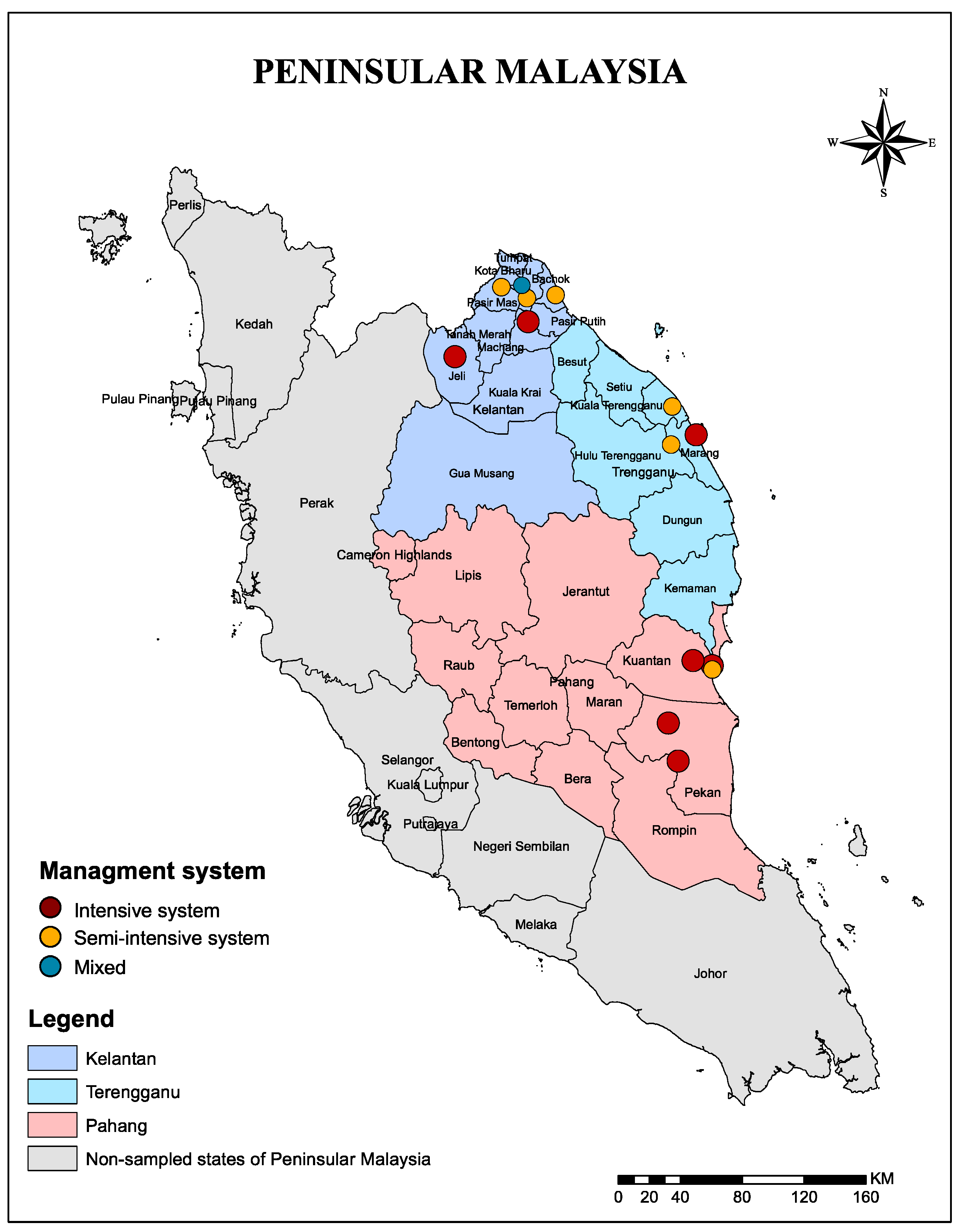
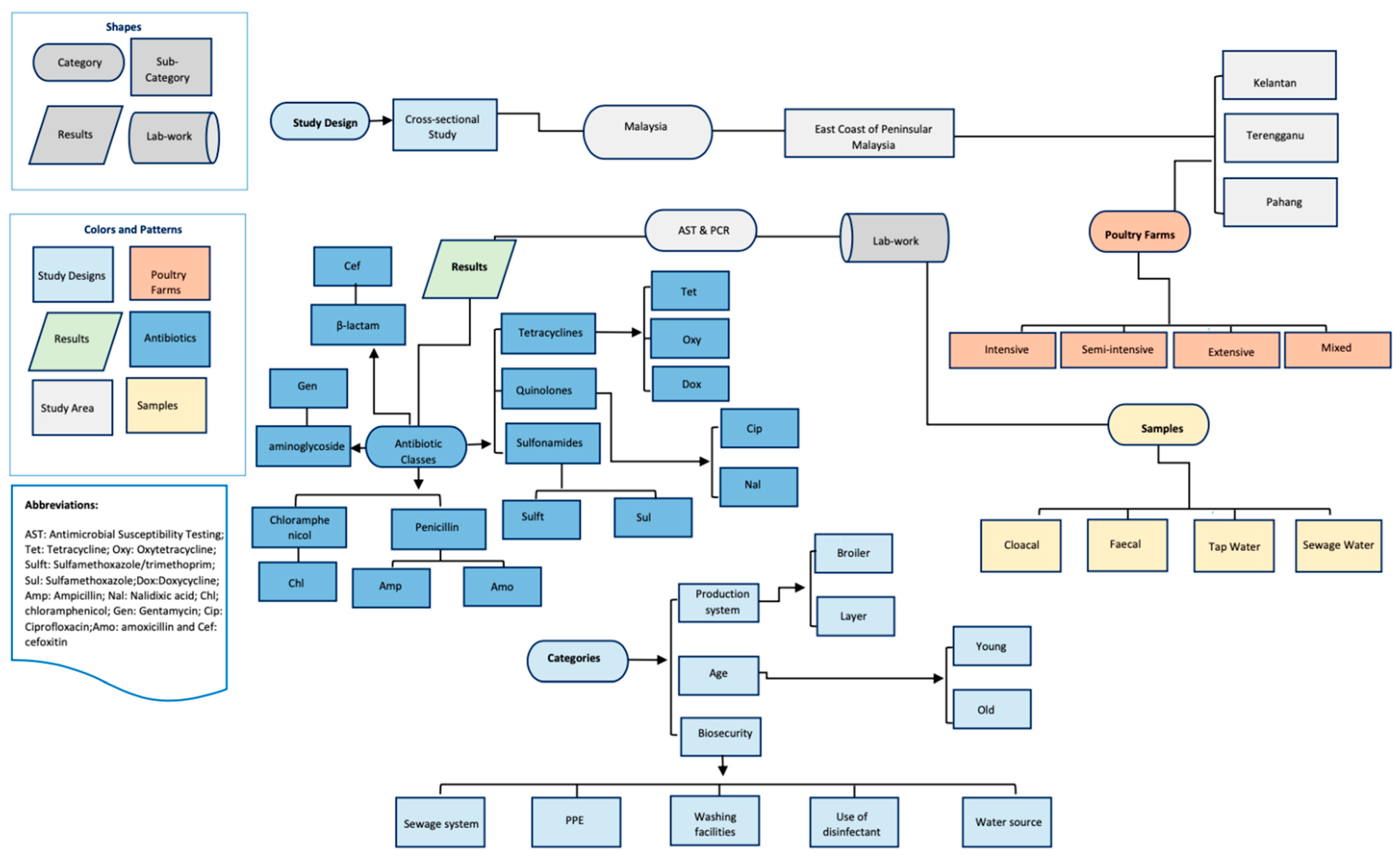
4.1. Study Area

The study was carried out between 8 February 2019 and 23 February 2020 in poultry farms located in three states of East coast peninsular Malaysia: Kelantan, Terengganu, and Pahang (Figure 4). These three states border the South China Sea and are dominated by a tropical climate which is characterized by humidity. There is a heavy monsoon season from November to March every year. The average temperature ranges from 21 to 32 °C. Average yearly rainfall falls is from 2032 mm to 2540 mm, with the wettest months being from November through January.

4.2. Study Design, Definitions, and Data Sources

We conducted a cross sectional survey targeting poultry farms in three states of east coast peninsular Malaysia that include Kelantan, Terengganu, and Pahang. A total of 371 samples (cloacal swabs = 259; faecal = 84; Sewage = 14, tap water = 14) were randomly collected. Farm characteristics including management, biosecurity, and disease history were collected using a semi-structured questionnaire. As such, 31 farmers that met strict inclusion criteria of keeping poultry farms and who responded to written consent were included in the analyses. Data pertaining to potential risk factors including management, biosecurity, and disease history were collected using semi-structured questionnaires. Similarly, antimicrobial usage data was obtained using a count-based approach, representing the use (yes/no) of an antimicrobial at the time of visit. Furthermore, sources from where antimicrobials and feed along with the source of water and the nature of their current sewage systems were collected (Table S1).
Regarding the management system, flock size, and sewage system, the following definitions and criteria were used:
  • Intensive management system is defined as mainly concentrated and often mechanized operations that use controlled-environment systems to provide the ideal thermal environment for the poultry.
  • Semi-intensive system is that which relies on natural airflow though the shed for ventilation.
  • Extensive system is mainly pasture-based and land-based where birds in the household flock are typically housed overnight in the shelter and are let out in the morning to forage during the day.
  • The criteria of the farm size included large-scale commercial farms that has more than ≥10,000 birds, medium-scale commercial farms that has more 5000–10,000, and small-scale farms where birds are often kept in single-age groups of >1000.
  • A poor sewage system is defined as that which retains high volumes of wastewater with low flow rate, blackish appearance, and sewage smell odour as a result of composing agricultural waste—probably as leakage from nearby irrigated effluent which is used for agricultural land application along with the presence of food waste, green waste, plastic, and heavy materials.
  • A good sewage system is that which has good drainage with no agricultural waste and relatively low heavy materials.
  • Excellent swage system is that which has significant drainage, no agriculture, and heavy materials.
Briefly, the cloacal samples were collected using sterile transport media; faecal samples using sterile containers and water samples using sterile water bottles and kept in a cooling box containing ice bags maintaining low temperature at (4°) and transferred to the lab within 24 h. All samples were collected according to standard operating procedures and good laboratory practices. A detailed study design is summarized in Figure 5.

4.3. Microbiological Testing

All cloacal swabs and fresh faecal samples were placed in Amies transport media, and transported on ice to the molecular biology laboratory, Universiti Malaysia Kelantan (UMK). Sewage tap water and surface water samples were transported in conical tubes, all on ice. Samples were enriched in buffered peptone water for 24 h and then plated onto eosin methylene blue agar (EMBA) and incubated for 24 h at 37 °C. Subsequently, five colonies were selected and sub-cultured on EMBA, before being further sub-cultured on Müller-Hinton agar and stored at −20 °C in cryovials. A single colony was picked at random from the plate for each original sample and biochemical tests including triple sugar iron agar, Simmon’s citrate agar, and motility-indole-lysine media were used for presumptive identification of E. coli isolates. All isolates were revived and inoculated onto Müller-Hinton plates before antimicrobial susceptibility testing.

4.4. Antimicrobial Susceptibility Testing

Isolates were tested for susceptibility against a panel of 12 antimicrobial agents perceived to be used frequently in both human and veterinary medicine in Malaysia. These antimicrobials included ampicillin (10 µg), amoxicillin-clavulanic acid (20/10 µg), chloramphenicol (30 µg), gentamicin (10 µg), tetracycline (30 µg), Oxytetracycline (30µg), doxycycline (30µg), trimethoprim-sulfamethoxazole (25 µg), nalidixic acid (30 µg), ciprofloxacin (5 µg), cefoxitin (30µg), and sulfonamides (300 µg) using the disc diffusion method (DDM) according to the Clinical and Laboratory Standards Institute guidelines [31]. Clinical and Laboratory Standards Institute guidelines were also used to determine as breakpoints for classifying isolates as sensitive, intermediate, or resistant to the drug [31]. Multidrug-resistant E. coli was defined as “non-susceptibility to at least one agent in three or more antimicrobial classes.” An antibiogram was defined as the combination of antimicrobials to which an isolate was resistant, and thus antibiogram length was defined as the number of antimicrobials to which an isolate was phenotypically resistance.

4.5. PCR

4.5.1. DNA Extraction of Escherichia Coli Isolates

E. coli isolates were sub-cultured overnight in Luria-Bertani broth and genomic DNA was extracted using a Presto™ Mini gDNA Bacteria Kit according to the manufacturer’s instructions.

4.5.2. Primers and PCR Assay for Specific Genes

The incidence of genes related to resistance to gentamicin (aac (3)-IV), tetracyclines (tet (A) and tet (B)), chloramphenicol (catA1 and cmlA), and sulfonamides (sul1) was determined by PCR. The set of primers used for each gene is shown in Table 6. PCR reactions were performed in a total volume of 25 mL using GoTaq1 Green Master Mix (Promega, USA), including 12.5 mL of GoTaq1 Green Master Mix, 1 mL of forward primer, 1 mL of reverse primers, 5.5 mL of nuclease-free water, and 5 mL of extracted DNA. Amplification reactions were carried out using a DNA thermocycler (Fisher Scientific UK, Loughborough, UK). PCR amplification was performed in duplicate. Amplified samples were analysed by electrophoresis in 1.5% agarose gel and were stained with ethidium bromide.

4.6. Statistical Analysis

Data were entered into Microsoft Excel spreadsheet and imported into SPSS version 25 and the R software (version 3.6.1) for statistical analysis. The data were sorted and checked for consistency and duplication. Data visualization were done in ArcGIS v. 10 (esri Inc., Redlands, CA, USA). The data focused on sets of variables that have been previously proposed or identified as risk factors for antimicrobial resistance [32,33]. Briefly, we have classified resistance as no resistance to antimicrobials detected in isolates and categorized the antimicrobials into their classes then identified which isolates were resistant to one or more specific classes. Classes of antimicrobials included tetracyclines, penicillins, aminoglycosides, quinolones, sulfonamides, third generation cephalosporins, and chloramphenicol. Prevalence of resistance of E.coli to a panel of 12 antimicrobials was also compared between four epidemiological samples that include cloaca, faecal, tap water, and sewage samples. Descriptive statistics for frequency of association between AMR and potential risk factors was performed. Selection of variables for inclusion in a logistic regression model were based on prior hypotheses and variables which were suggestive of an important effect from the descriptive analysis.

Supplementary Materials

The following are available online at https://0-www-mdpi-com.brum.beds.ac.uk/2079-6382/10/2/117/s1, Table S1: Characteristics of 31 farmers in Kelantan, Terengganu, and Pahang states, Malaysia.

Author Contributions

Conceptualization, A.Y.O. and I.O.; data curation, S.A.E. and D.S.; formal analysis, S.A.E., D.S. and A.Y.O.; funding acquisition, A.Y.O. and I.O.; investigation, S.A.E.; methodology, S.A.E. and D.S.; project administration, A.Y.O.; resources, I.O.; supervision, M.A.K., R.K., A.Y.O. and I.O.; validation, D.S. and A.Y.O.; visualization, A.Y.O.; writing—original draft, A.Y.O.; writing—review and editing, L.E., N.H., M.M.A.H., Y.A.S. and A.Y.O. All authors have read and agreed to the published version of the manuscript.

Funding

This research received research fund from Research Management Centre, Universiti Malaysia Kelantan (R/SGJP/A06.00/01625A/001/2018/000459/). David Simons, Linzy Elton, Najmul Haider, Muzamil Mahdi Abdel Hamid, Richard Kock and Abdinasir Yusuf Osman are members of the Pan African Network for Rapid Research, Response, and Preparedness for Infectious Diseases Epidemics consortium (PANDORA-ID-NET)—supported by the European and Developing Countries Clinical Trials Partnership (EDCTP2) programme (RIA2016E-1609. The funders had no role in the study design, data collection, analysis, interpretation or writing of the manuscript.

Institutional Review Board Statement

“This study was approved by Institutional Research Ethics Committee of the Faculty of Veterinary Medicine, University Malaysia Kelantan (UMK) (Ref: 12/2018).

Informed Consent Statement

Informed consent was obtained from all subjects involved in the study.

Data Availability Statement

The data for the present study will be available upon reasonable request.

Acknowledgments

David Simons, Linzy Elton, Najmul Haider, Muzamil Mahdi Abdel Hamid, Richard Kock and Abdinasir Yusuf Osman are members of the Pan African Network for Rapid Research, Response, and Preparedness for Infectious Diseases Epidemics consortium (PANDORA-ID-NET)—supported by the European and Developing Countries Clinical Trials Partnership (EDCTP2) programme (RIA2016E-1609). We acknowledge the LC-MS/MS Laboratory, Monash University, Malaysia and the support of the Faculty of Veterinary Medicine, Universiti Malaysia Kelantan with special reference to the field work assistance offered by Muhammad Luqman Nordin and Mohd Farhan Hanif. Abdinasir Yusuf Osman is partially supported by the IsDB, Saudi Arabia and the Centre for Global Public Health, Institute of Population Health Science, Queen Mary, University of London, UK.

Conflicts of Interest

The authors declare that they have no competing interests.

References

  1. Prestinaci, F.; Pezzotti, P.; Pantosti, A. Antimicrobial resistance: A global multifaceted phenomenon. Pathog. Glob. Health 2015, 109, 309–318. [Google Scholar] [CrossRef] [PubMed] [Green Version]
  2. Holmes, A.H.; Moore, L.S.; Sundsfjord, A.; Steinbakk, M.; Regmi, S.; Karkey, A.; Guerin, P.J.; Piddock, L.J. Understanding the mechanisms and drivers of antimicrobial resistance. Lancet 2016, 387, 176–187. [Google Scholar] [CrossRef]
  3. Winckler, C.; Grafe, A. Use of veterinary drugs in intensive animal production. J. Soils Sediments 2001, 1, 66. [Google Scholar] [CrossRef]
  4. Sun, P.; Barmaz, D.; Cabrera, M.L.; Pavlostathis, S.G.; Huang, C.-H. Detection and quantification of ionophore antibiotics in runoff, soil and poultry litter. J. Chromatogr. A 2013, 1312, 10–17. [Google Scholar] [CrossRef] [PubMed]
  5. Kay, P.; Blackwell, P.A.; Boxall, A.B. Transport of veterinary antibiotics in overland flow following the application of slurry to arable land. Chemosphere 2005, 59, 951–959. [Google Scholar] [CrossRef] [PubMed]
  6. Phillips, I. Withdrawal of growth-promoting antibiotics in Europe and its effects in relation to human health. Int. J. Antimicrob. Agents 2007, 30, 101–107. [Google Scholar] [CrossRef]
  7. Akbar, A.; Sitara, U.; Ali, I.; Khan, M.I.; Phadungchob, T.; Anal, A.K. Presence of Escherichia coli in poultry meat: A potential food safety threat. Int. Food Res. J. 2014, 21, 941. [Google Scholar]
  8. Van, T.T.H.; Chin, J.; Chapman, T.; Tran, L.T.; Coloe, P.J. Safety of raw meat and shellfish in Vietnam: An analysis of Escherichia coli isolations for antibiotic resistance and virulence genes. Int. J. Food Microbiol. 2008, 124, 217–223. [Google Scholar] [CrossRef]
  9. Acar, J.; Moulin, G. Antimicrobial resistance at farm level. Rev. Sci. Tech. 2006, 25, 775–792. [Google Scholar] [CrossRef]
  10. Sarkar, P.; Gould, I.M. Antimicrobial agents are societal drugs. Drugs 2006, 66, 893–901. [Google Scholar] [CrossRef]
  11. Hammerum, A.M.; Heuer, O.E.; Emborg, H.-D.; Bagger-Skjøt, L.; Jensen, V.F.; Rogues, A.-M.; Skov, R.L.; Agersø, Y.; Brandt, C.T.; Seyfarth, A.M. Danish integrated antimicrobial resistance monitoring and research program. Emerg. Infect. Dis. 2007, 13, 1633. [Google Scholar] [CrossRef] [PubMed]
  12. Mathew, A.G.; Cissell, R.; Liamthong, S. Antibiotic resistance in bacteria associated with food animals: A United States perspective of livestock production. Foodborne Pathog. Dis. 2007, 4, 115–133. [Google Scholar] [CrossRef] [PubMed] [Green Version]
  13. Prescott, J.F. Antimicrobial use in food and companion animals. Anim. Health Res. Rev. 2008, 9, 127. [Google Scholar] [CrossRef] [PubMed]
  14. Donado-Godoy, P.; Gardner, I.; Byrne, B.A.; Leon, M.; Perez-Gutierrez, E.; Ovalle, M.; Tafur, M.; Miller, W. Prevalence, risk factors, and antimicrobial resistance profiles of Salmonella from commercial broiler farms in two important poultry-producing regions of Colombia. J. Food Prot. 2012, 75, 874–883. [Google Scholar] [CrossRef]
  15. Planta, M.B. The role of poverty in antimicrobial resistance. J. Am. Board Fam. Med. 2007, 20, 533–539. [Google Scholar] [CrossRef]
  16. Abo-Amer, A.E.; Shobrak, M.Y.; Altalhi, A.D. Isolation and antimicrobial resistance of Escherichia coli isolated from farm chickens in Taif, Saudi Arabia. J. Glob. Antimicrob. Resist. 2018, 15, 65–68. [Google Scholar] [CrossRef]
  17. Messele, Y.E.; Abdi, R.D.; Yalew, S.T.; Tegegne, D.T.; Emeru, B.A.; Werid, G.M. Molecular determination of antimicrobial resistance in Escherichia coli isolated from raw meat in Addis Ababa and Bishoftu, Ethiopia. Ann. Clin. Microbiol. Antimicrob. 2017, 16, 55. [Google Scholar] [CrossRef] [Green Version]
  18. Torkan, S.; Khamesipour, F.; Anyanwu, M. Detection of virulence and antibacterial resistance genes in Salmonella isolates from diarrhoeic dogs in Iran. Rev. Méd. Vét. 2015, 166, 221–228. [Google Scholar]
  19. Aliyu, A.; Saleha, A.; Jalila, A.; Zunita, Z. Risk factors and spatial distribution of extended spectrum β-lactamase-producing-Escherichia coli at retail poultry meat markets in Malaysia: A cross-sectional study. BMC Public Health 2016, 16, 699. [Google Scholar] [CrossRef] [Green Version]
  20. Imtiaz, J.; Hashmi, I.; Saeed, A.; Qazi, I.; Arshad, M. Development of PCR Protocol for Detection of Escherichia Coli in Drinking Water. In Water Resources Management VII; Brebbia, C.A., Ed.; WIT PRESS: Southhampton, UK, 2013; pp. 225–232. [Google Scholar]
  21. Tang, K.L.; Caffrey, N.P.; Nóbrega, D.B.; Cork, S.C.; Ronksley, P.E.; Barkema, H.W.; Polachek, A.J.; Ganshorn, H.; Sharma, N.; Kellner, J.D. Restricting the use of antibiotics in food-producing animals and its associations with antibiotic resistance in food-producing animals and human beings: A systematic review and meta-analysis. Lancet Planet. Health 2017, 1, e316–e327. [Google Scholar] [CrossRef]
  22. Usui, M.; Ozawa, S.; Onozato, H.; Kuge, R.; Obata, Y.; Uemae, T.; Ngoc, P.T.; Heriyanto, A.; Chalemchaikit, T.; Makita, K. Antimicrobial susceptibility of indicator bacteria isolated from chickens in Southeast Asian countries (Vietnam, Indonesia and Thailand). J. Vet. Med. Sci. 2014, 76. [Google Scholar] [CrossRef] [PubMed] [Green Version]
  23. Nguyen, V.T.; Carrique-Mas, J.J.; Ngo, T.H.; Ho, H.M.; Ha, T.T.; Campbell, J.I.; Nguyen, T.N.; Hoang, N.N.; Pham, V.M.; Wagenaar, J.A. Prevalence and risk factors for carriage of antimicrobial-resistant Escherichia coli on household and small-scale chicken farms in the Mekong Delta of Vietnam. J. Antimicrob. Chemother. 2015, 70, 2144–2152. [Google Scholar] [PubMed] [Green Version]
  24. Shahaza, O.; Zakirah, S.; Muhammad, A.; Syamsyul, A.; Maswati, M. Antimicrobial resistance in veterinary clinical isolates of Escherichia coli from northern region of Peninsular Malaysia. Malays. J. Vet. Res. 2017, 8, 1–7. [Google Scholar]
  25. Cheong, Y.; Lim, V.; Jegathesan, M.; Suleiman, A. Antimicrobial resistance in 6 Malaysian general hospitals. Med. J. Malays. 1994, 49, 317–326. [Google Scholar]
  26. Aarestrup, F.M.; Woolhouse, M.E. Using sewage for surveillance of antimicrobial resistance. Science 2020, 367, 630–632. [Google Scholar] [CrossRef] [PubMed]
  27. Dangour, A.D.; Watson, L.; Cumming, O.; Boisson, S.; Che, Y.; Velleman, Y.; Cavill, S.; Allen, E.; Uauy, R. Interventions to improve water quality and supply, sanitation and hygiene practices, and their effects on the nutritional status of children. Cochrane Database Syst. Rev. 2013, 1. [Google Scholar] [CrossRef] [Green Version]
  28. Chuah, L.-O.; He, X.B.; Effarizah, M.E.; Syahariza, Z.A.; Shamila-Syuhada, A.K.; Rusul, G. Mislabelling of beef and poultry products sold in Malaysia. Food Control 2016, 62, 157–164. [Google Scholar] [CrossRef]
  29. Bong, C.W.; Chai, S.K.; Chai, L.C.; Wang, A.J.; Lee, C.W. Prevalence and characterization of Escherichia coli in the Kelantan River and its adjacent coastal waters. Water Supply 2020, 20, 930–942. [Google Scholar] [CrossRef]
  30. Alhaj, N.; Mariana, N.; Raha, A.; Ishak, Z. Prevalence of antibiotic resistance among Escherichia coli from different sources in Malaysia. Int. J. Poult. Sci. 2007, 6, 293–297. [Google Scholar] [CrossRef] [Green Version]
  31. Clinical and Laboratory Standards Institute. Performance Standards for Antimicrobial Susceptibility Testing, 26th ed; CLSI Supplement M100; CLSI: Wayne, PA, USA, 2016. [Google Scholar]
  32. Murphy, C.P.; Carson, C.; Smith, B.A.; Chapman, B.; Marrotte, J.; McCann, M.; Primeau, C.; Sharma, P.; Parmley, E.J. Factors potentially linked with the occurrence of antimicrobial resistance in selected bacteria from cattle, chickens and pigs: A scoping review of publications for use in modelling of antimicrobial resistance (IAM. AMR Project). Zoonoses Public Health 2018, 65, 957–971. [Google Scholar] [CrossRef]
  33. Ibrahim, R.A.; Cryer, T.L.; Lafi, S.Q.; Basha, E.-A.; Good, L.; Tarazi, Y.H. Identification of Escherichia coli from broiler chickens in Jordan, their antimicrobial resistance, gene characterization and the associated risk factors. BMC Vet. Res. 2019, 15, 159. [Google Scholar] [CrossRef] [PubMed]
Figure 1. Prevalence of antimicrobial-resistant Escherichia coli isolated from poultry farms collected from Kelantan, Terengganu, and Pahang poultry operations. Data are the number of samples (n = 371). Tet: Tetracycline; Oxy: Oxytetracycline; Sulft: Sulfamethoxazole/trimethoprim; Sul: Sulfamethoxazole; Dox: Doxycycline; Amp: Ampicillin; Nal: Nalidixic acid; Chl: chloramphenicol; Gen: Gentamycin; Cip: Ciprofloxacin; Amo: amoxicillin and Cef: cefoxitin.
Antibiotics 10 00117 g001
Figure 2. Prevalence of antimicrobial-resistant Escherichia coli isolated from poultry farms collected from Kelantan, Terengganu, and Pahang poultry operations. Data are the number of samples (n = 371).
Antibiotics 10 00117 g002
Figure 3. Percentage of antimicrobial-resistant Escherichia coli isolated from four epidemiological samples that include cloacal, faecal, tap water, and sewage collected from poultry farms. Data are the number of poultry samples (n = 371) in three states of east coast peninsular Malaysia. Tet: Tetracycline; Oxy: Oxytetracycline; Sulft: Sulfamethoxazole/trimethoprim; Sul: Sulfamethoxazole; Dox: Doxycycline; Amp: Ampicillin; Nal: Nalidixic acid; Chl: chloramphenicol; Gen: Gentamycin; Cip: Ciprofloxacin; Amo: amoxicillin and Cef: cefoxitin.
Antibiotics 10 00117 g003
Figure 4. A map showing location of the sampled states and exact location 14 poultry farms sampled and their management systems in Kelantan, Terengganu, and Pahang of east cast peninsular Malaysia. The map was created using ArcGIS v. 10 (esri Inc., Redlands, CA, USA).
Antibiotics 10 00117 g004
Figure 5. Diagrammatic representation of the study design.
Antibiotics 10 00117 g005
Table 1. Summary of risk factors of E.coli among poultry farms in the Kelantan, Terengganu and Pahang, Malaysia (n = 371) by using chi-square test.
Risk FactorsSamples TestedAffected (%)p-Value
Age 0.511
Young187123 (65.8%)
Adult184115 (62.5%)
Management system 0.541
Intensive187115 (61.5%)
Semi-intensive158105 (66.5%)
Mixed2618 (69.2%)
Production system 0.278
Broiler 212129 (60.8%)
Layer5335 (66%)
Mixed10674 (69.8%)
State 0.012
Kelantan158115 (72.8%)
Terengganu8046 (57.5%)
Pahang13377 (57.9%)
Districts 0.001
Bachok 5236 (69.2%)
Kota Bharu2618 (69.2%)
Machang2824 (85.7%)
Pasir Mas2614 (53.8%)
Jeli2623 (88.5%)
Kuantan7949 (62%)
Pekan5428 (51.9%)
kuala terengganu2620 (76.9%)
Marang5426 (48.1%)
Sample source 0.001
Cloaca swab259172 (66.4%)
Faecal Sample8458 (69%)
Sewage145 (35.7%)
Tape Water143 (21.4%)
Farm size 0.013
Small10477 (74%)
Medium188119 (63.2%)
Large7942 (53.2%)
Origin of the poultry 0.005
Local 2618 (69.2%)
Imported13371 (53.4%)
Both212149 (70.3%)
Table 2. Summary of prevalence of resistance to at least one antimicrobial and their associated risk factors.
Risk FactorsNo Antimicrobial Resistance
n = 137
Resistance to at least One Antimicrobial
n = 234
p-Value
Age 0.44
Young65 (47.4%)122 (52.1%)
Adult72 (52.6%)112 (47.9%)
Origin of the poultry 0.01
Local10 (7.3%)16 (6.8%)
Imported63 (46%)70 (29.9%)
Both64 (46.7%)148 (63.2%)
Management system 0.18
Intensive80 (58.4%)115 (49.1%)
Semi-intensive47 (34.3%)103 (44%)
Mixed10 (7.3%)16 (6.8%)
Production system 0.21
Broiler86 (62.8%)125 (53.4%)
Layer18 (13.1%)37 (15.8%)
Mixed33 (24.1%)72 (30.8%)
Farm size 0.02
Small29 (21.2%)75 (32.1%)
Medium70 (51.1%)117 (50%)
Large38 (27.7%)42 (17.9%)
Source of sample <0.001
Cloacal swab89 (65%)170 (72.6%)
Faecal sample26 (19%)58 (24.8%)
sewage9 (6.6%)5 (2.1%)
Tap water13 (9.5%)1 (0.4%)
Water source 0.02
Surface water37 (27%)69 (29.5%)
Bond water61 (44.5%)72 (30.8%)
Pump water39 (28.5%)93 (39.7%)
Sewage system 0.60
Excellent38 (27.7%)71 (30.3%)
Good82 (59.9%)128 (54.7%)
Poor17 (12.4%)35 (15%)
Feed source 0.53
Endogenous50 (36.5%)82 (35%)
Exogenous75 (54.7%)138 (59%)
Other12 (8.8%)14 (6%)
Table 3. Summary of univariate analysis of poultry samples for antimicrobial-resistant E. coli from poultry farms in east coast of Malaysia (n = 371 samples).
AntimicrobialsCloacal
n = 259
Faecal
n = 84
Sewage
n = 14
Tape Water
n = 14
p-Value
No identified resistance <0.001
No antimicrobial resistance89 (34.4%)26 (31%)9 (64.3%)13 (92.9%)
Resistance to at least one antimicrobial170 (65.6%)58 (69%)5 (35.7%)1 (7.1%)
Antimicrobial class resistance 0.003
No antimicrobial resistance89 (34.4%)26 (31%)9 (64.3%)13 (92.9%)
Resistant to 1 class4 (1.5%)1 (1.2%)0 (0%)0 (0%)
Resistant to 2 classes13 (5%)3 (3.6%)2 (14.3%)0 (0%)
Resistant to 3 classes46 (17.8%)19 (22.6%)2 (14.3%)1 (7.1%)
Resistant to 4 classes74 (28.6%)19 (22.6%)1 (7.1%)0 (0%)
Resistant to 5 or more classes33 (12.7%)16 (19%)0 (0%)0 (0%)
Source of antimicrobials 1
Drug supplier112 (43.2%)36 (42.9%)6 (42.9%)6 (42.9%)
Feed store147 (56.8%)48 (57.1%)8 (57.1%)8 (57.1%)
Tetracyclines <0.001
Not resistant93 (35.9%)26 (31%)9 (64.3%)13 (92.9%)
Resistant166 (64.1%)58 (69%)5 (35.7%)1 (7.1%)
Penicillins 0.048
Not resistant151 (58.3%)47 (56%)10 (71.4%)13 (92.9%)
Resistant108 (41.7%)37 (44%)4 (28.6%)1 (7.1%)
Aminoglycosides 0.246
Not resistant219 (84.6%)68 (81%)13 (92.9%)14 (100%)
Resistant40 (15.4%)16 (19%)1 (7.1%)0 (0%)
Quinolones 0.002
Not resistant142 (54.8%)49 (58.3%)13 (92.9%)13 (92.9%)
Resistant117 (45.2%)35 (41.7%)1 (7.1%)1 (7.1%)
Sulfonamides <0.001
Not resistant104 (40.2%)29 (34.5%)11 (78.6%)14 (100%)
Resistant155 (59.8%)55 (65.5%)3 (21.4%)0 (0%)
Cephelosporins 0.645
Not resistant246 (95%)79 (94%)14 (100%)14 (100%)
Resistant13 (5%)5 (6%)0 (0%)0 (0%)
Other classes 0.025
Not resistant224 (86.5%)65 (77.4%)14 (100%)14 (100%)
Resistant35 (13.5%)19 (22.6%)0 (0%)0 (0%)
Table 4. Univariate regression analysis of risk factors for antimicrobial-resistant E. coli from poultry farms in east coast of Malaysia (n = 238 samples).
VariablesOR2.5%97.5%Pr (>|z|)
Farms
Farm 110.202.9542.89<0.001***
Farm 22.720.918.530.07.
Farm 313.033.4665.56<0.001***
Farm 41.980.666.090.22
Farm 52.310.787.180.134
Farm 67.142.1627.160.002**
Farm 72.890.979.020.05.
Farm 8RefRefRefRef
Farm 94.611.4815.630.01*
Farm 103.821.2512.520.02*
Farm 111.160.383.530.78
Farm 121.060.343.250.91
Farm 135.661.7820.130.004**
Farm 142.260.776.870.13
Sample source
Cloaca swab24.834.82454.740.002**
Faecal sample29.05.35540.730.001**
Sewage7.220.95151.210.09.
Tap waterRefRefRefRef
Age
Young1.210.791.840.38
AdultRefRefRefRef
Poultry origin
Local1.440.613.500.41
Both2.081.323.260.001**
ImportedRefRefRefRef
Management system
Semi-intensive1.520.972.390.06.
Mixed1.110.482.650.80
IntensiveRefRefRefRef
Production system
Layer1.410.762.690.27
BroilerRefRefRef0.11
Mixed1.500.912.48Ref
Farm size
Small2.331.274.350.001**
Medium1.510.882.570.125
LargeRefRefRefRef
Water source
Surface water1.570.932.680.08.
Pump water2.021.223.360.01**
Bond water
Sewage system
Excellent0.910.441.810.786
Good0.750.391.420.398
PoorRefRefRefRef
Signif. codes: 0 ‘***’ 0.001 ‘**’ 0.01 ‘*’ 0.05 ‘.’ 0.1 ‘ ‘ 1.
Table 5. Multivariate regression analysis of risk factors for antimicrobial-resistant E. coli from poultry farms in east coast of Malaysia.
OR2.5%97.5%Pr (>|z|)
Cloaca swab26.505.08487.690.001**
Feacal sample30.925.63579.630.001**
Sewage7.430.96156.870.09.
Farm size Small2.501.334.770.004**
Farm size medium1.550.892.670.114
Signif. codes: 0 ‘***’ 0.001 ‘**’ 0.01 ‘*’ 0.05 ‘.’ 0.1 ‘ ‘ 1.
Table 6. Comparison for the detection of resistance genes from samples using PCR.
Antimicrobial Class/AgentResistance Gene% IsolatesTotal # Tested
Gentamicinaac(3)-IV12 (85.7%)14
Tetracyclinestet(A), tet(B)9 (64.2%)14
ChloramphenicolcatA12 (14.2%)14
Sulfonamidessul114 (100%)14
β-LactamsblaSHV6 (42.8%)14
TrimethoprimdhfrI4 (28.5%)14
Table 7. The set of primers used for each gene.
GenesPrimer Sequence(5′ to 3′)PCR ConditionProduct SizeReferences
β-LactamsF- CTATCGCCAGCAGGATCTGG
R- ATTTGCTGATTTCGCTCGGC
3 min at 95 °C; 35 cycles of 1 min at 94 °C, 90 s at 55 °C and 1 min at 72 °C; 10 min at 72 °C543[16]
Gentamicin aac(3)-IVF-CTTCAGGATGGCAAGTTGGT
R-TCATCTCGTTCTCCGCTCAT
3 min at 95 °C; 35 cycles of 1 min at 94 °C, 90 s at 55 °C and 1 min at 72 °C; 10 min at 72 °C286[17]
Sulfonamide sul1F- ACTGCAGGCTGGTGGTTATG
R- ACCGAGACCAATAGCGGAAG
3 min at 95 C; 35 cycles of 1 min at 94 C, 90 s at 55 °C and 1 min at 72 °C; 10 min at 72 °C271[8]
Tetracycline tet(A)F-CCTCAATTTCCTGACGGGCT
R-GGCAGAGCAGGGAAAGGAAT
3 min at 95 °C; 35 cycles of 1 min at 94 C, 90 s at 55 °C and 1 min at 72 °C; 10 min at 72 °C712[18]
Tetracycline tet(B)F-ACCACCTCAGCTTCTCAACG
R-GTAAAGCGATCCCACCACCA
3 min at 95 °C; 35 cycles of 1 min at 94 C, 90 s at 55 °C and 1 min at 72 °C; 10 min at 72 °C586[18]
Chloramphenicol catA1F- GAAAGACGGTGAGCTGGTGA
R- TAGCACCAGGCGTTTAAGGG
3 min at 95 °C; 35 cycles of 1 min at 94 °C, 90 s at 55 °C and 1 min at 72 °C; 10 min at 72 °C473[8]
Trimethoprim dhfrIF-AAGAATGGAGTTATCGGGAATG
R-GGGTAAAAACTGGCCTAAAATTG
15 min at 95 °C; 30 cycles of 30 s at 94 °C; 30 s at 58 °C; 1 min at 72 °C; 10 min 72 °C.391[8]
Ampicillin
CITM
F-TGGCCAGAACTGACAGGCAAA
R-TTTCTCCTGAACGTGGCTGGC
15 min at 95 °C; 30 cycles of 30 s at 94 °C; 30 s at 58 °C; 1 min at 72 °C; 10 min 72 °C.462[8]
E.coliF-TGACGTTACCCGCAGAAGAA
R- CTCCAATCCGGACTACGACG
3 min at 95 °C; 35 cycles of 15s at 95 °C, 90 s at 55 °C and 15s at 72 °C; 10 min at 72 °C832[19]
O157F-GTGTCCATTTATACGGACATCCATG
R-CCTATAACGTCATGCCAATATTGCC
2 min at 94 °C; 35 cycles of 30s at 94 °C, 30 s at 55 °C and 30s at 72 °C; 5 min at 72 °C292 [20]
Publisher’s Note: MDPI stays neutral with regard to jurisdictional claims in published maps and institutional affiliations.

