Article

The Prevalence and Clinical Significance of Anaerobic Bacteria in Major Liver Resection

Department of General, Visceral and Transplantation Surgery, University Hospital of Tuebingen, 72076 Tuebingen, Germany
*
Author to whom correspondence should be addressed.
Submission received: 6 January 2021 / Revised: 27 January 2021 / Accepted: 28 January 2021 / Published: 31 January 2021

Abstract

:
(1) Background: Anaerobic infections in hepatobiliary surgery have rarely been addressed. Whereas infectious complications during the perioperative phase of liver resections are common, there are very limited data on the prevalence and clinical role of anaerobes in this context. Given the risk of contaminated bile in liver resections, the goal of our study was to investigate the prevalence and outcome of anaerobic infections in major hepatectomies. (2) Methods: We retrospectively analyzed the charts of 245 consecutive major hepatectomies that were performed at the department of General, Visceral, and Transplantation Surgery of the University Hospital of Tuebingen between July 2017 and August 2020. All microbiological cultures were screened for the prevalence of anaerobic bacteria and the patients’ clinical characteristics and outcomes were evaluated. (3) Results: Of the 245 patients, 13 patients suffered from anaerobic infections. Seven had positive cultures from the biliary tract during the primary procedure, while six had positive culture results from samples obtained during the management of complications. Risk factors for anaerobic infections were preoperative biliary stenting (p = 0.002) and bile leaks (p = 0.009). All of these infections had to be treated by intervention and adjunct antibiotic treatment with broad spectrum antibiotics. (4) Conclusions: Anaerobic infections are rare in liver resections. Certain risk factors trigger the antibiotic coverage of anaerobes.

Graphical Abstract

1. Introduction

Liver resections are widely available procedures to cure benign and malignant diseases of the liver. After partial hepatectomy bile leaks and surgical site infections, complications are feared, since they drastically increase morbidity and mortality [1,2,3]. Liver resections are considered clean-contaminated procedures given the risk of preexisting bacterial and fungal colonization of the biliary system. The most common bacteria cultured from the bile duct and postoperative infectious complications are of gastrointestinal origin [4,5,6].
Anaerobic liver infections, however, are rare in hepatobiliary surgery. The most commonly described presentation of anaerobes in the liver is in the form of liver abscesses. These abscesses are frequently caused by acute or chronic abdominal inflammation or infection [7,8]. Recently, an emerging number of abscesses have been of iatrogenic origin, caused by percutaneous ablation of primary and secondary liver malignancies. Abscesses after ablation appear to develop in up to 2% of patients [9,10]. Anaerobic bacteria are common pathogens in the intestines. The liver and native biliary tract are generally uncommon habitats for anaerobic bacteria. However, they may be cultured from the biliary tract in up to 20% of patients in the presence of a biliary tract occlusion or stent [11,12]. Aside from biliary stents, bilioenteric anastomoses are considered a risk factor for anaerobic colonization and infection of the biliary tree (and liver). Therefore, the 2018 Tokyo Guidelines (TG2018) recommend covering anaerobic bacteria when treating cholangitis in the presence of bilioenteric anastomosis [13].
The department of General, Visceral, and Transplantation Surgery of the Tuebingen University Hospital is a Tertiary Care Academic Teaching facility that performs an average of ~200 liver resections as well as ~50 liver transplantations per year. Our center is specialized on complex liver resections with a clinical and scientific focus on biliary malignancies. [14,15]. A major hepatectomy/liver resection is defined as the removal of ≥3 liver segments [16]. Due to biliary obstruction of perihilar malignancies, patients often present with jaundice or have undergone preoperative stenting and are thus at risk of biliary tract infections [12]. Infectious complications after surgery are still a major risk factor for morbidity and mortality after liver resection. Surgical site and organ-space infections present in up to 20% of major hepatectomies [1,17]. The role of anaerobic bacteria is yet to be assessed. To our knowledge, there are neither sufficient data on the presence of anaerobes in intraoperative microbiological specimen from liver resections nor from surgical site infection (SSI) after hepatectomy. Even less is known about the presence of anaerobic bacteria’s antibiotic resistance to commonly used antibiotics after liver surgery.
The goal of this study was to evaluate the prevalence and clinical role of anaerobic bacteria in patients undergoing major liver resection for benign and malignant disease.

2. Results

2.1. Clinical Characteristics

We analyzed the charts of all consecutive patients that had undergone major liver resection at the department of General, Visceral, and Transplantation Surgery of the Tuebingen University Hospital from June 2017 to August 2020. We included all adult (age ≥ 16 years) consecutive patients that underwent laparotomy and had at least three liver segments removed. Pediatric patients, as well as patients that had minor liver resections, were excluded. Based on the preoperative diagnosis and intraoperative findings, material was sent for microbiological testing at the attending surgeon’s discretion.
During the study period, 245 patients met the inclusion criteria. Of these 245 patients, 76 had intraoperative material sent for microbiological testing, of which 49 showed growth of pathogenic bacteria. Furthermore, 102 of the 245 patients had culture-proven microbiological growth during the first 30 postoperative days (this includes the 49 that had growth on the intraoperative cultures). From these 102 patients, we were able to identify 13 patients that had anaerobic infections. Of these 13 patients, 7 patients had positive anaerobic cultures from specimens that were collected during under the primary surgical procedure. The remaining six patients had positive anaerobic cultures during revision (n = 5) procedures or from specimen collected from drains (n = 1). The median age was 60 years (Standard Deviation (SD) ±15, range 34–80). Five patients were male (38%), and eight were female (62%). Most patients underwent liver resection for malignancy (n = 9), while the other patients were treated for helminthic disease (n = 2), suspected cholangiocarcinoma (n = 1), and recurrent cholangitis after cholecystectomy (n = 1). Of the 13 patients, 9 patients received prolonged perioperative antibiotics for suspected contaminated biliary tract (n = 3) or risk of post-hepatectomy liver failure (PHLF) (n = 6). These patients had anaerobic coverage with metronidazole (n = 6) or piperacillin/tazobactam (n = 2) or meropenem (n = 1). For details, see Table 1.

2.2. Clinical Comparison of Anaerobic Infections

We compared the patient group that suffered from anaerobic infections to the control group, which consisted of the remaining 232 patients. Gender distribution (38% male vs. 56% male; p 0.204 Χ2) was similar in both groups. The same was true for age (60 years standard deviation (SD) ± 15 vs. 64 years SD ± 13; p 0.373 MWU) and body mass index (BMI) (25.2 kg/m2 SD ± 4.7 vs. 24.6 kg/m2 SD ± 4.6; p 0.415 MWU). There were similar rates of malignant (69% vs. 88%; p 0.052 Χ2) and infectious (8% vs. 15 %; p 0.429 Χ2) underlying disease. In the anaerobic group, there were significantly higher rates of preoperative stents (54% vs. 27%; p 0.000 Χ2), bile duct resections and reconstructions (69% vs. 32%; p 0.006 Χ2), and bile leaks (54% vs. 18%; p 0.001 Χ2). Mean operating time was longer (p 0.035 MWU) in the anaerobic group (likely due to the higher rate of bile duct reconstructions), as well as the length of stay (p 0.002 MWU). The attending surgeon decided whether to activate postoperative preventive measures to preemptively treat post-hepatectomy liver failure in 78% of patients of the anaerobic group and 63% of the control group (p 0.390 Χ2). For details, see Table 2.

2.3. Microbiological Results

Of the 245 patients in this study, 102 had positive microbiological cultures throughout their hospital stay and 13 patients grew anaerobic bacteria. Of these 13 patients, 7 patients had cultures from intraoperatively collected bile specimen. The remaining 6 patients had positive cultures from Bilioma (n = 2), perihepatic abscess (n = 1), subcutaneous abscess (n = 2), and enteric anastomotic leak (n = 1). Overall, there were 14 different strains of anaerobic bacteria cultured from these patients, and 1 patient had anaerobic bacteri, that we were unable to identify. Prevotella species was the most commonly isolated anaerobic species (n = 5; 33%), followed by Bacteroides strains (n = 3; 20%) and Finegoldia magna (n = 3; 20%). The latter was only cultured from specimen collected from “complications,” i.e., one bilioma (Figure 1), one perihepatic, and one subcutaneous abscess. Except for the perihepatic abscess caused by Finegoldia magna, all specimen that grew anaerobes showed polymicrobial growth of anaerobic and (multiple) aerobic bacteria. For details, see Table 3.

2.4. Risk Factor Analysis for Anaerobic Infection

Anaerobic infections were present in 13 out of 245 patients (5%). We performed a stepwise backward binary logistic regression to find risk factors for anaerobic infections in liver resection. As potential predictors, we chose age, BMI, diabetes mellitus 2, preoperative biliary stent, resection and reconstruction of the bile ducts, postoperative bile leak, extent of surgical procedure, American Society of Anesthesiologist (ASA) score, Model of End-Stage Liver Disease (MELD) score, activation of the in-house post-hepatectomy liver failure protocol, malignant disease, and infectious reason for surgery. In the final model, preoperative biliary stent and postoperative bile leak were identified as predictors for anaerobic infections, Χ2 (3) = 17.017, p 0.001; Cox & Snell 0.072; R2 Nagelkerke R2 0.212. Preoperative stent had an Odds Ratio of 7.790 (CI 95% 2.151–28.221; p 0.002), while bile leak had an Odds Ratio of 5.426 (CI 95% 1.535–19.182; p 0.009).

2.5. Treatment of Anaerobic Infections

Of the 13 patients, 7 patients had positive biliary cultures from intraoperative specimens gathered from the bile duct. Six of these patients had a stent, and the remaining were suffering from repeated cholangitis after complicative cholecystectomy and injury of the right hepatic artery. All of these patients had preemptive perioperative coverage of anaerobic bacteria with metronidazole or piperacillin/tazobactam. All of these patients had their bile ducts and or infected segments of the liver resected. None of these patients had recurrent anaerobic infection during their stay.
In the other six patients, the most common sites of origin of anaerobic bacteria were the skin and gastrointestinal tract in 50% and 50% of patients, respectively. In order to resolve the infectious complications, surgical source control was achieved by evacuation of the infected tissues via incision, resection and drainage. None of these complications occurred. Overall, the outcomes were favorable. The overall mortality in the cohort was 6%. The mortality within the group of patients that suffered from anaerobic infections was 15%. However, none of the deaths was attributed to septicemia from anaerobic bacteria.

3. Discussion

The role of anaerobic bacteria in infections of the liver is unclear. The most commonly described pathologies are (septic) liver abscesses originating from the biliary tract, gastrointestinal tract, or endocarditis [7,8]. In 1996, Huang et al. reported that up to 25% of liver abscesses grew anaerobic bacteria [8]. The widespread use of percutaneous ablation of liver tumors has also led to an increase in liver abscesses caused iatrogenically [9,10], especially if the patient has undergone previous bilioenteric anastomosis [18,19].
Data on the prevalence of anaerobic bacteria and their role in complications after liver resections are scarce. Surgical site and organ space infections after liver resections pose an increased risk to the patient and significantly prolong hospital stay [20]. We hereby present the first study to focus on the prevalence of anaerobes in microbiological specimen collected during major liver resection and complications following these procedures. We present a series of 245 consecutive patients who underwent major liver resection at our tertiary hepatobiliary center. We were able to identify seven patients with anaerobic bacteria in the bile duct, as well as six patients that developed complications and subsequent anaerobic infections. Unsurprisingly, the most common site of origin of these anaerobes was in the gastrointestinal tract. Therefore, a breach of the natural barrier at the duodenal papilla appears to be a risk factor for anaerobic colonization of the biliary tract, similar to fungal colonization of the bile duct [12]. The number of anaerobic infections appears to increase the longer a stent is in place [12]. Even though a recent meta-analysis suggests that there are less infections with percutaneous drainage of the biliary tract compared to endoscopic drainage, the rates of the bacterial colonization were high in both groups [5,21]. Biliary drainage prior to surgery leads to an increased risk of postoperative infection [22,23]. In a cohort of 475 patients who had undergone preoperative biliary drainage, Sugawara et al. reported that nearly 75% of patients had contaminated bile. Of these patients, 28.6% developed postoperative infectious complications. Bacteriobilia is an independent risk factor of postoperative infectious complications. However, compared to the endoscopic internal drainage, Sugawara et al. did not report a single positive anaerobic culture [5,12].
Perioperative anti-infective treatment in liver surgery is usually done as a single dose prior to incision, with repeated doses based on the length of the procedure and excretion kinetics of the substance used [24]. There are little data that support prolonged anti-infective treatment unless patients are operated in the setting of an infection [24]. Our in-house protocol recommends perioperative antibiotics for a minimum of 3 days in patients who had undergone preoperative stenting or recurrent cholangitis or are at an increased risk of post-hepatectomy liver failure. We frequently cultured bacteria from the bile that were not susceptible to first- and second-generation cephalosporins and thus elected to use third-generation cephalosporins for liver and bile duct surgery. All patients undergoing biliary tract surgery receive perioperative third-generation cephalosporins with metronidazole (and fluconazole in case of a stent). External drainage is very uncommon in our region. Therefore, our perioperative regimen is based on the known local resistance patterns. Whereas there are sufficient data to not recommend prolonged antibiotics in elective and “sterile” liver surgery, there are less data on what to do in the setting of a contaminated bile duct. Sudo et al. found an increased rate of antibiotic resistance in patients that had undergone biliary drainage compared to those who had not. This may in part be due to preoperative antibiotic treatment and stent placement in the setting of cholangitis. Whereas there is a risk of bias, we support their suggestion to take into account microbiological culture results from specimens gathered from external bile drainage whenever available [25]. A Japanese randomized controlled trial confirmed that culture-based targeted therapy leads to less surgical site infections [26]. The most frequently reported bacterial species are E. coli and Enterococcus [4,5,23]. There were no reports on anaerobic growth in these trials.
We hypothesized that infected biliomas are frequently colonized by anaerobic bacteria. In our study, we were able to culture anaerobic bacteria from two biliomas with polymicrobial infection. The bacterial spectrum of biliomas after liver surgery is considered to be similar to that of cholangitis. A single retrospective analysis of 32 biliomas (most of them after liver surgery) reported the microbiological spectrum of these patients. The authors isolated 121 different bacterial strains from 32 biliomas. Only four of these were anaerobes (all Clostridium perfringens) [4]. A higher rate of up to 10% of biliomas and liver abscesses infected with anaerobes was reported in liver transplant recipients [27,28]. To our knowledge, there is no larger study available that has focused on microbiological data on bilioma after liver resection. Thus, the incidence of anaerobic bacteria in this population is yet to be assessed.
Multicentric randomized trials need to be conducted to solve the question of the true prevalence of anaerobic infections in liver resections. Aside from bile and tissue cultures, new but not yet widely available diagnostic tools need to be introduced. A recent study by Dyrhovden et al. from Norway examined next generation sequencing from biliary samples in cholecystitis [11]. They reported up to 23% of anaerobic bacteria in bile samples from gallbladder aspirates and lower numbers from cultures. This suggests that we need to change the way we think about microbiological sampling during surgical procedures. There are no recent studies that have compared tissue and aspirate samples to swabs from infected tissues in abdominal surgery. Most studies comparing different sampling techniques have focused on chronic wounds [29] and infected musculoskeletal tissues [30]. Swabs from infected tissues are still a common specimen sampling in ORs worldwide. Clinicians and microbiologists will need to focus more on the proper collection and work up of samples from abdominal infections. Whereas the bile itself may be inhibitory to the growth of bacteria, we frequently had polymicrobial cultures [31]. The delayed growth of anaerobes and fungi furthermore may lead to therapeutic insecurity, especially when cultures grow bacteria days after surgery that are not covered by empiric antibiotics. Next-generation sequencing (NGS) potentially offers earlier results than cultures. In the near future, NGS may help in the decision process of when to stop empiric antibiotics early, thus preventing resistance formation and limiting unnecessary toxicity and costs.
Overall, merely 5% of our patients developed anaerobic infections. Assuming Dyrhovden’s results could be transferred to major hepatectomies, we are at risk of significant underdiagnosis [11]. In our patient cohort, preoperative biliary stents and postoperative bile leaks were predictors of anaerobic infection. This is similar to our department’s finding that anaerobic bacteria were more frequently isolated in cholecystitis patients who had undergone preoperative endoscopic retrograde cholangiopancreaticography [32]. The 2018 Tokyo Guidelines recommend covering anaerobes in the setting of cholangitis in patients that have biliary obstruction or bilioenteric anastomosis [13]. We therefore continue to empirically treat anaerobes in all hepatectomies involving biliary tract resections, preoperative stenting, and postoperative bilioma and bile leaks. Further prospective data are needed to support this approach.
The most commonly used antibiotics to treat anaerobes in abdominal infections are metronidazole, piperacillin/tazobactam, and carbapenems [13]. According to a 2020 review by Thabit, all three options have excellent penetration into the peritoneum as well as the biliary tract. However, since there are limited data on the biliary penetration of ertapenem and due to its intrinsic lack of activity against pseudomonas, meropenem or imipenem/cilastatin are likely to be preferred in infections of the liver [33]. Clindamycin, which is perceived to be a good candidate to treat anaerobes in the skin and “above the diaphragm,” has shown decreased activity against gastrointestinal aerobe and anaerobe pathogens [34] and has fallen out of the general surgeon’s favor for the increased risk of clostridium difficile infection [35] compared to other antibiotics.

Strengths and Limitations

To our knowledge, this is the first study to focus solely on the prevalence and clinical presentation of anaerobic bacteria in major liver resection. We presented data from 245 cases of major liver resections from our tertiary hepatobiliary surgery unit. We were able to identify biliary stenting and bile leaks as predictors for anaerobic infections. However, given the overall low prevalence of anaerobes, caution must be taken when generalizing these results. Overall, we were slightly surprised to find only 5% of anaerobic infections over this 3-year period. Given the high rate of cholangiocarcinoma in our cohort and the consecutive increased rate of bile duct obstructions and bile leaks, we expected to find a higher number of positive anaerobic cultures. Due to the retrospective character of this study, we were unable to optimize specimen sampling and were also unable to perform next-generation sequencing to correlate these results to those of our cultures.

4. Materials and Methods

4.1. Data Acquisition

We retrospectively screened our hospital information system for all patients who underwent major liver resection at the department of General, Visceral, and Transplantation Surgery of the University Hospital of Tuebingen, Germany. All adult patients (age ≥ 16 years) who underwent resection of at least 3 liver segments as an individual procedure at our center between January July 2017 and August 2020 were included in the final analysis. The medical reports of these patients were screened for intra- and perioperative microbiological cultures. Cultures were sent at the surgeon’s discretion, most commonly when there were clinical signs of chronic cholangitis or biliary stenting or intraoperative findings of abscesses or perforation. The study was performed on a consecutive database and was approved by the local ethics committee under the reference number 419/2020/BO.

4.2. Clinical Definitions

Major hepatectomy/liver resection is defined as the simultaneous resection of at least 3 segments of the liver. A right hepatectomy is the removal of Coinaud’s segments V-VIII, a left hepatectomy is the removal of Segments II-IV, a right Trisectionectomy is the removal of Segments IV-VIII, and a left Trisectionectomy is the removal of liver segments II-V and VIII according to the 2000 Brisbane terminology [16]. A bilioma is defined as an organized collection of bile, usually with association to the liver resection margin or to the bilioenteric anastomosis that is encapsulated and filled with bile.

4.3. Isolation and Identification of Strains

All samples were sent for both aerobic and anaerobic cultures. For the anaerobic cultures, we used a custom-built highly enriched and supplemented Sheep Blood Agar. Therefore, sheep blood (5% defibrinated, Source: Acila) was added to brain-heart infusion agar (Source: Oxoid) and then enriched with IsoVitalex enrichment (Source: BD) at 37 °C. The method has been recently described by our microbiology department [36]. The isolates were then identified using Matrix-Assisted Laser Desorption/Ionization-Time Of Flight (MALDI-TOF) mass spectrometry.

4.4. Statistics

Comparison between groups was carried out by the Chi-Square test (Χ2) or Fisher´s exact test (FET) for nominal variables and the Mann–Whitney U-test (MWU) for continuous variables, as appropriate. A probability of less than 0.05 was considered to be statistically significant. All p-values reported were the result of 2-sided testing. Where needed, Bonferroni correction was applied. Statistical analysis was carried out using IBM SPSS Statistics for Windows, Version 26.0 (IBM Corp., Armonk, NY, USA).

5. Conclusions

Anaerobic infections play an unknown rule in the setting of major hepatectomy. This study found an increased rate of anaerobic infection in patients that had undergone preoperative endoscopic biliary stenting. Furthermore, postoperative bile leaks appeared to be a predictor of anaerobic infections. Currently, there are little to no available trials that report the rate of anaerobic infections in hepatobiliary surgery, which may in part be due to the slow and difficult growth rate of anaerobes. Whereas the outcomes of the anaerobic infections in our cohort were favorable overall, all of our postoperative complications caused by anaerobic bacteria had to be treated interventionally and/or surgically in addition to the anti-infective medication. Further data is needed to understand the role of anaerobic bacteria in patients undergoing liver resections and to predict the presence and role of anaerobes in complications after hepatectomy. Newer diagnostic tools, such as next-generation sequencing, may aid in the early and precise identification of anaerobic strains.

Author Contributions

Conceptualization, J.S. and A.M.; methodology, J.S., S.B.; formal analysis, J.S., S.B.; data curation, J.S., A.M.; writing—original draft preparation, J.S., A.M.; writing—review and editing, S.N., A.K.; visualization, A.M.; supervision, R.L.; All authors have read and agreed to the published version of the manuscript.

Funding

This research received no external funding.

Institutional Review Board Statement

The study was conducted according to the guidelines of the Declaration of Helsinki, and approved by the Ethics Committee of the medical faculty of the University of Tuebingen under the protocol code 419/2020/BO; date of final approval 5 June 2020.

Informed Consent Statement

Patient consent was waived due to the analysis of pseudonymized data, according to the approved study protocol.

Data Availability Statement

The data presented in this study are available on request from the corresponding author. The data are not publicly available du the pseudonymized character of the data.

Acknowledgments

We acknowledge support by the Open Access Publishing Fund of the University of Tuebingen. We would like to thank Elke-Henriette Erdei, MSc in Mathematics, PhD Candidate at the Technical University of Dresden, Germany for her support with binary logistic regression and statistics.

Conflicts of Interest

The authors declare no conflict of interest.

Abbreviations

ASAAmerican Society of Anesthesiologist
CRLMColorectal Liver Metastasis
BMIBody Mass Index
CIConfidence Interval
FETFisher´s Exact Test
iCCAintrahepatic Cholangiocarcinoma
GBCAGallbladder Carcinoma
HCCHepatocellular Carcinoma
MALDI-TOFMatrix-Assisted Laser Desorption/Ionization-Time Of Flight
MELDModel of End-Stage Liver Disease
MWUMann-Whitney U-Test
NETNeuroendocrine Tumor
NGSNext-Generation Sequencing
phCCAperihilar Cholangiocarcinoma
PHLFPost-Hepatectomy Liver Failure
SDStandard Deviation
SSISurgical Site Infection
Χ2Chi-Square test

References

  1. Moreno Elola-Olaso, A.; Davenport, D.L.; Hundley, J.C.; Daily, M.F.; Gedaly, R. Predictors of surgical site infection after liver resection: A multicentre analysis using National Surgical Quality Improvement Program data. HPB (Oxford) 2012, 14, 136–141. [Google Scholar] [CrossRef] [PubMed] [Green Version]
  2. Yamashita, Y.-I.; Hamatsu, T.; Rikimaru, T.; Tanaka, S.; Shirabe, K.; Shimada, M.; Sugimachi, K. Bile leakage after hepatic resection. Ann. Surg. 2001, 233, 45–50. [Google Scholar] [CrossRef] [PubMed]
  3. Spetzler, V.N.; Schepers, M.; Pinnschmidt, H.O.; Fischer, L.; Nashan, B.; Li, J. The incidence and severity of post-hepatectomy bile leaks is affected by surgical indications, preoperative chemotherapy, and surgical procedures. Hepatobiliary Surg. Nutr. 2019, 8, 101–110. [Google Scholar] [CrossRef] [PubMed]
  4. Würstle, S.; Göß, A.; Spinner, C.D.; Huber, W.; Algül, H.; Schlag, C. A retrospective clinical and microbial analysis of 32 patients with bilomas. BMC Gastroenterol. 2019, 19, 50. [Google Scholar] [CrossRef] [PubMed]
  5. Sugawara, G.; Ebata, T.; Yokoyama, Y.; Igami, T.; Takahashi, Y.; Takara, D.; Nagino, M. The effect of preoperative biliary drainage on infectious complications after hepatobiliary resection with cholangiojejunostomy. Surgery 2013, 153, 200–210. [Google Scholar] [CrossRef] [PubMed]
  6. Xiong, Y.M.; Rao, X. Clinical and Microbiological Characteristics of Patients with Complicated Intra-abdominal Infections in Intensive Care Unit. Curr. Med. Sci. 2020, 40, 104–109. [Google Scholar] [CrossRef]
  7. Hansen, P.S.; Schønheyder, H.C. Pyogenic hepatic abscess. A 10-year population-based retrospective study. APMIS 1998, 106, 396–402. [Google Scholar] [CrossRef]
  8. Huang, C.J.; Pitt, H.A.; Lipsett, P.A.; Osterman, F.A., Jr.; Lillemoe, K.D.; Cameron, J.L.; Zuidema, G.D. Pyogenic hepatic abscess. Changing trends over 42 years. Ann. Surg. 1996, 223, 600–609. [Google Scholar] [CrossRef]
  9. Su, X.F.; Li, N.; Chen, X.F.; Zhang, L.; Yan, M. Incidence and Risk Factors for Liver Abscess After Thermal Ablation of Liver Neoplasm. Hepat. Mon. 2016, 16, e34588. [Google Scholar] [CrossRef] [Green Version]
  10. Giorgio, A.; Merola, M.G.; Montesarchio, L.; Merola, F.; Gatti, P.; Coppola, C.; Calisti, G. Percutaneous radiofrequency ablation of hepatocellular carcinoma in cirrhosis: Analysis of complications in a single centre over 20 years. Br. J. Radiol. 2017, 90, 20160804. [Google Scholar] [CrossRef]
  11. Dyrhovden, R.; Øvrebø, K.K.; Nordahl, M.V.; Nygaard, R.M.; Ulvestad, E.; Kommedal, Ø. Bacteria and fungi in acute cholecystitis. A prospective study comparing next generation sequencing to culture. J. Infect. 2020, 80, 16–23. [Google Scholar] [CrossRef] [PubMed] [Green Version]
  12. Schneider, J.; Hapfelmeier, A.; Fremd, J.; Schenk, P.; Obermeier, A.; Burgkart, R.; Weber, A. Biliary endoprosthesis: A prospective analysis of bacterial colonization and risk factors for sludge formation. PLoS ONE 2014, 9, e110112. [Google Scholar]
  13. Gomi, H.; Solomkin, J.S.; Schlossberg, D.; Okamoto, K.; Takada, T.; Strasberg, S.M.; Ukai, T.; Endo, I.; Iwashita, Y.; Hibi, T.; et al. Tokyo Guidelines 2018: Antimicrobial therapy for acute cholangitis and cholecystitis. J. Hepatobiliary Pancreat. Sci. 2018, 25, 3–16. [Google Scholar] [CrossRef] [PubMed]
  14. Capobianco, I.; Rolinger, J.; Nadalin, S. Resection for Klatskin tumors: Technical complexities and results. Transl. Gastroenterol. Hepatol. 2018, 3, 69. [Google Scholar] [CrossRef] [PubMed]
  15. Li, J.; Moustafa, M.; Linecker, M.; Lurje, G.; Capobianco, I.; Baumgart, J.; Nadalin, S. ALPPS for Locally Advanced Intrahepatic Cholangiocarcinoma: Did Aggressive Surgery Lead to the Oncological Benefit? An International Multi-center Study. Ann. Surg. Oncol. 2020, 27, 1372–1384. [Google Scholar] [CrossRef] [PubMed] [Green Version]
  16. Strasberg, S.M.; Belghiti, J.; Clavien, P.A.; Gadzijev, E.; Garden, J.O.; Lau, W.Y.; Strong, R.W. The Brisbane 2000 Terminology of Liver Anatomy and Resections. HPB 2000, 2, 333–339. [Google Scholar] [CrossRef]
  17. Bressan, A.K.; Isherwood, S.; Bathe, O.F.; Dixon, E.; Sutherland, F.R.; Ball, C.G. Preoperative Single-Dose Methylprednisolone Prevents Surgical Site Infections After Major Liver Resection: A Randomized Controlled Trial. Ann. Surg. 2020. Publish Ahead of Print. [Google Scholar] [CrossRef]
  18. Welch, B.T.; Schmitz, J.J.; Atwell, T.D.; McGauvran, A.M.; Kurup, A.N.; Callstrom, M.R.; Schmit, G.D. Evaluation of infectious complications following percutaneous liver ablation in patients with bilioenteric anastomoses. Abdom. Radiol. 2017, 42, 1579–1582. [Google Scholar] [CrossRef]
  19. Hoffmann, R.; Rempp, H.; Schmidt, D.; Pereira, P.L.; Claussen, C.D.; Clasen, S. Prolonged antibiotic prophylaxis in patients with bilioenteric anastomosis undergoing percutaneous radiofrequency ablation. J. Vasc. Interv. Radiol. 2012, 23, 545–551. [Google Scholar] [CrossRef]
  20. Kokudo, T.; Uldry, E.; Demartines, N.; Halkic, N. Risk factors for incisional and organ space surgical site infections after liver resection are different. World J. Surg. 2015, 39, 1185–1192. [Google Scholar] [CrossRef]
  21. Rizzo, A.; Ricci, A.D.; Frega, G.; Palloni, A.; De Lorenzo, S.; Abbati, F.; Brandi, G. How to Choose Between Percutaneous Transhepatic and Endoscopic Biliary Drainage in Malignant Obstructive Jaundice: An Updated Systematic Review and Meta-analysis. In Vivo 2020, 34, 1701–1714. [Google Scholar] [CrossRef] [PubMed]
  22. Ferrero, A.; Lo Tesoriere, R.; Viganò, L.; Caggiano, L.; Sgotto, E.; Capussotti, L. Preoperative biliary drainage increases infectious complications after hepatectomy for proximal bile duct tumor obstruction. World J. Surg. 2009, 33, 318–325. [Google Scholar] [CrossRef] [PubMed]
  23. Hochwald, S.N.; Burke, E.C.; Jarnagin, W.R.; Fong, Y.; Blumgart, L.H. Association of preoperative biliary stenting with increased postoperative infectious complications in proximal cholangiocarcinoma. Arch. Surg. 1999, 134, 261–266. [Google Scholar] [CrossRef] [PubMed] [Green Version]
  24. Shinkawa, H.; Tanaka, S.; Takemura, S.; Amano, R.; Kimura, K.; Nishioka, T.; Ito, T.; Miyazaki, T.; Ishihara, A.; Kubo, S. Giving short-term prophylactic antibiotics in patients undergoing open and laparoscopic hepatic resection. Ann. Gastroenterol. Surg. 2019, 3, 506–514. [Google Scholar] [CrossRef] [PubMed] [Green Version]
  25. Sudo, T.; Murakami, Y.; Uemura, K.; Hayashidani, Y.; Hashimoto, Y.; Ohge, H.; Sueda, T. Specific Antibiotic Prophylaxis Based on Bile Cultures Is Required to Prevent Postoperative Infectious Complications in Pancreatoduodenectomy Patients Who Have Undergone Preoperative Biliary Drainage. World J. Surg. 2007, 31, 2230–2235. [Google Scholar] [CrossRef] [Green Version]
  26. Okamura, K.; Tanaka, K.; Miura, T.; Nakanishi, Y.; Noji, T.; Nakamura, T. Randomized controlled trial of perioperative antimicrobial therapy based on the results of preoperative bile cultures in patients undergoing biliary reconstruction. J. Hepatobiliary Pancreat. Sci. 2017, 24, 382–393. [Google Scholar] [CrossRef] [Green Version]
  27. Said, A.; Safdar, N.; Lucey, M.R.; Knechtle, S.J.; D’Alessandro, A.; Musat, A.; Pirsch, J.; Kalayoglu, M.; Maki, D.G. Infected bilomas in liver transplant recipients, incidence, risk factors and implications for prevention. Am. J. Transplant. 2004, 4, 574–582. [Google Scholar] [CrossRef]
  28. Tachopoulou, O.A.; Vogt, D.P.; Henderson, J.M.; Baker, M.; Keys, T.F. Hepatic abscess after liver transplantation: 1990–2000. Transplantation 2003, 75, 79–83. [Google Scholar] [CrossRef]
  29. Copeland-Halperin, L.R.; Kaminsky, A.J.; Bluefeld, N.; Miraliakbari, R. Sample procurement for cultures of infected wounds: A systematic review. J. Wound Care 2016, 25, S4–S10. [Google Scholar] [CrossRef]
  30. Aggarwal, V.K.; Higuera, C.; Deirmengian, G.; Parvizi, J.; Austin, M.S. Swab cultures are not as effective as tissue cultures for diagnosis of periprosthetic joint infection. Clin. Orthop. Relat. Res. 2013, 471, 3196–3203. [Google Scholar] [CrossRef] [Green Version]
  31. Begley, M.; Gahan, C.G.; Hill, C. The interaction between bacteria and bile. FEMS Microbiol. Rev. 2005, 29, 625–651. [Google Scholar] [CrossRef] [PubMed] [Green Version]
  32. Strohäker, J.; Wiegand, L.; Beltzer, C.; Königsrainer, A.; Ladurner, R.; Meier, A. Clinical Presentation and Incidence of Anaerobic Bacteria in Surgically Treated Biliary Tract Infections and Cholecystitis. Antibiotics 2021, 10, 71. [Google Scholar] [CrossRef] [PubMed]
  33. Thabit, A.K. Antibiotics in the Biliary Tract: A Review of the Pharmacokinetics and Clinical Outcomes of Antibiotics Penetrating the Bile and Gallbladder Wall. Pharmacotherapy 2020, 40, 672–691. [Google Scholar] [CrossRef] [PubMed]
  34. Takesue, Y.; Kusachi, S.; Mikamo, H.; Sato, J.; Watanabe, A.; Kiyota, H.; Yanagihara, K. Antimicrobial susceptibility of common pathogens isolated from postoperative intra-abdominal infections in Japan. J. Infect. Chemother. 2018, 24, 330–340. [Google Scholar] [CrossRef] [PubMed] [Green Version]
  35. Brown, K.A.; Khanafer, N.; Daneman, N.; Fisman, D.N. Meta-analysis of antibiotics and the risk of community-associated Clostridium difficile infection. Antimicrob. Agents Chemother. 2013, 57, 2326–2332. [Google Scholar] [CrossRef] [Green Version]
  36. Buhl, M.; Willmann, M.; Liese, J.; Autenrieth, I.B.; Marschal, M. Prevotella colorans sp. nov., isolated from a human wound. Int. J. Syst. Evol. Microbiol. 2016, 66, 3005–3009. [Google Scholar] [CrossRef]
Figure 1. The CT scans of a patient that suffered from post-hepatectomy bilioma (white asterisk) originating from the remnant liver (RL). The left half of the image shows frontal/coronal plain, and the right half of the image shows the transverse plain.
Antibiotics 10 00139 g001
Table 1. The surgical procedures and outcomes, as well as the isolated anaerobic strains of the 13 patients that had positive anaerobic cultures. * Procedures according to Brisbane classification.
Surgical Details and Outcome
Age, GenderDiagnosisProcedureLocation of SpecimenAnaerobePreoperative StentComplicationsLength of StayOutcome
34 fiCCARight Trisectionectomy *
+ Resection of Extrahepatic bile duct and bilioenteric anastomosis
Bile duct
1st Procedure
Prevotella buccae
Prevotella melaninogenica
Yes 11dalive
72 mHCCRight Trisectionectomy
+ Resection of Extrahepatic bile duct and bilioenteric anastomosis
Bile duct
1st Procedure
Anaerobe not otherwise specifiedYesdeath from septic shock due to post-hepatectomy liver failure29ddead
44 wEmbryonal Sarcoma of the LiverRight Trisectionectomy
+ Segmental Colectomy
Anastomotic leak
Revision
Bacteroides vulgatusNodeath from multi-organ failure with bile leak and peritonitis
& post-hepatectomy liver failure
35ddead
38 mEchinococcus alveolarisRight Trisectionectomy
+ Resection of Extrahepatic bile duct and bilioenteric anastomosis
Bile duct
1st Procedure
Bifidobacterium animalisYes 28dalive
71 mphCCALeft Hepatectomy
+ Resection of Extrahepatic bile duct and bilioenteric anastomosis
Bile duct
1st Procedure
Bacteroides thetaiotaomicronYes 13dalive
78 fphCCALeft Hepatectomy
+ Resection of Extrahepatic bile duct and bilioenteric anastomosis
Bile duct
1st Procedure
Prevotella buccaeYes 20dalive
40 mEchinococcus granulosusRight HepatectomyAbscess perihepatic and subcutaneous
Revision
Finegoldia magnaNoperihepatic abscess, wound dehiscence29dalive
80 mKlatskin-mimicking LesionRight Trisectionectomy
+ Resection of Extrahepatic bile duct and bilioenteric anastomosis
Bilioma
CT-Drain
Prevotella melaninogenicaYesinfected bilioma35dalive
68 fGBCARight Trisectionectomy
+ Resection of Extrahepatic bile duct and bilioenteric anastomosis
Bile duct
1st Procedure
Bilophila wadsworthia
Veillonella parvula
Yesrevision laparotomy for postoperative bleeding31dalive
54 fRecurrent CholangitisRight Hepatectomy
+ Resection of Extrahepatic bile duct and bilioenteric anastomosis
Bile duct
1st Procedure
Bacteroides fragilisNo 8dalive
58 fiCCARight Trisectionectomy
+ Resection of Extrahepatic bile duct and bilioenteric anastomosis
Deep subcutaneous abscessPrevotella buccaeNorevision laparotomy for postoperative bleeding25dalive
60 fNET-MetastasisRight Trisectionectomy
+ Resection of Extrahepatic bile duct and bilioenteric anastomosis
Deep subcutaneous abscessFinegoldia magnaNorevision laparotomy for postoperative ileus36dalive
60 fCRLMLeft HepatectomyBilioma
Revision
Finegoldia magnaNorevision laparotomy for postoperative bile leak21dalive
Table 2. The comparison of the group of patients that grew anaerobic bacteria and the control group that did not.
Anaerobic Group vs. Control Group
Anaerobes
(n = 13)
No Anaerobes
(n = 232)
p
Gender m (Percentage)5 (38%)131 (56%)0.204
Median age in years60 (± 15)64 (± 13)0.373
Body mass index (BMI) in kg/m225.2 (± 4.7)24.6 (± 4.6)0.415
Diabetes3 (23%)35 (15%)0.439
Diagnosis
Malignant
Recurrent infections

9 (69%)
1 (8%)

204 (88%)
35 (15%)

0.052
0.429
Preoperative biliary stent7 (54%)85 (27%)0.000
Procedure
Right Hepatectomy
Right Trisectionectomy
Left Hepatectomy
Left Trisectionectomy
Central Resection

2 (15%)
8 (62%)
3 (23%)
0
0

91 (39%)
75 (32%)
33 (14%)
25 (11%)
5 (2%)

0.114


Median operating time in minutes
Median length of stay in days
292 (± 73)
28 (± 9)
247 (± 100)
13 (± 13)
0.035
0.002
Resection of extrahepatic bile duct9 (69%)75 (32%)0.006
Bile leak7 (54%)41 (18%)0.001
Elevated risk for PHLF10 (78%)147 (63%)0.390
Table 3. The isolated bacterial strains.
Microorganisms Isolated
n%
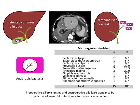
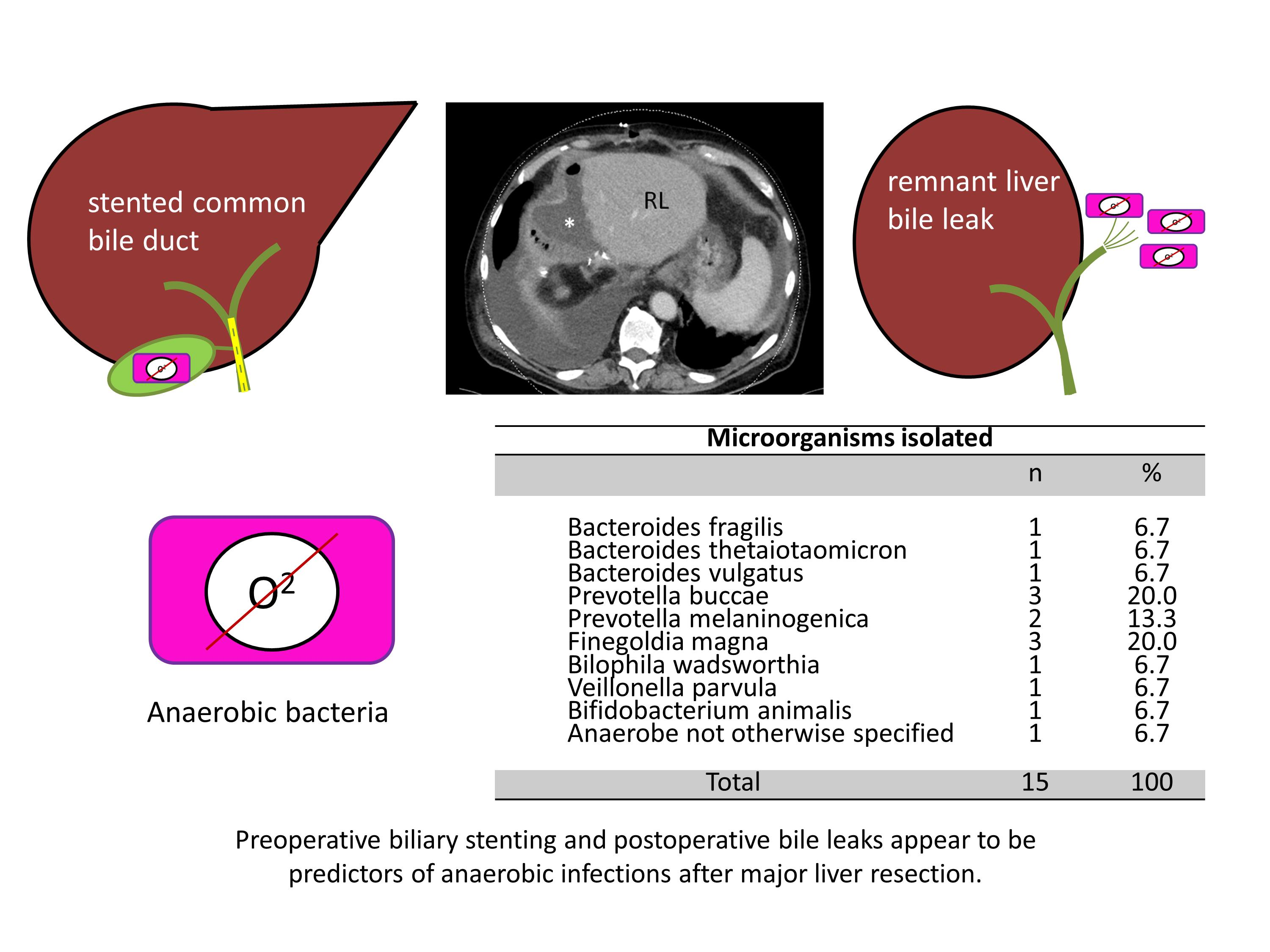
Bacteroides fragilis
Bacteroides thetaiotaomicron
Bacteroides vulgatus
Prevotella buccae
Prevotella melaninogenica
Finegoldia magna
Bilophila wadsworthia
Veillonella parvula
Bifidobacterium animalis
Anaerobe not otherwise specified
1
1
1
3
2
3
1
1
1
1
6.7
6.7
6.7
20.0
13.3
20.0
6.7
6.7
6.7
6.7
Total15100
Publisher’s Note: MDPI stays neutral with regard to jurisdictional claims in published maps and institutional affiliations.

