Article

Isolation and Characterization of Antibacterial Carotane Sesquiterpenes from Artemisia argyi Associated Endophytic Trichoderma virens QA-8

1
Key Laboratory of Experimental Marine Biology, Institute of Oceanology, Chinese Academy of Sciences, and Laboratory of Marine Biology and Biotechnology, Qingdao National Laboratory for Marine Science and Technology, Nanhai Road 7, Qingdao 266071, China
2
Yantai Institute of Coastal Zone Research, Chinese Academy of Sciences, Yantai 264003, China
3
College of Chemistry and Chemical Engineering, Hubei Normal University, Cihu Road 11, Huangshi 435002, China
4
Center for Ocean Mega-Science, Chinese Academy of Sciences, Nanhai Road 7, Qingdao 266071, China
*
Authors to whom correspondence should be addressed.
These authors contributed equally to this work.
Submission received: 14 January 2021 / Revised: 13 February 2021 / Accepted: 17 February 2021 / Published: 20 February 2021
(This article belongs to the Special Issue Novel Fungal Metabolites with Antimicrobial Activities)

Abstract

:
Carotane sesquiterpenes are commonly found in plants but are infrequently reported in the fungal kingdom. Chemical investigation of Trichoderma virens QA-8, an endophytic fungus associated with the inner root tissue of the grown medicinal herb Artemisia argyi H. Lév. and Vaniot, resulted in the isolation and characterization of five new carotane sesquiterpenes trichocarotins I–M (15), which have diverse substitution patterns, and seven known related analogues (612). The structures of these compounds were established on the basis of a detailed interpretation of their NMR and mass spectroscopic data, and the structures including the relative and absolute configurations of compounds 13, 5, 9, and 10 were confirmed by X-ray crystallographic analysis. In the antibacterial assays, all isolates exhibited potent activity against Escherichia coli EMBLC-1, with MIC values ranging from 0.5 to 32 µg/mL, while 7β-hydroxy CAF-603 (7) strongly inhibited Micrococcus luteus QDIO-3 (MIC = 0.5 µg/mL). Structure-activity relationships of these compounds were discussed. The results from this study demonstrate that the endophytic fungus T. virens QA-8 from the planted medicinal herb A. argyi is a rich source of antibacterial carotane sesquiterpenes, and some of them might be interesting for further study to be developed as novel antibacterial agents.

1. Introduction

Artemisia argyi H. Lév. and Vaniot, a traditional medicinal and edible plant in China, is reported to have pleiotropic bioactivities and has been used for the treatment of a variety of diseases, such as amenorrhea, bruising, dysmenorrhea, inflammation, jaundice, malaria, and metrorrhagia [1,2,3]. This plant has been widely cultivated as a medicinal herb in Qichun county, Hubei province, China. Artemisia argyi and its endophytic fungi (Trichoderma koningiopsis and T. virens) have been the source of a wide range of biologically active natural products [4,5,6,7,8,9]. A number of bioactive constituents such as flavonoids [4], polysaccharides [5], terpenes [6,7], and polyketides [8,9], have been reported from the plant A. argyi and its associated endophytic fungi. Several of these components have been investigated to show anticoagulation, antioxidant, antimicrobial, and anticancer activities [4,5,6,7,8,9,10].
Endophytic fungi have recently become an important source of new chemical substances with interesting biological activities. A wide range of compound classes such as alkaloids, quinones, phenols, polyketides, and terpenoids have been reported from various sourced endophytic fungi, and some of these compounds have been evidenced to possess significant biological properties such as antibacterial, anticancer, antifungal, antiinfections, antioxidant, antivirus, and enzymatic inhibitory activities [6,8,9,11,12,13]. Paclitaxel and penicillin, two famous chemicals that are well developed and marketed, could also be produced by endophytic fungi [12]. Based on the above results, we undertook a chemical investigation into the endophytic fungus from A. argyi which resulted in the identification of six cadinane-type sesquiterpenes trichocadinins B–G from Trichoderma virens QA-8, an endophytic fungus associated with the inner root tissue of A. argyi [6]. Further work on the additional portions of the culture extract led to the identification of 12 carotane sesquiterpenes (112) (Figure 1), with five new (trichocarotins I–M, 15) and seven known (612) [14,15,16,17] related analogues. Previous studies have shown that carotane sesquiterpenes have pleiotropic bioactivities, such as antifungal activity against Candida albicans strains, antifertility, anti-HIV activity, and inhibitory activity on the growth of etiolated wheat coleoptiles [14,17,18,19,20,21]. This paper describes the isolation, structure determination, stereochemical assignment, and antibacterial activities of the isolated compounds, and the results indicates that T. virens QA-8, an endophytic fungus from the planted medicinal herb A. argyi, has abundant antibacterial carotene sesquiterpenoids.

2. Results and Discussion

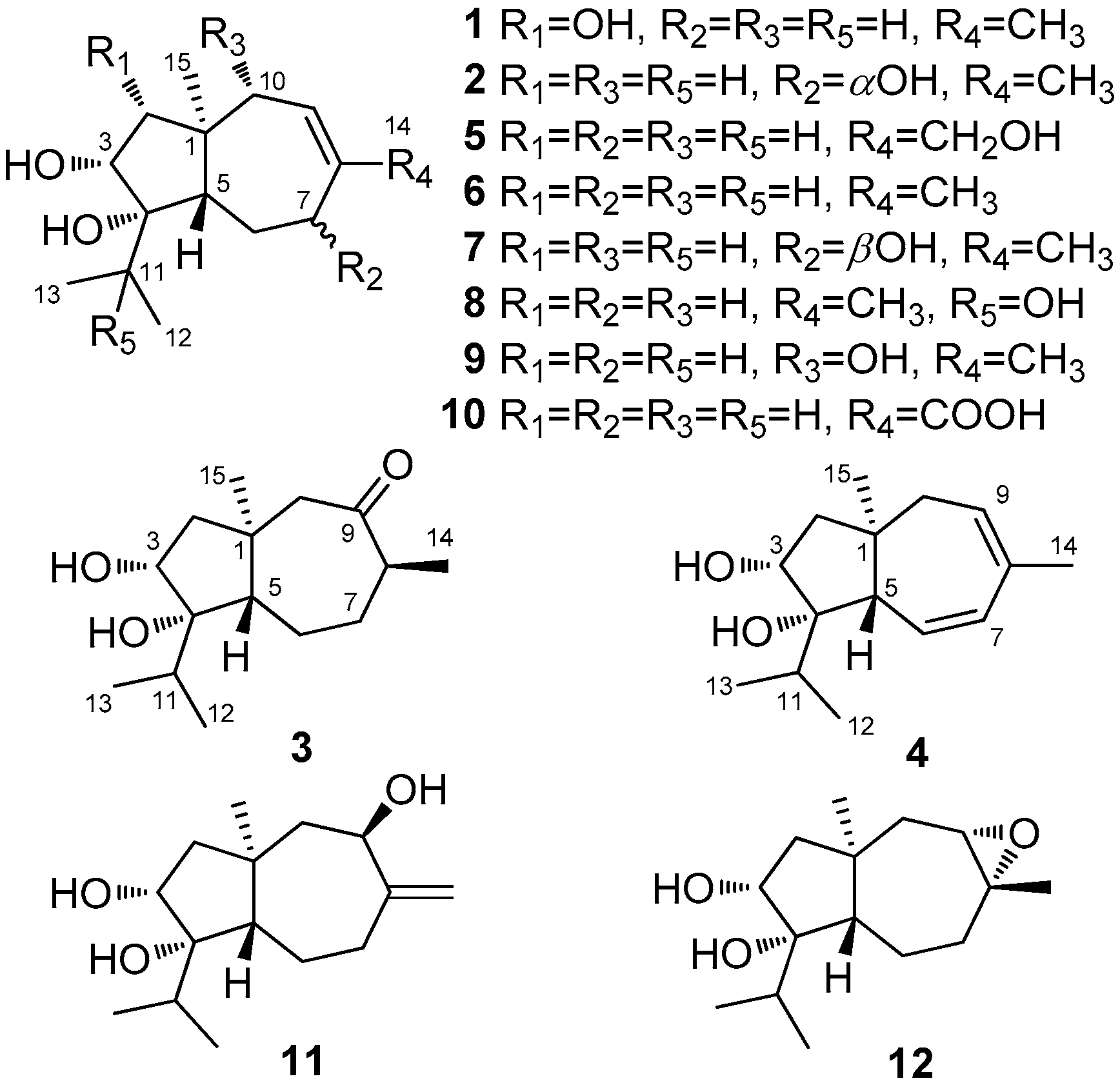
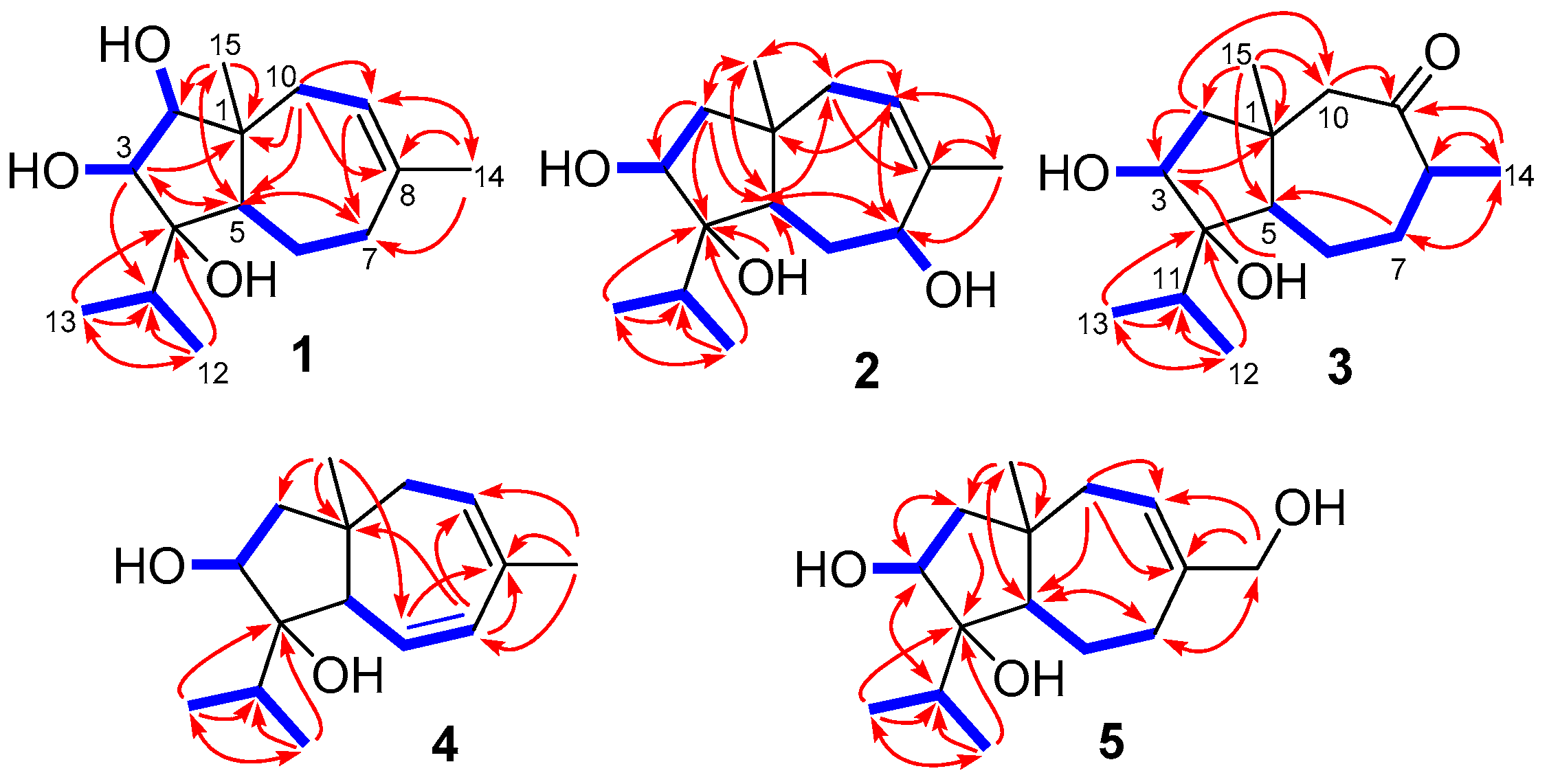
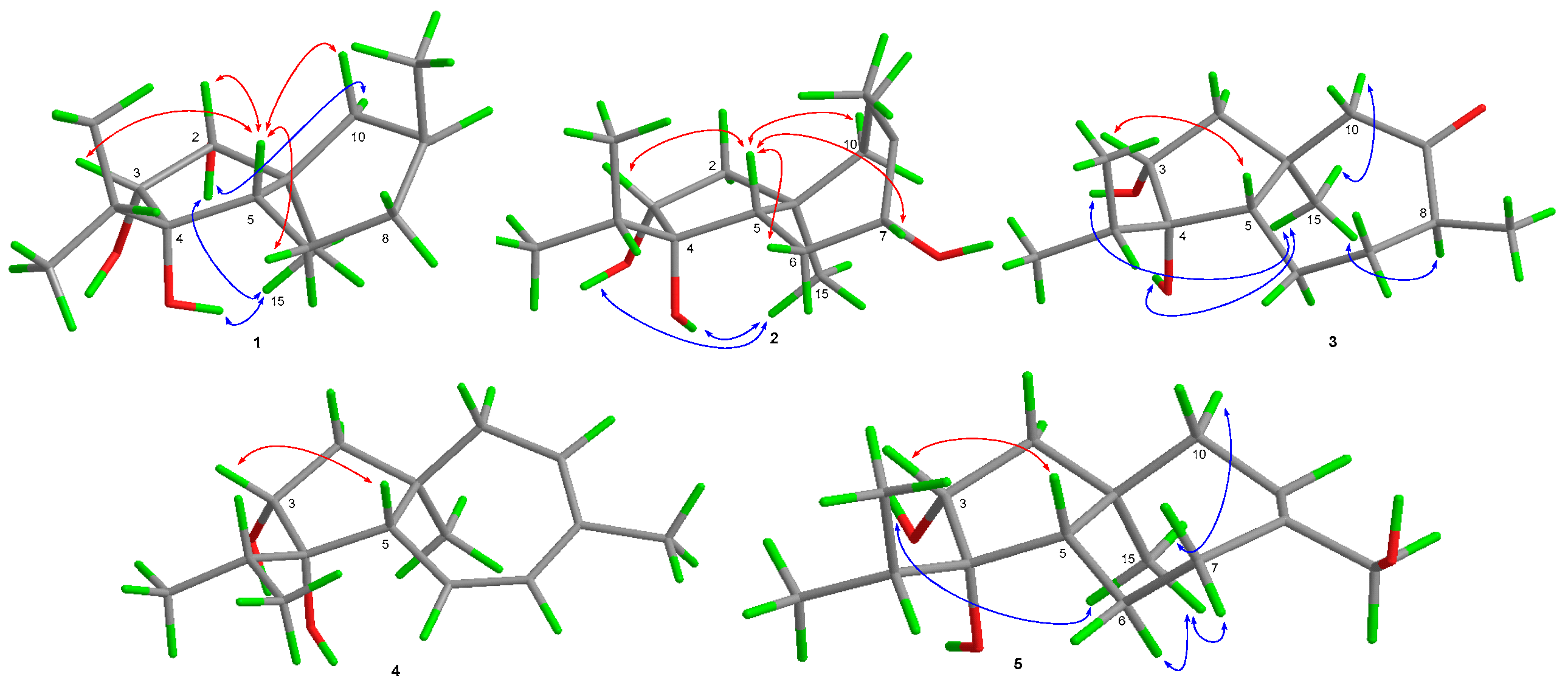
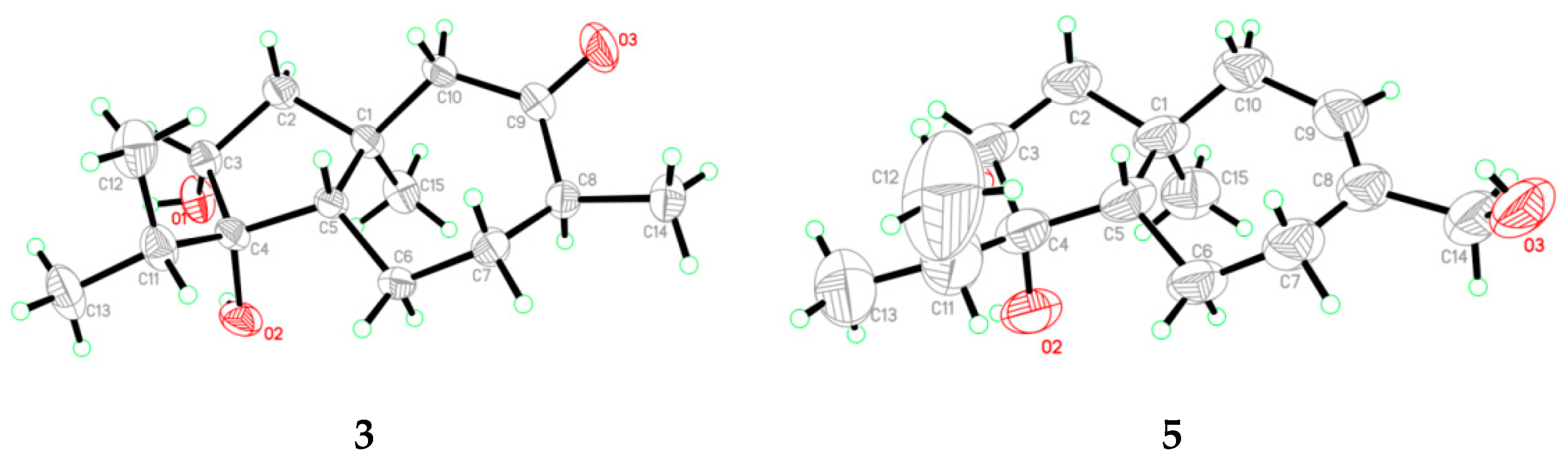
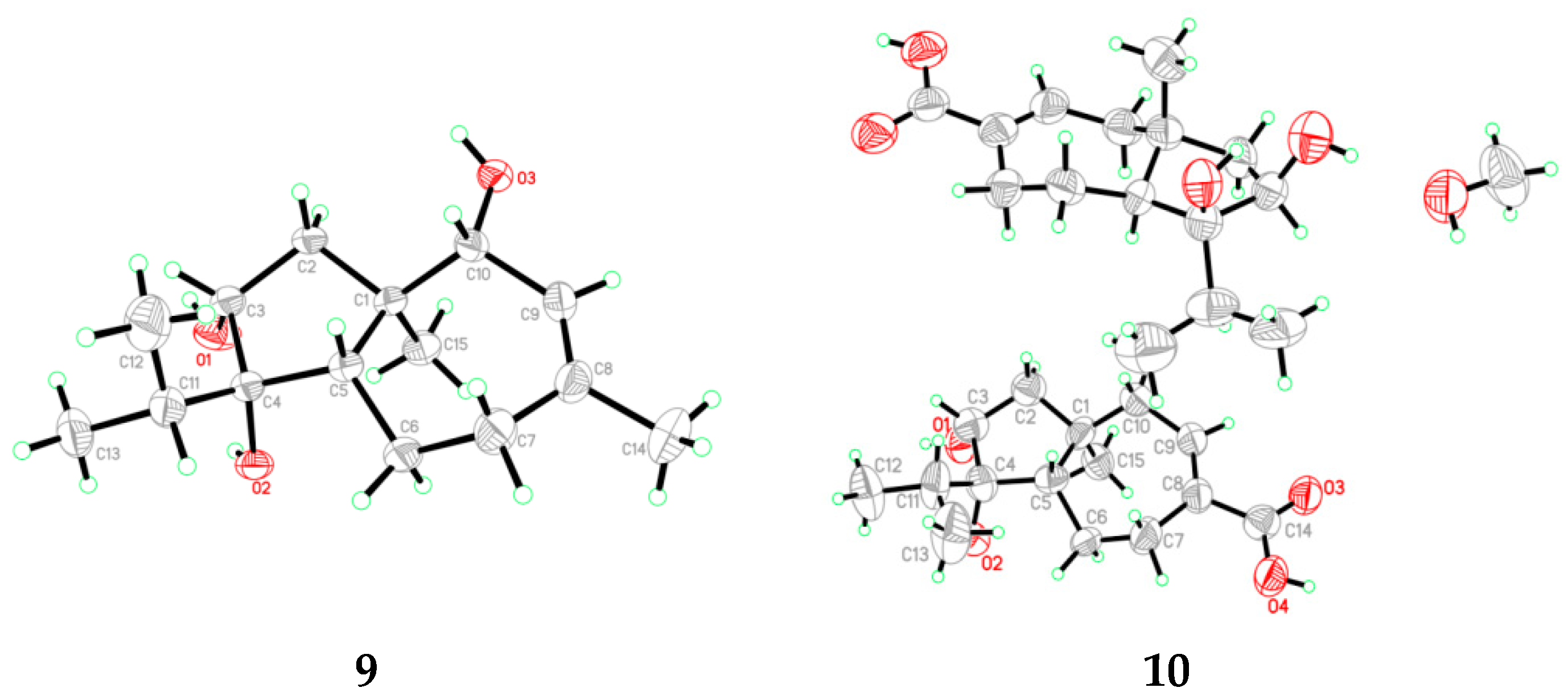
2.1. Structural Elucidation of the New Compounds

Compound 1 was purified as colorless crystals and its molecular formula was determined as C15H26O3 on the basis of HRESIMS data, implying three degrees of unsaturation. The 1H and 13C NMR data of 1 indicated the presence of four methyls, three methylenes, five methines (including one olefinic and two oxygenated), and three non-protonated carbons (including one olefinic and one oxygenated) (Table 1 and Table 2 and Figures S1 and S2 in the Supplementary Material). Comprehensive analysis of its 1H and 13C NMR data suggested that compound 1 belonged to carotane sesquiterpenes with structural similarity to that of CAF-603 (6, Figure 1) and differed from 6 mainly at the five-membered ring [14,15]. Compared to CAF-603 (6), resonances for the methylene group at δH/δC 1.39/50.3 (CH2-2) of 6 disappeared in that of 1. Instead, signals corresponding to an oxymethine group at δH/δC 3.09/78.4 (CH-2) were observed in the NMR spectra of 1. Additionally, a signal for an additional hydroxy group was observed at δH 4.16 (s, OH-2) in the 1H NMR spectrum of 1. These data suggested that the methylene group CH2-2 in 6 was replaced by an oxymethine group in 1. COSY and HMBC data (Figure 2) supported the above deduction. The planar structure of 1 was thus determined.
The relative configuration of 1 was determined by the observed NOEs from H3-15 to the protons of OH-2 and OH-4, from H-10α to the proton of OH-2, and from H-5 to H-2, H-3, and H-10β. The Cu Kα radiation single-crystal X-ray diffraction experiment resulted in Flack parameter 0.1(4) of 1, which allowed the establishment of its absolute configuration as 1S, 2R, 3R, 4S, and 5S (Figure 4). Thus, the structure of 1 was identified and named as trichocarotin I.
Trichocarotin J (2) was originally isolated as an amorphous powder. Its molecular formula was also determined as C15H26O3 based on the HRESIMS data. The 1H NMR spectrum of 2 indicated the presence of an olefinic methyl moiety, an aliphatic singlet methyl, and an isopropyl unit, which are typical structural features of the known carotane sesquiternene members. The 1H, 13C, and DEPT NMR data of 2 (Table 1 and Table 2 and Figures S8 and S9 in the Supplementary Material) showed almost identical spectral patterns to those of 7β-hydroxy CAF-603 (7) [15], with some minor variations for the chemical shifts of C-5 through C-9, and C-14 as well. Detailed inspection of the NMR data suggested that 2 is a diastereomer of 7, epimeric at C-7. This was supported by the observed NOEs from H-5 to H-3, H-6β, H-7, and H-10β, and from H3-15 to the protons of OH-3 and OH-4 (Figure 3). Upon slow evaporation of the solvent (MeOH:H2O = 8:1) by storage in a refrigerator, quality single crystals of compound 2 were obtained. The structure and absolute configuration of 2 were further confirmed by a single-crystal X-ray diffraction experiment using Cu Kα radiation (Figure 4). The Flack parameter 0.06(4) allowed for the establishment of the absolute configuration of 2 as 1R, 3R, 4S, 5S, and 7R.
The molecular formula of trichocarotin K (3) was determined to be C15H26O3 by HRESIMS. Its 1H and 13C NMR data revealed the presence of four methyls, four methylenes, four methines (one oxygenated), and three non-protonated (one oxygenated and one ketone) carbons (Table 1 and Table 2 and Figures S15 and S16 in the Supplementary Material). A detailed comparison of NMR data revealed that compound 3 differed from CAF-603 (6) [14,15] mainly at the seven-membered ring, and that resonances corresponding to the double bond (C-8 and CH-9) in 6 disappeared in that of 3, while signals for an aliphatic methine (CH-8) and for a keto group (C-9) were observed in the NMR spectra of 3 (Table 1 and Table 2). HMBC correlations from H-14 to C-7, C-8, and C-9 confirmed this deduction. Other COSY and HMBC correlations (Figure 2) further confirmed the planar structure of 3.
The key NOE correlations from H3-15 to H-8, H-10α, and to the protons of OH-3 and OH-4, and from H-3 to H-5 determined the relative configuration of 1 (Figure 3). Upon slow evaporation of the solvent (MeOH) by storing the sample in a refrigerator, quality single crystals of 3 were obtained, and the absolute configuration of 3 was thus determined as 1S, 3R, 4S, 5S, and 8S by X-ray diffraction analysis (Figure 5).
Compound 4 was isolated as a colorless oil and its molecular formula was determined as C15H24O2 by HRESIMS. Its 1H and 13C NMR data revealed the presence of four methyls, two methylenes, six methines (one oxygenated and three olefinic), and three non-protonated (one oxygenated and one olefinic) carbons (Table 1 and Table 2 and Figures S22 and S23 in the Supplementary Material). These data suggested that 4 had the same carbon skeleton as that of CAF-603 (6) [14,15]. Actually, compound 4 was a C-6 and C-7 deprotonated product of CAF-603. This was verified by chemical shifts and COSY correlations of H-6 with H-5 and H-7 (Figure 2). In the NOESY experiments, the NOE from H-3 to H-5 indicated the co-facial orientation of these groups (Figure 3). However, no other diagnostic NOEs were observed, and thus the relative configuration could not be characterized by NOESY experiments. According to the literature reports [14,15,16], the relative configuration of all the congeners of CAF-603 was deduced to be same at the five-membered ring. The absolute configuration of 4 was tentatively deduced as 1R, 3R, 4S, 5S on the basis of biogenic considerations. Thus, the structure of compound 4 was characterized and was named trichocarotin L.
Trichocarotin M (5), initially obtained as a colorless waxy solid, was determined to possess the molecular formula C15H26O3, based on HRESIMS data. The 1H and 13C NMR data of 5 (Table 1 and Table 2 and Figures S29 and S30 in the Supplementary Material) closely resembled those of 14-hydroxy CAF-603 (trichocarane B), whose relative configuration was determined by NOESY spectrum [17], but its absolute configuration was not clarified. The relative configuration of 5 was deduced to be the same as that of 14-hydroxy CAF-603 based on the NOESY data (Figure 3). However, the optical rotation of compound 5 ([α]25 D +16 (c 0.25, CHCl3)) has an opposite sign to that of 14-hydroxy CAF-603 ([α]25 D –28.0 (c 0.20, CHCl3)) [17]. Thus, trichocarotin J (5) might be the enantiomer of 14-hydroxy CAF-603. To further confirm this deduction we turned our efforts to a crystallographic study of this compound. Upon slow evaporation of the solvent (MeOH:H2O = 15:1) by storage in a refrigerator, quality single crystals of compound 5 were obtained, and the relative and absolute configuration of 5 was therefore established by a single-crystal X-ray diffraction experiment using Cu Kα radiation (Figure 5). The Flack parameter 0.3(2) of 5 allowed the unambiguous confirmation of the absolute configuration of 5 as 1R, 3R, 4S, and 5S, and named trichocarotin M, which is an enantiomer of 14-hydroxy CAF-603.
In addition to new compounds 15, seven known analogues including CAF-603 (6) [14,15], 7β-hydroxy CAF-603 (7) [15], trichocarotins E–H (811) [16], and trichocarane A (12) [17] were also isolated and identified. Their structures were elucidated by comparing their NMR data with those reported in the literature. It deserve to mention that the absolute configurations of compounds 9 and 10 were previously assigned solely from a biogenetic perspective [16], but in the present study the single-crystal X-ray diffraction (Figure 6) was used to confirm their absolute configurations.

2.2. Antibacterial Activities of the Isolated Compounds

Compounds were assayed for their antibacterial activities against human pathogens Escherichia coli EMBLC-1 and Micrococcus luteus QDIO-3. Statistical analysis by ANOVA for various concentrations when assessed for E. coli and M. luteus showed potent inhibitory growth both at concentrations ≥ MICs when compared to that at concentrations < MICs, and the MICs were obtained. Significance of all the statistical tests was predetermined at p < 0.05. As a result, each of these compounds showed strong inhibitory activity against E. coli, with MIC values ranging from 0.5 to 16 µg/mL, and the activity of compounds 35, 8, and 11 are as active as that of the positive control (chloramphenicol, MIC = 0.5 µg/mL) (Table 3). In addition, compounds 68 showed potent activity against M. luteus with MIC values of 4, 0.5, and 2 µg/mL, respectively, and the activity of compound 7 was stronger than that of chloramphenicol (MIC = 1 µg/mL).
Structure-activity relationship analysis revealed that the OH substitution at C-11 increased the activities against both E. coli and M. luteus (8 vs. 6), while the OH group at C-14 increased the activity against E. coli and decreased the activity against M. luteus (5 vs. 6). Similarly, the 8,9-epoxy group at the seven-membered ring also increased the activity against E. coli and decreased the activity against M. luteus (12 vs. 6).

3. Materials and Methods

3.1. General Experimental Procedures

The general experimental procedures and the apparatus used in the current work are the same as that described in our previous report [6]: an SGW X-4 micro-melting-point apparatus, an Optical Activity AA-55 polarimeter, a PuXi TU-1810 UV-visible spectrophotometer, a JASCO J-715 spectropolarimeter, a Bruker Avance 500 spectrometer, an API QSTAR Pulsar 1 mass spectrometer, analytical HPLC: a Dionex HPLC system, equipped with P680 pump (Dionex), silica gel GF254 precoated plates, 100–200 mesh and 200–300 mesh silica gel, 40–63 μm RP-18 reverse-phase Si gel, and Sephadex LH-20.

3.2. Plant and Fungal Materials

The fungus T. virens QA-8 was isolated from the fresh inner root tissue of the Compositae medical plants A. argyi collected at Qichun, Hubei Province, in central China in July 2014 and was identified by analysis of its ITS region of the rDNA. The primers used for PCR are ITS1 (5′-TCCGTAGGTGAACCTGCGG-3′) and ITS4 (5′-TCCTCCGCTTATTGATATGC-3′), and the total length of sequenced ITS is 616bp. The BLAST search showed that the amplified ITS sequence (GenBank accession no. MK224593) has 100% homology with other members of the genus T. virens (compared with MT256290.1). The strain QA-8 is cryopreserved at −80 °C in 20% aqueous glycerol at the Key Laboratory of Experimental Marine Biology, Institute of Oceanology of the Chinese Academy of Sciences (IOCAS).

3.3. Fermentation, Extraction and Isolation

The culture of T. virens QA-8 was grown on PDA medium at 28 °C for 7 days and were then inoculated in a 1L-Erlenmeyer flask containing solid rice medium consisting of 70 g rice, 0.1 g corn flour, 0.3 g peptone, and 100 mL distilled water. After 30 days, the whole fermented cultures (180 flasks) were extracted with EtOAc and the combined EtOAc solution was concentrated under reduced pressure to yield 97.4g extract. The extract was fractionated by Si gel vacuum liquid chromatography (VLC) to yield 10 fractions (Frs. 1–10). Purification of Fr. 3 (3.0 g), Fr. 4 (3.7 g), Fr. 5 (11.0 g), and Fr. 6 (22.7 g) by reversed-phase column chromatography (CC) over Lobar LiChroprep RP-18 with a MeOH-H2O gradient (from 10:90 to 100:0) yielded six subfractions (Frs. 3.1–3.6), nine subfractions (Frs. 4.1–4.9), 10 subfractions (Frs. 5.1–5.10), and 15 subfractions (Frs. 6.1–6.15), respectively. Fr. 3.5 (341 mg) was further purified by CC on Si gel eluting with a PE–acetone gradient (from 50:1 to 10:1) to obtain compound 6 (185.9 mg). Fr. 4.9 (325 mg) was purified by CC on Si gel eluting with a CH2Cl2–acetone gradient (from 20:1 to 5:1) and then by preparative TLC (plate: 20 × 20 cm, developing solvents: PE–EtOAc, 1:1) to yield compounds 3 (7.9 mg) and 10 (18.3 mg). Fr. 5.5 (436 mg) was purified by CC on Si gel eluting with a CH2Cl2−acetone gradient (from 100:1 to 20:1) and then purified by CC on Sephadex LH-20 (MeOH) to give compounds 7 (3.6 mg), 8 (8.9 mg) and 12 (9.3 mg), respectively. Fr. 5.8 (275 mg) was purified by CC on Si gel eluting with a CH2Cl2−acetone gradient (from 100:1 to 10:1) and then purified by CC on Sephadex LH-20 (MeOH) and preparative TLC (plate: 20 × 20 cm, developing solvents: PE/EtOAc, 1:1) yielded compounds 2 (7.2 mg) and 9 (5.5 mg). Fr. 6.2 (136 mg) was further purified by preparative TLC (plate: 20 × 20 cm, developing solvents: CH2Cl2− EtOAc, 5:1) to afford compound 1 (6.5 mg). Fr. 6.7 (94 mg) was purified by CC on Sephadex LH-20 (MeOH) to give compound 11 (5.8 mg). Fr. 6.8 (582 mg) was purified by CC on Si gel eluting with a CH2Cl2−MeOH gradient (from 200:1 to 10:1) and then purified by preparative TLC (plate: 20 × 20 cm, developing solvents: PE/acetone/ acetic acid, 3:1:0.04) to obtain compounds 4 (8.9 mg) and 5 (12.0 mg).

3.4. Spectroscopic Data

Trichocarotin I (1): colorless crystals; mp 99–101 °C; [α]25 D –5.56 (c 0.18, MeOH); ECD (3.15 mM, MeOH) λmaxε) 215 (–3.35), 249 (+1.75) nm; 1H and 13C NMR data, Table 1 and Table 2; HRESIMS m/z 253.1812 [M – H] (calcd for C15H25O3, 253.1809).
Trichocarotin J (2): colorless crystals; mp 88–90 °C; [α]25 D –10.64 (c 0.47, MeOH); ECD (4.33 mM, MeOH) λmaxε) 223 (–28.47), 262 (+20.03) nm; 1H and 13C NMR data, Table 1 and Table 2; HRESIMS m/z 253.1810 [M – H] (calcd for C15H25O3, 253.1809).
Trichocarotin K (3): colorless crystals; mp 92–93 °C; [α]25 D +98.41 (c 0.63, MeOH); ECD (5.12 mM, MeOH) λmaxε) 219 (+5.15), 234 (+5.39), 294 (+7.79) nm; 1H and 13C NMR data, Table 1 and Table 2; HRESIMS m/z 253.1808 [M – H] (calcd for C15H25O3, 253.1809).
Trichocarotin L (4): colorless oil; [α]25 D +26.32 (c 0.19, MeOH); ECD (7.20 mM, MeOH) λmaxε) 243 (–10.60) nm; 1H and 13C NMR data, Table 1 and Table 2; HRESIMS m/z 235.1709 [M – H] (calcd for C15H23O2, 235.1704).
Trichocarotin M (5): colorless crystals; mp 131–133 °C; [α]25 D +85.92 (c 0.71, MeOH) or [α]25 D +16.00 (c 0.25, CHCl3); ECD (5.51 mM, MeOH) λmaxε) 209 (–4.84), 220 (+2.44), 240 (–1.72) nm; 1H and 13C NMR data, Table 1 and Table 2; HRESIMS m/z 299.1869 [M + HCOO] (calcd for C16H27O5, 299.1864).

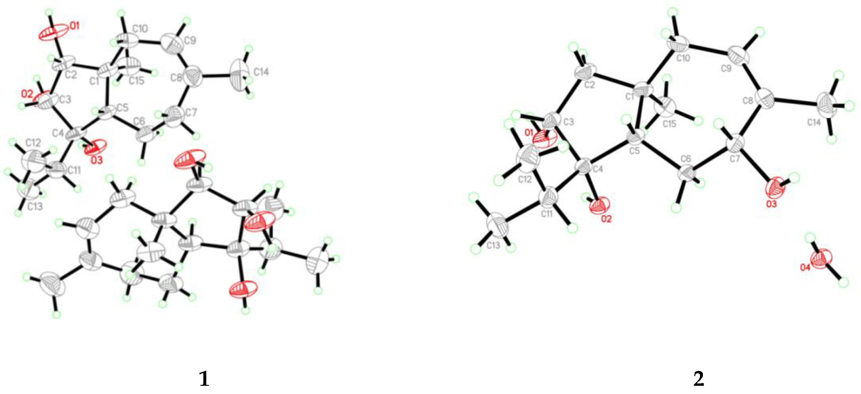
3.5. X-Ray Crystallographic Analysis of Compounds 1–3, 5, 9, and 10

The crystallographic data were collected on a Bruker Smart1000 or Bruker D8 Venture CCD diffractometer. The data were corrected for absorption by using the program SADABS [22,23]. The structures were solved by direct methods with the SHELXTL software package [24]. The structures were refined by full-matrix least-squares techniques [25].
Crystal data for compound1: C15H26O3, F.W. = 254.36, Monoclinic space group P2(1), unit cell dimensions a = 6.9632(6) Å, b = 12.8384(13) Å, c = 16.8664(18) Å, V = 1506.5(3) Å3, α = γ = 90°, β = 92.3770(10)°, Z = 4, dcalcd = 1.121 mg/m3, crystal dimensions 0.36 × 0.08 × 0.05 mm3, µ = 0.605 mm–1, F(000) = 560. The 3967 measurements yielded 2231 independent reflections. The final refinement gave R1 = 0.0649 and wR2 = 0.1385 [I > 2σ(I)]. The Flack parameter was 0.1(4).
Crystal data for compound2: C15H26O3·H2O, F.W. = 272.37, Orthorhombic space group P2(1)2(1)2(1), unit cell dimensions a = 7.7040(9) Å, b = 13.802(4) Å, c = 14.5355(16) Å, V = 1545.6(5) Å3, α = β = γ = 90°, Z = 4, dcalcd = 1.171 mg/m3, crystal dimensions 0.18 × 0.16 × 0.12 mm3, µ = 0.668 mm–1, F(000) = 600. The 2796 measurements yielded 2746 independent reflections. The final refinement gave R1 = 0.0293 and wR2 = 0.0804 [I > 2σ(I)]. The Flack parameter was 0.06(4).
Crystal data for compound3: C15H26O3, F.W. = 254.36, Orthorhombic space group P2(1)2(1)2(1), unit cell dimensions a = 7.4144(10) Å, b = 7.5400(10) Å, c = 26.394(4) Å, V = 1475.5(4) Å3, α = β = γ = 90°, Z = 4, dcalcd = 1.145 mg/m3, crystal size 0.22 × 0.20 × 0.18 mm, µ = 0.617 mm–1, F(000) = 560. The 2675 measurements yielded 2588 independent reflections after. The final refinement gave R1 = 0.0347 and wR2 = 0.1193 [I > 2σ(I)]. The Flack parameter was −0.1(3).
Crystal data for compound5: C15H26O3, F.W. = 254.36, monoclinic space group P2(1), unit cell dimensions a = 7.6721(12) Å, b = 7.4046(12) Å, c = 13.155(3) Å, V = 746.3(3) Å3, α = γ = 90°, β = 92.961(14)°, Z = 2, dcalcd = 1.132 mg/m3, crystal dimensions 0.16 × 0.15 × 0.14 mm, µ = 0.610 mm–1, F(000) = 280. The 2201 measurements yielded 1459 independent reflections. The final refinement gave R1 = 0.0542 and wR2 = 0.1530 [I > 2σ(I)]. The Flack parameter was 0.3(2) in the final refinement for all 2201 reflections with 1459 Friedel pairs.
Crystal data for compound9: C15H26O3, F.W. = 254.36, Orthorhombic space group P2(1)2(1)2(1), unit cell dimensions a = 6.9592(2) Å, b = 11.0588(4) Å, c = 19.4762(9) Å, V = 1498.90(10) Å3, α = β = γ = 90°, Z = 4, dcalcd = 1.127 mg/m3, crystal dimensions 0.30 × 0.10 × 0.07 mm3, µ = 0.608 mm–1, F(000) = 560. The 2603 measurements yielded 2124 independent reflections. The final refinement gave R1 = 0.0403 and wR2 = 0.0908 [I > 2σ(I)]. The Flack parameter was 0.1(3).
Crystal data for compound10: 2C15H24O4·CH3OH, F.W. = 568.73, Orthorhombic space group P2(1)2(1)2(1), unit cell dimensions a = 7.2324(7) Å, b = 15.2789(15) Å, c = 28.581(3) Å, V = 3158.3(5) Å3, α = β = γ = 90°, Z = 4, dcalcd = 1.196 mg/m3, crystal size 0.21 × 0.08 × 0.03 mm, µ = 0.704 mm–1, F(000) = 1240. The 5499 measurements yielded 1880 independent reflections. The final refinement gave R1 = 0.1114 and wR2 = 0.2422 [I > 2σ(I)]. The Flack parameter was 0.0(7) in the final refinement for all 5499 reflections with 1880 Friedel pairs.

3.6. Antibacterial Assays

Antibacterial evaluation against human pathogens E. coli EMBLC-1 and M. luteus QDIO-3 was carried out by the microplate assay with three repetitions [26]. E. coli and M. luteus (95 μL × 5 × 105 CFU/mL per well) were cultured at 37 °C in LB medium containing 1% peptone, 0.5% yeast extract, 1% NaCl and distilled water with 5 μL various concentrations of compounds 112 in each well of 96-well plates for 24 h. The pathogens used in the assays were obtained from the Institute of Oceanology, Chinese Academy of Sciences. Chloramphenicol was used as positive control.

4. Conclusions

In summary, five new carotane sesquiterpenes trichocarotins I–M (15) and seven known analogues (612) were identified from the culture extract of endophytic fungus Trichoderma virens QA-8. Their structures were elucidated by a detailed interpretation of the spectroscopic data and the structures and absolute configurations of compounds 13, 5, 9, and 10 were confirmed by X-ray crystallographic analysis. The crystal structures of the known compounds 9 and 10 are reported for the first time. The absolute configurations of this kind of sesquiterpene were barely presumed by biosynthesis in previously published reports [16], and some of them were not even determined [14,17], but in the present study single crystal X-ray diffraction was used to confirm their absolute configurations. Compounds 35, 8, and 11 showed inhibitory activity against E. coli (MIC = 0.5 µg/mL) and compound 7 showed the strongest activity against M. luteus (MIC = 0.5 µg/mL), which are similar to or stronger than that of the positive control. Our results suggested that some of these compounds could be interesting and could lead compounds into a further development of novel antibacterial agents.

Supplementary Materials

The following are available online at https://0-www-mdpi-com.brum.beds.ac.uk/2079-6382/10/2/213/s1, Figures S1–S35: Selected 1D and 2D NMR and HRESIMS spectra of 15; Figures S36 and S37: Crystal packing of 1, 9 and 10.

Author Contributions

Conceptualization, D.-J.W., X.-W.Z., N.-Y.J., and B.-G.W.; methodology, B.-G.W.; validation, D.-J.W., X.-W.Z., N.-Y.J., B.-G.W., and X.-M.L.; collection of the plants and isolation of the fungus, X.-S.S. and Y.-P.S.; formal analysis, X.-S.S., Y.-P.S., L.-H.M., S.-Q.Y., and X.-M.L.; investigation, X.-S.S. and Y.-P.S.; writing—original draft preparation, X.-S.S. and L.-H.M.; writing—review and editing, B.-G.W.; supervision, N.-Y.J., B.-G.W. and X.-M.L.; project administration, X.-M.L.; funding acquisition, X.-M.L. All authors have read and agreed to the published version of the manuscript.

Funding

Financial support from the National Natural Science Foundation of China is gratefully acknowledged (31870328, to X.-M.L.).

Data Availability Statement

The data presented in this study is included in the supplementary material and is available online at https://0-www-mdpi-com.brum.beds.ac.uk/xxx/s1.

Acknowledgments

B.-G.W. and N.-Y.J. appreciate the support of Taishan Scholar Program from Shandong Province of China (ts201511060 and tsqn201909164) as well as the support of Shuangbai Plan from Yantai Municipal City (2018-3).

Conflicts of Interest

The authors declare no conflict of interest.

References

  1. Song, X.W.; Wen, X.; He, J.W.; Zhao, H.; Li, S.M.; Wang, M.Y. Phytochemical components and biological activities of Artemisia argyi. J. Funct. Foods 2019, 52, 648–662. [Google Scholar] [CrossRef]
  2. Nakasugi, T.; Nakashima, M.; Komai, K. Antimutagens in gaiyou (Artemisia argyi levl. et vant.). J. Agric. Food Chem. 2000, 48, 3256–3266. [Google Scholar] [CrossRef] [PubMed]
  3. Zhang, L.B.; Duan, J.A.; Lv, J.L. Phytochemistry and bioactivities of sesquiterpenoids from the Artemisia species. J. Chin. Pharm. Sci. 2017, 26, 317–334. [Google Scholar] [CrossRef]
  4. Lv, J.L.; Li, Z.Z.; Zhang, L.B. Two new flavonoids from Artemisia argyi with their anticoagulation activities. Nat. Prod. Res. 2018, 32, 632–639. [Google Scholar] [CrossRef] [PubMed]
  5. Zhang, P.; Shi, B.; Li, T.; Xu, Y.; Jin, X.; Guo, X.; Yan, S. Immunomodulatory effect of Artemisia argyi polysaccharide on peripheral blood leucocyte of broiler chickens. J. Anim. Physiol. Anim. Nutr. 2018, 1, 1–8. [Google Scholar] [CrossRef] [PubMed]
  6. Shi, X.S.; Meng, L.H.; Li, X.M.; Li, X.; Wang, D.J.; Li, H.L.; Zhou, X.W.; Wang, B.G. Trichocadinins B−G: Antimicrobial cadinane sesquiterpenes from Trichoderma virens QA-8, an endophytic fungus obtained from the medicinal plant Artemisia argyi. J. Nat. Prod. 2019, 82, 2470–2476. [Google Scholar] [CrossRef] [PubMed]
  7. Zhang, L.; Yang, Y.M.; Wang, S.X.; Ren, Z.; Cheng, Y.X. Three new sesquiterpenoids with cytotoxic activity from Artemisia argyi. Nat. Prod. Res. 2019, 2, 1–7. [Google Scholar] [CrossRef]
  8. Shi, X.S.; Wang, D.J.; Li, X.M.; Li, H.L.; Meng, L.H.; Li, X.; Pi, Y.; Zhou, X.W.; Wang, B.G. Antimicrobial polyketides from Trichoderma koningiopsis QA-3, an endophytic fungus obtained from the medicinal plant Artemisia argyi. RSC Adv. 2017, 7, 51335–51342. [Google Scholar] [CrossRef] [Green Version]
  9. Shi, X.S.; Li, H.L.; Li, X.M.; Wang, D.J.; Li, X.; Meng, L.H.; Zhou, X.W.; Wang, B.G. Highly oxygenated polyketides produced by Trichoderma koningiopsis QA-3, an endophytic fungus obtained from the fresh roots of the medicinal plant Artemisia argyi. Bioorg. Chem. 2020, 94, 103448. [Google Scholar] [CrossRef]
  10. Zhang, X.W.; Wang, S.; Tu, P.F.; Zeng, K.W. Sesquiterpene lactone from Artemisia argyi induces gastric carcinoma cell apoptosis via activating NADPH oxidase/reactive oxygen species/ mitochondrial pathway. Eur. J. Pharmacol. 2018, 837, 164–170. [Google Scholar] [CrossRef]
  11. Anoumedem, E.G.M.; Mountessou, B.Y.G.; Kouam, S.F.; Narmani, A.; Surup, F. Simplicilones A and B isolated from the endophytic fungus Simplicillium subtropicum SPC3. Antibiotics 2020, 9, 753. [Google Scholar] [CrossRef] [PubMed]
  12. Manganyi, M.C.; Ateba, C.N. Untapped potentials of endophytic fungi: A review of novel bioactive compounds with biological applications. Microorganisms 2020, 8, 1934. [Google Scholar] [CrossRef]
  13. Das, M.; Prakash, H.S.; Nalini, M.S. Antibacterial metabolites from Bipolaris specifera, an endophytic fungus from the endemic medicinal plant, Zingiber nimmonii (J. Graham) Dalzell. Biotech 2020, 10, 317–324. [Google Scholar] [CrossRef]
  14. Watanabe, N.; Yamagishi, M.; Mizutani, T.; Kondoh, H.; Omura, S.; Hanada, K.; Kushida, K. CAF-603: A new antifungal carotane sesquiterpene isolation and structure elucidation. J. Nat. Prod. 1990, 53, 1176–1181. [Google Scholar] [CrossRef] [PubMed]
  15. Yasumura, R.; Ashtekar, K.D.; Tonouchi, A.; Nehira, T.; Borhan, B.; Hashimoto, M. 7-β- and 10-β-Hydroxylated congeners of CAF-603; elucidation of absolute configuration of CAF-603 family, and their SAR studies in the anti-fungal activity. Tetrahedron 2013, 69, 9469–9474. [Google Scholar] [CrossRef]
  16. Shi, Z.Z.; Fang, S.T.; Miao, F.P.; Yin, X.L.; Ji, N.Y. Trichocarotins A−H and trichocadinin A, nine sesquiterpenes from the marine-alga-epiphytic fungus Trichoderma virens. Bioorg. Chem. 2018, 81, 319–325. [Google Scholar] [CrossRef]
  17. Macías, F.A.; Varela, R.M.; Simonet, A.M.; Cutler, H.G.; Cutler, S.J.; Eden, M.A.; Hill, R.A. Bioactive carotanes from Trichoderma virens. J. Nat. Prod. 2000, 63, 1197–1200. [Google Scholar] [CrossRef] [PubMed]
  18. Ondeyka, J.G.; Ball, R.G.; Garcia, M.L.; Dombrowski, A.W.; Sabnis, G.; Kaczorowski, G.J.; Zink, D.L.; Bills, G.F.; Goetz, M.A.; Schmalhofer, W.A.; et al. A carotane sesquiterpene as a potent modulator of the Maxi-K channel from Arthrinium phaesospermum. Bioorg. Med. Chem. Lett. 1995, 5, 733–734. [Google Scholar] [CrossRef]
  19. Lee, S.H.; Hensens, O.D.; Helms, G.L.; Liesch, J.M.; Zink, D.L.; Giacobbe, R.A.; Bills, G.F.; Stevens-Miles, S.; Garcia, M.L.; Schmalhofer, W.A.; et al. L-735,334, a novel sesquiterpenoid potassium channel-agonist from Trichoderma virens. J. Nat. Prod. 2004, 58, 1822–1828. [Google Scholar] [CrossRef]
  20. Singh, M.M.; Agnihotri, A.; Garg, S.N.; Agarwal, S.K.; Gupta, D.N.; Keshri, G.; Kamboj, V.P. Antifertility and hormonal properties of certain carotane sesquiterpenes of Ferula jaeschkeana. Planta Med. 1988, 54, 492–494. [Google Scholar] [CrossRef] [PubMed]
  21. Zhao, Y.X.; Zhou, J.; Wang, Q.; Dai, H.F.; Zheng, Y.T.; Ma, Q.Y.; Zhang, X.; Huang, S.Z. Anti-HIV terpenoids from Daphne aurantiaca Diels. Stems. RSC Adv. 2015, 5, 80254–80263. [Google Scholar] [CrossRef]
  22. Cambridge Crystallographic Data Centre. Crystallographic Data of Compounds 1-3, 5, 9, and 10 as CCDCs1890738, 2014253, 2014254, 1967785, 1890742, and 1890734. Available online: http://www.ccdc.cam.ac.uk/data_request/cif (accessed on 8 February 2021).
  23. Sheldrick, G.M. SADABS, Software for Empirical Absorption Correction; University of Gottingen: Gottingen, Germany, 1996. [Google Scholar]
  24. Sheldrick, G.M. SHELXTL, Structure Determination Software Programs; Bruker Analytical X-ray System Inc.: Madison, WI, USA, 1997. [Google Scholar]
  25. Sheldrick, G.M. SHELXL, Program for the Refinement of Crystal Structures; University of Gottingen: Gottingen, Germany, 2014. [Google Scholar]
  26. Abdulwahab, H.G.; Harras, M.F.; El Menofy, N.G.; Hegab, A.M.; Essa, B.M.; Selim, A.A.; Sakr, T.M.; El-Zahabi, H.S.A. Novel thiobarbiturates as potent urease inhibitors with potential antibacterial activity: Design, synthesis, radiolabeling and biodistribution study. Bioorg. Med. Chem. 2020, 28, 115759. [Google Scholar] [CrossRef] [PubMed]
Figure 1. Structures of compounds 112.
Antibiotics 10 00213 g001
Figure 2. Key HMBC (arrows) and COSY (bold lines) correlations of compounds 15.
Antibiotics 10 00213 g002
Figure 3. Key NOE correlations of compounds 15.
Antibiotics 10 00213 g003
Figure 4. X-ray crystallographic structures of compounds 1 and 2.
Antibiotics 10 00213 g004
Figure 5. X-ray crystallographic structures of compounds 3 and 5.
Antibiotics 10 00213 g005
Figure 6. X-ray crystallographic structures of compounds 9 and 10.
Antibiotics 10 00213 g006
Table 1. 1H NMR Data for Compounds 15 in DMSO-d6 (500 MHz).
No.12345
23.09, d (7.0)1.41, m (overlap)1.50, m1.64, m (overlap)1.49, m (overlap)
33.63, d (7.0)3.88, m3.88, dd (7.2, 1.6)3.87, m3.88, t (4.4)
51.26, dd (11.4, 1.7)1.41, m (overlap)1.66, m1.64, m (overlap)1.36, m
1.49, m1.56, m1.78, m5.80, d (11.2)1.30, m
6β1.37, m1.49, m1.78, m 1.49, m (overlap)
2.00, m (overlap)4.03, m1.43, m5.39, d (11.2)1.81, m (overlap)
7β2.00, m (overlap) 1.43, m 2.11, dd (14.9, 3.2)
8 2.38, m
95.32, br d (7.0)5.26, m 5.57, m5.50, br d (8.2)
10α1.59, dd (14.4, 1.2)1.87, dd (14.0, 9.1)2.43, d (14.4)2.18, m1.81, m (overlap)
10β2.07, dd (14.4, 8.9)1.68, m2.30, d (14.4)2.18, m1.99, dd (14.4, 8.9)
111.66, m1.74, m1.70, m1.64, m (overlap)1.69, m
120.79, d (6.9)0.81, d (6.9)0.79, d (6.9)0.85, d (6.9)0.81, d (6.9)
130.85, d (6.7)0.88, d (6.9)0.86, d (6.7)0.78, d (6.9)0.87, d (6.7)
141.70, s1.71, s0.96, d (6.9)1.74, s3.76, s
150.71, s0.88, s1.06, s0.98, s0.92, s
2-OH4.16, s
3-OH5.39, s5.21, d (4.2)5.35, s
4-OH3.53, s3.69, s3.76, s 3.60, br s
7-OH 4.66, d (5.1)
Table 2. 13C NMR Data for Compounds 15 in DMSO-d6 (125 MHz).
No.12345
144.5, C40.7, C40.2, C44.6, C41.6, C
278.4, CH49.6, CH250.3, CH247.9, CH250.0, CH2
370.6, CH70.1, CH70.4, CH71.8, CH70.4, CH
479.0, C82.5, C82.1, C82.1, C82.6, C
554.3, CH53.5, CH55.4, CH47.2, CH57.7, CH
620.2, CH231.7, CH218.4, CH2143.8, CH21.1, CH2
734.1, CH272.6, CH33.0, CH2125.3, CH30.0, CH2
8138.4, C143.6, C46.3, CH129.0, C142.6, C
9122.3, CH119.6, CH214.3, C127.4, CH121.4, CH
1040.0, CH241.5, CH257.3, CH227.0, CH242.1, CH2
1134.8, CH34.4, CH34.7, CH35.0, CH34.7, CH
1217.6, CH317.7, CH317.6, CH317.7, CH317.7, CH3
1317.1, CH317.2, CH317.1, CH317.3, CH317.1, CH3
1427.1, CH321.1, CH322.8, CH326.9, CH366.9, CH2
1513.9, CH320.9, CH323.2, CH321.4, CH320.9, CH3
Table 3. Antibacterial activity of compounds 1–12 (MIC, μg/mL).
No.123456789101112Chloramphenicol
EC16320.50.50.516160.516160.580.5
ML32840.523281
EC: E. coli. ML: M. luteus; –: no activity (MIC > 64 µg/mL).
Publisher’s Note: MDPI stays neutral with regard to jurisdictional claims in published maps and institutional affiliations.

