Article

Multimodal Interventions to Prevent and Control Carbapenem-Resistant Enterobacteriaceae and Extended-Spectrum β-Lactamase Producer-Associated Infections at a Tertiary Care Hospital in Egypt

by
Noha A. Kamel
1,
Khaled M. Elsayed
1,
Mohamed F. Awad
2,
Khaled M. Aboshanab
3,* and
Mervat I. El Borhamy
1,4
1
Department of Microbiology, Faculty of Pharmacy, Misr International University (MIU), Cairo P.O. Box 19648, Egypt
2
Department of Biology, College of Science, Taif University, P.O. Box 11099, Taif 21944, Saudi Arabia
3
Department of Microbiology and Immunology, Faculty of Pharmacy, Ain Shams University, Organization of African Unity St., Cairo P.O. Box 11566, Egypt
4
International Medical Center, Clinical Microbiology Laboratory, Cairo P.O. Box 11451, Egypt
*
Author to whom correspondence should be addressed.
Submission received: 7 April 2021 / Revised: 23 April 2021 / Accepted: 27 April 2021 / Published: 30 April 2021
(This article belongs to the Special Issue Antimicrobial Resistance in Gram-Negative Bacteria, 2nd Edition)

Abstract

:
The current rise of multidrug-resistant (MDR) Gram-negative Enterobacteriaceae including the extended-spectrum β-lactamase (ESBL)-producing organisms and carbapenem-resistant Enterobacteriaceae (CRE) has been increasingly reported worldwide, posing new challenges to health care facilities. Accordingly, we evaluated the impact of multimodal infection control interventions at one of the major tertiary healthcare settings in Egypt for the aim of combating infections by the respective pathogens. During the 6-month pre-intervention period, the incidence rate of CRE and ESBL-producing clinical cultures were 1.3 and 0.8/1000 patient days, respectively. During the post-intervention period, the incidence of CRE and ESBL producers continued to decrease, reaching 0.5 and 0.28/1000 patient days, respectively. The susceptibility rate to carbapenems among ESBL producers ranged from 91.4% (ertapenem) to 98.3% (imipenem), amikacin (93%), gentamicin (56.9%), and tobramycin (46.6%). CRE showed the highest resistance pattern toward all of the tested β-lactams and aminoglycosides, ranging from 87.3% to 94.5%. Both CRE and ESBL producers showed a high susceptibility rate (greater than 85.5%) to colistin and tigecycline. In conclusion, our findings revealed the effectiveness of implementing multidisciplinary approaches in controlling and treating infections elicited by CRE and ESBL producers.

1. Introduction

Globally, multidrug-resistant (MDR) organisms are considered to be one of the major public health issues within healthcare settings [1]. Infections with MDR organisms or the rising, extensive, drug-resistant Gram-negative bacteria including the carbapenem-resistant Enterobacteriaceae (CRE) and extended-spectrum β-lactamase (ESBL) producers remain the most challenging. This is because of their limited treatment options, their rapid spread among healthcare environments causing outbreaks associated with high mortality rates and their associated prolonged hospital stays with a high economic burden [2,3,4]. It is also estimated that by 2030, more than half of Klebsiella pneumoniae and Escherichia coli will be resistant to third-generation cephalosporins [5]. Additionally, the widespread distribution of Enterobacteriaceae in nature and gastrointestinal tracts has facilitated their extensive spread among the ecosystem, causing community and hospital-acquired infections [6,7]. Additionally, prior hospitalization with a prolonged stay and underlying disease conditions makes patients vulnerable to infections [8]. Consequently, preventing the spread of MDR is a major health issue that requires instant efforts to rapidly react against the deadly superbugs. To enable the containment of MDR Gram-negative pathogens (GNP), early detection of CRE—especially for carbapenemase producers and ESBL producers—through targeted patient screening, infection prevention and control measures (IPC) and applying antimicrobial stewardship programs were of proven value for weakening the impact of resistant pathogens among healthcare settings [9,10]. For earlier recognition of the colonization state that usually precedes or co-exists with MDR Gram-negative bacteria, rectal surveillance cultures were examined. Such active surveillance programs play a pivotal role in identifying high-risk patients for developing subsequent infections and shed a light on the importance of earlier implantation of IPC measures [11]. The application of core IPC, including antimicrobial stewardship programs, contact precautions, environmental cleaning and staff education should be applied to minimize the risk of MDR spread between or within health care settings [11,12]. Moreover, MDR carriers, especially CRE, should be accommodated in isolated rooms whenever possible [13]. Therefore, the aim of this study was to evaluate the effect of applying the core IPC measures on the incidence rate of MDR GNP-associated infections including CRE and ESBL producers in one of the major tertiary healthcare settings in Egypt, with the aim of combating these nightmare pathogens.

2. Results

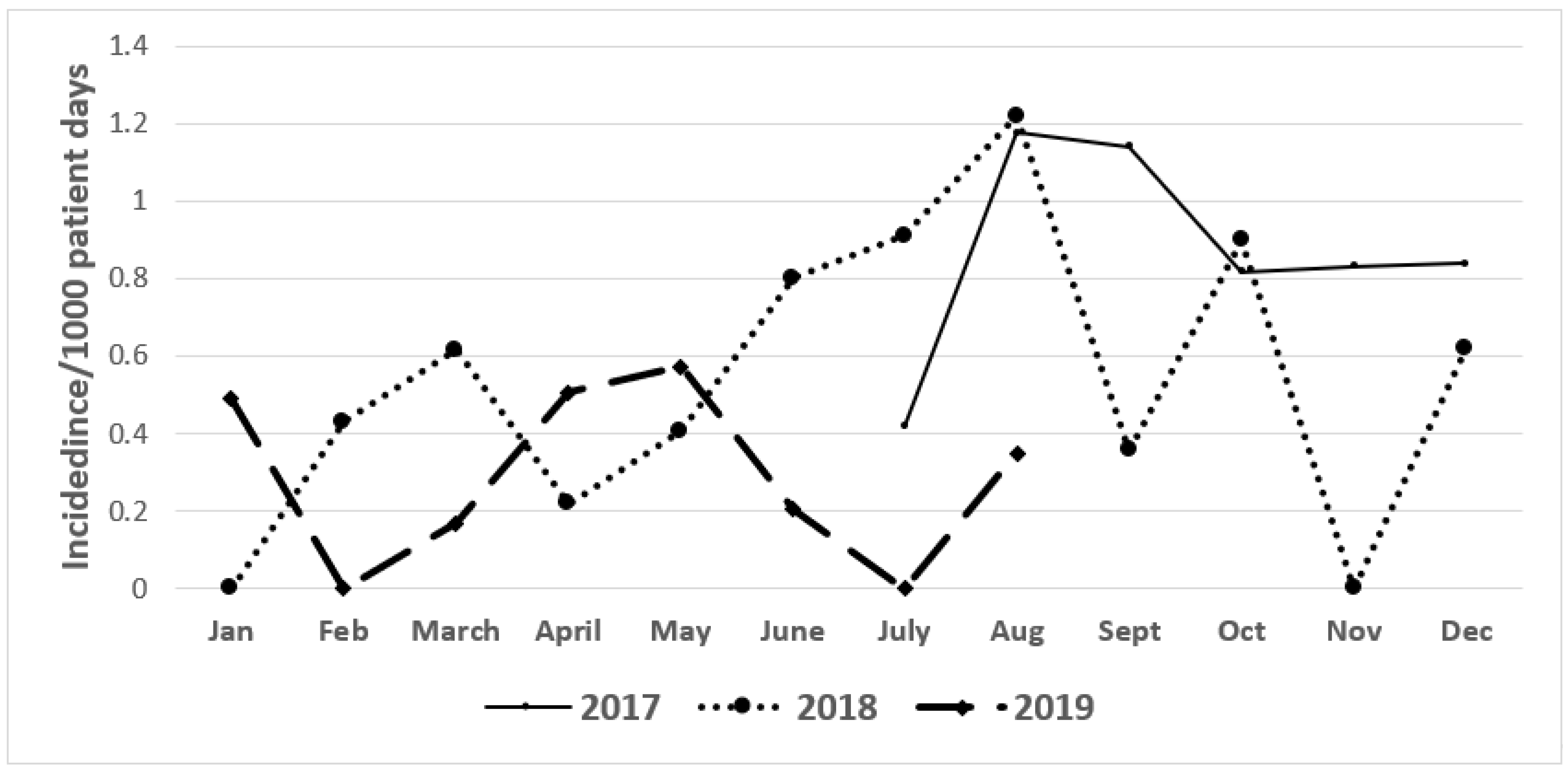
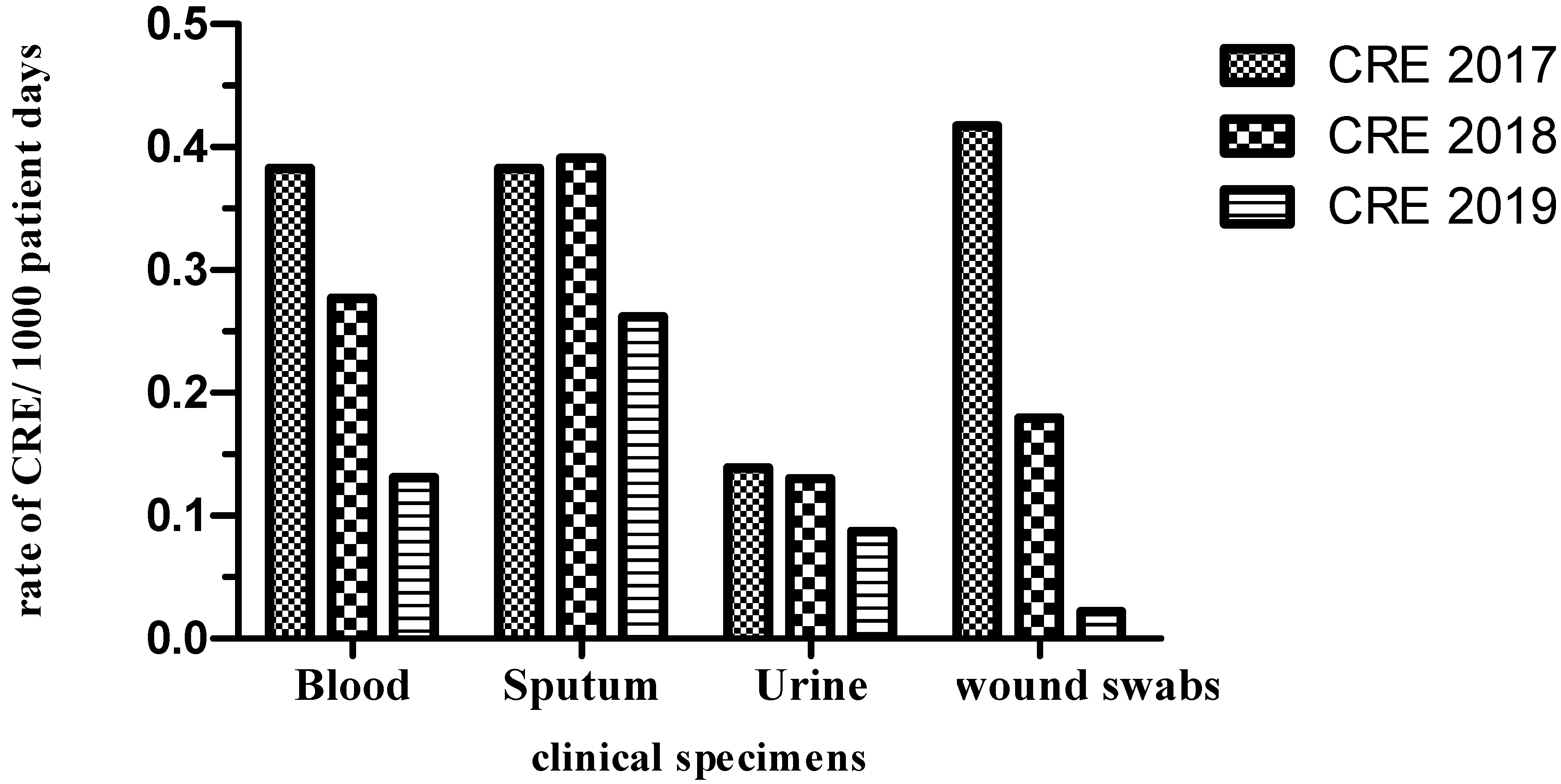
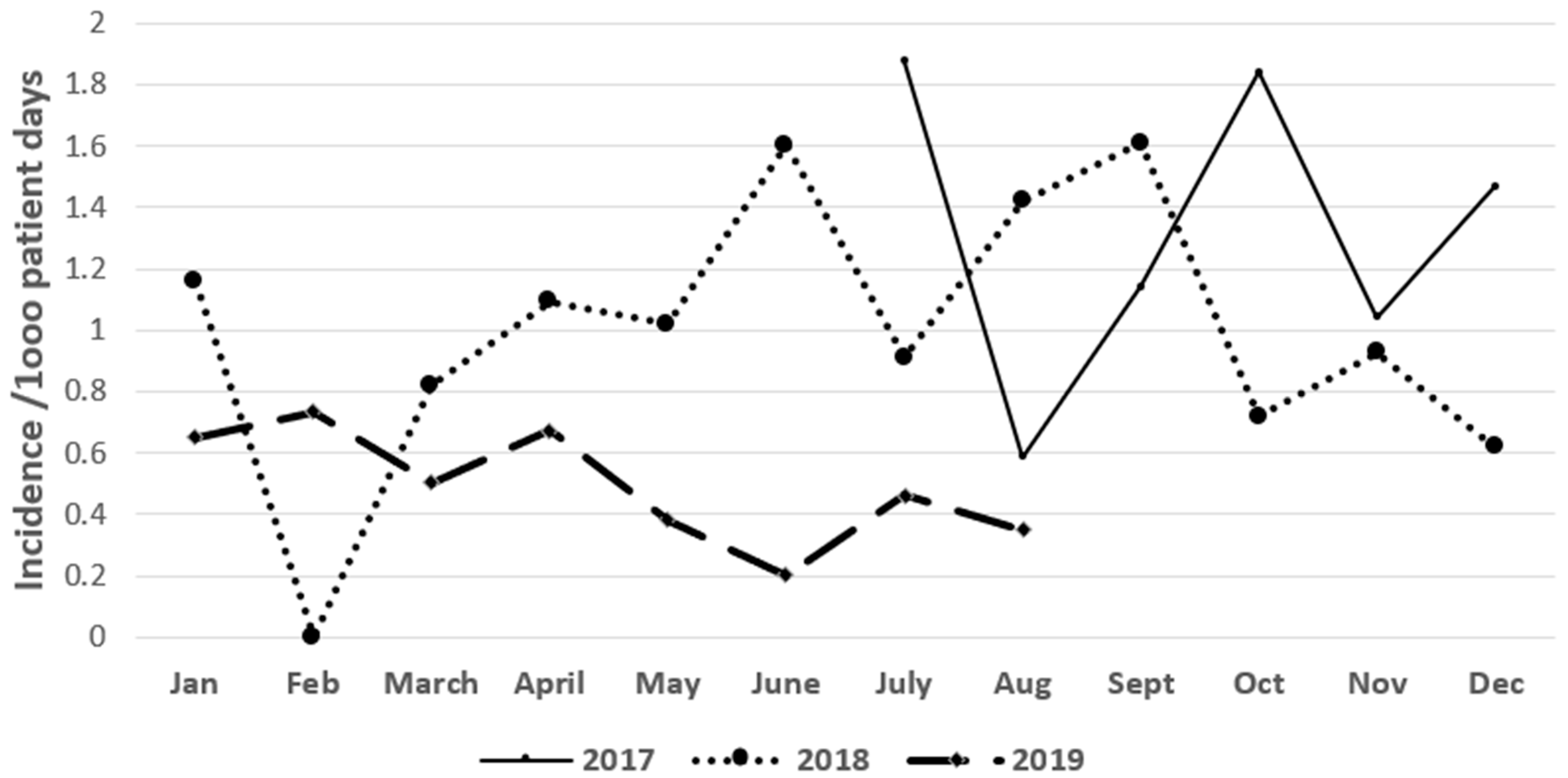
2.1. Incidence of CRE and ESBL Producers (2017–2019)

During the 6-month pre-intervention period from June 2017 to December 2017, the incidence (median rate) of CRE and ESBL-producing clinical cultures were 1.3 and 0.8/1000 patient days, respectively. During the post-intervention period (January 2018–August 2019), the incidence of CRE and ESBL producers continued to decrease, reaching 0.5 and 0.28/1000 patient days, respectively (Figure 1 and Figure 2).
The rate of distribution of CRE and ESBL producers among the different clinical specimens per 1000 patient days is shown in Figure 3 and Figure 4. The incidence of CRE for a 2-year period post-intervention decreased from 0.27 to 0.13 (blood), 0.39 to 0.26 (sputum), 0.13 to 0.08 (urine) and 0.17 to 0.02 (wound). The incidence of ESBL producers during the 2-year post-intervention period also decreased from 0.17 to 0.08 (blood), 0.11 to 0.06 (sputum), 0.17 to 0.02 (urine) and 0.06 to 0 (wound). Overall, 253 patients were screened for CRE and ESBL colonization within 2 days of their respective International Medical Center (IMC) admissions. Of the 253 patients screened, 58 (23%) and 87 (34%) were colonized with CRE and ESBL producers, respectively. Both CRE and ESBL producers were positive for one or more of CR and ESBL genes as determined by the agarose gel electrophoresis and DNA sequencing of the PCR products. Among the positively screened patients, 21 patients (36%) and 24 patients (27.5%) developed subsequent infection with CRE and ESBL producers, respectively. Out of the 195 and 166 patients negatively screened for CRE and ESBL producers, 22 (11%) and 23 (14%) developed subsequent clinical infections, respectively. Out of the 58 and 87 positively screened CRE and ESBL producers, 33 (57%) and 29 (33.3%) developed subsequent clinical infections, respectively. Out of 195 and 166 negatively screened CRE and ESBL producers, 33 (17%) and 25 (15%) developed clinical infections, respectively.
Out of 66 clinical specimens positive for CRE, about 41% were community-acquired infections (CAIs) and only 9% were hospital-acquired infections (HAIs). Out of 54 clinical specimens positive for ESBLs, 50% were CAIs and about 4% were HAIs.

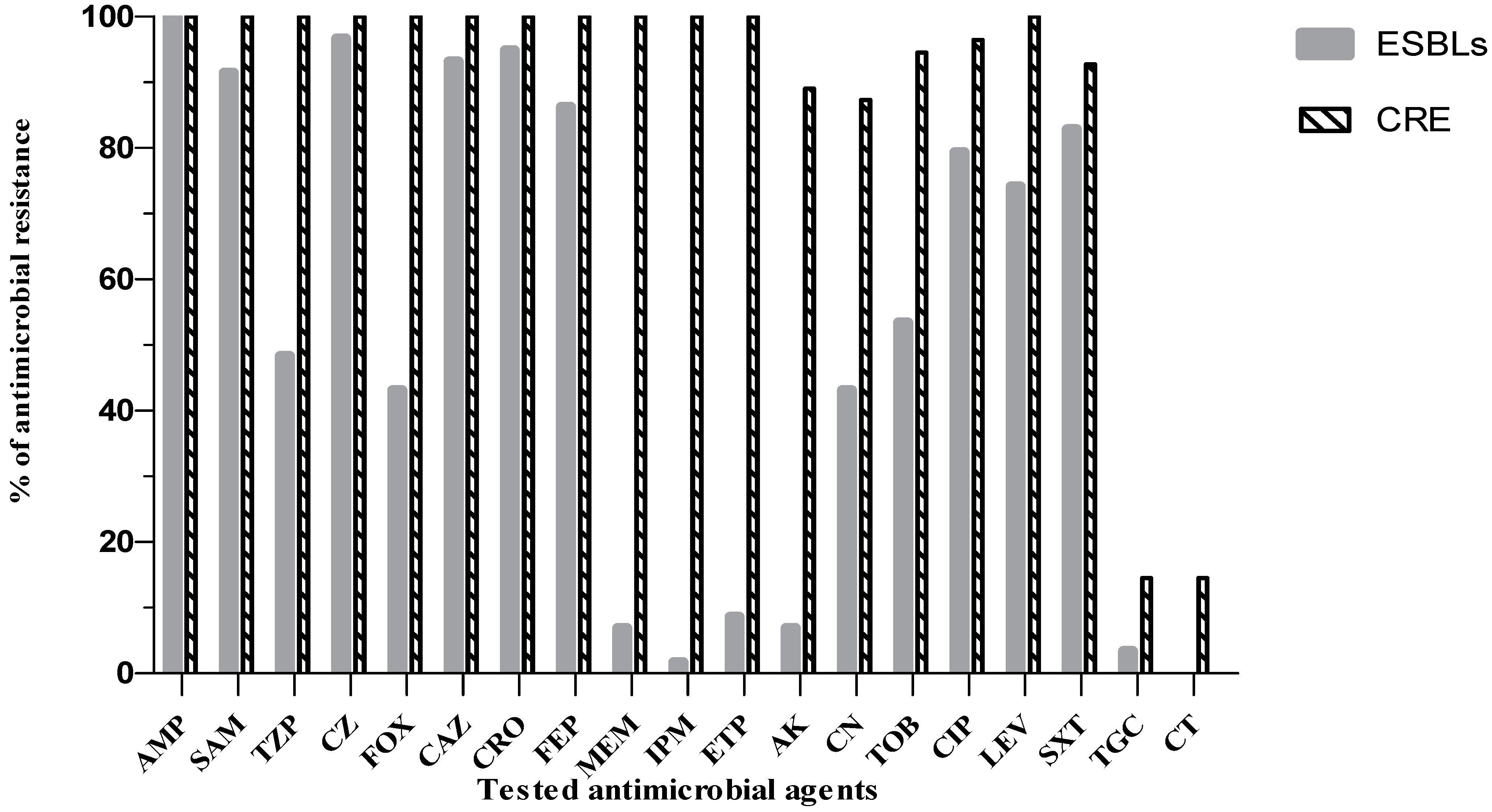
2.2. Antibiogram Analysis of CRE and ESBL Producers

The most commonly recovered CRE and ESBL producers were Klebsiella pneumoniae (80%) and E. coli (67.7%). As depicted in Figure 5, the susceptibility rates to carbapenems among ESBL producers ranged from 91.4% (ertapenem) to 98.3% (imipenem). The other tested β-lactam group showed reduced susceptibility that exceeded 86.2%, except for cefoxitin (43.1%) and piperacillin/tazobactam (48.2%). On the other hand, CRE showed the highest resistance pattern toward all of the tested β-lactam groups. Among ESBL producers, the susceptibility rates to amikacin, gentamicin and tobramycin were 93.1%, 56.9% and 46.6%, respectively. In contrast, CRE had recorded a high resistant pattern toward the tested aminoglycosides ranging from 87.3% to 94.5%. Both groups of MDR organisms showed reduced susceptibility toward tested quinolones and sulfamethoxazole/trimethoprim. The overall susceptibility rates to colistin and tigecycline were greater than 85.5% among CRE and ESBL producers.
The mortality rate within patients infected with ESBL-producing bacteria was 30%, whereas within patients infected with CRE, it was 67%. The highest mortality rates associated with CRE infections were recovered from respiratory tract infections (19, 49%), followed by bloodstream infections (6, 15%), urinary tract infections (8, 21%), surgical site infections (4, 10%), and others including gastrointestinal infections (2, 5%). The highest mortality rates associated with ESBL producers associated infections were recovered from bloodstream infections followed by respiratory tract infections and urinary tract infections.

3. Discussion

Like other developing countries, Egypt has limited data regarding the surveillance system that can track the burden of infectious diseases. Moreover, incomplete data about the etiology of Gram-negative, MDR-associated infections, including pneumonia, bloodstream and genitourinary infections could underestimate the problem by at least 50% [14]. Accordingly, this study was undertaken in a tertiary care hospital to examine the effect of implementing care bundles on the improvement of patient health.
An “all or none approach” was used where each element of the bundle was implemented collectively to achieve the most favorable outcomes [14,15]. A multimodal IPC intervention, such as applying antimicrobial stewardship programs, controlling the source of infection coupled with environmental cleaning and compliance were collectively evaluated. Our study revealed that baseline ESBL producers and CRE rates were 0.8 and 1.3 in the pre-intervention period, and 0.28 and 0.5 in the post-intervention period, respectively. Our results were slightly higher than that reported by a recent study where the ESBL producers and CRE rates were 0.70 and 0.11 in the pre-intervention period and 0.11 and 0.09 in the post-intervention period, respectively [16]. Such discrepancies between these results could be attributed to different sample size and the assessment of diverse interventions that hinders the contribution of infection control measures. Although targeted screening and active surveillance are quite far beyond the capacity of most facilities especially in developing countries, the application of such simple and cost-effective interventions will control the spread of MDR organisms among health care settings as reported by other studies [17,18].
Unlike MDR Gram-positive organisms such as Staphylococcus aureus and Enterococcus spp., active screening as part of a transmission control program for the MDR Gram-negative pathogens remains controversial. However, the European Society of Clinical Microbiology and Infectious Diseases has strongly recommended that such intervention be followed by contact precautions in epidemic settings [18]. Our results reveal that active rectal screening for CRE and ESBL producers is feasible, with a rate of colonization of 23% and 34%, respectively. Our data were similar to other recent studies, with a rate of CRE colonization reaching 21% [4] and ESBL colonization reaching 30.6% among hospitalized patients [19]. Additionally, our data showed that 36% and 27.5% of the positively screened patients had developed subsequent infection with CRE and ESBL producers, respectively. A higher rate of subsequent CRE infections (47%) was reported by another study, ensuring that rectal colonization could be an important epidemiological risk factor for the development of subsequent CRE infection [20]. Concerning ESBL-producers fecal carriers, one study also found that 27% of patients had acquired infections at an ICU [21]. These findings highlight the contribution of colonizing enterobacterial isolates as one of the potential endogenous reservoirs in the human intestine for subsequent infections.
Our study revealed that about 41% of the positively screened CRE patients had acquired CRE infections from community settings and 9% from hospital settings. Concerning positively screened cases with ESBL producers, our data revealed that 50% were CAIs and about 4% were HAIs. The level of CAIs with CRE and ESBL producers was slightly higher than other studies that ranged from 12% to 30% [22,23], respectively, and slightly lower than a recent study in India, where 64.5% suffered from community-acquired ESBL producers infections [24]. Our findings assert that MDR pathogens, especially ESBL producers and CRE, have been more recently recognized in community settings even without prior hospital exposure. Hence, clinicians should give attention to the emerging public health problem by identifying predictive risk factors that could improve patient management. Interestingly, a low incidence of HAIs by CRE and ESBL producers was recorded by our study compared to other previous international studies. Such a promising decline in levels could be related to intense infection-control programs that were applied at our tertiary care hospital.
In this study, the mortality rate within patients infected with CRE was 2.2 times higher than that of ESBLs producing bacteria. Carbapenem-resistant non-bacteremic infections (respiratory tract, urinary tract and wound infections) were associated with 85% mortality and 49% of patients with respiratory tract infections evolved into death. Similar results were reported by Tumbarello et al., as the mortality rate reached 40% among patients with lower respiratory tract infections caused by CR Klebsiella pneumoniae [25]. On the other hand, our data showed that the mortality rate among patients infected with ESBL producers that caused bloodstream infections reached 32%. Similar results were also recently reported where the mortality rate of those who suffered from bloodstream infection due to ESBL-producing E. coli was 30.8% [26]. However, we should put into consideration that mortality might also be due to the severity of the underlying disease and its co-morbidity.
In the past few decades, the incidence of MDR Gram-negative associated infections have increased dramatically with international disparity. Thus, regional investigation of antimicrobial resistance patterns coupled with pathogen stratification is of paramount importance to create, estimate and update empirical and exiting treatment guidelines. Overall, our data revealed that the most common pathogens for CRE and ESBL producers cases were Klebsiella spp. (80%) and E. coli (68%), respectively. Such findings were almost constituent with other findings reporting a high prevalence of Klebsiella spp. ranging from 71% [27] to 85% [28] for CRE cases and a high prevalence of E. coli reaching 60% among ESBL-producers cases [29]. Regarding the susceptibility profile of ESBL producers producing isolates, our data showed a high resistance pattern for third and fourth generation cephalosporins ranging from 86% to 95%, fluoroquinolones between 74% and 79% and trimethoprim/sulfamethoxazole at 83%. This high pattern of co-resistance could be attributed to a relatively large plasmid that encodes ESBL producers enzymes and genes for other non-β-lactam antibiotics, especially for fluoroquinolones, co-trimoxazole and/or aminoglycosides. However, in accordance with other studies, the majority of ESBL producers showed an enhanced sensitivity pattern toward carbapenems, amikacin, colistin [30,31,32] and tigecycline [33]. In comparison to ESBL producers, CRE antibiogram analysis was more severe and complicated, providing evidence that the latter strains are usually resistant to many other classes of antibiotics in clinical practice [34]. With the exception of colistin and tigecycline, CRE showed a high level of resistance (89% to 100%) to other tested antimicrobial classes. Like our findings, other studies have also reported that tigecycline and colistin were the most effective antimicrobial agents against CRE [34,35]; however, literature has reported that tigecycline alone was associated with a high mortality rate and decreased clinical efficacy [36], while renal impairment and the appearance of hetero-resistance were the main drawbacks of colistin monotherapy [37]. In support of this data, clinicians should examine the use of such mainstay antimicrobials agents in combinations to lessen the emergence of high-level resistant phenotypes.

4. Materials and Methods

4.1. Study Design

A prospective study was conducted at the International Medical Center (IMC) from July 2017 to June 2019 to screen carriers or patients infected with ESBL-producing Gram-negative pathogens and CRE. The IMC is an acute care, tertiary hospital with around 800 beds, 10 different ICUs and 3 different wards (oncology, surgical and medical). Authorized members of an infection control team were assigned to collect the microbiological records including the identified culture with its sensitivity pattern from different clinical specimens, including blood, sputum, urine and wound swabs among all ICUs and wards of IMC. The study protocol was reviewed and approved by the institutional ethics committee, Faculty of Pharmacy, Ain Shams University (ENREC-ASU-2018-72). This study was conducted in accordance with the ethical principles stated in the Declaration of Helsinki.

4.2. Microbiological Procedures and Definitions

Rectal swab specimens were collected within 48 hours of ICU admission to assess bacterial colonization from 253 studied participants over a period of 6 months, from July 2017 to December 2017. Swabs were inserted 1 cm into the rectum and then rotated 360°. Amies transport medium was used for immediate transformation for further laboratory analysis. For the rapid detection of CRE and ESBL producers from potentially colonized rectal specimens, the HB&L carbapenemase and ESBL producers-AmpC kits (Alifax, Padua, Italy) were used, respectively. Kits were read by a ALIFAX microbiology line analyzer (Alifax, Padua, Italy) that depends on laser-scattering technology for the automatic detection and enumeration of tested broth vials. Briefly, the laser beam passes through a glass bottle containing the reagent (HB&L carbapenamase or ESBL producers-AmpC) in addition to the specimen and a selective supplement broth that contain certain antibacterial and antifungals agents. For CRE and ESBL detection, the broth was combined with carbapenem and certain 3rd-generation cephalosporins, respectively, to inhibit non-carbapenem resistant Enterobacteriaceae spp. and non-ESBL. The patented technology enabled us to monitor real-time growth curves and positive or negative results were reported based on a calculation of colony-forming unit per mL. As recommended by the manufacturer, we have used the automated system, Vitek-2 (bioMérieux, Marcy L’Etoile; Lyon, France) for the identification and detection of antimicrobial breakpoints.
By definition, the tested strains were considered MDR if they displayed resistance to 3 or more antimicrobial classes, i.e., susceptible to not more than 1 of the following classes by disk diffusion method: aminoglycosides (10 µg), carbapenems (10 µg), cephalosporins (30 µg), fluoroquinolones (5 µg) or penicillin (10 µg) [38]. Susceptibility to polymyxins, sulbactam, sulfonamides and tetracycline were not considered in the definition. The rate of MDR organisms was calculated monthly by dividing the number of MDR by the total number of patient days and multiplying by 1000 [39]. Admitted persons were classified into colonized and cases with infections according to previously published literature [40]. Infections with CRE and ESBL producers were defined as the detection of Enterobacteriaceae isolates resistant to carbapenem antibiotics and 3rd-generation cephalosporins, respectively, with additional signs and symptoms of infection as determined by the clinicians. Detected infection from days 1–3 upon IMC admission was counted as community-acquired (CAIs), while later detections counted as hospital-acquired (HAIs) [41]. The recovered bacterial isolates were tested for susceptibility to the antimicrobial agents recommended by the Clinical and Laboratory Standards Institute (CLSI, 2018) for each bacterial species [42].

4.3. Molecular Identification of ESBL and CR Genes

4.3.1. Extraction of Chromosomal DNA from CRE and ESBL Producers

Overnight cultures of the phenotypically confirmed CRE or ESBL-producing isolates were grown in Luria Bertani broth containing 16 μg/mL meropenem or ampicillin, respectively. The chromosomal DNA was carried out using a GeneJET DNA extraction kit (Thermo Fisher Scientific, Waltham, MA, USA). The extracted chromosomal DNA was analyzed via agarose gel electrophoresis and visualized by a UV transilluminator [43].

4.3.2. PCR Detection of ESBL and CR Genes

The extracted chromosomal DNA of each isolate was used as a template for PCR using the appropriate primers synthesized by Invitrogen (Thermo Fisher Scientific, Waltham, MA, USA), and Dream Taq Green PCR Master Mix (Thermo Fisher Scientific, Waltham, MA, USA). The annealing temperatures (Ta) and primers for ESBL genes—including blaCTX-m, blaTEM and blaSHV as well as for 5 of the major CR genes, including blaKPC, blaNDM, blaVIM, blaOXA-48 and blaIMP—are shown in Table 1. The amplified PCR products were analyzed using agarose gel electrophoresis and the size of DNA fragments was determined by comparing them to a 100 bp DNA ladder (GeneRuler 100 bp DNA ladder, Thermo Fisher Scientific, Waltham, MA, USA).

4.3.3. Sequencing of Selected PCR Products

Some PCR products of amplified genes were sent for sequencing at Macrogen Inc. (Seoul, Korea) using an Applied Biosystems 3730XL sequencer. The assembly of the obtained forward and reverse sequence files into the final consensus sequence was completed using BioEdit v7.2.5 software [50]. The open reading frames (ORFs) of the final contigs were detected using FramePlot 2.3.2 [51]. The sequencing data were analyzed using the basic local alignment search tool.

4.4. Infection Control and Preventitive Interventions

A multidisciplinary team approach, including an infection control department, microbiology laboratory and clinical pharmacy was necessary to develop and implement strategies to control the spread of CRE and ESBL producers among the hospital settings. A pilot study was undertaken for 6 months from July 2017 to December 2017 and a prospective period from January 2018 to August 2019. The following core IPC measures were implemented during the pilot study and enhanced during the second part of the study.

4.4.1. Contact Isolation

The infection control team had trained, monitored and provided feedback on hand hygiene and the use of gloves and gowns [52,53]. In case of carriage or infection with CRE, patient and staff cohorting were applied and such patients were preferably placed in a single room [54,55].

4.4.2. Environmental Cleaning

The infection control team checked that beds and the surroundings were disinfected by chloride detergents. Cultures from different surfaces were randomly taken 2 times during the pilot study [56]. On a daily basis, the new checklist containing all items in the devices bundle was audited by the infection-control team.

4.4.3. Chlorhexidine Bathing

On a routine daily basis, 2% chlorhexidine gluconate (impregnated cloth) was used for whole-body bathing by certified nursing assistants [57,58].

4.4.4. Antimicrobial Stewardship Programs

The clinical pharmacy department started to update the list of restricted antimicrobials and the checklist of compliance. Restricted broad-spectrum antibiotics including 3rd or 4th-generation cephalosporins, carbapenems and fluoroquinolones were only prescribed after approval by a designated infectious diseases physician. Additionally, the clinical department communicated with the microbiology laboratory to update the list of antibiotic combinations and a new pocket pamphlet was prepared to promote the antimicrobial stewardship program.

4.4.5. Statistical Analyses

Data analyses were performed using GraphPad Prism (version 6). Qualitative variables were expressed as frequency and percentages. The incidence density was calculated as infections per 1000 patient days (stratified by infection type).

5. Conclusions

Being a carrier or infected with MDR Enterobacterales, particularly ESBL producers and CRE, requires prompt action by health care settings to control transmission and optimize patient management protocols. The establishment of a multidisciplinary approach, including screening, contact isolation, environmental cleaning and applying antimicrobial stewardship programs showed promising results in reducing the incidence of CRE and ESBL producers per 1000 patient days. Our findings revealed the effectiveness of multi-modal IPC approaches (controlling the source of infection, environmental cleaning and applying antimicrobial stewardship programs) in controlling and preventing infections elicited by CRE and ESBL producers, and hence they will be of great value for guiding clinicians in implementing effective treatment options against the respective clinically relevant pathogens.

Author Contributions

Conceptualization, N.A.K., K.M.E., M.F.A., K.M.A. and M.I.E.B.; methodology, N.A.K., K.M.E., M.F.A., K.M.A. and M.I.E.B.; writing—original draft preparation, N.A.K., K.M.E. and M.I.E.B.; writing—review and editing, M.F.A., K.M.A. and M.I.E.B.; supervision, K.M.A. and M.I.E.B.; statistical analysis, K.M.A. and M.F.A.; funding acquisition, N.A.K., K.M.E., M.F.A., K.M.A. and M.I.E.B. All authors have read and agreed to the published version of the manuscript.

Funding

The current work was funded by Taif University Researchers Supporting Project number (TURSP-2020/111), Taif University, Taif, Saudi Arabia.

Institutional Review Board Statement

The study was conducted according to the guidelines of the Declaration of Helsinki and approved by the Faculty of Pharmacy, Ain Shams University Research Ethics Committee (ENREC-ASU-2018-72).

Informed Consent Statement

Informed consent was obtained from all subjects involved in the study. Written informed consent has been obtained from the patient(s) to publish this paper.

Acknowledgments

The authors express their appreciation to the International Medical Center, Clinical Microbiology Laboratory, Cairo, Egypt for administrative and laboratory support including clinical specimens and utensils used for experiments as well as to Taif University, Taif, Saudi Arabia, for funding current work by Taif University Researchers Supporting Project number (TURSP-2020/111) at Taif University.

Conflicts of Interest

The authors declare no conflict of interest.

References

  1. Podschun, R.; Ullmann, U. Klebsiella Spp. as Nosocomial Pathogens: Epidemiology, Taxonomy, Typing Methods, and Pathogenicity Factors. Clin. Microbiol. Rev. 1998, 11, 589–603. [Google Scholar] [CrossRef] [Green Version]
  2. Navon-Venezia, S.; Kondratyeva, K.; Carattoli, A. Klebsiella Pneumoniae: A Major Worldwide Source and Shuttle for Antibiotic Resistance. FEMS Microbiol. Rev. 2017, 41, 252–275. [Google Scholar] [CrossRef]
  3. Mil-Homens, D.; Martins, M.; Barbosa, J.; Serafim, G.; Sarmento, M.J.; Pires, R.F.; Rodrigues, V.; Bonifácio, V.D.B.; Pinto, S.N. Carbapenem-Resistant Klebsiella Pneumoniae Clinical Isolates: In Vivo Virulence Assessment in Galleria Mellonella and Potential Therapeutics by Polycationic Oligoethyleneimine. Antibiotics 2021, 10, 56. [Google Scholar] [CrossRef] [PubMed]
  4. Abdelwahab, R.; Yasir, M.; Godfrey, R.E.; Christie, G.S.; Element, S.J.; Saville, F.; Hassan, E.A.; Ahmed, E.H.; Abu-Faddan, N.H.; Daef, E.A.; et al. Antimicrobial Resistance and Gene Regulation in Enteroaggregative Escherichia coli from Egyptian Children with Diarrhoea: Similarities and Differences. Virulence 2021, 12, 57–74. [Google Scholar] [CrossRef] [PubMed]
  5. Bassetti, M.; Vena, A.; Croxatto, A.; Righi, E.; Guery, B. How to Manage Pseudomonas Aeruginosa Infections. Drugs Context 2018, 7. [Google Scholar] [CrossRef] [PubMed]
  6. Breijyeh, Z.; Jubeh, B.; Karaman, R. Resistance of Gram-Negative Bacteria to Current Antibacterial Agents and Approaches to Resolve It. Molecules 2020, 25, 1340. [Google Scholar] [CrossRef] [Green Version]
  7. Aslam, B.; Wang, W.; Arshad, M.I.; Khurshid, M.; Muzammil, S.; Rasool, M.H.; Nisar, M.A.; Alvi, R.F.; Aslam, M.A.; Qamar, M.U.; et al. Antibiotic Resistance: A Rundown of a Global Crisis. Infect. Drug Resist. 2018, 11, 1645–1658. [Google Scholar] [CrossRef] [PubMed] [Green Version]
  8. Davies, J.; Davies, D. Origins and Evolution of Antibiotic Resistance. Microbiol. Mol. Biol. Rev. 2010, 74, 417–433. [Google Scholar] [CrossRef] [Green Version]
  9. Sultan, I.; Rahman, S.; Jan, A.T.; Siddiqui, M.T.; Mondal, A.H.; Haq, Q.M.R. Antibiotics, Resistome and Resistance Mechanisms: A Bacterial Perspective. Front. Microbiol. 2018, 9. [Google Scholar] [CrossRef] [Green Version]
  10. Christaki, E.; Marcou, M.; Tofarides, A. Antimicrobial Resistance in Bacteria: Mechanisms, Evolution, and Persistence. J. Mol. Evol. 2020, 88, 26–40. [Google Scholar] [CrossRef]
  11. Tacconelli, E.; Cataldo, M.A.; Dancer, S.J.; De Angelis, G.; Falcone, M.; Frank, U.; Kahlmeter, G.; Pan, A.; Petrosillo, N.; Rodríguez-Baño, J.; et al. European Society of Clinical Microbiology. ESCMID guidelines for the management of the infection control measures to reduce transmission of multidrug-resistant Gram-negative bacteria in hospitalized patients. Clin. Microbiol. Infect. 2014, 20, 1–55. [Google Scholar] [CrossRef] [PubMed] [Green Version]
  12. Redondo-Salvo, S.; Fernández-López, R.; Ruiz, R.; Vielva, L.; de Toro, M.; Rocha, E.P.C.; Garcillán-Barcia, M.P.; de la Cruz, F. Pathways for Horizontal Gene Transfer in Bacteria Revealed by a Global Map of Their Plasmids. Nat. Commun. 2020, 11, 3602. [Google Scholar] [CrossRef] [PubMed]
  13. Peterson, E.; Kaur, P. Antibiotic Resistance Mechanisms in Bacteria: Relationships Between Resistance Determinants of Antibiotic Producers, Environmental Bacteria, and Clinical Pathogens. Front. Microbiol. 2018, 9. [Google Scholar] [CrossRef] [PubMed]
  14. Talaat, M.; El-Shokry, M.; El-Kholy, J.; Ismail, G.; Kotb, S.; Hafez, S.; Attia, E.; Lessa, F.C. National surveillance of health care-associated infections in Egypt: Developing a sustainable program in a resource-limited country. Am. J. Infect. Control 2016, 44, 1296–1301. [Google Scholar] [CrossRef] [Green Version]
  15. Abushaheen, M.A.; Fatani, A.J.; Alosaimi, M.; Mansy, W.; George, M.; Acharya, S.; Rathod, S.; Divakar, D.D.; Jhugroo, C.; Vellappally, S.; et al. Antimicrobial Resistance, Mechanisms and Its Clinical Significance. Dis. Mon. 2020, 66, 100971. [Google Scholar] [CrossRef]
  16. Du, D.; Wang-Kan, X.; Neuberger, A.; Veen, H.W.; van Pos, K.M.; Piddock, L.J.V.; Luisi, B.F. Multidrug Efflux Pumps: Structure, Function and Regulation. Nat. Rev. Microbiol. 2018, 16, 523–539. [Google Scholar] [CrossRef] [PubMed]
  17. Kumar, A.; Schweizer, H.P. Bacterial Resistance to Antibiotics: Active Efflux and Reduced Uptake. Adv. Drug Deliv. Rev. 2005, 57, 1486–1513. [Google Scholar] [CrossRef] [PubMed]
  18. Alibert, S.; N’gompaza Diarra, J.; Hernandez, J.; Stutzmann, A.; Fouad, M.; Boyer, G.; Pagès, J.-M. Multidrug Efflux Pumps and Their Role in Antibiotic and Antiseptic Resistance: A Pharmacodynamic Perspective. Expert Opin. Drug Metab. Toxicol. 2017, 13, 301–309. [Google Scholar] [CrossRef]
  19. King, D.T.; Sobhanifar, S.; Strynadka, N.C.J. The Mechanisms of Resistance to β-Lactam Antibiotics. In Handbook of Antimicrobial Resistance; Springer: New York, NY, USA, 2017; pp. 177–201. [Google Scholar]
  20. Tooke, C.L.; Hinchliffe, P.; Bragginton, E.C.; Colenso, C.K.; Hirvonen, V.H.A.; Takebayashi, Y.; Spencer, J. β-Lactamases and β-Lactamase Inhibitors in the 21st Century. J. Mol. Biol. 2019, 431, 3472–3500. [Google Scholar] [CrossRef]
  21. Mojica, M.F.; Bonomo, R.A.; Fast, W. B1-Metallo-Beta-Lactamases: Where Do We Stand? Curr. Drug Targets 2016, 17, 1029–1050. [Google Scholar] [CrossRef]
  22. Bush, K.; Bradford, P.A. Interplay between β-Lactamases and New β-Lactamase Inhibitors. Nat. Rev. Microbiol. 2019, 17, 295–306. [Google Scholar] [CrossRef] [PubMed]
  23. Kotra, L.P.; Haddad, J.; Mobashery, S. Aminoglycosides: Perspectives on Mechanisms of Action and Resistance and Strategies to Counter Resistance. Antimicrob. Agents Chemother. 2000, 44, 3249–3256. [Google Scholar] [CrossRef] [PubMed] [Green Version]
  24. Ramirez, M.S.; Tolmasky, M.E. Amikacin: Uses, Resistance, and Prospects for Inhibition. Molecules 2017, 22, 2267. [Google Scholar] [CrossRef] [PubMed] [Green Version]
  25. Park, C.H.; Robicsek, A.; Jacoby, G.A.; Sahm, D.; Hooper, D.C. Prevalence in the United States of Aac(6′)-Ib-Cr Encoding a Ciprofloxacin-Modifying Enzyme. AAC 2006, 50, 3953–3955. [Google Scholar] [CrossRef] [PubMed] [Green Version]
  26. World Health Organization WHO Publishes List of Bacteria for Which New Antibiotics Are Urgently Needed. Available online: https://www.who.int/news/item/27-02-2017-who-publishes-list-of-bacteria-for-which-new-antibiotics-are-urgently-needed (accessed on 10 April 2021).
  27. Duan, N.; Du, J.; Huang, C.; Li, H. Microbial Distribution and Antibiotic Susceptibility of Lower Respiratory Tract Infections Patients From Pediatric Ward, Adult Respiratory Ward, and Respiratory Intensive Care Unit. Front. Microbiol. 2020, 11. [Google Scholar] [CrossRef] [PubMed]
  28. Roth, G.A.; Abate, D.; Abate, K.H.; Abay, S.M.; Abbafati, C.; Abbasi, N.; Abbastabar, H.; Abd-Allah, F.; Abdela, J.; Abdelalim, A.; et al. Global, Regional, and National Age-Sex-Specific Mortality for 282 Causes of Death in 195 Countries and Territories, 1980–2017: A Systematic Analysis for the Global Burden of Disease Study 2017. Lancet 2018, 392, 1736–1788. [Google Scholar] [CrossRef] [Green Version]
  29. Troeger, C.; Blacker, B.; Khalil, I.A.; Rao, P.C.; Cao, J.; Zimsen, S.R.M.; Albertson, S.B.; Deshpande, A.; Farag, T.; Abebe, Z.; et al. Estimates of the Global, Regional, and National Morbidity, Mortality, and Aetiologies of Lower Respiratory Infections in 195 Countries, 1990–2016: A Systematic Analysis for the Global Burden of Disease Study 2016. Lancet Infect. Dis. 2018, 18, 1191–1210. [Google Scholar] [CrossRef] [Green Version]
  30. Jit, M.; Ng, D.H.L.; Luangasanatip, N.; Sandmann, F.; Atkins, K.E.; Robotham, J.V.; Pouwels, K.B. Quantifying the Economic Cost of Antibiotic Resistance and the Impact of Related Interventions: Rapid Methodological Review, Conceptual Framework and Recommendations for Future Studies. BMC Med. 2020, 18. [Google Scholar] [CrossRef] [Green Version]
  31. Ventola, C.L. The Antibiotic Resistance Crisis. P T 2015, 40, 277–283. [Google Scholar]
  32. Li, B.; Webster, T.J. Bacteria Antibiotic Resistance: New Challenges and Opportunities for Implant-Associated Orthopaedic Infections. J. Orthop. Res. 2018, 36, 22. [Google Scholar] [CrossRef] [Green Version]
  33. Center for Disease Control and Prevention (CDC). Antibiotic Resistance Threats in the United States, 2013; 2013. Available online: https://www.cdc.gov/ (accessed on 10 April 2021).
  34. Golkar, Z.; Bagasra, O.; Pace, D.G. Bacteriophage Therapy: A Potential Solution for the Antibiotic Resistance Crisis. J. Infect. Dev. Ctries 2014, 8, 129–136. [Google Scholar] [CrossRef] [PubMed]
  35. Center for Disease Control and Prevention (CDC). Antibiotic Resistance Threats in the United States, 2019; 2019. Available online: https://www.cdc.gov/ (accessed on 10 April 2021).
  36. Siwakoti, S.; Subedi, A.; Sharma, A.; Baral, R.; Bhattarai, N.R.; Khanal, B. Incidence and Outcomes of Multidrug-Resistant Gram-Negative Bacteria Infections in Intensive Care Unit from Nepal- a Prospective Cohort Study. Antimicrob. Resist. Infect. Control. 2018, 7. [Google Scholar] [CrossRef] [PubMed] [Green Version]
  37. El-Sokkary, R.H.; Ramadan, R.A.; El-Shabrawy, M.; El-Korashi, L.A.; Elhawary, A.; Embarak, S.; Tash, R.M.E.; Elantouny, N.G. Community Acquired Pneumonia among Adult Patients at an Egyptian University Hospital: Bacterial Etiology, Susceptibility Profile and Evaluation of the Response to Initial Empiric Antibiotic Therapy. Infect. Drug Resist. 2018, 11, 2141–2150. [Google Scholar] [CrossRef] [PubMed] [Green Version]
  38. Tadesse, B.T.; Ashley, E.A.; Ongarello, S.; Havumaki, J.; Wijegoonewardena, M.; González, I.J.; Dittrich, S. Antimicrobial Resistance in Africa: A Systematic Review. BMC Infect. Dis. 2017, 17, 616. [Google Scholar] [CrossRef]
  39. Singh, S.; Sharma, A.; Nag, V.L. Bacterial Pathogens from Lower Respiratory Tract Infections: A Study from Western Rajasthan. J. Fam. Med. Prim. Care 2020, 9, 1407. [Google Scholar] [CrossRef]
  40. Vijay, S.; Dalela, G. Prevalence of LRTI in Patients Presenting with Productive Cough and Their Antibiotic Resistance Pattern. J. Clin. Diagn. Res. 2016, 10, DC09–DC12. [Google Scholar] [CrossRef]
  41. Kang, H.; Zheng, W.; Kong, Z.; Jiang, F.; Gu, B.; Ma, P.; Ma, X. Disease Burden and Molecular Epidemiology of Carbapenem-Resistant Klebsiella Pneumonia Infection in a Tertiary Hospital in China. Ann. Transl. Med. 2020, 8. [Google Scholar] [CrossRef]
  42. Tian, D.; Pan, F.; Wang, C.; Sun, Y.; Zhang, H. Resistance Phenotype and Clinical Molecular Epidemiology of Carbapenem-Resistant Klebsiella Pneumoniae among Pediatric Patients in Shanghai. Infect. Drug Resist. 2018, 11, 1935–1943. [Google Scholar] [CrossRef] [PubMed] [Green Version]
  43. Sambrook, J.; Fritsch, E.F.; Maniatis, T. Molecular Cloning: A Laboratory Manual; Cold Spring Harbor Laboratory Press: Cold Spring Harbor, NY, USA, 1989. [Google Scholar]
  44. Doyle, D.; Peirano, G.; Lascols, C.; Lloyd, T.; Church, D.L.; Pitout, J.D.D. Laboratory Detection of Enterobacteriaceae That Produce Carbapenemases. J. Clin. Microbiol. 2012, 50, 3877–3880. [Google Scholar] [CrossRef] [PubMed] [Green Version]
  45. Nordmann, P.; Poirel, L.; Carrer, A.; Toleman, M.A.; Walsh, T.R. How To Detect NDM-1 Producers. J. Clin. Microbiol. 2011, 49, 718–721. [Google Scholar] [CrossRef] [Green Version]
  46. Poirel, L.; Potron, A.; Nordmann, P. OXA-48-like carbapenemases: The phantom menace. J. Antimicrob. Chemother. 2012, 67, 1597–1606. [Google Scholar] [CrossRef] [PubMed] [Green Version]
  47. Woodford, N.; Tierno, P.M.; Young, K.; Tysall, L.; Palepou, M.-F.I.; Ward, E.; Painter, R.E.; Suber, D.F.; Shungu, D.; Silver, L.L.; et al. Outbreak of Klebsiella pneumoniae Producing a New Carbapenem-Hydrolyzing Class A β-Lactamase, KPC-3, in a New York Medical Center. Antimicrob. Agents Chemother. 2004, 48, 4793–4799. [Google Scholar] [CrossRef] [PubMed] [Green Version]
  48. Bonnet, R.; Dutour, C.; Sampaio, J.; Chanal, C.; Sirot, D.; Labia, R.; De Champs, C.; Sirot, J. Novel cefotaximase (CTX-M-16) with increased catalytic efficiency due to substitution Asp-240→ Gly. Antimicrob. Agents Chemother. 2001, 45, 2269–2275. [Google Scholar] [CrossRef] [PubMed] [Green Version]
  49. Rasheed, J.; Jay, C.; Metchock, B.; Berkowitz, F.; Weigel, L.; Crellin, J.; Steward, C.; Hill, B.; Medeiros, A.; Tenover, F.J.A.A.; et al. Evolution of extended-spectrum beta-lactam resistance (SHV-8) in a strain of Escherichia coli during multiple episodes of bacteremia. Antimicrob. Agents Chemother. 1997, 41, 647–653. [Google Scholar] [CrossRef] [PubMed] [Green Version]
  50. Hall, T.A. BioEdit: A user-friendly biological sequence alignment editor and analysis program for Windows 95/98/NT. Nucleic Acids Symp. Ser. 1999, 41, 95–98. [Google Scholar]
  51. Ishikawa, J. FramePlot: A new implementation of the Frame analysis for predicting protein-coding regions in bacterial DNA with a high G+C content. FEMS Microbiol. Lett. 1999, 174, 251–253. [Google Scholar] [CrossRef] [PubMed]
  52. Zhang, Y.; Wang, Q.; Yin, Y.; Chen, H.; Jin, L.; Gu, B.; Xie, L.; Yang, C.; Ma, X.; Li, H.; et al. Epidemiology of Carbapenem-Resistant Enterobacteriaceae Infections: Report from the China CRE Network. Antimicrob. Agents Chemother. 2018, 62. [Google Scholar] [CrossRef] [Green Version]
  53. Buehrle, D.J.; Shields, R.K.; Clarke, L.G.; Potoski, B.A.; Clancy, C.J.; Nguyen, M.H. Carbapenem-Resistant Pseudomonas Aeruginosa Bacteremia: Risk Factors for Mortality and Microbiologic Treatment Failure. Antimicrob. Agents Chemother. 2016, 61. [Google Scholar] [CrossRef] [Green Version]
  54. Gao, B.; Li, X.; Yang, F.; Chen, W.; Zhao, Y.; Bai, G.; Zhang, Z. Molecular Epidemiology and Risk Factors of Ventilator-Associated Pneumonia Infection Caused by Carbapenem-Resistant Enterobacteriaceae. Front. Pharmacol. 2019, 10, 262. [Google Scholar] [CrossRef]
  55. Kumudunie, W.G.M.; Wijesooriya, L.I.; Wijayasinghe, Y.S. Comparison of Four Low-Cost Carbapenemase Detection Tests and a Proposal of an Algorithm for Early Detection of Carbapenemase-Producing Enterobacteriaceae in Resource-Limited Settings. PLoS ONE 2021, 16, e0245290. [Google Scholar] [CrossRef]
  56. Perez, F.; El Chakhtoura, N.G.; Papp-Wallace, K.; Wilson, B.M.; Bonomo, R.A. Treatment Options for Infections Caused by Carbapenem-Resistant Enterobacteriaceae: Can We Apply “Precision Medicine” to Antimicrobial Chemotherapy? Expert Opin. Pharmacother. 2016, 17, 761–781. [Google Scholar] [CrossRef] [PubMed] [Green Version]
  57. Limbago, B.M.; Rasheed, J.K.; Anderson, K.F.; Zhu, W.; Kitchel, B.; Watz, N.; Munro, S.; Gans, H.; Banaei, N.; Kallen, A.J. IMP-Producing Carbapenem-Resistant Klebsiella Pneumoniae in the United States. J. Clin. Microbiol. 2011, 49, 4239–4245. [Google Scholar] [CrossRef] [PubMed] [Green Version]
  58. Mushtaq, S.; Vickers, A.; Doumith, M.; Ellington, M.J.; Woodford, N.; Livermore, D.M. Activity of β-Lactam/Taniborbactam (VNRX-5133) Combinations against Carbapenem-Resistant Gram-Negative Bacteria. J. Antimicrob. Chemother. 2021, 76, 160–170. [Google Scholar] [CrossRef] [PubMed]
Figure 1. Incidence rate of CRE from the period 2017–2019.
Antibiotics 10 00509 g001
Figure 2. Incidence rate of ESBL producers from the period 2017–2019.
Antibiotics 10 00509 g002
Figure 3. Rate of CRE distribution among various clinical specimens per 1000 patient days over the period 2017–2019.
Antibiotics 10 00509 g003
Figure 4. Rate of ESBL producers distribution among various clinical specimens per 1000 patient days over the period 2017–2019.
Antibiotics 10 00509 g004
Figure 5. Antibiogram analysis of CRE and ESBL producers against various antimicrobial agents. AMP: Ampicillin, SAM: Ampicillin/Sulbactam, TZP: Piperacillin/Tazobactam, CZ: Cefazolin, FOX: Cefoxitin, CAZ: Ceftazidime, CRO: Ceftriaxone, FEP: Cefepime, MEM: Meropenem, IPM: Imipenem, ETP: Ertapenem, AK: Amikacin. CN: Gentamicin, TOB: Tobramycin, CIP: Ciprofloxacin, LEV: Levofloxacin, SXT: Trimethoprim/Sulfamethoxazole, TGC: Tigecycline, CT: Colistin.
Antibiotics 10 00509 g005
Table 1. Primers, expected PCR product sizes and annealing temperatures (Ta) of the tested genes.
GenePrimerPrimer Sequence (5’ → 3’)Expected PCR Product Size (bp)Ta (°C)References
blaKPCPfTGTCACTGTATCGCCGTC101150[44]
PrCTCAGTGCTCTACAGAAAACC
blaNDMPfGGTTTGGCGATCTGGTTTTC621[45]
PrCGGAATGGCTCATCACGAT
blaVIMPfTCTACATGACCGCGTCTGTC74850[46]
PrTGTGCTTTGACAACGTTCGC
blaOXA-48PfGCGTGGTTAAGGATGAACAC438[44]
PrCATCAAGTTCAACCCAACCG
blaIMPPfCTACCGCAGCAGAGTCTTTG58750[47]
PrAACCAGTTTTGCCTTACCAT
blaCTX-mPfCGCTTTGCGATGTGCAG55047[48]
PrACCGCGATATCGTTGGT
blaTEMPfATGAGTATTCAACATTTCCG86747[49]
PrCTGACAGTTACCAATGCTTA
blaSHVPfGGTTATGCGTTATATTCGCC86747[49]
PrTTAGCGTTGCCAGTGCTC
Notes: blaKPC, blaNDM, blaVIM, blaOXA-48 and blaIMP: gene codes for KPC, NDM, VIM, OXA-48-like, and IMP carbapenemases, respectively. blaCTX-m: gene coding for cefotaxime (CTX-M) extended-spectrum β-lactamase, blaTEM: gene coding for TEM extended-spectrum β-lactamase, blaSHV: gene coding for SHV extended-spectrum β-lactamase, Pf: forward primer, Pr: reverse primer, Ta: annealing temperature.
Publisher’s Note: MDPI stays neutral with regard to jurisdictional claims in published maps and institutional affiliations.

