Article

Evaluation of the Photodynamic Therapy with Curcumin on L. braziliensis and L. major Amastigotes

by
André Henrique Correia Pereira
,
Luciana Maria Cortez Marcolino
,
Juliana Guerra Pinto
and
Juliana Ferreira-Strixino
*
Photobios–Photobiology Applied to Health, University of Vale do Paraíba, Urbanova, São José dos Campos 2911, SP, Brazil
*
Author to whom correspondence should be addressed.
Submission received: 31 March 2021 / Revised: 29 April 2021 / Accepted: 29 April 2021 / Published: 25 May 2021
(This article belongs to the Special Issue New and Innovative Applications of Antimicrobial Photodynamic Therapy)

Abstract

:
Cutaneous leishmaniasis (CL) is a neglected disease prevalent in tropical countries with the ability to cause skin lesions. Photodynamic therapy (PDT) represents a specific and topical option for the treatment of these lesions. This study evaluated the response of macrophages infected with L. braziliensis and L. major to PDT with curcumin. Curcumin concentrations were evaluated in serial dilutions from 500.0 to 7.8 µg/mL using LED (λ = 450 ± 5 nm), with a light dose of 10 J/cm2. The Trypan blue viability test, ultrastructural analysis by scanning electron microscopy (SEM), mitochondrial polarity by Rhodamine 123 (Rho 123), curcumin internalization by confocal microscopy, and counting of the recovered parasites after the PDT treatment were performed. The lowest concentrations of curcumin (15.6 and 7.8 µg/mL) presented photodynamic inactivation. Cell destruction and internalization of curcumin in both macrophages and intracellular parasites were observed in microscopy techniques. In addition, an increase in mitochondrial membrane polarity and a decrease in the number of parasites recovered was observed in the PDT groups. This study indicates that PDT with curcumin has the potential to inactivate infected macrophages and might act as a basis for future in vivo studies using the parameters herein discussed.

1. Introduction

Cutaneous leishmaniasis (CL) is a neglected disease caused by a protozoan of the genus Leishmania spp., typical of tropical regions and commonly present in underdeveloped countries. WHO places CL as one of the 10 most important infectious diseases worldwide, being present in more than 85 countries, and worsening the situation of people who, due to the development of the disease, may have to leave their professional occupations due to the physical and psychosocial disorders they face [1,2,3].
The treatment of CL is done through the administration of pentavalent antimonials such as Pentosan® and Glucantime®, or even with the use of Amphotericin B in more severe cases. However, these drugs present several side effects such as nausea, vomiting, and cardiac disorders, among others, besides presenting restrictions on their use by patients with heart problems or pregnant women [4,5].
Photodynamic therapy (PDT) is an alternative to the conventional treatment for CL, standing out for being non-invasive and presenting very few application restrictions for the patients. PDT is based on the inactivation of infected cells through the production of reactive oxygen species (ROS) from the photochemical process that results from the interaction between a photosensitizer (PS) and light at a wavelength suitable to it releasing oxygen [6,7].
CL is potentially more harmful to the most impoverished populations in developing countries, since its transmission usually affects individuals with poor access to health conditions, basic sanitation, and low purchasing power. Therefore, PDT may represent an important alternative since it presents minimum side effects and can be easily implemented. Clinical applications of PDT in CL treatment using PS, such as methyl-ALA, is widely reported, indicating the potential of this therapy [8,9,10]. However, it is important to consider that if the treatment cost is low, the access to the therapy for the poorest population will be greater, especially in underdeveloped countries such as Brazil, Colombia, Afghanistan, Peru, and Bolivia [1,11].
A feature that could make the application of PDT a less expensive method is the use of a low-cost and easily obtained PS, such as curcumin. This orange pigment compound is derived from Curcuma longa and is currently used as an oral supplement in the management of various dermatological conditions [12]. The spectrum of light absorption is between 430 and 500 nm, allowing PDT with curcumin to be applied superficially to skin lesions caused by Leishmania [13,14]. However, studies evaluating the use of this PS are still scarce, especially those involving the co-culture of macrophages, a condition that most closely resembles the clinical development of CL.
Considering the scarcity of studies evaluating the effects of curcumin as PS in the treatment of CL, this work aimed to evaluate the in vitro response of PDT with curcumin in the amastigotes of L. braziliensis and L. major, analyzing the inactivation of the infected macrophages, the interaction of the cells with curcumin, the cell morphology after the therapy, the mitochondrial alterations, and the retrieval of Leishmania after PDT.

2. Results

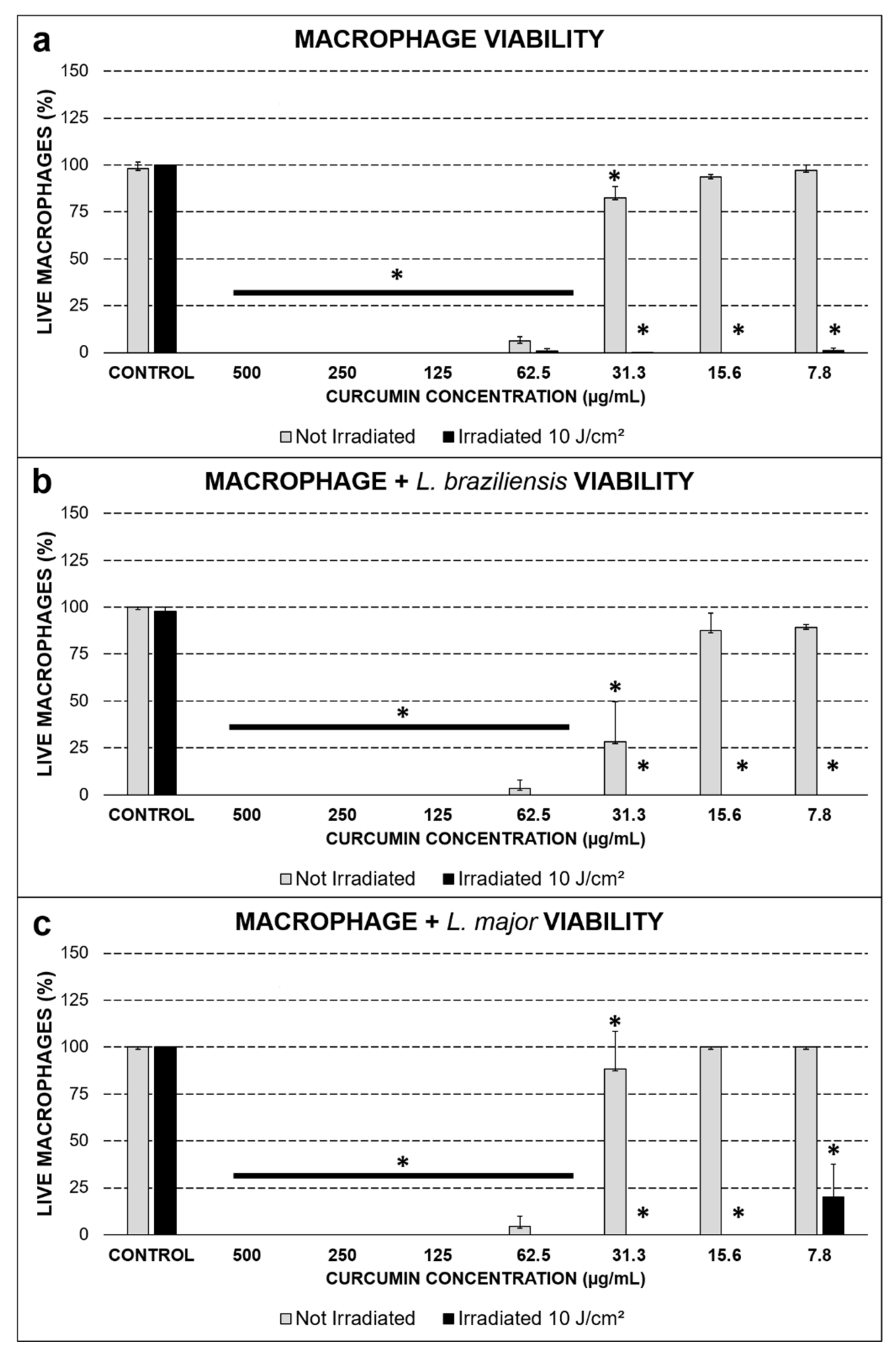
2.1. Viability Assessed by the Trypan Blue Exclusion Test

In the viability test, the cytotoxic effect of curcumin in the dark was assessed at 500.0, 250.0, 125.0, and 62.5 µg/mL. This behavior was repeated when the uninfected macrophages were evaluated, as well as those infected with the two species of Leishmania, indicating that these concentrations were not interesting for in vitro application, since only curcumin in the dark resulted in cell death. However, at low PS concentrations, the viability of the groups kept in the dark was higher than 80% (p ≤ 0.01) at 15.6 and 7.8 µg/mL, and when submitted to PDT, the groups maintained viability below 5%, even reaching the complete inactivation of the macrophages infected with L. braziliensis and L. major. At the lowest concentration evaluated (7.8 µg/mL), the macrophages infected with L. major were slightly more resistant to PDT under these parameters (Figure 1).

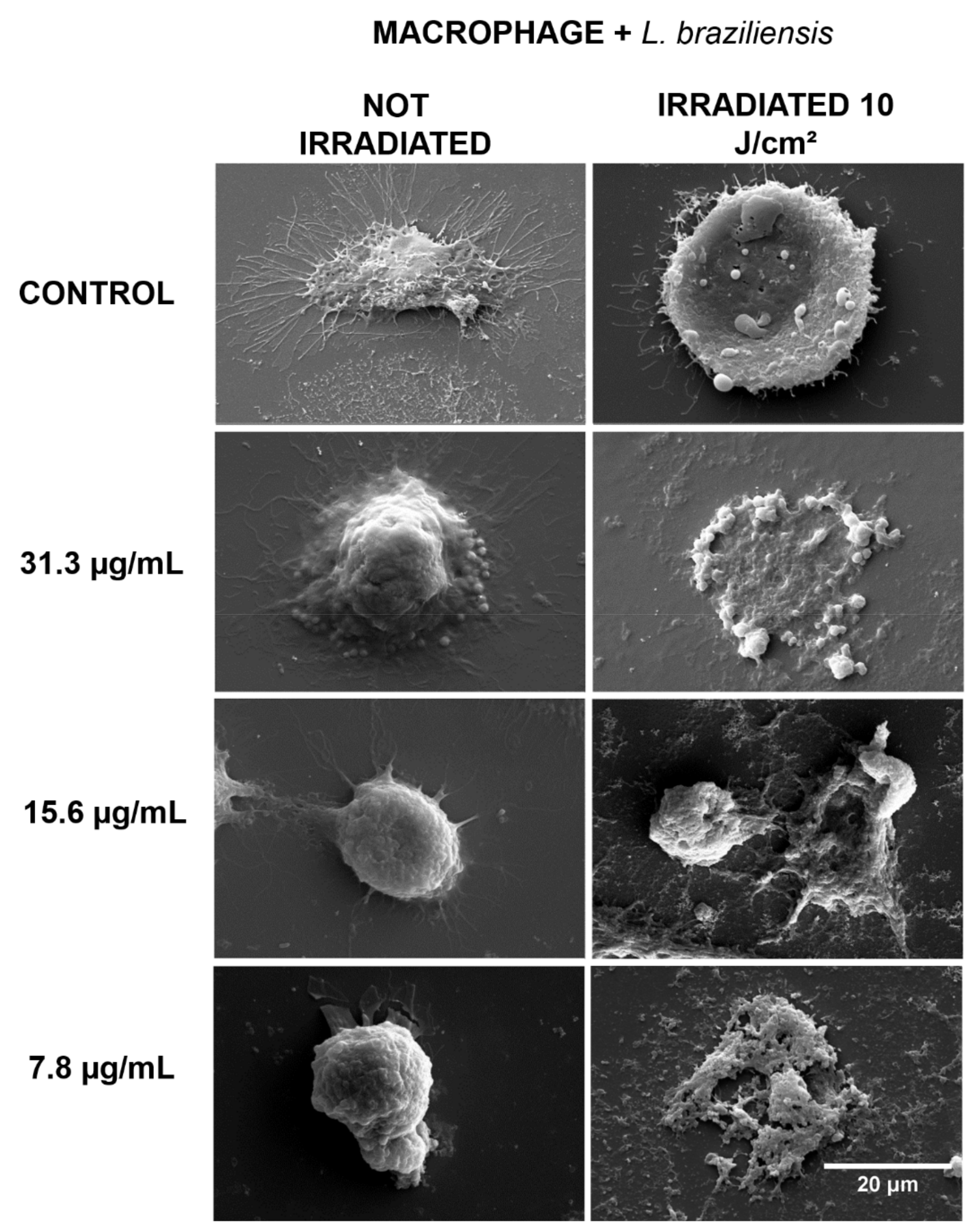
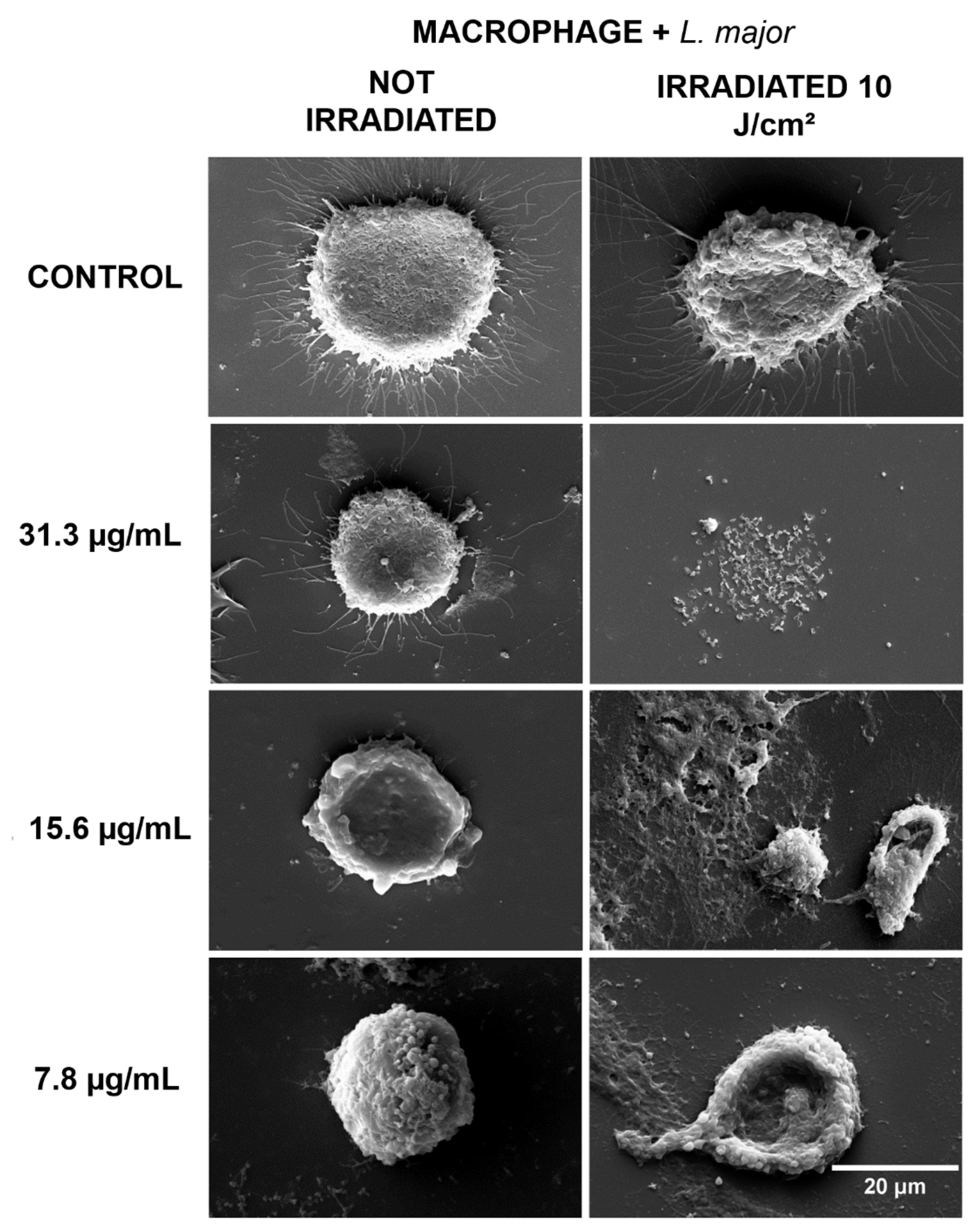
2.2. Morphological Evaluation by Scanning Electron Microscopy

Considering the results obtained in the viability test, concentrations of 31.3, 15.6, and 7.8 µg/mL were selected to carry out the evaluation of cell morphology. In the groups kept in the dark, the characteristic morphology was observed, with no visible damage detected. In the groups that received PDT, intense cell destruction was observed, indicating that the changes occurred due to the action of this substance (Figure 2).
Regarding the macrophages infected with L. braziliensis (Figure 3) and L. major (Figure 4) that were kept in the dark, a higher number of cytoplasmic prolongations was observed in the cells from the non-irradiated groups, in a process expected for phagocytic cells, such as macrophages, in the presence of foreign organisms like Leishmania. In the groups that received PDT, intense cellular destruction of infected macrophages was observed, confirming the inactivation promoted by PDT, as shown in the results of cell viability.

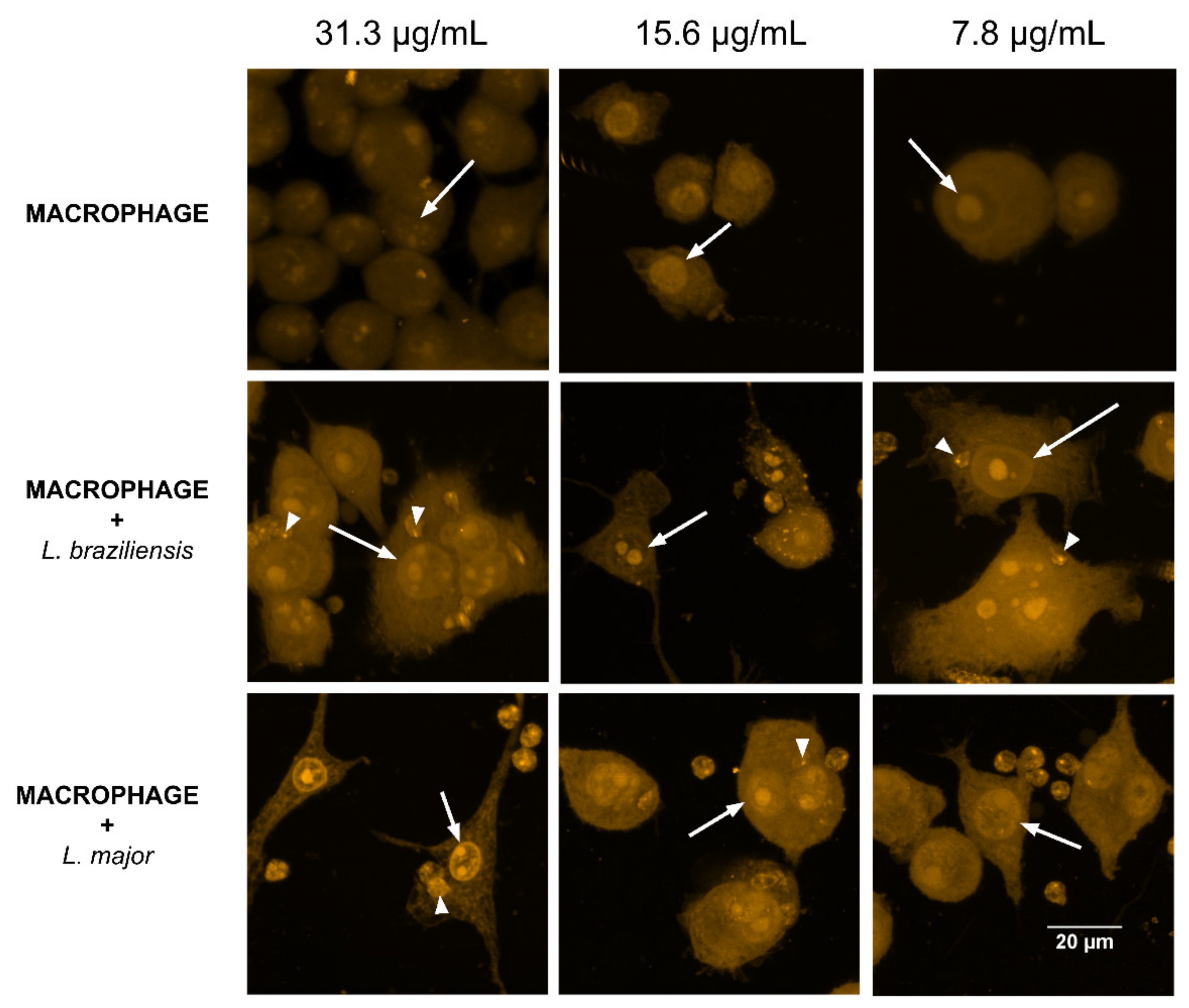
2.3. Internalization of Curcumin in Isolated and Leishmania-Infected Macrophages

The internalization of curcumin in concentrations that presented the best response in the viability test was evaluated. The analysis of the images (Figure 5) allowed us to observe that at all three concentrations curcumin was internalized in the cells since no fluorescent markers were used except for this substance. The PS distribution was observed throughout the cell cytoplasm and in the cell nucleus, suggesting that besides being dispersed in the cell, curcumin can cross the nuclear membrane (arrows). Considering that several biomolecules can be the target of interaction with the ROS produced by PDT, DNA becomes yet another cellular target for inactivation in these evaluated cells. Additionally, an accumulation of curcumin can be observed in the amastigotes internalized by macrophages infected with both species of Leishmania (arrowheads). This result is very important since amastigotes internalized by macrophages can also be a direct target of the ROS produced.

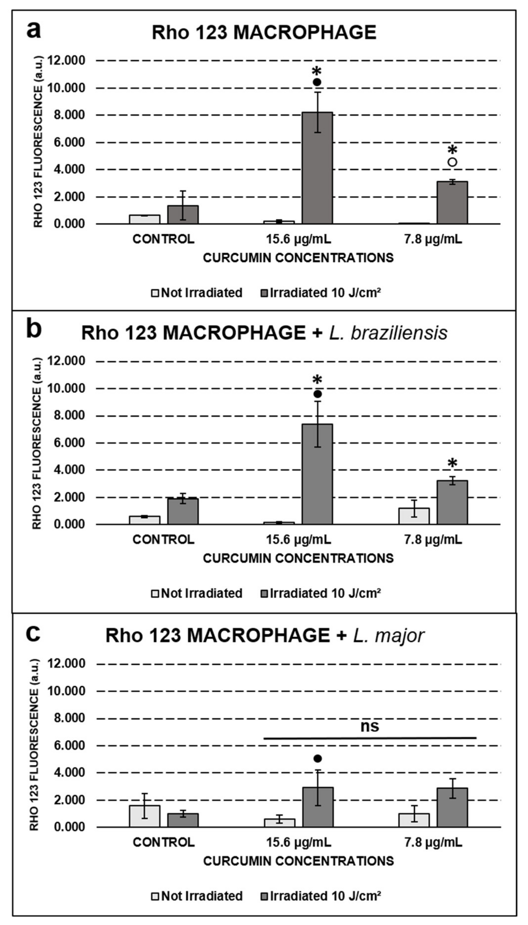
2.4. Qualitative and Quantitative Assessment of Mitochondrial Activity by Rho 123

Rho 123 is a compound that actively interacts with the mitochondrial membrane, presenting high fluorescence emissions in a broad variety of active membranes and low fluorescence in low active ones. This study showed that the controls presented less mitochondrial activity than the groups submitted to PDT (p ≤ 0.01), both for uninfected macrophages and for those infected with L. braziliensis. The fluorescence emitted by the control group was not statistically different (p > 0.05) from that produced by the treated groups incubated only with curcumin for the macrophages infected with L. major. When comparing groups submitted to PDT, mitochondrial polarization by Rho 123 was concentration dependent (p ≤ 0.01), showing higher fluorescence at 15.6 than at 7.8 µg/mL, for macrophages both uninfected and infected with L. braziliensis (Figure 6a,b).

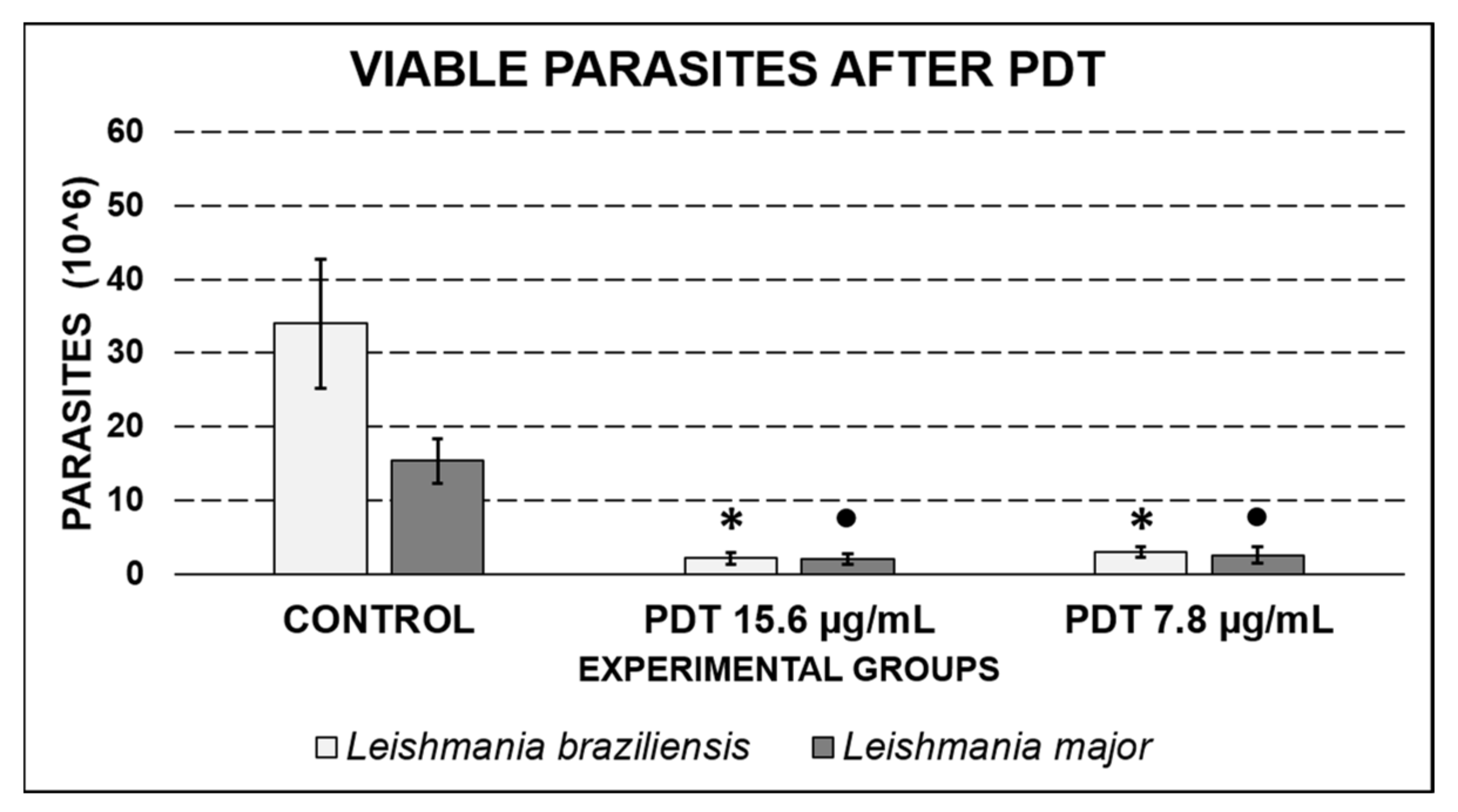
2.5. Retrieval of Leishmania after PDT Exposure

The recovery test of the post-PDT amastigote was carried out in order to verify the impact of PDT on the growth of the parasites. As can be seen in Figure 7, the control groups for L. braziliensis and L. major presented 35 × 106 and 15 × 106 cells/mL, respectively, while the groups submitted to PDT exhibited fewer than 5 × 10⁶ cells/mL (statistically significant decrease, p ≤ 0.01). There were no statistical differences in the quantification of post-PDT parasites when comparing the curcumin concentrations of 15.6 and 7.8 µg/mL (p > 0.05), a condition observed for both the evaluated species.

3. Discussion

This study showed that PDT reduced the viability of RAW 264.7 macrophages infected with L. braziliensis and L. major. In addition, we tried to interpret the mechanisms that resulted in the death of these cells, considering the potential of the mitochondrial membrane and the internalization of curcumin in the evaluated macrophages. While there is a considerable number of in vitro studies that involve the application of PDT in Leishmania promastigotes, especially using photosensitizers such as methylene blue and different phthalocyanines [15,16,17,18,19,20,21], the literature involving the application of PDT with curcumin is scarce, especially when it comes to assays with infected macrophages. Such a model is closer to the expected in clinical applications and in vivo, being an important contribution to the knowledge in this area.
This study showed that PDT induced a viability reduction in the groups with curcumin at 31.3, 15.6, and 7.8 µg/mL. A previous study by our research group demonstrated that macrophages infected with Leishmania were more sensitive to the treatment with lower concentrations of PS than parasites at the promastigote phase [22]. In order to maintain the integrity of the tissue adjacent to CL lesions, the effectiveness of the proposed protocol at low curcumin concentrations is a promising result.
In addition, cells infected with L. braziliensis were more sensitive to the treatment than cells infected with L. major, a result similar to the one observed by Oliveira and collaborators (2015), who recorded a higher efficacy of the therapy for promastigotes of L. braziliensis than for those of L. amazonensis [18]. This finding is positive for countries in South America, especially Brazil, where the L. braziliensis is the main species responsible for CL cases.
The SEM images showed the typical cell morphology of macrophages in the controls and in the groups treated only with curcumin, while in the groups treated with PDT, there was intense morphological change and cell destruction. Cell lysis, the loss of the rounded morphology with cytoplasmic extensions typical of macrophages, and the presence of cell fragments in the fields indicate the destruction of these cells caused by the action of PDT. Morphological changes such as the loss of the characteristic spindle-shaped morphology and the loss of cell attachments such as flagella have been described in several studies with different photosensitizers [23,24,25]. However, the literature lacks the same type of evaluation regarding infected macrophages and especially studies using curcumin. It is worth mentioning that curcumin alone was not able to cause morphological changes in the cells, at the concentrations tested, confirming its low cytotoxicity at low concentrations.
A concern arising from the results obtained in this study was to assess whether the destroyed macrophages would release viable amastigotes after the PDT treatment. The recovery test for parasites after PDT showed a lower growth rate for these parasites compared to the untreated control, indicating that the therapy was able to inactivate both the parasitized macrophages and the internalized amastigotes. Andrade and collaborators (2018) observed a 40% reduction in the number of intracellular amastigote parasites after treatment with PDT. Zinc porphyrin was used as a PS and irradiation was set at a wavelength of 455 ± 20 nm with a fluence of 90 and 180 J/cm2. They also demonstrated that the viability of the internalized amastigotes was compromised during the action of PDT [21].
The results obtained in the internalization of curcumin by infected macrophages reinforced the potential for photodynamic inactivation of infected macrophages and internalized amastigotes, due to the fluorescence signal of curcumin observed in intracellular parasites. Considering the small diffusion capacity and the short half-life of the ROS formed in the photochemical process of PDT, the location of the PS is closely related to its place of action [26,27,28]. In addition, the observed result reiterates what was found in our previous work [23], the binding of curcumin to the cell nucleus due to its affinity with DNA, as proposed in other studies [29,30,31].
The assessment of the mitochondrial membrane potential (Δψm) was made through the marking with Rho 123, which showed higher intensity in the groups treated with PDT. Rho 123 can be used as a marker for Δψm, indicating that in the fully active and undamaged membranes, this potential will be higher compared to the damaged ones [32,33,34].
Fedyaeva and colleagues (2014) found a correlation between the increased mitochondrial ROS production and the hyperpolarization of the mitochondrial membrane during the temperature rise [35]. ROS are produced by the mitochondrial route in order to signal and activate antioxidant cellular mechanisms. However, the high production of these species may end up resulting in a stimulation of the apoptotic cell death route [36]. Therefore, the 18-h time used in this study to evaluate the samples could perhaps coincide with this type of cellular response, not ruling out subsequent cell death from apoptosis, since most cells were not viable in the Trypan blue viability test.
The results also indicate that the macrophages infected with L. major did not show differences in Δψm compared to the control group. This response may be related to the different antioxidant capacities of Leishmania species. After all, macrophages infected with L. major were also less susceptible to PDT at lower concentrations of curcumin. A study performed by Tasbihi et al. demonstrated the presence of different rates of expression of mitochondrial proteins in L. tropica compared with L. braziliensis, a trait that ensures resistance to the drugs routinely used against leishmaniasis [37]. Considering this information, different species of Leishmania with different rates and types of antioxidant mitochondrial protein expression, such as superoxide dismutase (SOD), for example, may respond differently to PDT [38].
Although curcumin has presented toxicity to macrophages at concentrations higher than 15.6 µg/mL, the finding of greater resistance in macrophages infected with L. major has also been observed previously in a study by Silva and collaborators [18]. Lower viability rates were observed after the PDT treatment in macrophages infected by L. braziliensis compared to those infected by L. amazonensis. In the present study, it was observed that the changes in cell viability were confirmed by the morphological changes observed by the SEM images. The intense destruction of the cell is probably linked to the wide distribution of curcumin in its cytoplasm and nucleus of the cell, and its possible interaction with the mitochondria is revealed by the change in the patterns of Rho 123.
Since Leishmania is a parasite that establishes itself inside the cell and manages to evade the defense system of the host, the observation that a large number of the internalized parasites was killed by PDT after a single application is an important finding. If the therapy eliminated only the host macrophages, there would be a risk of releasing viable parasites to infect new cells. However, this study showed that alternate applications of PDT could completely eliminate the parasites from the local lesions.

4. Materials and Methods

4.1. Parasite Culture

L. braziliensis (M2904) and L. major (LV39) strains were used. Initially, the promastigote forms of the parasites were maintained in liver infusion tryptose (LIT) medium supplemented with 10% fetal bovine serum (FBS), 1% penicillin/streptomycin solution, 2 mM glutamine, 1% urine, and 2.5 mg/mL of hemine, and were peaked weekly according to the log phase of growth.

4.2. Cultivation of Macrophages

The macrophage strain RAW 264.7 acquired from the Rio de Janeiro cell bank was kept in 200 µL of Dulbecco’s Modified Eagle’s Medium (DMEM) supplemented with 10% FBS and 1% penicillin/streptomycin solution, and stored in an incubator at 37 °C with 5% CO2.

4.3. Macrophage Infection

For the macrophage infection, these cells interacted with Leishmania strains in the proportion of 10:1. First, 1 × 10⁴ macrophages were counted and transferred to each well, with a total of 24 well plates, and left for adhesion for 12 h in an incubator at 37 °C and 5% CO2. After that period, the parasites were counted and 1 × 10⁵ promastigotes were added to each well containing macrophages. The internalization period lasted for 24 h in an incubator at 37 °C and 5% CO2. Finally, the evaluation of the internalization was done by visualization in an inverted microscope Zeiss Axio Vert. A1.

4.4. Curcumin Preparation

Curcumin (PDT Pharma) stock solution was initially diluted in dimethylsulfoxide (DMSO) at the concentration of 100 mg/mL. Subsequently, a 1:10 dilution in alcohol was made to obtain a 10 mg/mL curcumin solution in DMSO/alcohol. The work solutions for the experiments were prepared by diluting the stock solution in phosphate-buffered saline (PBS), and the highest concentration was diluted in a 1:20 ratio. The final proportion of DMSO in the working solutions was lower than 1%.

4.5. Photodynamic Therapy

All the tests were performed in triplicate, keeping the experimental groups sheltered from light. The experimental groups were divided into non-irradiated (control and curcumin groups without irradiation at different concentrations) and irradiated (irradiated control group and PDT groups with curcumin at different concentrations). After preparing the infected culture described in topics 4.2 and 4.3, 200 µL of working solutions of curcumin were added directly into culture wells without medium, remaining in incubation (37 °C and 5% CO2) for 1 h. The control and irradiated control groups were incubated with PBS under the same conditions. An LED-based device (Biotable Irrad/LED) was used for irradiation, with a wavelength of 450 ± 5 nm, power of 3 W per LED, irradiance of 36 mW/cm2, and fluence of 10 J/cm2. The irradiation time was set up in 278 s. The viability and mitochondrial potential tests were carried out 18 h after the irradiation of the experimental groups. Until then, the cells were incubated with 200 µL DMEM in an incubator at 37 °C and 5% CO2.

4.6. Viability Assessed by the Trypan Blue Exclusion Test

The Trypan blue exclusion test is based on the property of living cells to eliminate the dye, while dead cells lose the selective ability of their membranes and tend to accumulate the compound in the intracellular medium. The test was performed 18 h after the PDT application, adding a Trypan blue solution (0.2%) followed by a five-minute incubation period. Viability was assessed by counting the number of living and dead cells in 10 different random fields for each well.

4.7. Morphological Evaluation by Scanning Electron Microscopy

The three lowest curcumin concentrations evaluated in the viability test were selected to assess the morphology of the macrophages after PDT using SEM. The cells observed under SEM were initially fixed with 2.5% glutaraldehyde solution diluted in PBS with 0.9% saline and pH 7.4. After fixation and post-fixation with osmium tetroxide solution, dehydration was carried out, immersing the coverslips for 10 min successively in baths with 30, 40, 50, 60, 70, 80, 90, and 100% ethanol, the latter being repeated twice. Finally, a drop of hexamethyldisilazane (HMDS) was added, followed by drying in an incubator.
The samples were metalized with gold and analyzed using a scanning electron microscope Zeiss EVO MA 10.

4.8. Curcumin Internalization in Macrophages Isolated and Infected with Leishmania

The process of curcumin internalization started with the culture and infection of macrophages performed in glass coverslips arranged in 24 well plates. The internalization of curcumin at 31.3, 15.6, and 7.8 µg/mL was evaluated. The incubation time was one hour, followed by washing with PBS and fixing with 4% paraformaldehyde for 15 min at room temperature. After fixing, the coverslips were again washed with PBS and mounted on glass slides. The analysis was performed under a Zeiss LSM 700 confocal microscope.

4.9. Evaluation of Mitochondrial Activity by Rhodamine 123 Fluorescent Staining

Rho 123 is a cationic fluorescent green dye that tends to accumulate in the mitochondria without having a cytotoxic effect, being used to assess the potential of the mitochondrial membrane in apoptotic cells. For this evaluation, macrophages were grown in circular coverslips and subsequently infected with Leishmania species evaluated in this study. After a period of incubation of 18 h after PDT, the groups were labeled with 20.0 µg/mL of Rho 123 for 40 min at room temperature. Subsequently, the slides were prepared using Prolong Gold Antifade with DAPI (Thermo Fisher) and evaluated using a Zeiss LSM 700 confocal microscope. The quantification of the emitted fluorescence was performed in triplicate using the ImageJ software, maintaining the microscope acquisition parameters for all the images obtained in the experiment.

4.10. Retrieval of Leishmania after PDT

In order to evaluate the proliferative capacity of the remaining parasites in the culture after PDT application, the parasites found in the groups treated with PDT (curcumin at 15.6 and 7.8 µg/mL) were retrieved, quantified, and compared to the control group. In order to do so, macrophages were lysed with a 0.05% SDS solution dissolved in supplemented LIT medium and subsequently incubated in an incubator at 27 °C for 48 h. After the incubation period, the promastigotes obtained were counted in a Neubauer chamber.

4.11. Statistical Analysis

The ANOVA test was applied using the BioEstat 5.0 software from Mamirauá Institute (Tefé-AM, Brazil), used for all the quantitative analysis of this study. All tests were performed in three independent triplicates.

5. Conclusions

Therefore, curcumin showed a good distribution inside the macrophages, as well as in the amastigotes within them, at the tested concentrations, after an hour of incubation. PDT with curcumin was able to reduce the viability of amastigotes within the macrophages, besides changing their mitochondrial activity, which resulted in a significant impact on the morphology of these cells, causing intense destruction. Although this study is an in vitro evaluation of PDT with curcumin and in vivo studies are required to improve this research field, an interesting point is the low recovery of parasites after the PDT treatment, which allowed us to infer that the use of PDT associated with curcumin, at the tested parameters, was effective in eliminating the parasites and reducing the parasitic load with a single application. Protocol adaptations for in vivo application, serial applications of PDT, and development of specific pharmaceutical formulations for topical applications of this treatment, may be future fields of study to advance this research.

Author Contributions

Conceptualization, J.F.-S., A.H.C.P., L.M.C.M., and J.G.P.; methodology, A.H.C.P., L.M.C.M., and J.G.P.; validation, J.F.-S., A.H.C.P., L.M.C.M., and J.G.P.; formal analysis, A.H.C.P., L.M.C.M., and J.G.P.; investigation, A.H.C.P., L.M.C.M., and J.G.P.; resources, J.F.-S.; writing—original draft preparation, A.H.C.P.; writing—review and editing, L.M.C.M., J.G.P., and J.F.-S.; supervision, J.F.-S.; project administration, J.F.-S.; funding acquisition, J.F.-S. All authors have read and agreed to the published version of the manuscript.

Funding

This research was funded by São Paulo State Research Support Foundation (FAPESP), grant number 2016/12211-4, CEPOF, grant number 2013/07276-1, FINEP, grant number 01.13.0275/00, and Coordination for the Improvement of Higher Education Personnel—Brazil (CAPES), grant number Code 001.

Data Availability Statement

The data presented in this study are available on request from the corresponding author.

Acknowledgments

The authors would like to thank the Research and Development Institute of the University of Vale do Paraíba.

Conflicts of Interest

The authors declare no conflict of interest.

References

  1. Alvar, J.; Yactayo, S.; Bern, C. Leishmaniasis and poverty. Trends Parasitol. 2006, 22, 552–557. [Google Scholar] [CrossRef]
  2. Aronson, N.; Herwaldt, B.L.; Libman, M.; Pearson, R.; Lopez-velez, R.; Weina, P.; Carvalho, E.M.; Ephros, M.; Jeronimo, S.; Magill, A. Diagnosis and Treatment of Leishmaniasis: Clinical Practice Guidelines by the Infectious Diseases Society of America ( IDSA ) and the American Society of Tropical Medicine and Hygiene ( ASTMH ). Clin. Infect. Dis. 2016, 63, e202–e264. [Google Scholar] [CrossRef]
  3. Von Stebut, E. Leishmaniasis. J. Ger. Soc. Dermatol. 2015, 191–201. [Google Scholar] [CrossRef]
  4. Abadir, A.; Patel, A.; Haider, S. Systemic therapy of New World cutaneous leishmaniasis: A case report and review article. Can. J. Infect. Dis. Med. Microbiol. 2010, 21, 79–84. [Google Scholar] [CrossRef] [PubMed] [Green Version]
  5. Khatami, A.; Firooz, A.; Gorouhi, F.; Dowlati, Y.; Arabia, S. Treatment of acute Old World cutaneous leishmaniasis: A systematic review of the randomized controlled trials. J. Am. Acad. Dermatol. 2007, 57, 335e1–335e29. [Google Scholar] [CrossRef] [PubMed]
  6. Baptista, M.S.; Wainwright, M. Photodynamic antimicrobial chemotherapy (PACT) for the treatment of malaria, leishmaniasis and trypanosomiasis. Braz. J. Med. Biol. Res. 2011, 44, 1–10. [Google Scholar] [CrossRef] [PubMed] [Green Version]
  7. Van der Snoek, E.M.; Robinson, D.J.; Van Hellemond, J.J.; Neumann, H.A.M. A review of photodynamic therapy in cutaneous leishmaniasis. J. Eur. Acad. Dermatol. Venereol. 2008, 22, 918–922. [Google Scholar] [CrossRef]
  8. Slape, D.R.-M.; Kim, E.N.-Y.; Weller, P.; Gupta, M. Leishmania tropica successfully treated with photodynamic therapy. Australas. J. Dermatol. 2019, 60, e64–e65. [Google Scholar] [CrossRef]
  9. Goldin, H.; Kohen, S.; Taxy, J.; Libman, M.; Cibull, T.; Billick, K. Leishmania tropica infection of the ear treated with photodynamic therapy. JAAD Case Rep. 2020, 6, 514–517. [Google Scholar] [CrossRef]
  10. Khan, A.; Khan, K.; Biradar, G.; Khan, M. Treatment of Cutaneous Leishmaniasis with Photodynamic Therapy: The First Case Report from Khyber Pakhthunkhwa (KPK), Swat Pakistan. Clin. Dermatol. Open Access J. 2020, 5, 120–124. [Google Scholar] [CrossRef]
  11. Sunyoto, T.; Boelaert, M.; Meheus, F. Understanding the economic impact of leishmaniasis on households in endemic countries: A systematic review. Expert Rev. Anti Infect. Ther. 2019, 17, 57–69. [Google Scholar] [CrossRef]
  12. Nistico, S.; Tamburi, F.; Bennardo, L.; Dastoli, S.; Schipani, G.; Caro, G.; Fortuna, M.C.; Rossi, A. Treatment of telogen effluvium using a dietary supplement containing Boswellia serrata, Curcuma longa, and Vitis vinifera: Results of an observational study. Dermatol. Ther. 2019, 32, 6–8. [Google Scholar] [CrossRef] [PubMed]
  13. Okwor, I.; Uzonna, J. Social and economic burden of human leishmaniasis. Am. J. Trop. Med. Hyg. 2016, 94, 489–493. [Google Scholar] [CrossRef] [PubMed] [Green Version]
  14. De Oliveira, S.; João, E.; Monteiro, J.; Neto, M.B.; Pinheiro, A. Leishmanicidal effect of antiparasitic photodynamic therapy—ApPDT on infected macrophages. Lasers Med. Sci. 2017, 32, 1959–1964. [Google Scholar] [CrossRef]
  15. Aureliano, D.P.; Ribeiro, M.S.; Angelo, J.; Lindoso, L.; Pogliani, F.C.; Sellera, F.P.; Song, D.; Baptista, M.S. Treatment and Control of Leishmaniasis Using Photodynamic Therapy. In Leishmaniasis-Trends in Epidemiology, Diagnosis and Treatment [Internet]; Intech: Rijeka, Croatia; pp. 393–412.
  16. Pinto, J.G.; de Martins, J.F.S.; Pereira, A.H.C.; Mittmann, J.; Raniero, L.J.; Ferreira-Strixino, J. Evaluation of methylene blue as photosensitizer in promastigotes of Leishmania major and Leishmania braziliensis. Photodiagn. Photodyn. Ther. 2017, 18, 325–330. [Google Scholar] [CrossRef]
  17. Song, D.; Lindoso, J.A.L.; Oyafuso, L.K.; Kanashiro, E.H.Y.; Cardoso, J.L.; Uchoa, A.F.; Tardivo, J.P.; Baptista, M.S. Photodynamic therapy using methylene blue to treat cutaneous leishmaniasis. Photomed. Laser Surg. 2011, 29, 711–715. [Google Scholar] [CrossRef]
  18. De Silva, E.P.O.; Mittmann, J.; Ferreira, V.T.P.; Cardoso, M.A.G.; Beltrame, M. Photodynamic effects of zinc phthalocyanines on intracellular amastigotes of Leishmania amazonensis and Leishmania braziliensis. Lasers Med. Sci. 2015, 30, 347–354. [Google Scholar] [CrossRef] [PubMed]
  19. Nesi-reis, V.; Navasconi, T.R.; Lera-nosone, D.S.S.L.; Oliveira, E.L.; Barbosa, P.M.; Caetano, W.; Silveira, T.G.V.; Aristides, S.M.A.; Hioka, N.; Lonardoni, M.V.C. Photodiagnosis and Photodynamic Therapy Phototoxic effect of aluminium-chlorine and aluminium-hydroxide phthalocyanines on Leishmania (l.) amazonensis. Photodiagn. Photodyn. Ther. 2018, 21, 239–245. [Google Scholar] [CrossRef]
  20. Pinto, J.G.; Pacheco-Soares, C.; Mittmann, J. Assessment of Leishmania major and Leishmania braziliensis promastigote viability after photodynamic treatment with aluminum phthalocyanine tetrasulfonate (AlPcS4). J. Venom. Anim. Toxins Incl. Trop. Dis. 2011, 17, 300–307. [Google Scholar]
  21. Andrade, C.G.; Figueiredo, R.C.B.Q.; Ribeiro, K.R.C.; Souza, L.I.O.; Santos, B.S.; Ribeiro, M.S.; Fontes, A. Photodynamic effect of zinc porphyrin on the promastigote and amastigote forms of Leishmania braziliensis. Photochem. Photobiol. Sci. 2018, 17, 482–490. [Google Scholar] [CrossRef]
  22. Pinto, J.G.; Marcolino, L.M.C.; Ferreira-Strixino, J. Photodynamic activity of Photogem® in Leishmania promastigotes and infected macrophages. Future Microbiol. 2021, 16, 95–106. [Google Scholar] [CrossRef]
  23. Pinto, J.G.; Fontana, L.C.; de Oliveira, M.A.; Kurachi, C.; Raniero, L.J.; Ferreira-Strixino, J. In vitro evaluation of photodynamic therapy using curcumin on Leishmania major and Leishmania braziliensis. Lasers Med. Sci. 2016, 31, 883–890. [Google Scholar] [CrossRef] [PubMed]
  24. De Souza, C.; Carvalho, J.A.; Abreu, A.S.; De, L.P.; Ambrósio, J.A.R.; Junior, M.B.; de Oliveira, M.A.; Mittmann, J.; Simioni, A.R.; de Souza, C.; et al. Polyelectrolytic gelatin nanoparticles as a drug delivery system for the promastigote form of Leishmania amazonensis treatment. J. Biomater. Sci. Polym. Ed. 2021, 32, 1–21. [Google Scholar] [CrossRef]
  25. Pinto, J.G.; Pereira, A.H.C.; de Oliveira, M.A.; Kurachi, C.; Raniero, L.J.; Ferreira-Strixino, J. Chlorin E6 phototoxicity in L. major and L. braziliensis promastigotes—In vitro study. Photodiagnosis Photodyn. Ther. 2016, 15, 19–24. [Google Scholar] [CrossRef]
  26. Awad, M.M.; Tovmasyan, A.; Craik, J.D.; Batinic-Haberle, I.; Benov, L.T. Important cellular targets for antimicrobial photodynamic therapy. Appl. Microbiol. Biotechnol. 2016, 100, 7679–7688. [Google Scholar] [CrossRef] [PubMed]
  27. Oliveira, C.S.; Turchiello, R.; Kowaltowski, A.J.; Indig, G.L.; Baptista, M.S. Major determinants of photoinduced cell death: Subcellular localization versus photosensitization efficiency. Free Radic. Biol. Med. 2011, 51, 824–833. [Google Scholar] [CrossRef]
  28. Ogilby, P.R. Singlet oxygen: There is indeed something new under the sun. Chem. Soc. Rev. 2010, 39, 3181–3209. [Google Scholar] [CrossRef]
  29. Nafisi, S.; Adelzadeh, M.; Norouzi, Z.; Sarbolouki, M.N. Curcumin Binding to DNA and RNA. DNA Cell Biol. 2009, 28, 201–208. [Google Scholar] [CrossRef] [PubMed]
  30. Gupta, S.C.; Prasad, S.; Kim, H.; Patchva, S.; Webb, L.J.; Priyadarsini, K.; Aggarwal, B.B. Multitargeting by curcumin as revealed by molecular interaction studies. Nat. Prod. Rep. 2011, 28, 1937–1955. [Google Scholar] [CrossRef] [Green Version]
  31. Basu, A.; Kumar, G.S. Biophysical studies on curcumin—Deoxyribonucleic acid interaction: Spectroscopic and calorimetric approach. Int. J. Biol. Macromol. 2013, 62, 257–264. [Google Scholar] [CrossRef]
  32. Di Lisa, F.; Bernardi, P. Mitochondrial function as a determinant of recovery or death in cell response to injury. Mol. Cell. Biochem. 1998, 184, 379–391. [Google Scholar] [CrossRef]
  33. Petit, P.X.; O’Connor, J.E.; Grunwald, D.; Brown, S.C. Analysis of the membrane potential of rat- and mouse-liver mitochondria by flow cytometry and possible applications. Eur. J. Biochem. 1990, 194, 389–397. [Google Scholar] [CrossRef] [PubMed]
  34. Huang, M.; Camara, A.K.S.; Stowe, D.F.; Qi, F.; Beard, D.A. Mitochondrial inner membrane electrophysiology assessed by rhodamine-123 transport and fluorescence. Ann. Biomed. Eng. 2007, 35, 1276–1285. [Google Scholar] [CrossRef] [PubMed]
  35. Fedyaeva, A.V.; Stepanov, A.V.; Lyubushkina, I.V.; Pobezhimova, T.P.; Rikhvanov, E.G. Heat Shock Induces Production of Reactive Oxygen Species and Increases Inner Mitochondrial Membrane Potential in Winter Wheat Cells. Russ. Biokhimiya 2014, 79, 1202–1210. [Google Scholar] [CrossRef] [PubMed]
  36. Murphy, M.P. How mitochondria produce reactive oxygen species. Biochem. J. 2009, 417, 1–13. [Google Scholar] [CrossRef] [PubMed] [Green Version]
  37. Tasbihi, M.; Shekari, F.; Hajjaran, H.; Khanmohammadi, M.; Hadighi, R. Comparative mitochondrial proteomics of Leishmania tropica clinical isolates resistant and sensitive to meglumine antimoniate. Parasitol. Res. 2020, 119, 1857–1871. [Google Scholar] [CrossRef]
  38. Yue, J.; Shen, Y.; Liang, L.; Cong, L.; Xu, W.; Shi, W.; Liang, C.; Xu, S. Revealing Mitochondrial Microenvironmental Evolution Triggered by Photodynamic Therapy. Anal. Chem. 2020, 92, 6081–6087. [Google Scholar] [CrossRef]
Figure 1. Viability test by the Trypan blue dye exclusion test, (a) uninfected macrophages, (b) macrophages infected with L. braziliensis, and (c) macrophages infected with L. major. Values are expressed as mean ± SD. The * symbol represents a statistical difference (p ≤ 0.01) between the control group and the other groups indicated with the same symbol. The horizontal lines represent the comparison of all of the groups below them.
Antibiotics 10 00634 g001
Figure 2. SEM of uninfected macrophages, with non-irradiated and irradiated groups. Control: cells that were not incubated with curcumin. The 31.3, 15.6, and 7.8 µg/mL groups were incubated with curcumin at these concentrations. Magnification of 2000×.
Antibiotics 10 00634 g002
Figure 3. SEM for macrophages infected with L. braziliensis, with the non-irradiated and irradiated groups. Control: cells that were not incubated with curcumin. The 31.3, 15.6, and 7.8 µg/mL groups were incubated with curcumin at these concentrations. Magnification of 2000×.
Antibiotics 10 00634 g003
Figure 4. SEM for macrophages infected with L. major, with the non-irradiated and irradiated groups. Control: cells that were not incubated with curcumin. The 31.3, 15.6, and 7.8 µg/mL groups were incubated with curcumin at these concentrations. Magnification of 2000×.
Antibiotics 10 00634 g004
Figure 5. Internalization of curcumin by uninfected and infected macrophages with L. braziliensis and L. major at concentrations of 31.3, 15.6, and 7.8 µg/mL. Arrows indicate the nucleus of the macrophages, while the arrowheads indicate the amastigotes of L. braziliensis and L. major internalized in the macrophages.
Antibiotics 10 00634 g005
Figure 6. Quantification of Rho 123 fluorescence for (a) uninfected macrophages, (b) macrophages infected with L. braziliensis, and (c) macrophages infected with L. major. Values are expressed as mean ± SD. The * symbol represents a statistically significant difference (p ≤ 0.01) between the indicated groups and the non-irradiated control. The symbol “●” represents a statistically significant difference between the irradiated and non-irradiated groups at the curcumin concentration of 15.6 µg/mL. The symbol “○” represents a statistically significant difference between the irradiated and non-irradiated groups at the curcumin concentration of 7.8 µg/mL. The symbol “ns” represents the absence of a statistically significant difference (p > 0.05) between the control and all groups below the horizontal line.
Antibiotics 10 00634 g006
Figure 7. Quantitative comparison between control groups and those submitted to PDT with curcumin concentrations of 15.6 and 7.8 µg/mL for L. braziliensis and L. major. Values are expressed as mean ± SD. The symbol * represents a statistically significant difference (p ≤ 0.01) between the non-irradiated control group and the other groups for L. braziliensis. The symbol ● represents a statistically significant difference (p ≤ 0.01) between the non-irradiated control group and the other groups for L. major.
Antibiotics 10 00634 g007
Publisher’s Note: MDPI stays neutral with regard to jurisdictional claims in published maps and institutional affiliations.

