Article

HPLC-PDA-ESI-MS/MS Profiling and Anti-Biofilm Potential of Eucalyptussideroxylon Flowers

by
Mona M. Okba
1,†,
Riham A. El-Shiekh
1,†,
Mohammed Abu-Elghait
2,*,
Mansour Sobeh
3 and
Rehab M. S. Ashour
1,*
1
Department of Pharmacognosy, Faculty of Pharmacy, Cairo University, Cairo 11562, Egypt
2
Department of Botany and Microbiology, Faculty of Science, Al-azhar University, Cairo 11884, Egypt
3
AgroBioSciences Research Division, Mohammed VI Polytechnic University, Ben-Guerir 43150, Morocco
*
Authors to whom correspondence should be addressed.
These authors contributed equally to this work.
Submission received: 21 May 2021 / Revised: 17 June 2021 / Accepted: 18 June 2021 / Published: 23 June 2021

Abstract

:
The development of multidrug-resistant bacterial strains is a worldwide emerging problem that needs a global solution. Exploring new natural antibiofilm agents is one of the most important alternative therapies in combating bacterial infections. This study aimed at testing the antimicrobial potential of Eucalyptus sideroxylon flowers extract (ESFE) against Bacillus subtilis, Staphylococcus aureus, Escherichia coli, Pseudomonas aeruginosa and Candida albicans prior to testing the antibiofilm activity against S. aureus, P. aeruginosa and C. albicans. ESFE demonstrated antimicrobial activity and promising inhibition activity against methicillin-resistant S. aureus (MRSA) biofilm formation up to 95.9% (p < 0.05) at a concentration of 0.05 mg/mL and eradicated C. albicans biofilm formation up to 71.2% (p < 0.05) at a concentration of 0.7 mg/mL. LC-MS analysis allowed the tentative identification of eighty-three secondary metabolites: 21 phloroglucinol, 18 terpenes, 16 flavonoids, 7 oleuropeic acid derivatives, 7 ellagic acid derivatives, 6 gallic acid derivatives, 3 phenolic acids, 3 fatty acids and 2 miscellaneous. In conclusion, E. sideroxylon is a rich source of effective constituents that promote its valorization as a promising candidate in the management of multidrug-resistant bacterial infections.

1. Introduction

Several pathogenic bacteria and fungi form a polymeric matrix called a biofilm. The latter increases the resistance of its aggregated cells to different groups of antibiotics and the host’s immune system elements by preventing their penetration through the bacterial surfaces [1,2]. A biofilm complex contains several molecules, such as extracellular DNA, lipids, proteins, and polysaccharides, which get absorbed into the implants and biotic surfaces to provide the initial attachment to the bacterial cell in order to succeed in the infection process [3].
The regulation of biofilm formation is considered one of the effective solutions against the worldwide emerging problem of increased bacterial resistance to antimicrobials [4]. Plant extracts have been reported to regulate biofilm formation and inhibit quorum sensing (QS) [5]. Various studies have explored different natural secondary metabolites to prevent the formation of biofilms in Gram-negative and Gram-positive bacteria, in addition to the dimorphic pathogenic yeast [4,6,7,8].
Eucalyptus is a diverse genus, with about 800 species and subspecies, of Myrtaceae flowering shrubs and trees. Eucalyptus is native to Australia and is widely distributed all over the world. It represents the second most widely spread plant genus [9]. Eucalyptus species were successfully introduced in many countries due to their commercial, medicinal and ornamental uses. Eucalyptus species are reported to be used traditionally in wound healing and in the treatment of fungal infections [10]. However, there is a lack of enough scientific data to support the mechanism by which Eucalyptus species can be used as antimicrobial agents.
Eucalyptus sideroxylon Cunn. ex Woolls is known as iron wood, mugga, and red-iron bark [11,12]. It has been previously tested for its effective use as alternative medicine in treating various bacterial and fungal infections [13,14]. Several secondary metabolites have been characterized from the plant, including phloroglucinols, flavonoids, tannins, oleuropeic acid glucose esters, sterols and triterpenes [15,16]. Our team previously identified 13 phloroglucinols in an extract of its leaves [15]. In an attempt to localize and quantify the phloroglucinols in different Eucalyptus species, it was found that the E. sideroxylon flowers’ tendency to accumulate phloroglucinols is about 5 times compared to the leaves. This was the sole study that traced E. sideroxylon flower phloroglucinols [17]. Apart from the essential oil, this is the first detailed chemical profile of E. sideroxylon flowers.
In the current work, the anti-biofilm potential of E. sideroxylon flowers against two strong biofilm forming bacteria (Pseudomonas aeruginosa and Staphylococcus aureus) in addition to the pathogenic yeast Candida albicans was investigated. The antimicrobial activities against other pathogens were also tested. A complete map of the E. sideroxylon flower’s secondary metabolites was also studied using LC-MS/MS.

2. Material and Methods

2.1. Plant Material and Extraction

Eucalyptus sideroxylon Cunn. ex Woolls flowers were collected in May 2018, from El-Kobba Palace, Cairo, Egypt. A voucher specimen (No. 05.06.19.I) was deposited in the museum of Pharmacognosy Department, Faculty of Pharmacy, Cairo University [15,16]. The air-dried flowers were ground, extracted by maceration in methanol and filtered. The combined methanol extract was evaporated under reduced pressure at temperature not exceeding 50 °C till dryness, yielding the crude E. sideroxylon flowers extract (ESFE).

2.2. Strains and Culture Conditions

For testing the antimicrobial and antibiofilm potentials, Staphylococcus aureus (ACL51) clinical strain, which is a potent biofilm-producing isolate that was subjected to 16SrRNA analysis, was used. The susceptibility profile of this strain was investigated using the VITEK®2 automated system (BioMerieux, Marcy-l’Étoile, France) and denoted as the methicillin-resistant Staphylococcus aureus (MRSA) strain. In addition, a coded methicillin-sensitive Staphylococcus aureus MSSA (ATCC 29213) strain as well as Bacillus subtilis (ATCC 6051), Pseudomonas aeruginosa (ATCC 27853), Escherichia coli (ATCC 35218) and Candida albicans (ATCC 90028) strains were used in this study.

2.3. Anti-Microbial Assay

The antimicrobial activity of ESFE was determined by Agar diffusion assay [18,19]. Muller-Hinton agar (MHA) (Oxoid, Hampshire, UK) and Tryptic soy agar (TSA) (Oxoid, Hampshire, UK) media were used to evaluate the growth of the tested bacterial and yeast strains. Microbial strains were cultured in TSA media by streaking and incubated for 24 h at 37 °C. Then after, 2–5 single colonies of the growth were suspended in melted MHA media and cooled to 50 °C with a density of 1.5 × 10⁸ CFU/mL (turbidity of 0.5 McFarland standards). They were then equally distributed into Petri dishes and left to solidify. A well with a diameter of 8 mm was punched with a sterile cork-borer, and 100 μL ESFE dissolved in 5% DMSO (final concentration in the well) was introduced into the well. The agar plates were incubated at 37 °C for 24 h. After incubation, the growth inhibition zone was determined using a digital caliper. The diameters of the zones were recorded in mm. The results were recorded according to CLSI guidelines [20].

2.4. Determination of the Minimum Inhibitory Concentration (MIC) and Minimum Bactericidal Concentration (MBC)

Microdilution assay using microtiter plates (MTP) was employed to test the MIC of ESFE against the tested organisms according to standard methods [20,21], with minor modifications. Two-fold serial dilutions of ESFE in DMSO (1% final concentration in the well) from 25 to 0.09 mg/mL w/v were distributed in a 96-well microtiter plate (SPL Life Sciences Co., Ltd, Pocheon-si, South Korea) and mixed with the specific microbial culture inoculated with the test organisms from 1:100 diluted overnight cultures (final inoculum size 106 CFU/mL). After that, the plates were incubated with shaking at 120 rpm for 24 h in 37 °C. The MIC was calculated according to CLSI guidelines [20], as the lowest ESFE concentration causing 100% inhibition of the test organisms compared to the positive and negative control. The cell density was measured using a microplate reader at 620 nm (Tecan Elx800, Fitchburg, WI, USA). After incubation, the culture suspension was diluted and inoculated on tryptic soy agar (TSA) plates, incubated overnight at 37 °C and then the colony-forming units (CFUs) were counted. The MBC was determined as the lowest concentration of ESFE required to kill all bacterial cells [22].

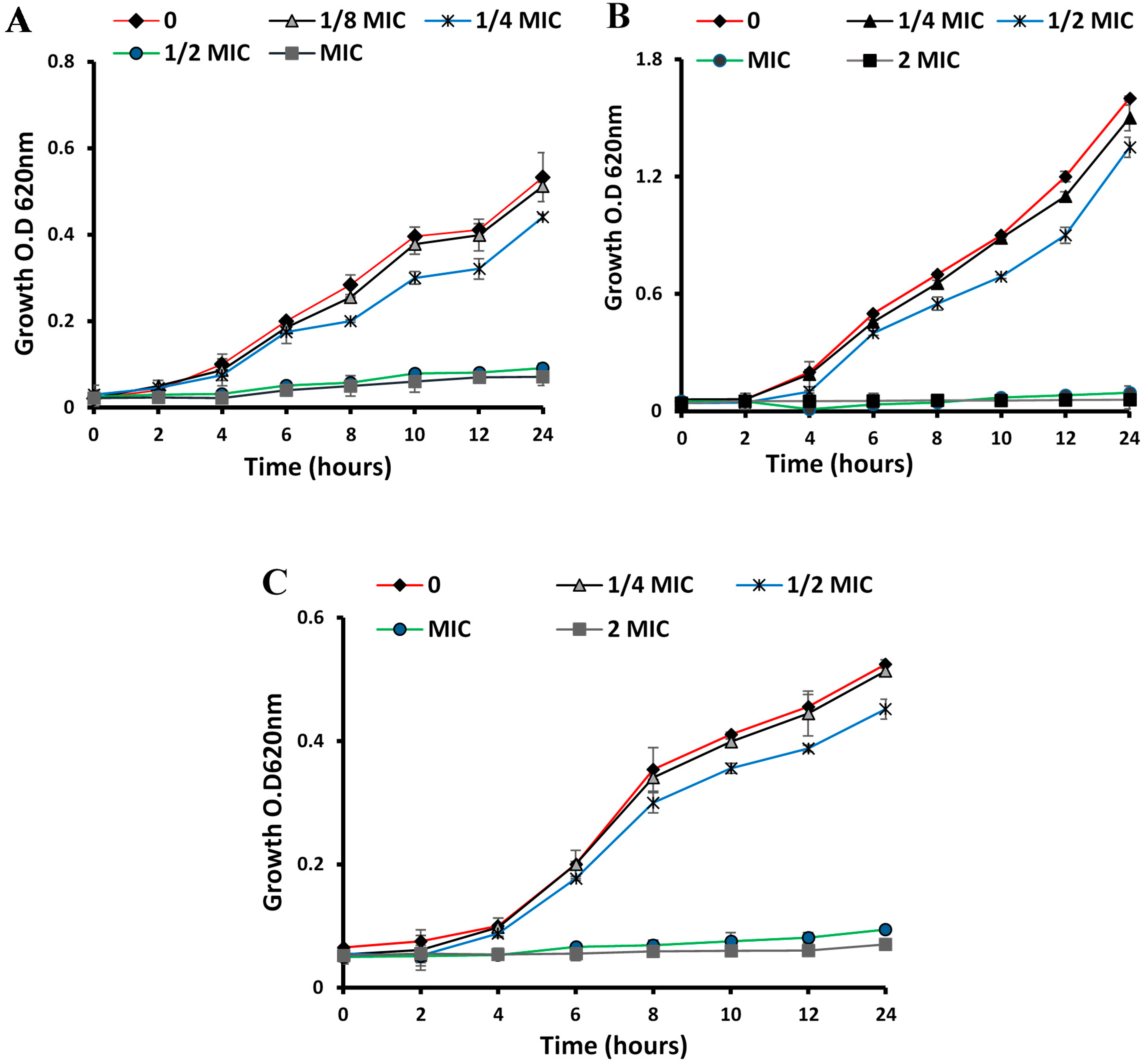
2.5. Time–Kill Curves

Time–kill curves were used to monitor bacterial growth and death over a gradient of ESFE concentrations versus time [22]. To assess the antimicrobial potential of ESFE on S. aureus, P. aeruginosa, Bacillus subtilis, Escherichia coli and C. albicans, the bacterial suspension (1 × 10⁶ CFU/mL) was mixed with the tested sample at concentrations of 0, 1/4, 1/2, 1 and 2 MIC and then incubated at 37 °C at 150 rpm. O.D. was determined spectrophotometrically at 620 nm every 2 h during the first 12 h and then after 24 h.

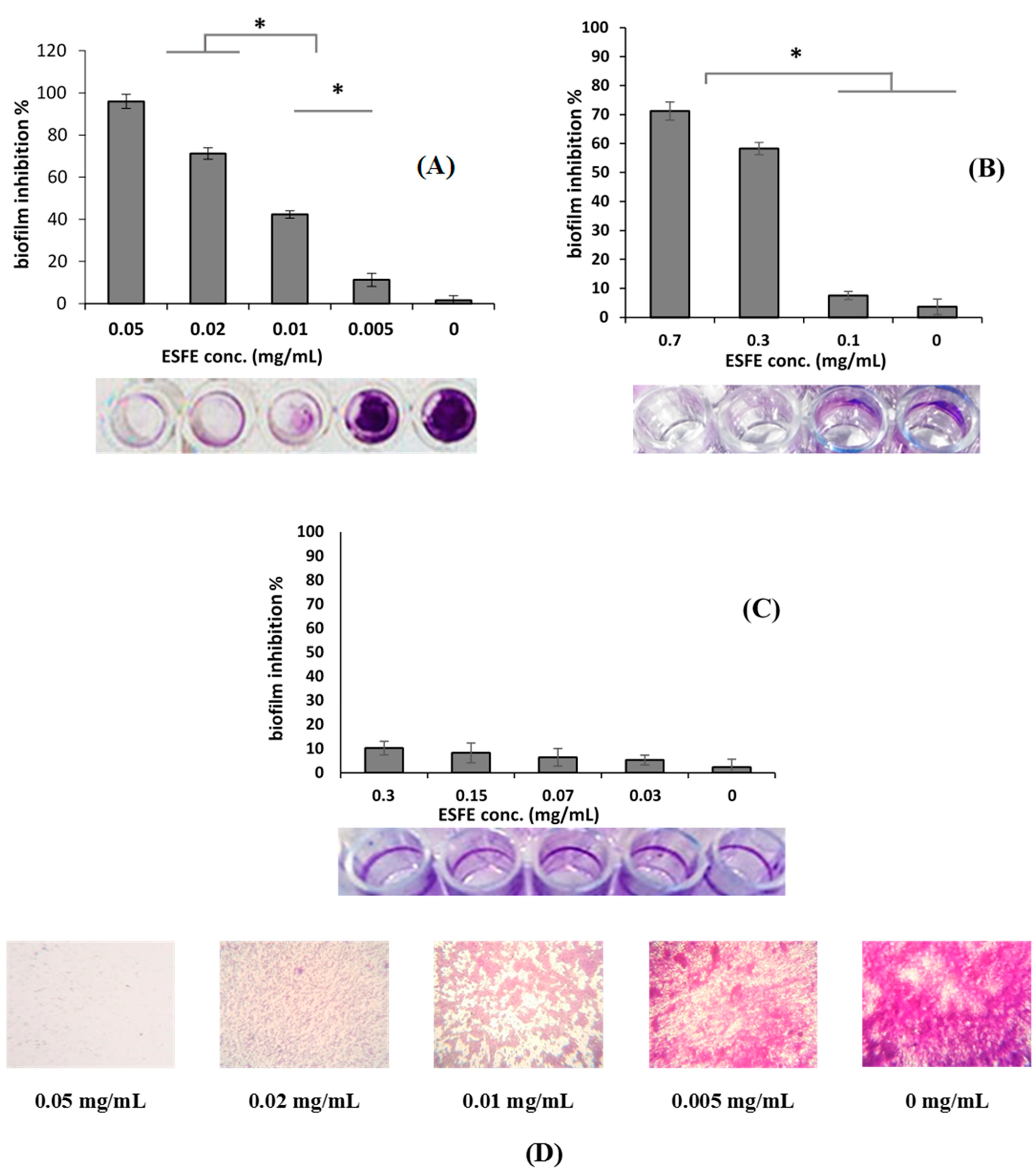
2.6. Biofilm Inhibition Assay and MBIC

To evaluate the inhibitory potential of ESFE on bacterial and yeast biofilm formation, the MTP method was used against the S. aureus clinical isolate, P. aeruginosa and C. albicans [21,23]. Briefly, gradient concentrations (0.5–0.05, 2.5–0.3 and 6.0–0.7 mg/mL for S. aureus, P. aeruginosa and C. albicans, respectively) of ESFE was distributed into a flat-bottomed MTP with a tryptic soy broth media (TSB) supplemented with glucose (1%). An overnight culture of the test organisms was 1:100 diluted in TSB to an inoculum size of 1 × 10⁵ CFU/mL and loaded onto MTP and incubated for 48 h at 37 °C. O.D. was measured at 620 nm after the incubation period prior to the transfer of planktonic cells from the plates. The well content was then transferred without troubling the formed biofilms and the MTP wells were washed 3 times with phosphate-buffered saline (PBS) at pH 7.4 to remove the residue of the floated unbounded cells. The formed biofilm was then fixed with 200 μL methanol (95%) for 10 min in all wells. Crystal violet (0.3% w/v) was then added to each well using a multi-channel micropipette (CAPP, Berlin, Germany) and the plates were incubated at room temperature for 15 min. After that, the excess of crystal violet stain was removed, and the plates were washed with distilled sterile water. Finally, the crystal violet bound with biofilm was examined at this point and then photographed by an inverted microscope (Olympus Ck40, Tokyo, Japan) at 150×. For the quantitative determination of biofilm formation, 30% acetic acid was added to the wells and the color absorbance was measured at O.D540nm by an automated microplate reader (Tecan Elx800, USA). The treated and the untreated wells were compared. For the estimation of the minimum biofilm inhibitory concentration (MBIC), an overnight culture of the test organism was adjusted to achieve an inoculum size of a 0.5 McFarland standard (1.5 × 10⁸ CFU/mL), and 150 μL of the media inoculated with the test organism was added to the wells and then incubated at 37 °C for 24 h. After incubation, the plates were washed 3 times with PBS (pH 7.4) to remove the planktonic unbounded bacteria and left to dry under aseptic conditions in an inverted position. Then 100 µL of the gradient dilutions of the ESFE in TSB media were transferred into the dried wells with the perfectly performing biofilms. The MTP were incubated at 37 °C for 20 h. For the determination of minimum biofilm inhibitory concentrations, all contents of the 96-well plates were cultured on TSA plates prior to washing with PBS and staining with crystal violet. The TSA plates were then incubated at 37 °C for 24 h. The MBIC value was considered as the concentration at which there was no growth of bacterial cells. [24].

2.7. HPLC-PDA-ESI-MS/MS

The ESFE extract was analyzed utilizing a ThermoFinnigan LCQ-Duo ion trap mass spectrometer (ThermoElectron Corporation, Waltham, MA, USA) coupled with an ESI source (ThermoQuest Corporation, Austin, TX, USA) [25]. A ThermoFinnigan HPLC system using a Discovery HS F5 column (15 cm × 4.6 mm ID, 5 µm particles, Sigma-Aldrich Co Steinheim, Germany) was used. Water and acetonitrile (ACN) (Sigma-Aldrich GmbH, Berlin, Germany) (0.1% formic acid each) were used as a mobile phase adopting the same method reported in [26,27]. The autosampler surveyor ThermoQuest was used to inject the sample and Xcalibur software (XcaliburTM 2.0.7, Thermo Fischer Scientific, Waltham, MA, USA) was used to control the system. The MS operated in the negative mode [26,27].

2.8. Statistical Analyses

Three replicates were performed for each assay, and all resulted values were the averages of three independent experiments. To analyze the differences between a sample and the corresponding control, Student’s t-test was used. Differences were considered significant if the p values were <0.05.

3. Results

3.1. Antibacterial Activities

3.1.1. Anti-Microbial, MIC and MBC of ESFE

ESFE displayed broad spectrum antimicrobial potential against Gram-positive and Gram-negative bacteria in addition to the yeast, C. albicans. ESFE growth inhibitory potential, reflected by the diameter of the inhibition zone, against Gram-positive S. aureus and B. subtilis bacteria was superior to its inhibitory potential against Gram-negative E. coli and P. aeruginosa (Table 1).
The inhibition zone reached 20 mm in diameter in the case of Gram-positive species at a low concentration of ESFE. There was no significant difference between the ESFE inhibitory potential against MRSA and MSSA. On the other hand, Gram-negative bacteria required a higher concentration of ESFE (up to 2.5 mg/mL) to obtain an inhibitory effect (Table 1). Moreover, a higher concentration of ESFE (5 mg/mL) was required to inhibit the growth of C. albicans with an inhibition zone diameter of 9 mm. MICs were determined against the previous organisms. The lowest MIC value (0.5 mg/mL) was recorded against MRSA and MSSA while the highest MIC value (3 mg/mL) was recorded against C. albicans. The MIC and MBC values against the Gram-positive, Gram-negative and yeast strains are listed in Table 2. The time–kill curve showed that a 1/4 MIC of ESFE had no growth inhibitory potential against the three biofilm-forming organisms: S. aureus ACL51 (MRSA), P. aeruginosa and C. albicans (Figure 1). Thus, this concentration was selected for the antibiofilm assay.

3.1.2. Biofilm Inhibition Activity

ESFE demonstrated robust biofilm inhibitory activities against MRSA and C. albicans at sublethal concentrations (p < 0.05; Figure 2). ESFE exhibited significant dose-dependent inhibition of MRSA biofilm formation up to 95.9% (p < 0.05) at a concentration of 0.05 mg/mL. At a concentration of 0.7 mg/mL, the ability of ESFE to eradicate C. albicans biofilm formation reached 71.2% (p < 0.05) in a dose-dependent manner. On the other hand, the formation of P. aeruginosa biofilm was not affected by the sub-lethal dose of ESFE (0.3–0.03 mg/mL). The MBIC against MRSA and C. albicans was 0.01 and 0.3 mg/mL, respectively.

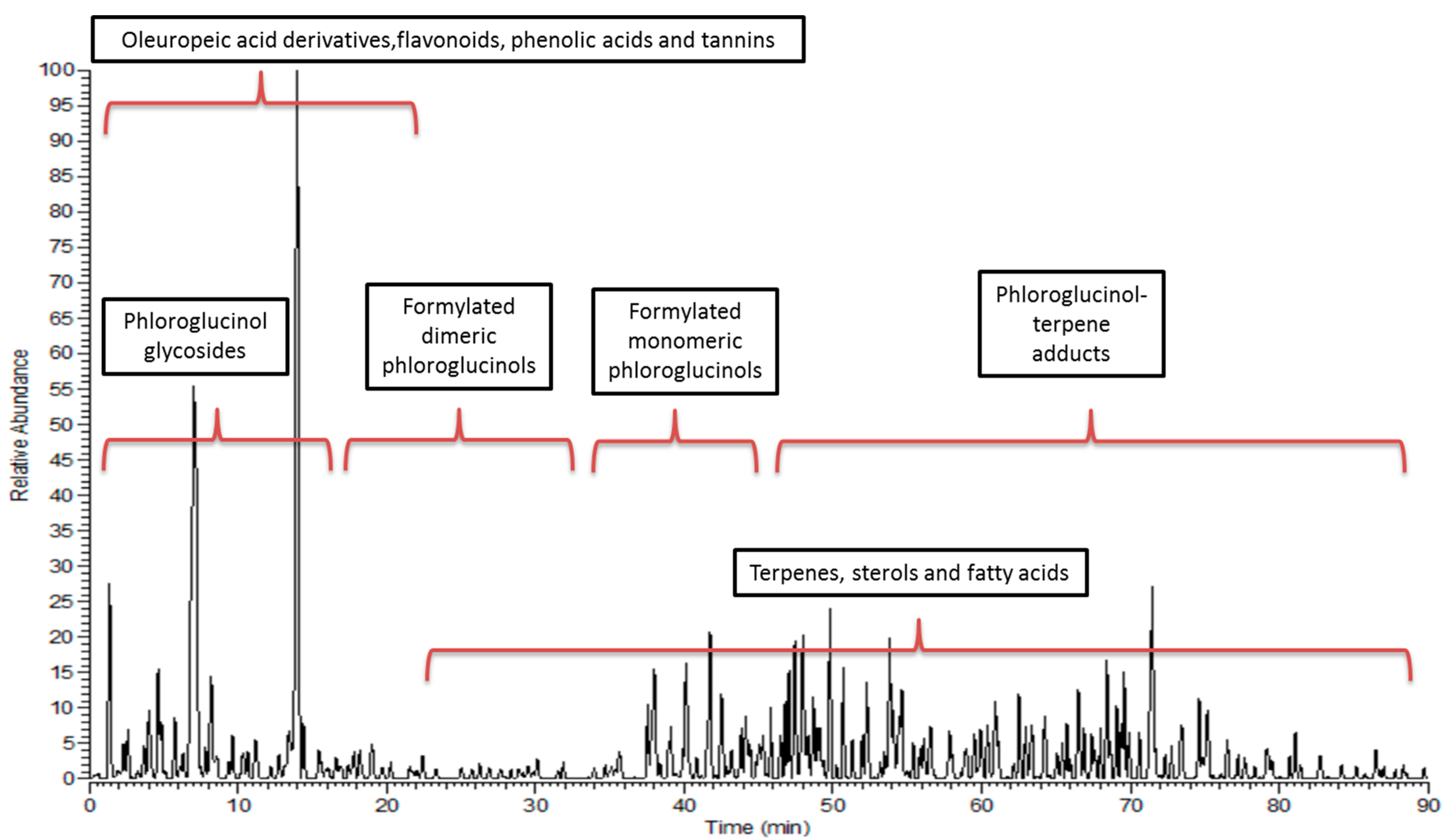
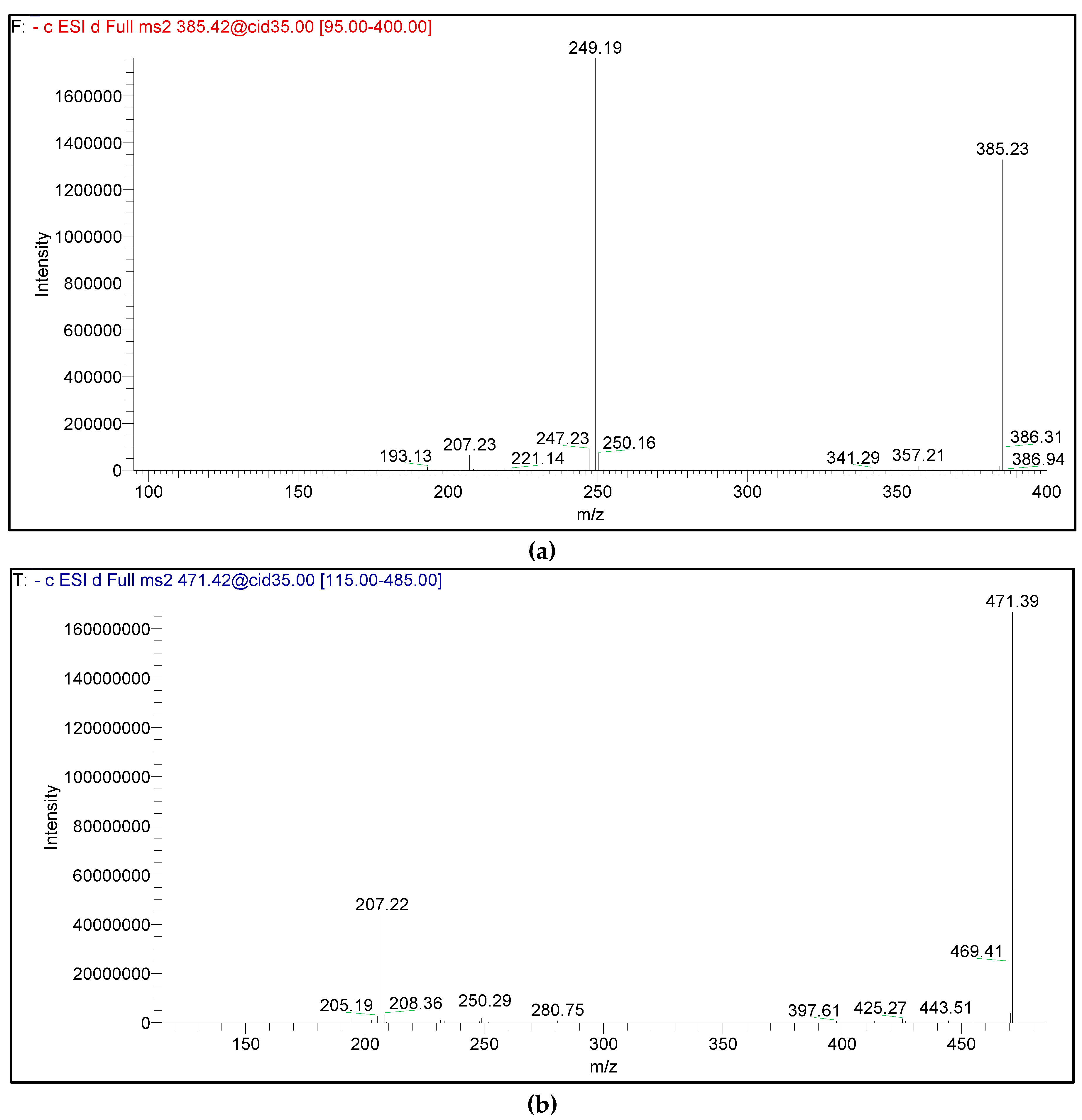
3.2. Metabolic Profiling

The HPLC-PDA-ESI-MS/MS analysis of the ESFE revealed the tentative identification of 83 compounds: 21 phloroglucinol, 18 terpenes, 16 flavonoids, 7 oleuropeic acid derivatives, 7 ellagic acid derivatives, 6 gallic acid derivatives, 3 phenolic acids, 3 fatty acids and 2 miscellaneous. The observed molecular weights, retention times (rt), fragment ions and chemical class of each compound, and their identities, are presented in Table 3. Figure 3 represents the LC-MS profile of the extract.
The identified metabolites are a complex mixture of several formylated phloroglucinols (FPs), polyphenolics (Hydrolyzable tannins, phenolic acids, flavanone glycoside, and oleuropeic acid derivatives) and terpenoids. The peaks’ identities were predicted using an in-house metabolite database, according to the parent masses and retention times and based on the comparison of their mass spectra with the reported data from the genus Eucalyptus, as well as the Plant Dictionary MS Database and METLIN.
Phloroglucinols: Twenty-one phloroglucinols were identified. They included three formylated monomeric phloroglucinols, five formylated dimeric phloroglucinols, four phloroglucinol glycosides and nine phloroglucinol-terpene adducts (phloroglucinol meroterpenoids). Compound (6) was tentatively identified as sideroxylonal A/B/C. Its molecular ion peak [MH] appeared at m/z 499. Upon fragmentation, it gave an ion at m/z 471 and at m/z 453 due to loss of CO (28 dalton) and subsequent loss of the H2O moiety (18 dalton). Additionally, a daughter ion at m/z 249 corresponding to the isopentyl diformyl phloroglucinol moiety was also detected. These isomers were eluted at retention times of 4.66, 8.41 and 10.96 min, which show that several isomers of sideroxylonal were present in ESFE. This comes in agreement with the findings of Moore et al. [45], who reported that macrocarpals and sideroxylonals are the most common groups of FPs reported in the genus Eucalyptus.
Phloroglucinol-terpene adducts (Euglobals and macrocarpals) were also detected. The identified euglobal (21) was detected at m/z 385. It was characterized by an intense fragment daughter ion at m/z 249 and a less intense ion at m/z 207 (Figure 4a) [46]. On the other hand, the tentatively identified macrocarpals (13, 14, 17 and 20) are sesquiterpene adducts with molecular ions detected at m/z 489, 471 and 453, respectively. Upon fragmentation, they yielded an intense product ion at m/z 207, with a weaker product ion at m/z 249 (Figure 4b) [15,46]. The fragment produced at m/z 249 in macrocarpals corresponds to the isopentyl diformyl phloroglucinol moiety that resulted from the typical cleavage of the sesquiterpene moiety.
Phloroglucinol glycosides were found as well. Myrciaphenone B (9), a phloroglucinol glycoside attached to the galloyl moiety, and thus the 169 dalton daughter ion corresponding to the galloyl moiety, was detected. Another daughter ion at m/z 331 due to the loss of the galloyl and hexose moieties [M−H-169-162] was also observed. The other three tentatively identified phloroglucinol glycosides were eucalmainoside A, B and C. The molecular ion of eucalmainoside A (10) was detected at 301 corresponding to 2-methylphloroglucinol-O-β-D-glucopyranoside. The molecular ions of compounds (12 and 11) at m/z 315 and 329 exceeded that of eucalmainoside A (10) by 14 (CH3 moiety) and 28 (CHO moiety) dalton, respectively, corresponding to 2,4-dimethylphloroglucinol- O-β-D-glucopyranoside (eucalmainoside B) and 2,4,6-trihydroxy- 3-methylbenzaldehyde-2-O-β-D-glucopyranoside (eucalmainoside C), respectively.
Oleuropeic acid derivatives: Seven oleuropeic acid derivatives were tentatively identified (22–28). All of them are oleuropeic acid-containing carbohydrates (oleuropeic acid glucosides); i.e., carbohydrate monoterpene esters. There are two types of oleuropeic acid glucosides characterized in the studied extract. The first type contains a terpene moiety other than oleuropeic acid and is represented by globulusin A (23). It is a β-glucopyranose ester of gallic acid and 2-hydroxy-1,8-cineol. The second type contains a polyphenolic moiety. This type is represented by the cypellocarpines (24,25 and 28); a group of oleuropeic acid glucosides that differs only in the phenolic moiety incorporated in their structures. Globulusin A/B (23 and 27) are monoterpene glycosides conjugated with gallic acid. Globulusin A (23) fragmentation showed peaks at m/z 313 [M − H-galloyl], 169 [M-hydroxyl cineole-162] and 151 [M − H-162-169]. Daughter ions at m/z 313 (oleuropeic acid methyl ester), 169 (galloyl moiety), 151 [M − H-162-169] and 183 (oleuropeic acid methyl ester) were detected due to fragmentation of globulusin B (27). The fragmentation pattern of globulusin A and globulusin B is in accordance with what was previously reported in [34]. The MS spectrum of Compound (26) showed a prominent fragment (m/z at 463) corresponding to the loss of the oleuropeic acid moiety of eucalmaidin D or cypellogin A/B. Compound (26) fragmentation will be discussed under the flavonoids section.
Flavonoids and their glycosides: The sixteen characterized flavonoids are 11 flavonols (29–31,33,34,36,38–42), 4 flavones (35,37,43–44) and a flavanone (32). The predominating class was the flavonols. They were characterized, in the spectra of their deprotonated glycosides, by ions corresponding to the deprotonated aglycones at m/z 285, 301 and 315 (for kaempferol, quercetin and isorhamnetin, respectively), generated by the loss of the sugar units. Furthermore, fragment ions at m/z 255, 271 and 285 were detected corresponding to [M-H-CO-H], resulting from fragmentation of the kaempferol, quercetin and isorhamnetin aglycones, respectively [47]. A total of 16 flavonoids were tentatively identified, of which eleven were O-glycosides, one was C-glycoside (37) and four were flavonoid aglycones (41–44); this in addition to eucalmaidin D, quercetin-4’-O-(6-O-oleuropeoyl)-R-D-glucopyranoside (26), which was listed under the abovementioned oleuropeic acid derivatives class. Eucalmaidin D (26) is a rare example of an α-configured glucoside found in nature. It was detected before in the leaves of the same plant [15] and was isolated from the leaves of E. maideni as well [48]. The nature of the sugars in O-glycosides could be revealed from the elimination of the sugar residue. The primary sugar can be identified as hexose, pentose, and deoxyhexose if the intermediate ion appears at 162, 132, and 146 dalton from molecular ions, respectively [49].
Phenolic acids: Three phenolic acids (45–47) were tentatively identified in ESFE, namely, gallic, chlorogenic/neochlorogenic and ferulic acid. Their fragmentation pattern is in agreement with that reported for phenolic acids in [50], where all of them were previously reported from the genus Eucalyptus [37].
Gallic acid derivatives: Results revealed the presence of six gallic acid derivatives, three of which are gallic acid glycosides (48, 49 and 52), in addition to myrciaphenone B (9), globulusin A (23) and globulusin B (27) discussed before under the phloroglucinol glycosides and oleuropeic acid derivatives sections. Product ions due to the loss of the galloyl moieties [M − H-169] and galloyl moiety (169 dalton) were observed in their mass spectra (Table 3).
Ellagic acid derivatives: Compound (54) was assigned to ellagic acid. It was characterized by [M-H] at m/z 301. Its MS/MS fragmentation was typical to the reported ellagic acid pattern [16]. The molecular ions of Compounds (58–60) exceeded that of ellagic acid by 14, 28 and 42 dalton, corresponding to the extra methyl groups (1-CH3, 2-CH3 and 3-CH3). Their fragmentation was characterized by a loss of 15 dalton [M-CH3-H] due to the loss of the methyl radical. They were tentatively identified as methylellagic, dimethylellagic, and trimethylellagic acids.
Triterpenes and sterols: About 18 triterpenoids were characterized: oleananes (75 and 76), ursanes (62,64–68, and 69) and lupanes (63), representing the classes of pentacyclic triterpenes. Compound (62) was assigned to asiatic acid lactone ((2a,3b)-2,3,23-trihydroxy-13,28-epoxyurs-11-en-28-one). This is the third report of the presence of this pentacyclic triterpenoid with an 11-en-28,13β-olide structure in nature after being detected in E. camaldulensis [51] and E. sidroxylon leaves [15]. Four pentacyclic triterpenes with an O-p coumaroyl substitution were detected. The observed daughter ions at m/z 455, 485, 437 and 471 in the MS spectra of (69,70,73 and 74), respectively, were due to the loss of 162 dalton; corresponding to losing the O-p coumaroyl group at C3. The [M-H] of Compounds (70) and (74) (647 and 633, respectively) exceeded that of O-p coumaroyl maslinic/alphitolic acid (69) by 30 and 16 dalton, respectively. This indicates an extra OCH3 in Compound (70) and OH in Compound (74), which, in turn, confirms their identification as eucalyptic acid (Eucalyptolic) (70) and O-p coumaroyl tormentic acid (74). The observed daughter ion in Compound (70) at m/z 617 is due to the loss of the OCH3 group [M-H-OCH3].
Fatty acids: Three oxygenated fatty acids (saturated and unsaturated) were annotated. Their fragmentation was in agreement with that of the hydroxylated fatty acids [52,53]. Compounds (79) and (81) with molecular ion peaks at m/z 329 and m/z 295 were assigned to trihydroxy octadecenoic and hydroxy octadecadienoic. The mass difference of 2 × 16 dalton between Compounds (79) and (81) suggested an extra two hydroxyl groups in addition to a 2 dalton difference corresponding to the saturation of one of the double bonds of (81). Our identification was confirmed by the appearance of a product ion at m/z 277 [M-H-H2O] corresponding to the loss of a water molecule.
Miscellaneous compounds: Vomifoliol (82), a monoterpene derivative related to abscisic acid, and withanolide A (83) were tentatively identified. Withanolide is a naturally occurring C-28 steroidal lactone with an ergostane-type skeleton.

4. Discussion

The inhibition of biofilm is considered an important way for the treatment of bacterial infections [8]. Searching for bacterial virulence factors inhibitors is very important due to the well-known ability of pathogens to develop different mechanisms of antibiotics resistance. Exploring phyto-constituents seems necessary to avoid synthetic drugs’ toxic side effects [54]. Results showed that ESFE had broad spectrum antibacterial activity against Gram-positive and Gram-negative bacteria and yeast. The Gram-positive bacteria S. aureus and B. subtilis bacteria were more susceptible than the Gram-negative bacteria E. coli and P. aeruginosa to the inhibitory activity of ESFE, while C. albicans showed the smallest inhibition zone. Furthermore, at sub-lethal concentrations, ESFE showed significant anti-biofilm activity against MRSA and C. albicans (95.9 and 71.2% inhibition, respectively) in a dose-dependent manner.
The observed higher potency of ESFE towards Gram-positive bacteria than Gram-negative bacteria is in accordance with that reported for three Eucalyptus species (E. globulus, E. radiate and E. citriodora) against different Gram-positive (VRE and MRSA) and Gram-negative (P. aeruginosa, E. coli, Klebsiella pneumonia and Acineto) bacteria [55]. The tested sample was active with variable degrees against Gram-positive bacteria with regards to their chemical constituents. However, the tested samples were almost inactive against multidrug-resistant Gram-negative bacteria due to the differences in cell wall sub-structures. In the same stream, E. globulus and E. camaldulensis inhibitory activity was evaluated by micro-atmosphere, aromatogramme and germs in suspension assays against E. coli and S. aureus. They demonstrated an inhibitory effect on both bacteria, but to a lesser extent on E. coli [56]. The observed anti-Candida potential of ESFE matched that reported about the significant activity of E. globulus and E. citriodora against several Candida species [57].
HPLC-PDA-ESI-MS/MS results revealed that ESFE is rich in phloroglucinols (twenty-one secondary metabolites) and flavonoids (sixteen secondary metabolites) in addition to several phenolic acid derivatives. Consequently, the obtained potent antimicrobial potency might be due to the intrinsic acidic characters of this phenolic metabolites, which create a lethal antibacterial environment [58].
The anti-biofilm activity of phloroglucinols and flavonoids is well documented [7,59,60]. Their phenolic nature could be responsible for such activity. Many mechanisms were suggested for the antibiofilm activity of plant phenolics, including modulation of bacterial cells communication, interference with motility, surface hydrophobicity and charge and downregulation of the genes responsible for the biofilm formation [59,61,62].
The most common acylpholoroglucinols in Eucalyptus are the diformyl monomeric phloroglucinols, phloroglucinol glycosides, dimeric acylphloroglucinols, phloroglucinol-sesquiterpene and acylphloroglucinol-monoterpene adducts [31]. All these classes were detected in the E. sideroxylon flowers. It was found to be rich in sideroxylonals, macrocarpals and euglobals.
The antibacterial activity of the pholoroglucinol, eucalyptin A, was previously evaluated against S. epidermidis, S. aureus and P. aeruginosa. It showed potent antimicrobial activity against the two biofilm-producing strains S. epidermidis and S. aureus, with MIC values of 1.7 and 3.5 mg/mL, respectively, but was inactive against the Gram-negative P. aeruginosa [63].
The desmethyl eucalyptin detected in ESFE was proven to exhibit bacteriostatic activity against methicillin-resistant and sensitive S. aureus strains (MRSA and MSSA). It also exhibited potent antibiofilm potential at sub-MICs and inhibited staphyloxanthin biosynthesis and decreased the survival rates of MRSA (73.1%) and MSSA (54.6%). Disintegration of the outer membrane, irregular shape of the cells and leakage of cytoplasm were also observed after treatment of MSSA with desmethyl eucalyptin and its methylated derivative, eucalyptin [7].
The monoterpene acid glucose ester, eucaglobulin, was previously isolated from E. globulus leaves [34,64,65] and fruits [66] and was reported to exert significant inhibition against C. albicans, E. coli and S. aureus [65]. The flower extract of E. sideroxylon is a rich source of hydrolyzable tannins (6 gallic and 7 ellagic acid derivatives). This is in accordance with two recent studies that reported the presence of several hydrolysable tannins in the bark of E. sideroxylon [16] and determined their quantity [67]. The S. aureus biofilm inhibitory activity of gallic [68] and ellagic acids [69] at subinhibitory concentrations was also reported. It was reported that ellagic acid/ellagic acid derivatives could limit biofilm formation of S. aureus to an extent that could be correlated with the increased antibiotic susceptibility [70]. Gallic and ellagic acids also inhibited biofilm formation of E. coli [71,72,73].
The potent antibiofilm activity of ESFE against MRSA and C. albicans might be attributed to the synergistic effects of all secondary metabolites identified by HPLC-PDA-ESI-MS/MS, including phloroglucinols, OAG, flavonoids and tannins. It is worthy to mention that Myrtaceae phloroglucinols may act as biofilm inhibitors more effectively in their pure state rather that in the crude extract [7]. Chemical synthesis studies have attempted to produce phloroglucinols [28]; however, this was a difficult and costly process. Consequently, it is highly recommended to prepare E. sideroxylon flower phloroglucinol-rich extracts or to isolate and structurally elucidate its phloroglucinols, to be studied as a nucleus for exploring novel anti-biofilm agents.

5. Conclusions

This study highlights the promising role of E. sideroxylon flowers as an anti-biofilm agent against both Gram-positive bacteria and yeast. The HPLC-PDA-MS/MS fingerprint of the E. sideroxylon flowers represents the first complete secondary metabolites profile of the plant. The efficacy of the extract against MRSA and C. albicans could prove greater efficacy after further in vivo and clinical studies. The combination of E. sideroxylon flower extract with other natural antibiofilm agents could reduce the risk of bacterial and yeast strain resistance to various synthetic drugs.

Author Contributions

M.M.O., R.A.E.-S. and R.M.S.A.: HPLC and mass data analysis, interpretation and characterized the chemical composition; M.A.-E. performed the microbiological study and analyzed its results; M.S. revised the chemical composition of the extract. All authors have read and agreed to the published version of the manuscript.

Funding

This research received no external funding.

Acknowledgments

The authors would like to thank M. Wink, Heidelberg University, for giving us the opportunity to collect LC-MS data at his lab and the Royal Botanic Gardens Herbarium, Kew, Richmond, Surrey, United Kingdom, for identifying and authenticating the plant.

Conflicts of Interest

The authors report no conflict of interest.

References

  1. Khan, F.; Pham, D.T.N.; Oloketuyi, S.F.; Manivasagan, P.; Oh, J.; Kim, Y.-M. Chitosan and Their Derivatives: Antibiofilm Drugs Against Pathogenic Bacteria. Colloids Surf. B Biointerfaces 2020, 185, 110627. [Google Scholar] [CrossRef]
  2. Desouky, S.E.; El-Gamal, M.S.; Mohammed, A.F.; Abu-Elghait, M.A. Determination of Some Virulence Factors in Staphylococus Spp. Isolated from Clinical Samples of Different Egyptian Patients. World Appl. Sci. J. 2014, 32, 731–740. [Google Scholar]
  3. Otto, M. Staphylococcal Biofilms. Microbiol. Spectr. 2019, 6, 699–711. [Google Scholar]
  4. Kaur, S.; Sharma, N.; Aanchal, A.G.; Sharma, A.; Sharma, A.; Sharma, V. Anti-biofilm Potential of Aqueous Eucalyptus Leaf Extract against Nosocomial Pathogens: Staphylococcus and Pseudomonas Aeruginosa. Pharm. Innov. J. 2018, 7, 425–432. [Google Scholar]
  5. Lu, L.; Hu, W.; Tian, Z.; Yuan, D.; Yi, G.; Zhou, Y.; Cheng, Q.; Zhu, J.; Li, M. Developing Natural Products as Potential Anti-Biofilm Agents. Chin. Med. 2019, 14, 1–17. [Google Scholar] [CrossRef] [Green Version]
  6. Barriuso, J. Quorum Sensing Mechanisms in Fungi. AIMS Microbiol. 2015, 1, 37–47. [Google Scholar] [CrossRef]
  7. Shehabeldine, A.M.; Ashour, R.M.; Okba, M.M.; Saber, F.R. Callistemon Citrinus Bioactive Metabolites as New Inhibitors of Methicillin-Resistant Staphylococcus Aureus Biofilm Formation. J. Ethnopharmacol. 2020, 254, 112669. [Google Scholar] [CrossRef]
  8. Mostafa, I.; Abbas, H.A.; Ashour, M.L.; Yasri, A.; El-Shazly, A.M.; Wink, M.; Sobeh, M. Polyphenols from Salix Tetrasperma Impair Virulence and Inhibit Quorum Sensing of Pseudomonas Aeruginosa. Molecules 2020, 25, 1341. [Google Scholar] [CrossRef] [Green Version]
  9. Mohamed, G.A.; Ibrahim, S.R. Eucalyptone G, a New Phloroglucinol Derivative and Other Constituents from Eucalyptus Globulus Labill. Arkivoc 2007, 15, 281–291. [Google Scholar] [CrossRef] [Green Version]
  10. Bhuyan, D.J.; Vuong, Q.V.; Chalmers, A.C.; van Altena, I.A.; Bowyer, M.C.; Scarlett, C.J. Phytochemical, Antibacterial and Antifungal Properties of an Aqueous Extract of Eucalyptus Microcorys Leaves. S. Afr. J. Bot. 2017, 112, 180–185. [Google Scholar] [CrossRef]
  11. Bailey, L.H. Manual of Cultivated Plants; The Macmillan Company: New York, NY, USA, 1958. [Google Scholar]
  12. Umberto Quattrocchi, F.L.S. World Dictionary of Plant Names; Harvard University and Harvard University Herbaria: Cambridge, MA, USA, 1999; Volume 2. [Google Scholar]
  13. Ashour, H.M. Antibacterial, Antifungal, and Anticancer Activities of Volatile Oils and Extracts from Stems, Leaves, and Flowers of Eucalyptus Sideroxylon and Eucalyptus Torquata. Cancer Biol. Ther. 2008, 7, 399–403. [Google Scholar] [CrossRef] [PubMed] [Green Version]
  14. Elaissi, A.; Salah, K.H.; Mabrouk, S.; Larbi, K.M.; Chemli, R.; Harzallah-Skhiri, F. Antibacterial Activity and Chemical Composition of 20 Eucalyptus Species’ Essential Oils. Food Chem. 2011, 129, 1427–1434. [Google Scholar] [CrossRef]
  15. Okba, M.M.; El Gedaily, R.A.; Ashour, R.M. UPLC–PDA–ESI–qTOF-MS Profiling and Potent Anti-HSV-II Activity of Eucalyptus Sideroxylon Leaves. J. Chromatogr. B 2017, 1068, 335–342. [Google Scholar] [CrossRef] [PubMed]
  16. Ashour, R.M.; Okba, M.M.; Menze, E.T.; El Gedaily, R.A. Eucalyptus Sideroxylon Bark Anti-inflammatory Potential, Its UPLC-PDA-ESI-qTOF-MS Profiling, and Isolation of a New Phloroglucinol. J. Chromatogr. Sci. 2019, 57, 565–574. [Google Scholar] [CrossRef]
  17. Santos, B.M.d.; Zibrandtsen, J.F.; Gunbilig, D.; Sørensen, M.; Cozzi, F.; Boughton, B.A.; Heskes, A.M.; Neilson, E.H.J. Quantification and Localization of Formylated Phloroglucinol Compounds (Fpcs) in Eucalyptus Species. Front. Plant Sci. 2019, 10, 186. [Google Scholar] [CrossRef]
  18. Hamed, A.; Abdel-Razek, A.S.; Araby, M.; Abu-Elghait, M.; El-Hosari, D.G.; Frese, M.; Soliman, H.S.; Stammler, H.G.; Sewald, N.; Shaaban, M. Meleagrin from Marine Fungus Emericella Dentata Nq45: Crystal Structure and Diverse Biological Activity Studies. Nat. Prod. Res. 2020, 1–9. [Google Scholar] [CrossRef]
  19. Stankov, S.; Fidan, H.; Stefanova, G.; Kostova, I.; Damyanova, S.; Dimitrova-Dyulgerova, I.; Ercisli, S.; Stoyanova, A. Chemical Composition and Antimicrobial Activity of Essential Oil from Aerial Part (Leaves and Fruit) of Eucalyptus gomphocephala DC. J. Essent. Oil Plants 2020, 23, 1–9. [Google Scholar] [CrossRef]
  20. CLSI. Performance Standards for Antimicrobial Susceptibility Testing; CLSI document M100-S20; Clinical Laboratory Standards Institute: Wayne, PA, USA, 2011. [Google Scholar]
  21. Abdelhameed, R.M.; Abu-Elghait, M.; El-Shahat, M. Hybrid Three Mofs Composites (ZIF-67@ ZIF-8@ MIL-125-NH2): Enhancement the Biological and Visible-Light Photocatalytic Activity. J. Environ. Chem. Eng. 2020, 8, 104107. [Google Scholar] [CrossRef]
  22. Qian, W.; Liu, M.; Fu, Y.; Zhang, J.; Liu, W.; Li, J.; Li, X.; Li, Y.; Wang, T. Antimicrobial Mechanism of Luteolin against Staphylococcus Aureus and Listeria Monocytogenes and Its Antibiofilm Properties. Microb. Pathog. 2020, 142, 104056. [Google Scholar] [CrossRef] [PubMed]
  23. Desouky, S.E.; Hassan, S.E.; El-gamal, M.S.; Ragab, M.A.E.T.I.; Emam, M. Effect of Salvia Egypticae and Foeniculum Vulgara Extracts on Quorum Sensing and Biofilm Formation of Methicillin Resistant/Sensitive Staphylococcus Aureus Isolates. World J. Pharm. Med. Res. 2017, 3, 466–475. [Google Scholar]
  24. Černohorská, L.; Votava, M. Antibiotic Synergy against Biofilm-Forming Pseudomonas Aeruginosa. Folia Microbiol. 2008, 53, 57. [Google Scholar] [CrossRef]
  25. Sobeh, M.; Mahmoud, M.F.; Sabry, O.M.; Adel, R.; Dmirieh, M.; El-Shazly, A.M.; Wink, M. HPLC-PDA-MS/MS Characterization of Bioactive Secondary Metabolites from Turraea Fischeri Bark Extract and Its Antioxidant and Hepatoprotective Activities in Vivo. Molecules 2017, 22, 2089. [Google Scholar] [CrossRef] [PubMed] [Green Version]
  26. El-Hawary, S.S.; Mubarek, M.M.; Lotfy, R.A.; Hassan, A.R.; Sobeh, M.; Okba, M.M. Validation of Antidiabetic Potential of Gymnocarpos decandrus Forssk. Nat. Prod. Res. 2020, 1–6. [Google Scholar] [CrossRef]
  27. El-Hawary, S.S.; Sobeh, M.; Badr, W.K.; Abdelfattah, M.A.; Ali, Z.Y.; El-Tantawy, M.E.; Rabeh, M.A.; Wink, M. HPLC-PDA-MS/MS Profiling of Secondary Metabolites from Opuntia Ficus-Indica Cladode, Peel and Fruit Pulp Extracts and Their Antioxidant, Neuroprotective Effect in Rats with Aluminum Chloride Induced Neurotoxicity. Saudi J. Biol. Sci. 2020, 27, 2829–2838. [Google Scholar] [CrossRef]
  28. Singh, I.P.; Sidana, J.; Bharate, S.B.; Foley, W.J. Phloroglucinol Compounds of Natural Origin: Synthetic Aspects. Nat. Prod. Rep. 2010, 27, 393–416. [Google Scholar] [CrossRef]
  29. Qin, X.-J.; Feng, M.-Y.; Liu, H.; Ni, W.; Rauwolf, T.; Porco, J.A., Jr.; Yan, H.; He, L.; Liu, H.-Y. Eucalyptusdimers A–C, Dimeric Phloroglucinol–Phellandrene Meroterpenoids from Eucalyptus robusta. Org. Lett. 2018, 20, 5066–5070. [Google Scholar] [CrossRef] [PubMed] [Green Version]
  30. Rojas-Garbanzo, C.; Zimmermann, B.F.; Schulze-Kaysers, N.; Schieber, A. Characterization of Phenolic and Other Polar Compounds in Peel and Flesh of Pink Guava (Psidium Guajava L. Cv.‘Criolla’) by Ultra-High Performance Liquid Chromatography with Diode Array and Mass Spectrometric Detection. Food Res. Int. 2017, 100, 445–453. [Google Scholar] [CrossRef]
  31. Brezáni, V.; Šmejkal, K. Secondary Metabolites Isolated from the Genus Eucalyptus. Curr. Trends Med. Chem. 2013, 7, 65–75. [Google Scholar]
  32. Gurbuz, P.; Baran, M.Y.; Demirezer, L.O.; Guvenalp, Z.; Kuruuzum-Uz, A. Phenylacylated-Flavonoids from Peucedanum chryseum. Rev. Bras. Farmacogn. 2018, 28, 228–230. [Google Scholar] [CrossRef]
  33. Yamakoshi, Y.; Murata, M.; Shimizu, A.; Homma, S. Isolation and Characterization of Macrocarpals BG Antibacterial Compounds from Eucalyptus Macrocarpa. Biosci. Biotechnol. Biochem. 1992, 56, 1570–1576. [Google Scholar] [CrossRef] [Green Version]
  34. Hasegawa, T.; Takano, F.; Takata, T.; Niiyama, M.; Ohta, T. Bioactive Monoterpene Glycosides Conjugated with Gallic Acid from the Leaves of Eucalyptus Globulus. Phytochemistry 2008, 69, 747–753. [Google Scholar] [CrossRef]
  35. Ito, H.; Koreishi, M.; Tokuda, H.; Nishino, H.; Yoshida, T. Cypellocarpins A−C, Phenol Glycosides Esterified with Oleuropeic Acid, from Eucalyptus Cypellocarpa. J. Nat. Prod. 2000, 63, 1253–1257. [Google Scholar] [CrossRef]
  36. Chen, C.; Zhang, H.; Xiao, W.; Yong, Z.-P.; Bai, N. High-Performance Liquid Chromatographic Fingerprint Analysis for Different Origins of Sea Buckthorn Berries. J. Chromatogr. A 2007, 1154, 250–259. [Google Scholar] [CrossRef] [PubMed]
  37. González-Burgos, E.; Liaudanskas, M.; Viškelis, J.; Žvikas, V.; Janulis, V.; Gómez-Serranillos, M.P. Antioxidant Activity, Neuroprotective Properties and Bioactive Constituents Analysis of Varying Polarity Extracts from Eucalyptus Globulus Leaves. J. Food Drug Anal. 2018, 26, 1293–1302. [Google Scholar] [CrossRef]
  38. Al-Sayed, E.; Martiskainen, O.; Bobrowska-Hägerstrand, M.; Sinkkonen, J.; Törnquist, K.; Pihlaja, K.; Ayoub, N.; Singab, A.-N. Phenolic Compounds from Eucalyptus Gomphocephala with Potential Cytotoxic and Antioxidant Activities. Nat. Prod. Commun. 2010, 5, 1934578X1000501025. [Google Scholar] [CrossRef] [Green Version]
  39. Saraf, I.; Marsh, K.J.; Vir, S.; Foley, W.J.; Singh, I.P. Quantitative Analysis of Various B-ring Unsubstituted and Substituted Flavonoids in Ten Australian Species of Eucalyptus. Nat. Prod. Commun. 2017, 12, 1934578X1701201109. [Google Scholar] [CrossRef] [Green Version]
  40. Wollenweber, E.; Kohorst, G. Epicuticular Leaf Flavonoids from Eucalyptus Species and from Kalmia Latifolia. Zeitschrift Nat. C 1981, 36, 913–915. [Google Scholar] [CrossRef]
  41. Ghareeb, M.A.; Sobeh, M.; El-Maadawy, W.H.; Mohammed, H.S.; Khalil, H.; Botros, S.; Wink, M. Chemical Profiling of Polyphenolics in Eucalyptus Globulus and Evaluation of Its Hepato–Renal Protective Potential against Cyclophosphamide Induced Toxicity in Mice. Antioxidants 2019, 8, 415. [Google Scholar] [CrossRef] [PubMed] [Green Version]
  42. Abu-Reidah, I.M.; Ali-Shtayeh, M.S.; Jamous, R.M.; Arráez-Román, D.; Segura-Carretero, A. HPLC–DAD–ESI-MS/MS Screening of Bioactive Components from Rhus Coriaria L. (Sumac) Fruits. Food Chem. 2015, 166, 179–191. [Google Scholar] [CrossRef] [Green Version]
  43. Yang, X.-W.; Guo, Q.-M. Studies on Chemical Constituents in Fruits of Eucalyptus Globulus. China J. Chin. Mater. Med. 2007, 32, 496–500. [Google Scholar]
  44. Jamal, A.; Yaacob, W.; Din, L.B. A Chemical Study on Phyllanthus Reticulatus. J. Phys. Sci. 2008, 19, 45–50. [Google Scholar]
  45. Moore, B.D.; Wallis, I.R.; Palá-Paúl, J.; Brophy, J.J.; Willis, R.H.; Foley, W.J. Antiherbivore Chemistry of Eucalyptus—Cues and Deterrents for Marsupial Folivores. J. Chem. Ecol. 2004, 30, 1743–1769. [Google Scholar] [CrossRef] [PubMed]
  46. Eyles, A.; Davies, N.W.; Mohammed, C. Novel Detection of Formylated Phloroglucinol Compounds (Fpcs) in the Wound Wood of Eucalyptus Globulus and E. Nitens. J. Chem. Ecol. 2003, 29, 881–898. [Google Scholar] [CrossRef] [PubMed]
  47. Ablajan, K.; Abliz, Z.; Shang, X.Y.; He, J.M.; Zhang, R.P.; Shi, J.G. Structural Characterization of Flavonol 3, 7-Di-O-Glycosides and Determination of the Glycosylation Position by Using Negative Ion Electrospray Ionization Tandem Mass Spectrometry. J. Mass Spectrom. 2006, 41, 352–360. [Google Scholar] [CrossRef] [PubMed]
  48. Tian, L.-W.; Zhang, Y.-J.; Wang, Y.-F.; Lai, C.-C.; Yang, C.-R. Eucalmaidins A−E, (+)-Oleuropeic Acid Derivatives from the Fresh Leaves of Eucalyptus Maideni. J. Nat. Prod. 2009, 72, 1608–1611. [Google Scholar] [CrossRef] [PubMed]
  49. Davis, B.D.; Brodbelt, J.S. Determination of the Glycosylation Site of Flavonoid Monoglucosides by Metal Complexation and Tandem Mass Spectrometry. J. Am. Soc. Mass Spectrom. 2004, 15, 1287–1299. [Google Scholar] [CrossRef] [Green Version]
  50. Fathoni, A.; Saepudin, E.; Cahyana, A.; Rahayu, D.; Haib, J. Identification of Nonvolatile Compounds in Clove (Syzygium aromaticum) from Manado. AIP Conference Proceedings. 2017. Available online: https://aip.scitation.org/doi/abs/10.1063/1.4991183 (accessed on 18 June 2021).
  51. Tsiri, D.; Aligiannis, N.; Graikou, K.; Spyropoulos, C.; Chinou, I. Triterpenoids from Eucalyptus Camaldulensis Dehnh. Tissue Cultures. Helv. Chim. Acta 2008, 91, 2110–2114. [Google Scholar] [CrossRef]
  52. Wheelan, P.; Zirrolli, J.A.; Murphy, R.C. Low-Energy Fast Atom Bombardment Tandem Mass Spectrometry of Monohydroxy Substituted Unsaturated Fatty Acids. Biol. Mass Spectrom. 1993, 22, 465–473. [Google Scholar] [CrossRef]
  53. Kerwin, J.L.; Wiens, A.M.; Ericsson, L.H. Identification of Fatty Acids by Electrospray Mass Spectrometry and Tandem Mass Spectrometry. J. Mass Spectrom. 1996, 31, 184–192. [Google Scholar] [CrossRef]
  54. Scott, E.N.; Gescher, A.J.; Steward, W.P.; Brown, K. Development of Dietary Phytochemical Chemopreventive Agents: Biomarkers and Choice of Dose for Early Clinical Trials. Cancer Prev. Res. 2009, 2, 525–530. [Google Scholar] [CrossRef] [Green Version]
  55. Mulyaningsih, S.; Sporer, F.; Reichling, J.; Wink, M. Antibacterial Activity of Essential Oils from Eucalyptus and of Selected Components against Multidrug-Resistant Bacterial Pathogens. Pharm. Biol. 2011, 49, 893–899. [Google Scholar] [CrossRef]
  56. Ghalem, B.R.; Mohamed, B. Antibacterial Activity of Leaf Essential Oils of Eucalyptus Globulus and Eucalyptus Camaldulensis. Afr. J. Pharm. Pharmacol. 2008, 2, 211–215. [Google Scholar]
  57. Barbosa, J.P.; de Oliveira, T.R.; Puppin, D.d.G.P.B.; Teixeira, A.L.; Boni, G.C.; de Feiria, S.N.B.; Höfling, J.F. Anti-Candida Activity of Essential Oils from Eucalyptus Species. A Preliminary Study. Oral Health 2018, 8, 1–5. [Google Scholar]
  58. Miklasińska-Majdanik, M.; Kępa, M.; Wojtyczka, R.D.; Idzik, D.; Wąsik, T.J. Phenolic Compounds Diminish Antibiotic Resistance of Staphylococcus Aureus Clinical Strains. Int. J. Environ. Res. Public Health 2018, 15, 2321. [Google Scholar] [CrossRef] [PubMed] [Green Version]
  59. Vikram, A.; Jayaprakasha, G.K.; Jesudhasan, P.; Pillai, S.; Patil, B. Suppression of Bacterial Cell–Cell Signalling, Biofilm Formation and Type Iii Secretion System by Citrus Flavonoids. J. Appl. Microbiol. 2010, 109, 515–527. [Google Scholar] [CrossRef] [PubMed]
  60. Sarkisian, S.A.; Janssen, M.; Matta, H.; Henry, G.; LaPlante, K.L.; Rowley, D.C. Inhibition of Bacterial Growth and Biofilm Production by Constituents from Hypericum spp. Phytother. Res. 2012, 26, 1012–1016. [Google Scholar] [CrossRef] [PubMed] [Green Version]
  61. Monte, J.; Abreu, A.C.; Borges, A.; Simões, L.C.; Simões, M. Antimicrobial Activity of Selected Phytochemicals against Escherichia Coli and Staphylococcus Aureus and Their Biofilms. Pathogens 2014, 3, 473–498. [Google Scholar] [CrossRef] [PubMed] [Green Version]
  62. Kim, Y.-G.; Lee, J.-H.; Gwon, G.; Kim, S.-I.; Park, J.G.; Lee, J. Essential Oils and Eugenols Inhibit Biofilm Formation and the Virulence of Escherichia Coli O157: H7. Sci. Rep. 2016, 6, 1–11. [Google Scholar] [CrossRef] [Green Version]
  63. Shou, Q.; Smith, J.E.; Mon, H.; Brkljača, Z.; Smith, A.-S.; Smith, D.M.; Griesser, H.J.; Wohlmuth, H. Rhodomyrtals A–D, Four Unusual Phloroglucinol-Sesquiterpene Adducts from Rhodomyrtus Psidioides. RSC Adv. 2014, 4, 13514–13517. [Google Scholar] [CrossRef] [Green Version]
  64. Hou, A.; Liu, Y.; Lin, Z.W.; Sun, H. Eucaglobulin, a New Complex of Gallotannin and Monoterpene from Eucalyptus Globulus. Chin. Chem. Lett. 1998, 9, 541–543. [Google Scholar]
  65. Hou, A.-J.; Liu, Y.-Z.; Yang, H.; Lin, Z.-W.; Sun, H.-D. Hydrolyzable Tannins and Related Polyphenols from Eucalyptus Globulus. J. Asian Nat. Prod. Res. 2000, 2, 205–212. [Google Scholar] [CrossRef] [PubMed]
  66. Boulekbache-Makhlouf, L.; Meudec, E.; Chibane, M.; Mazauric, J.-P.; Slimani, S.; Henry, M.; Cheynier, V.; Madani, K. Analysis by High-Performance Liquid Chromatography Diode Array Detection Mass Spectrometry of Phenolic Compounds in Fruit of Eucalyptus Globulus Cultivated in Algeria. J. Agric. Food Chem. 2010, 58, 12615–12624. [Google Scholar] [CrossRef]
  67. Miranda, I.; Lima, L.; Quilhó, T.; Knapic, S.; Pereira, H. The Bark of Eucalyptus Sideroxylon as a Source of Phenolic Extracts with Anti-Oxidant Properties. Ind. Crop. Prod. 2016, 82, 81–87. [Google Scholar] [CrossRef]
  68. Luís, Â.; Silva, F.; Sousa, S.; Duarte, A.P.; Domingues, F. Antistaphylococcal and Biofilm Inhibitory Activities of Gallic, Caffeic, and Chlorogenic Acids. Biofouling 2014, 30, 69–79. [Google Scholar] [CrossRef] [PubMed]
  69. Bakkiyaraj, D.; Nandhini, J.R.; Malathy, B.; Pandian, S.K. The Anti-Biofilm Potential of Pomegranate (Punica Granatum L.) Extract Against Human Bacterial and Fungal Pathogens. Biofouling 2013, 29, 929–937. [Google Scholar] [CrossRef]
  70. Quave, C.L.; Estévez-Carmona, M.; Compadre, C.M.; Hobby, G.; Hendrickson, H.; Beenken, K.E.; Smeltzer, M.S. Ellagic Acid Derivatives from Rubus Ulmifolius Inhibit Staphylococcus Aureus Biofilm Formation and Improve Response to Antibiotics. PLoS ONE 2012, 7, e28737. [Google Scholar] [CrossRef] [Green Version]
  71. Kang, M.-S.; Oh, J.-S.; Kang, I.-C.; Hong, S.-J.; Choi, C.-H. Inhibitory Effect of Methyl Gallate and Gallic Acid on Oral Bacteria. J. Microbiol. 2008, 46, 744–750. [Google Scholar] [CrossRef]
  72. Shao, D.; Li, J.; Li, J.; Tang, R.; Liu, L.; Shi, J.; Huang, Q.; Yang, H. Inhibition of Gallic Acid on the Growth and Biofilm Formation of Escherichia Coli and Streptococcus Mutans. J. Food Sci. 2015, 80, M1299–M1305. [Google Scholar] [CrossRef]
  73. Hancock, V.; Dahl, M.; Vejborg, R.M.; Klemm, P. Dietary Plant Components Ellagic Acid and Tannic Acid Inhibit Escherichia Coli Biofilm Formation. J. Med. Microbiol. 2010, 59, 496–498. [Google Scholar] [CrossRef] [PubMed] [Green Version]
Figure 1. Time–kill curves illustrating the effect of different concentrations of E. sideroxylon flower extract on the growth of (A) S. aureus ACL51 (MRSA), (B) P. aeruginosa ATCC 27853, and (C) C. albicans ATCC 90028 every 2 h during the first 12 h and then after 24 h.
Antibiotics 10 00761 g001
Figure 2. Biofilm inhibition percentage of E. sideroxylon flower extract (ESFE) against the biofilm formation of (A) S. aureus ACL51 (MRSA), (B) C. albicans ATCC 90028, and (C) P. aeruginosa ATCC 27853. (D) Microscopic images (×150) illustrate the effect of different sub-MICs of ESFE on the biofilm formation of the highly producing biofilm S. aureus ACL51 (MRSA). Significant differences are indicated by * p < 0.05.
Antibiotics 10 00761 g002
Figure 3. LC-MS/MS profile of the methanolic extract of E. sideroxylon flowers.
Antibiotics 10 00761 g003
Figure 4. MS-MS product ions of [M−H] ions at (a) m/z 385 of monoterpene hydrocarbons euglobals (b) m/z 471 of sesquiterpene alcohols macrocarpals.
Antibiotics 10 00761 g004
Table 1. Antimicrobial potential of ESFE.
StrainESFE (mg/mL)Gentamicin
(10 µg/mL)
201052.51.250.60.12
Gram positiveMSSA++++++++++++++++++
MRSA++++++++++++++++++
B. subtilis+++++++++++++++++
Gram negativeE. coli+++++++++++
P. aeruginosa+++++++++++++
YeastC. albicans+++++++
ESFE: E. sideroxylon flower extract; MRSA: methicillin-resistant Staphylococcus aureus; MSSA: methicillin-sensitive S. aureus; Zone of inhibition −: < 5 mm, +: 5–9 mm, ++: 10–19 mm, +++: > 20 mm.
Table 2. MIC and MBC of ESFE against the tested organisms.
StrainESFE (mg/mL)
MICMBC
Gram positiveMSSA0.51.0
MRSA0.51.0
B. subtilis1.22.5
Gram negativeE. coli1.22.5
P. aeruginosa1.22.5
YeastC. albicans36
ESFE: Eucalyptus sideroxylon flower extract; MBC: minimum bactericidal concentration; MIC: minimum inhibitory concentration, MRSA: methicillin-resistant Staphylococcus aureus; MSSA: methicillin-sensitive S. aureus.
Table 3. Secondary metabolites identified in the methanolic extract of E. sideroxylon flowers.
No.IdentificationRt (min)[M−H]Main FragmentsRef.
Phloroglucinol
Formylated monomeric phloroglucinols
1Jensenone27.98265249, 193, 165, 149
2Grandinol37.98251236, 167[28]
3Homograndinol40.73265250, 207
Formylated dimeric phloroglucinols
4Dehydro-eucalyptusdimer C7.72, 10.88725563, 441, 423, 361, 207[29]
5Eucalyptusdimer A/B10.28713609, 503, 489, 457, 207[29]
6Sideroxylonal A/B/C4.66, 8.41, 10.96499471, 453, 423, 207, 165[15]
7Loxophlebal A8.97471281, 249, 207[28]
8Eucalyprobusone A27.96459319, 251, 249, 209, 181[29]
Phloroglucinol glycosides
9Myrciaphenone B 1.47481331, 319, 301, 183, 163[30]
10Eucalmainoside A6.30301257, 229, 183, 177, 169[31]
11Eucalmainoside C/Myrciaphenone A15.35329229, 183, 171, 169, 167[32]
12Eucalmainoside B19.61315301, 249, 183, 169, 151[31]
Phloroglucinol-terpene adducts (phloroglucinol meroterpenoids)
13Macrocarpal E/Eucalyptone/Eucalyptals B/E44.71, 63.04485471, 409, 439, 373, 207[15]
14Macrocarpal J/I39.69489471, 324, 249, 207
15Eucalrobusone R/O35.84469423, 249, 207
16(Iso)leptospermone43.56265250, 207, 112
17Macrocarpal A/B/D/K/H/L-Eucalyptin A/B45.30, 47.87471469, 453, 385, 249, 207
18Eucalyptal A/C/Eucalrobusone D49.55467453, 249, 207
19Eucalyptone G58.44675453, 397, 250, 207[9]
20Macrocarpal C/G61.95, 62.99453428, 407, 250, 207, 165[33]
21Euglobals G1-G12/R63.39, 63.79385249, 207[15]
Oleuropeic acid derivatives
22Galloylglucose2.01331331, 313, 169[34]
23Globulusin A 4.08483313, 353, 183, 169, 151
24Cypellocarpin B22.66537453, 385, 209, 183, 191[35]
25Cypellocarpin C (Camaldulenside)24.67519353, 335, 245, 205, 183
26Eucalmaidin D/Cypellogin A/B17.78, 23.54629519, 469, 463, 301, 183[15]
27Globulusin B/Eucaglobulin/Cypellocarpin A 28.27497437, 331, 313, 183, 169[34]
28Dihydrocypellocarpine C29.10521489,441, 353, 279, 160[35]
Flavonoids and flavonoid glycosides
29Quercetin O-sophoroside7.97625463, 301, 271, 151[36]
30Quercetin rutinoside (Rutin)10.28609463, 301, 271[31]
31Quercetin O-arabinopyranoside-gallate11.92585301, 269[37]
32Hydroxytetramethoxy-flavone-O-glucopyranoside12.05519447, 353, 335, 205[15]
33Isorhamnetin O-rutinoside (Narcissin)12.50623315, 300, 285, 271, 255[37]
34Quercetin O-glucopyranoside-gallate13.55615301, 271[38]
35Luteolin O-rutinoside (Scolymoside)16.80593429, 285[37]
36Quercetin O-arabinofuranoside/Quercetin O-arabinopyranoside19.61, 24.67433301, 271
37Homoorientin (Isoorientin)20.95447315
38Quercetin O-glucopyranoside 22.24463301, 271, 151
39Quercetin O-rhamnoside22.34447301, 271
40Kaempherol O-glucopyranoside/Luteolin O- glucopyranoside25.59447285, 255
41Trimethoxykaempferol26.53327309, 283, 255[9]
42Isorhamnetin27.55315300, 285, 151, 107[37]
43Desmethyl eucalyptin 31.92311297, 293, 267, 249[39]
44Sideroxylin38.17311296, 249, 207[40]
Phenolic acids
45Gallic acid2.01169169, 125[28]
46Chlorogenic/Neochlorogenic acid2.21 2.25353233, 191[37]
47Ferulic acid28.13193165
Gallic acid derivatives
48O-galloyl-O-HHDP-glucose1.54633463, 301, 275, 169 [41]
49Tri-O-galloylglucose2.45635483, 477, 465, 169 [41]
50Epicatechin gallate3.21441271, 169[37]
51Tellimagradin I3.35785634, 617, 301, 169[28]
52Coumaroyl-digalloylhexoside5.07629463, 459, 313, 169[42]
53Sinapaldehyde14.32207179, 161[28]
Ellagic acid derivatives
54Ellagic acid2.17301273, 257, 229[37]
55Methylellagic acid acetyl hexoside4.53503373, 315, 313, 183[43]
56Ellagic acid deoxyhexoside5.00447315, 301, 261, 185[16]
57Dimethylellagic acid hexoside1.51475327, 301[28]
58Methylellagic acid5.86315300, 269, 180[16]
59Dimethyl ellagic acid11.42329315, 163
60Trimethyl ellagic acid11.74343328, 315, 249
Terpenes
61Hydroxy-O-acetylhydroshengmanol-O-xylopyranoside38.11695649, 533, 520, 225[15]
62Asiatic acid lactone39.88485403, 433, 251, 207
63Betulin40.14, 42.48, 49443443, 399, 165
64Hydroxy ursolic/betulinic acid 45.30, 47.93, 59.77471453, 427, 380
65Euscaphic/asiatic/arjunolic acid45.56487469, 453, 423, 207
66Trihydroxy-oxoursenoic acid46.63473454, 375, 311
67Nor-ursene-diol47.20427301, 297, 207
68Acetyl ursolic/Acetyl oleanolic acid/Acetobetulinic acid48.33497485, 249, 207
69O-Coumaroyl maslinic/Alhitolic acid48.40, 48.53, 49.22617574, 471, 455, 453, 249
70Eucalyptic (eucalyptolic) acid49.27647632, 617, 497, 485, 397
71Lupeol acetate57.17467439, 249, 209[44]
72Eucalyptanoic acid57.62453249, 207[15]
73Bryocoumaric acid61.91599555, 469, 437, 385, 249
74O-coumaroyl tormentic acid62.04633471, 453, 207
75Ursolic/Oleanolic/betulinic acid62.78, 65.38455398, 251, 249, 207
764-Methoxycinnamoyloleanolic acid methyl ester63.51629614, 585, 485, 249
77Ursolic acid lactone63.55453325
78Nor triterpene64.45453385, 249
Fatty acids
79Trihydroxy octadecenoic acid50.23, 67.72329311, 293, 275, 229[15]
80Hydroxy tetracosanoic acid50.27, 57.44383363, 326, 309, 272
81Hydroxy octadecadienoic acid82.44295277, 171
Miscellaneous
82Vomifoliol 25.61223208, 139[28]
83Withanolide A65.63469425, 249, 205[15]
Publisher’s Note: MDPI stays neutral with regard to jurisdictional claims in published maps and institutional affiliations.

