Article

Effectiveness of Natural Antioxidants against SARS-CoV-2? Insights from the In-Silico World

1
Department of Chemistry, Chemical Engineering and Biotechnology, Donghua University, Shanghai 201620, China
2
Institute of Chemistry, University of Sargodha, Sargodha 41600, Pakistan
3
Department of Zoology, University of Sargodha, Sargodha 41600, Pakistan
4
Department of Medical Biology, Faculty of Medicine, Nigde Omer Halisdemir University, Nigde 51240, Turkey
5
Department of Food Processing, Bahçe Vocational School, Osmaniye Korkut Ata University, Osmaniye 80000, Turkey
6
Department of Chemistry, University of Sargodha, Bhakkar Campus, Bhakkar 30000, Pakistan
7
School of Health and Life Sciences, Teesside University, Middlesbrough TS1 3BA, UK
8
National Horizons Centre, Teesside University, Darlington DL1 1HG, UK
9
Med-X Research Institute, School of Biomedical Engineering, Shanghai Jiao Tong University, Shanghai 201620, China
10
School of Biological Sciences, University of the Punjab, Lahore 54600, Pakistan
*
Authors to whom correspondence should be addressed.
Submission received: 1 August 2021 / Revised: 14 August 2021 / Accepted: 16 August 2021 / Published: 20 August 2021

Abstract

:
The SARS CoV-2 pandemic has affected millions of people around the globe. Despite many efforts to find some effective medicines against SARS CoV-2, no established therapeutics are available yet. The use of phytochemicals as antiviral agents provides hope against the proliferation of SARS-CoV-2. Several natural compounds were analyzed by virtual screening against six SARS CoV-2 protein targets using molecular docking simulations in the present study. More than a hundred plant-derived secondary metabolites have been docked, including alkaloids, flavonoids, coumarins, and steroids. SARS CoV-2 protein targets include Main protease (MPro), Papain-like protease (PLpro), RNA-dependent RNA polymerase (RdRp), Spike glycoprotein (S), Helicase (Nsp13), and E-Channel protein. Phytochemicals were evaluated by molecular docking, and MD simulations were performed using the YASARA structure using a modified genetic algorithm and AMBER03 force field. Binding energies and dissociation constants allowed the identification of potentially active compounds. Ligand-protein interactions provide an insight into the mechanism and potential of identified compounds. Glycyrrhizin and its metabolite 18-β-glycyrrhetinic acid have shown a strong binding affinity for MPro, helicase, RdRp, spike, and E-channel proteins, while a flavonoid Baicalin also strongly binds against PLpro and RdRp. The use of identified phytochemicals may help to speed up the drug development and provide natural protection against SARS-CoV-2.

1. Introduction

SARS-CoV-2 (Severe Acute Respiratory Syndrome Coronavirus 2) originated in the Wuhan province of Central China in December 2019 [1] and its disease, COVID-19, was declared as a pandemic on 11 March 2020, after the infection spread globally [2]. This disease has affected more than 204 million people and claimed more than 4.31 million lives around the globe [3]. Thanks to vaccines, the recovery rate from COVID-19 is getting better from 70 to 90%, but the battle between the virus and humans is continued. Unfortunately, despite all efforts and worldwide scientific contributions, no specific medicines are available. The SARS-CoV-2 variants have worsened the situation, especially in countries like India, USA, and Brazil, where COVID-19 infections have crossed 77 million [3]. The rise of the SARS-CoV-2 delta variant requires better disease management as vaccinated people, including adults and children, seem to be susceptible to infection. Though the mass vaccination in the last months has decreased the severity of the disease [4], there is still a need for effective and broadly accessible remedies. Global vaccination and the emergence of a COVID-19 free world may take years, especially in the least developed countries. Moreover, new SARS-CoV-2 variants may affect the vaccination process by reducing the efficacy and efficiency of the developed vaccines. Furthermore, neutralizing/spike antibodies responses from vaccines are not sufficient to protect the people with health complications/comorbidities from COVID-19 [5]. Herbal medicines and natural therapeutics consisting of phytochemical extracts provide safe interventions to treat various viral infections using novel mechanisms. Secondary metabolites from plants and mushrooms have shown different biological effects including antioxidants, antimicrobials, and anticancer activities [6,7,8]. Natural antivirals with the least toxicity are the best choice against coronaviruses [2,9,10,11]. Many of these phytochemicals not only provide antiviral activity but also act as antioxidants to tame interleukin storm and ROS damage during COVID-19.
SARS-CoV-2 is a beta coronavirus that uses its glycosylated surface spike (S) protein to interact with the angiotensin-converting enzyme II (ACE2) receptors present in the host cells. SARS-CoV-2 attacks the lower respiratory tract to cause viral pneumonia. It can infect the gastrointestinal tract, heart, kidney, liver, and central nervous system, leading to multiple organ failure [2]. The common symptoms of virus infection include high fever, dry cough, muscle pain, fatigue, diarrhea, and shortness of breath, while the complications of COVID-19 include Acute respiratory distress syndrome (ARDS), sepsis, kidney failure, and cardiac injury [12,13,14]. The federal drug authority (FDA) takes an average of 12 years to approve a drug or vaccine for any disease [15]. Therefore, a number of antivirals, antibiotics, and other FDA-approved drugs have been repurposed to treat COVID-19 [16]. A combination of an anti-arthritis drug, Hydroxychloroquine, and an antibiotic, azithromycin, was reported as a treatment of choice for COVID-19 due to their additional antiviral properties against many viruses, including Ebola, HIV, and SARS [17,18,19,20,21,22,23,24].
Later, the same drugs were found to increase the mortality rate in the COVID-19 patients [25,26] by causing arrhythmia and heart failure [27,28]. Monoclonal antibodies like Tocilizumab and Sarilumab [29,30,31], convalescent blood plasma [32,33], Camostat mesylate [34], β-interferon [35], Corticosteroids [36] have also been used against COVID-19 patients, but these treatments are costly as well as coming with long term side effects. Recently, a non-FDA-approved antiviral drug, Remdesivir, has been allowed for emergency use against COVID-19, but the drug is costly and still requires large non-randomized trials for safe use [37]. Most of these therapeutics target SAS-CoV-2 key proteins, including main protease (MPro), Papain-like protease (PLPro), RNA-dependent RNA polymerase (RdRNA polymerase), Spike glycoprotein (S), and helicase (Figure 1). In silico studies provide a quicker method to screen large libraries of those naturally existing in a few hours to days. In this study, we have screened more than a hundred natural compounds including Phenolics, Flavonoids, Saponins, Steroids, against six different proteins of SARS-CoV-2, with an aim to identify potential bioactivity, using molecular docking and molecular dynamic simulations (MDS).

2. Results

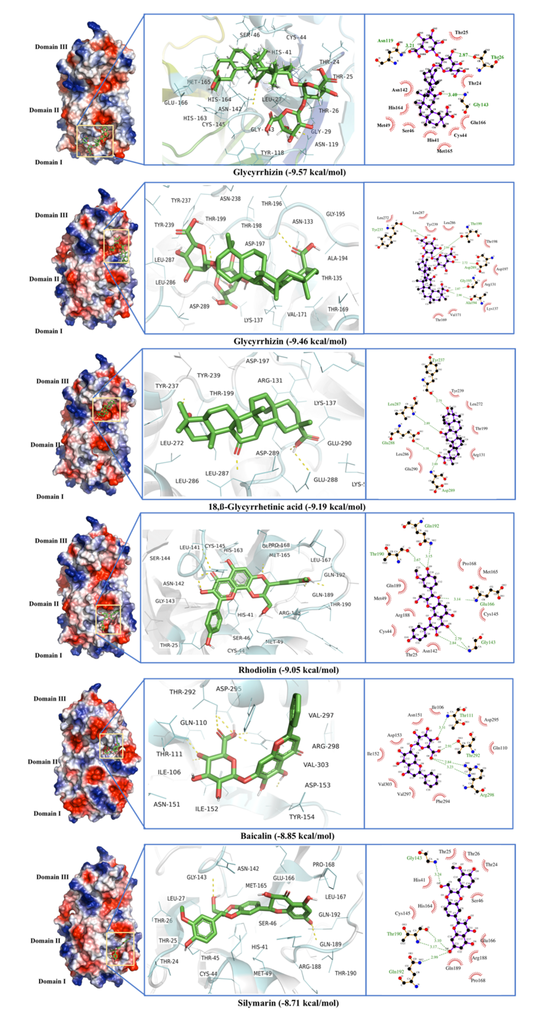
2.1. Main Protease (MPro)

The phytochemical ligands in this investigation were screened against MPro. The ligands with the best docking against the Main Protease are listed in Table 1. Glycyrrhizin, 18,β-Glycyrrhetinic acid, Rhodiolin, Baicalin, and Silymarin were the best five ligands with high binding energies and dissociation constants (Figure 2). Glycyrrhizin showed binding to MPro at two different binding sites with the highest binding scores of −9.57 and −9.46 kcal·mol−1 and a dissociation constant of 0.11 and 0.76 µM. The best Glycyrrhizin binding site is the conventional MPro active site located between domains I and II. This site mainly consists of Thr24, Thr25, Thr26, Leu27, His41, Cys45, Ser46, Met49, Tyr118, Asn119, Asn142, Gly143, Cys145, His163, His164, Met165, Gly166, and Gln189, where Thr26 and Gly143 formed H-bonds with the Glycyrrhizin (Figure 2, Table 1). His41 and Cys145 are the key residues involved in the enzyme active site and have been previously defined in MPro active site by an X-ray crystallographic structure (PDB ID 6WQF) obtained at room temperature [38]. The His41 and Cys145 form a catalytic dyad that interacts with the bound ligand. Other amino acids in the proposed active site Ser46, Leu141, Asn142, Glu166, Pro168, Gln189, Thr190, and Ala191 [38,39] were found involved in the stabilization of the enzyme active site.
The allosteric binding sites for Glycyrrhizin were also found between domains II and III and included the residues Arg131, Lys137, Asp197, Thr198, Thr199, Tyr239, Leu271, Asn274, Gly275, Met276, Asn277, Gly278, Arg279, Ala285, Leu286, Leu287, Glu288, Asp289, and Glu29 where Asp197, Thr199, and Ala285 form H-bonds with Glycyrrhizin (Figure 2, Table 1). The Glycyrrhizin binding to this active site may disrupt the native enzyme structure and affects its activity. This distal pocket has also been reported as a promising inhibitor binding site [40,41]. This site consists of both a loop region and β-strands.
The 18,β-Glycyrrhetinic acid, a derivative of Glycyrrhizin, showed distal allosteric binding with an energy of -9.19 kcal·mol−1 and dissociation constant of 0.35 µM. The active residues for 18,β-Glycyrrhetinic acid include Lys5, Arg131, Lys137, Asp197, Thr199, Tyr237, Tyr239, Leu272, Leu286, Leu287, Glu288, Asp289, and Glu290. Among these residues, Tyr237, Leu287, Glu288, and Asp289 formed H-bonds (Figure 2, Table 1). Although 18,β-Glycyrrhetinic acid does not directly bind to the proposed site of the enzyme, it interacts with the residues which seem present at the dimer interface of the main protease [42] making the binding more interesting to explore (Figure 2).
Other ligands with prominent binding to the main protease include Rhodiolin and silymarin that target the enzyme catalytic site with binding energies of −9.05 and −8.81 kcal·mol−1, while the Baicalin binding site includes some of the distal site residues, including Ile106, Gln110, Thr111, Asn151, Ile152, Asp153, Thr292, Phe294, Val297, Arg298, and Val203 (Figure 2, Table 1). The strong binding of Baicalin to these distal amino acids may reduce the enzyme activity. The worst docking ligands include methyl tridecanoate (binding energy −4.01 kcal·mol−1), Margaric acid (binding energy −3.91 kcal·mol−1), and docosanoic acid (binding energy −3.86 kcal·mol−1) (Table S1).
The initial MD simulation in the case of Glycyrrhizin (active site binding) shows that an equilibrium was achieved after 25 ns, so simulations were limited to 30 ns. The RMSD and RMSF values show the flexible residues in two regions, one from residues Arg40-Asp56 (Domain I) and from Ile136 to Asp153 (Domain II) (Figure 3). This shows the ligand interactions with the active site residues of the enzyme. The fluctuations between Tyr237 and Gly251 show that Glycyrrhizin binding to the active site may also induce a conformational change in other parts of the enzyme. This conformational change may be involved in reducing the enzyme activity. The Rhodiolin-MPro complex showed huge fluctuation around Ile281 to Val296 while minor fluctuations in a loop region around Val91 to Pro96 (Figure 3). The radius of gyration (R) at the end of the MD simulation shows the compactness and stability of the Glycyrrhizin- and rhodiolin-enzyme complexes (Figure S1).

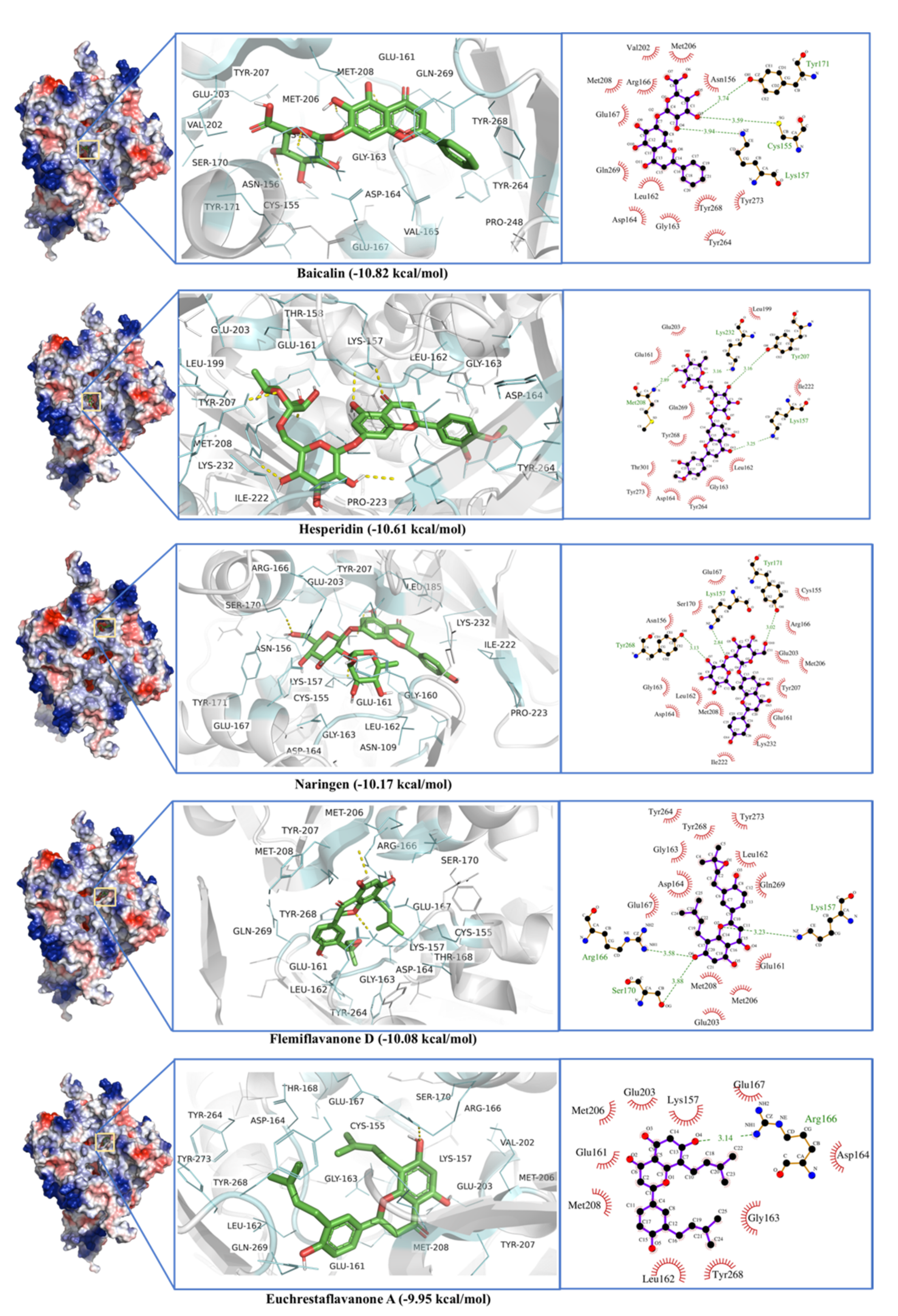
2.2. Papain-Like Protease

Like the MPro, Papain-like Protease is involved in diverse functions that make it a potential drug target [2]. The ligands with the best docking with PLPro are listed in Table 1. Most of the ligands indirectly interacted with the active site triad Cys111, His272, and Asp286 [43] by binding around the enzyme active site (Figure 4). These triad residues are involved in enzyme activity [43]. Baicalin showed a strong binding with the binding energy of 10.82 kcal·mol−1 with a dissociation constant of 10 nM. The low dissociation constant shows strong ligand binding even at low concentrations. The active site residues include Arg166, Ser170, Val202, Glu203, Met206, Tyr207, Met208, Cys155, Asn156, Lys157, Glu161, Leu162, Gly163, Asp164, Val165, Glu167, Tyr171, Pro248, Tyr264, Tyr268, Gln269, and Tyr273 (Figure 4). Baicalin shows π-π interactions with Tyr268, while H-bonding is observed with Cys155, Ly157, and Tyr171 (Figure 4). Previously, a binding site in crystal structure of papain-like protease (PLPro) (PDB ID 6WX4) was defined by the residues; Trp106, Asn109, Tyr112, Cys111, Leu162, Asp164, Val165, Arg166, Me208, Pro247, Pro248, Tyr264, Gln269, Cys270, Gly271, His272, Tyr273, and Thr301 [44]. This shows although Biacalin does not directly interact with the catalytic triad, it binds in the vicinity of the enzyme active site very strongly and impairs its proper functioning.
Hesperidin showed the second-best docking with PLpro with a binding energy of −10.61 kcal.mol−1 and a dissociation constant of 0.02 µM. The active site residues include Arg183, Leu185, Leu199, Glu203, Met206, Tyr207, Met208, Gly209, Phe216, Val220, Gln221, Ile222, Pro223, Lys232, Lys157, Thr158, Glu161, Leu162, Gly163, Asp164, Val165, Glu167, Pro248, Tyr264, Tyr268, Gln269, Cys270, Tyr273, and Thr301. Among these active site residues, Lys157, Tyr207, Met208, and Lys232 formed H-bond with Hesperidin (Figure 4). Lys157, Arg166, and Tyr268 seem to be involved in all top five ligand-enzyme interactions.
The worst docking ligands include 6,9,12-octadecatrienoic acid (binding energy −4.85 kcal.mol−1), docosanoic acid (binding energy −4.64 kcal.mol−1), and acetohydroxamic acid (binding energy −4.13 kcal.mol−1). These compounds were unable to bind the proposed active site (Table S1).
MD simulations of 30 ns were run for Baicalin, Hesperidin, and Solophenol, where considerable RMSD and RMSF fluctuations were found in the active site residues of PLPro (Figure 5). In the case of Baicalin, strong binding and low dissociation constant for the Baicalin-enzyme complex is also confirmed by MD simulations. N-terminal region from Val21 to Pro46 is the most flexible region, while regions including Tyr71 to Asp76, Leu101 to Gln121, Gly266 to Ile276, and Thr281 to Lys292 seem to be involved in Biacalin-PLpro interactions (Figure 5). The Hesperidin-enzyme complex Radius of gyration (Rg) for Solophenol increased in 15–30 ns of MD simulation but Baicalin and Hesperidin, due to strong binding, show the Rg similar to the native enzyme (Figure S2).

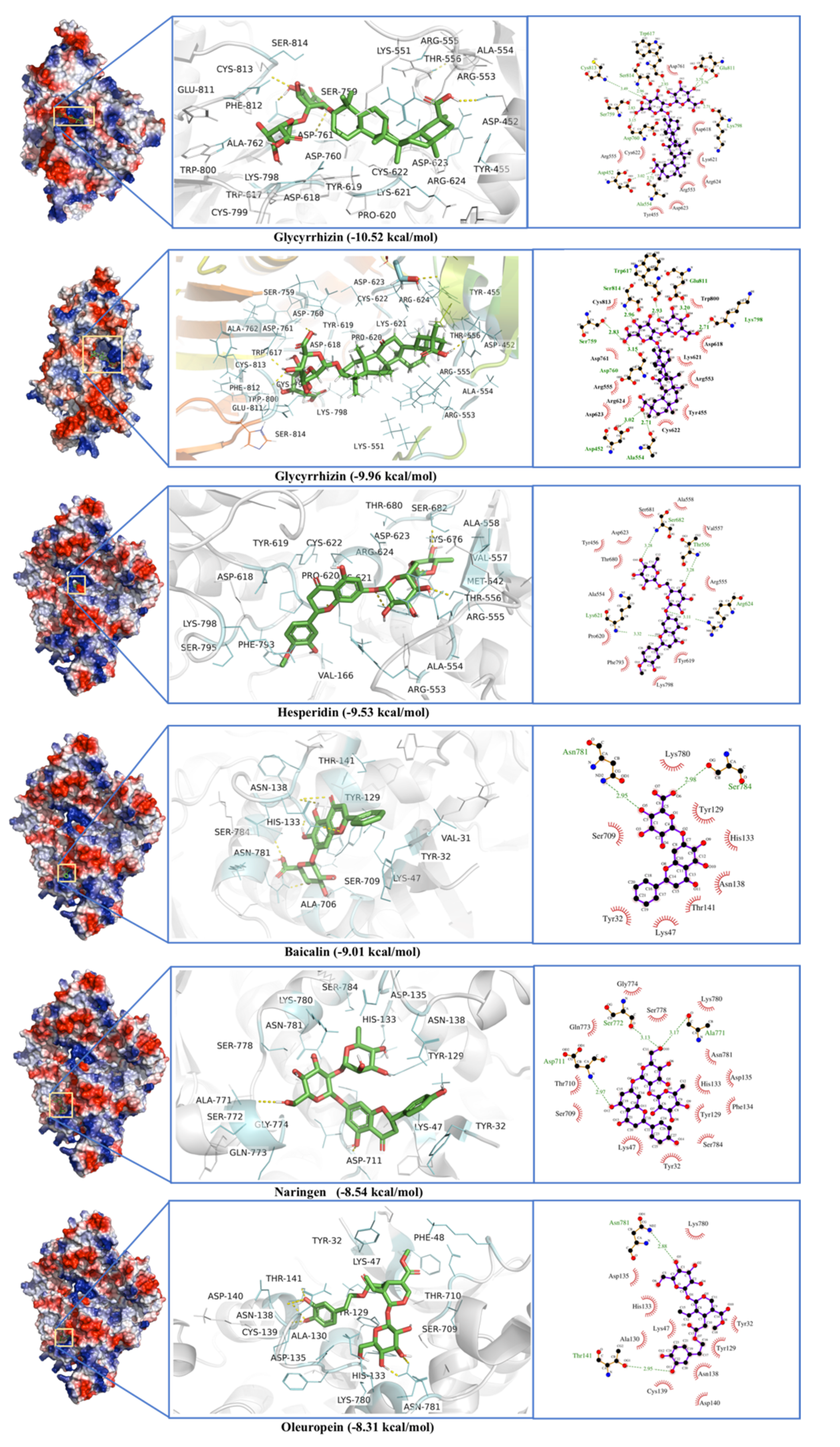
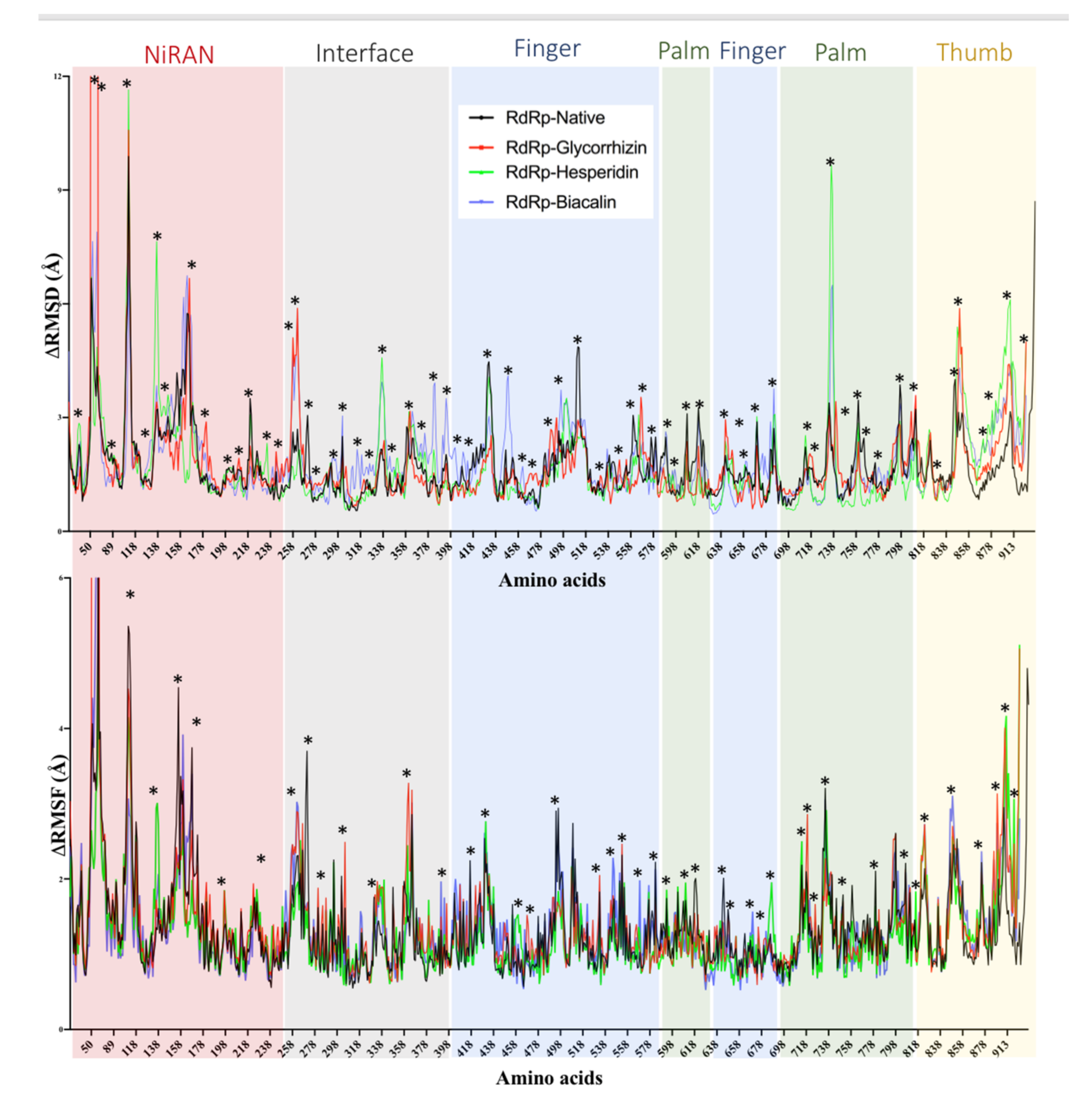
2.3. RNA-Dependent RNA Polymerase (RdRp)

The RNA-dependent RNA Polymerase (RdRp), the main replication enzyme for SARS-CoV-2, was screened against a library of phytochemicals (Table S1). Glycyrrhizin showed the strongest binding to RdRp. Interestingly, Glycyrrhizin strongly interacts with RdRp at two different binding sites with binding energies of −10.27 and −9.96 kcal·mol−1 with dissociation constants of 0.03 and 0.05 µM (Table 1, Figure 6). The key residues of the Glycyrrhizin-enzyme complex include Asp452, Tyr455, Lys551, Arg553, Ala554, Arg555, Thr556, Trp617, Asp618, Tyr619, Pro620, Lys 621, Cys622, Asp623, Arg624, Ser759, Asp760, Asp761, Ala762, Lys798, Cys799, Trp800, Glu811, Phe812, Cys813 Ser814, where Asp452, Ala554, Trp617, Asp760, Lys798, Glu811, Cys813, and Ser814 are involved in H-binding (Figure 6). The formation of multiple H-bonds stabilizes the Glycyrrhizin-RdRp interactions.
The second binding site for Glycyrrhizin includes the residues; Ala46, Lys47, Tyr129, His133, Phe134, Asp135, Met615, Ser709, Thr710, Asp711, Phe766, Ala771, Ser 72, Gln773, Gly774, Ser778, Ile779, Lys780, Asn781, Ser784, and Thr801. Among these residues, Tyr129, His133, Ser709, Asp711, and Asn781 form H-bonds (Figure 6). The binding site of RNA-dependent RNA Polymerase (RdRp) has already been defined by using cryo-EM structures [45] where key catalytic site residues are Lys500, Ser501, Asn507, Lys545, Arg555, Asp618, Ser759, Asp760, Asp761, Cys813, Ser814, and Gln815. It seems that Glycyrrhizin interacts with these residues with a high binding affinity (multiple H-bonds) and may impair the RdRp interactions with the RNA.
Hesperidin, though not interacting with the proposed RdRp active site, binds near to the enzyme active site, where Thr556, Lys621, Arg624, and Ser628 form H-bonds and Phe793 shows π-π interactions. Hesperidin showed a binding energy of −9.53 kcal·mol−1 and with a dissociation constant of 0.1 µM. Baicalin, Naringen, and Oleuropein bind to a completely different site in RdRp, where Thr141, Asn781, and Ser784 are key residues (Figure 6). Baicalin showed the best binding energy of −9.01 kcal·mol−1 with a dissociation constant of 0.12 µM. Active site residues are Phe35, Asp36, Ile37, Tyr38, Asn39, Phe48, Leu49, Lys50, Thr51, Asn52, Val204, Thr206, Asp208, Asn209, Asp218, Asp221, Tyr728. Among these residues, Tyr38, Asn209, Asp218, and Tyr728 formed H-bond (Figure 6).
The worst binders include 11-eicosenoic acid (binding energy −4.11 kcal·mol−1), Oleic acid (binding energy −3.95 kcal·mol−1), and heneicosanoic acid (binding energy −3.76 kcal·mol−1) (Table S1).
A 20 ns MD simulation validates the Glycyrrhizin, Hesperidin, and baicalin interactions with RdRP. Glycyrrhizin and Baicalin show RMSD changes in the interface region consisting of residue from Val258 to Leu270, in the finger region Asp481 to Tyr515, Thr738 to Tyr770 (Figure 7). The RMSD and RMSF fluctuations seem larger in Baicalin in comparison to Glycyrrhizin and Hesperidin. In the thumb region, substantial RMSD changes were observed in all three complexes. All three ligand-enzyme complexes seem more stable in terms of potential energy than native enzymes (Figure S3).

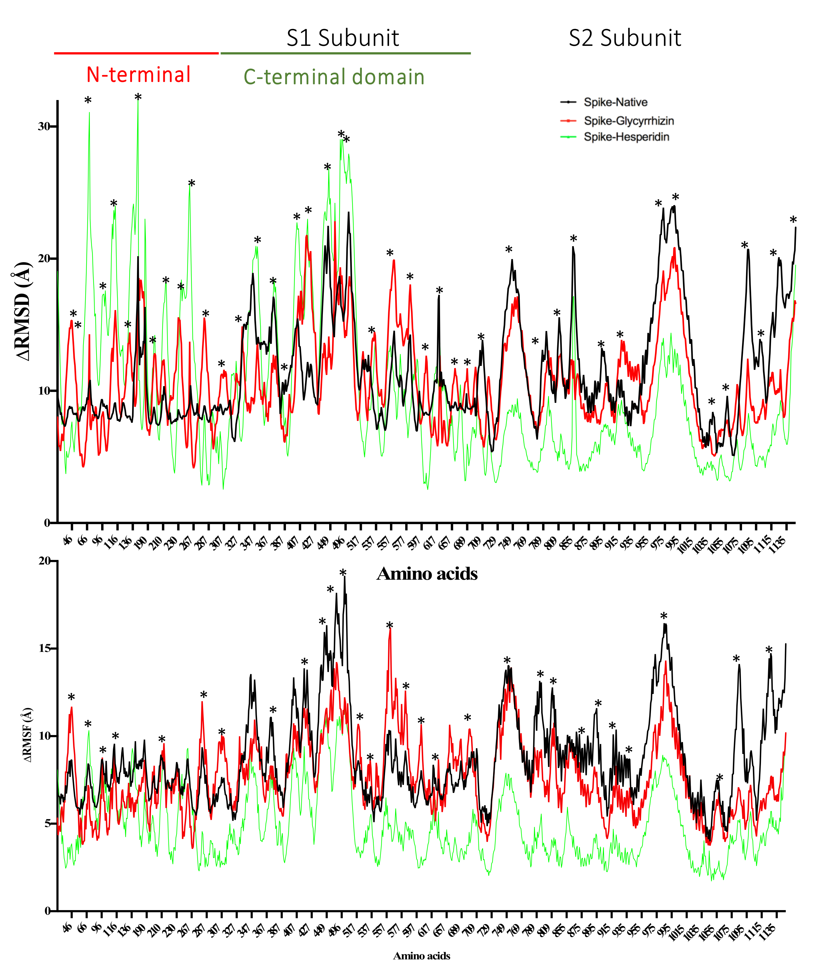
2.4. Spike Glycoprotein

The phytochemical ligands were screened against Spike Glycoprotein’s with both open and closed states of the protein. The ligands with the best docking for spike glycoprotein are listed in Table 1. Glycyrrhizin showed the best binding energy of −9.29 and −9.49 kcal·mol−1 and a dissociation constant of 0.16 and 0.11 µM for the open and close state of spike glycoproteins, respectively. The active site residues for the open state include Val47, His49, Lys304, Met740, Tyr741, Ile742, Cys743, Gly744, Asp745, Phe855, Asn856, Val963, Lys964, Leu966, Ser967, Ser975, Val976, Leu977, Asn978, and Arg1000. H-bonds and π-π interactions were observed in the Glycyrrhizin-Spike protein interactions that show strong binding of the ligand. Among these residues of the active site, Gly744, Asp745, and Arg1000 formed H-bonds, and Tyr741, Phe855 show π-π interactions in an open protein state (Figure 8a). Glycyrrhizin interacts with Tyr741, Ile742, Cys743, Gly744, Asp745, Phe855, Asn856, Val963, Lys964, Leu966, Ser967, Ser975, Val976, Leu977, Asn978, and Arg1000 residues in the close spike protein state, while Tyr741, Gly744, Asp745, and Arg1000 formed H-bonds in the closed state of spike glycoprotein (Figure 8b). This shows Glycyrrhizin’s interactions with the S2 subunit of the spike protein.
Rhodiolin and Hesperidin bind to the N-terminal domain of spike protein and show the second-best binding energies, i.e., −8.68 and −8.53 kcal·mol−1 and dissociation constants, i.e., 0.43 and 0.56 µM for open and states, respectively. Active site residues for Rhodiolin were Arg102, Gly103, Trp104, Ile119, Asn121, Val126, Ile128, Phe168, Tyr170, Ser172, Arg190, Phe192, Ile203, His207, Leu226, Val227, Asp228, Leu229, where Arg102 and Asn121 formed H-bonds (Figure 8a). Hesperidin shows interactions with residues, i.e., Tyr38, Asp40, Lys41, Val42, Phe43, Arg44, Lys206, Phe220, Ser221, Ala222, Glu224, Pro225, Leu226, Tyr279, Gly283, and Thr284, where Ala222 forms a H-bond (Figure 8b).
The worst docking ligands include stearic acid (binding energy −3.38 kcal·mol−1), oleic acid (binding energy −3.26 kcal·mol−1), and docosanoic acid (binding energy −3.21 kcal·mol−1) for open state spike glycoprotein (Table S1). The worst docking ligands include Nervonic acid (binding energy −3.92 kcal·mol−1), octadec-9-enyl icosanoate (binding energy −3.87 kcal·mol−1), and Tetracosanoic acid (binding energy −3.71 kcal·mol−1) for close state spike glycoprotein (Table S1).
MD simulation with a closed state of spike protein shows that Glycyrrhizin-enzyme complexes are more stable than Hesperidin-enzyme complexes in terms of potential energy and Rg (Figure S4). RMSD and RMSF fluctuations in N-terminal domain subunit 1 validate the hesperidin interactions with the residue present in this region (Figure 9). Glycyrrhizin and Hesperidin also show large fluctuations in the S2 areas of spike protein (729–769 and 955–1035 a.a).

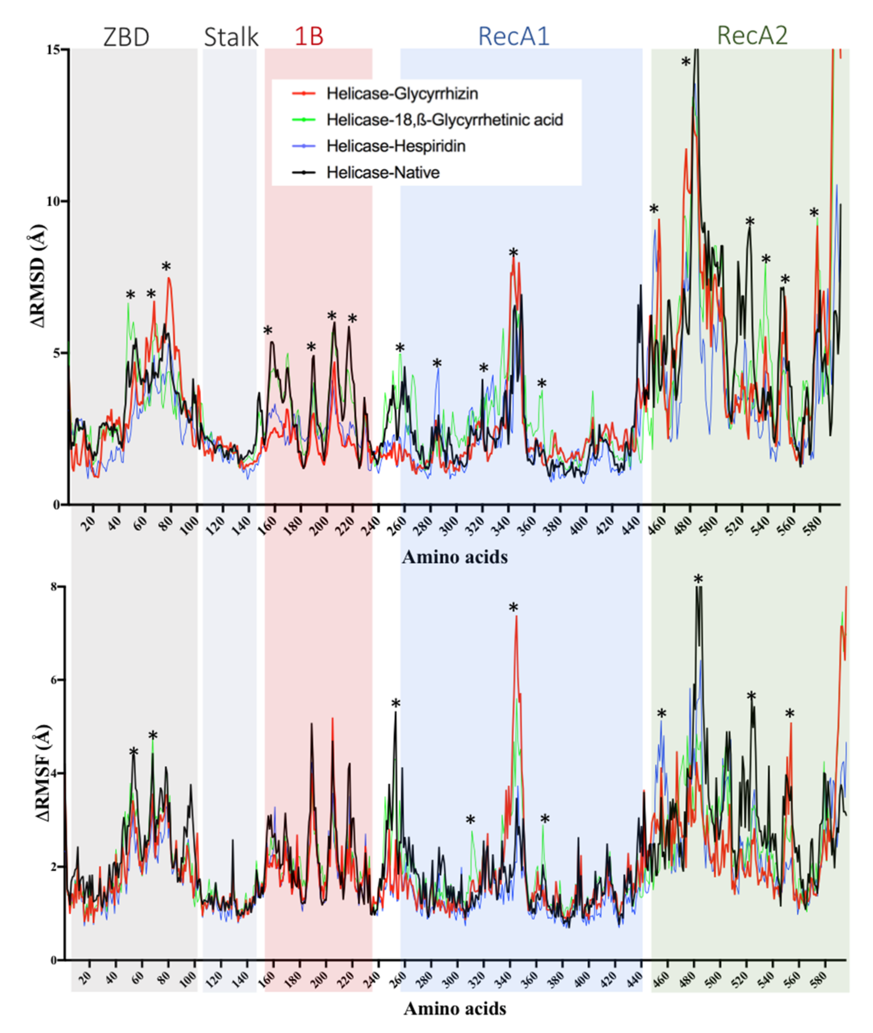
2.5. Helicase (Nsp13) Protein

Phytochemicals ligands were docked against Helicase (Nsp13) protein and Glycyrrhizin, β-Glycyrrhetinic Acid, Solophenol A, Hesperidin, and Baicalin were found to be the best docking ligands (Table 1). Interestingly, Glycyrrhizin showed very strong interactions with the helicase with a binding energy of −11.57 kcal·mol−1 and a dissociation constant of 3 nM. Active site residues are Pro175, Leu176, Asn177, Lys202, Leu405, Pro406, Ala407, Pro408, Arg409, Leu412, Thr413, Gly415, Thr416, Leu417, Phe422, Ser485, Ser486, Pro514, Tyr515, Asn516, Asn519, Thr532, Val533, Asp534, His554, Asn557, Asn559, and Arg560. Among active site six residues, including Asn177, Lys202, Ser485, Asn516, Asp534, and Arg560 form H-bond (Figure 10). Allosteric binding for Glycyrrhizin was observed in a region between RecA1 and RecA2 domains. The same was found to be true in the case of MD simulation, where fluctuations in RMSF and RMSD were observed in a region between RecA1 and RecA2 (Figure 11). The Glycyrrhizin derivative, 18,β-Glycyrrhetinic acid, also showed strong interactions with the second-best binding energy of −9.91 kcal·mol−1 and a dissociation constant of 54 nM. The active site residues are Ala4, Val6, Arg15, Arg21, Arg22, Pro23, Phe24, Arg129, Leu132, Phe133, Glu136, Pro234, Leu235, and Ser236. Among active site residues, Arg15 formed two H-bond. Hesperidin shows the binding energy of −8.93 kcal·mol−1 binds to enzyme active site where three H-bonds were observed for Lys146, His230, and Arg339 (Figure 10). The worst docking ligands include octadec-9-enyl icosanoat (binding energy −4.30 kcal·mol−1), docosanoic acid (binding energy −4.16 kcal·mol−1), and acetohydroxamic acid (binding energy −4.11 kcal·mol−1) for open state spike glycoprotein. Some of these compounds were even unable to bind the proposed active site (Table S1).

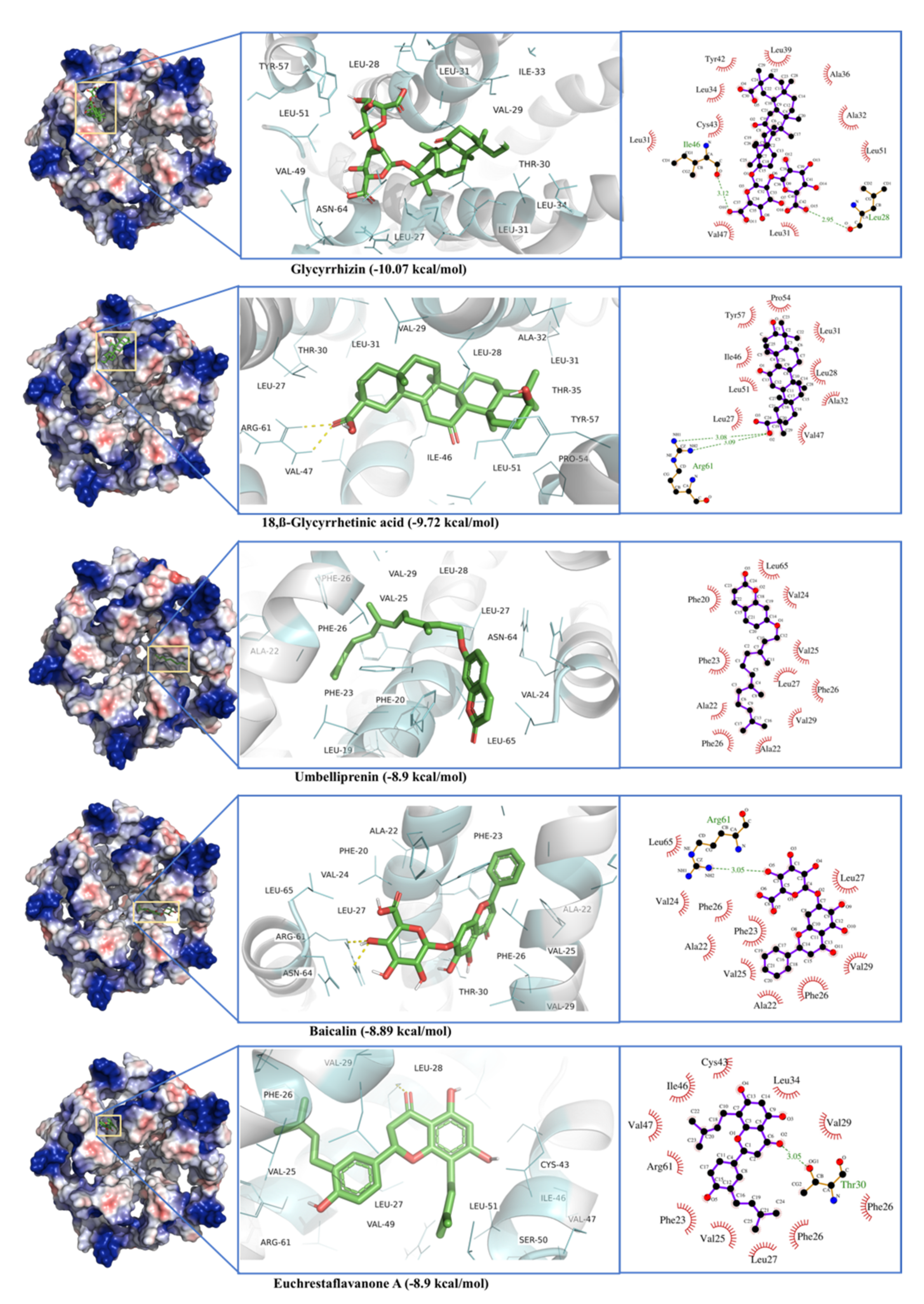
2.6. E-Channel (Envelop Small Membrane Protein)

The phytochemical ligands were docked against E-channel protein are listed in Table 1. Glycyrrhizin showed the best binding score of −10.07 kcal·mol−1 and dissociation constant of 0.04 µM. Glycyrrhizin interacts with the active site residues present in different chains including Arg61, and Asn64 from Chain A, Leu28, Val29, Leu31, Ala32, Ile33, Ala36, and Arg38 from Chain D, Leu27, Thr30, Leu31, Leu34, Leu37, Leu39, Tyr42, Cys43, Ile46, Val47, Val49, Ser50, Leu51, Pro54, and Tyr57 from Chain E. Among these active site residues, Leu28 and Ile46 formed H-bonds (Figure 12). 18,β-Glycyrrhetinic acid showed the second-best bind energy of −9.72 kcal·mol−1 with a dissociation constant of 0.07 µM. Active site residues are Arg61 (Chain A), Leu28, Val29, Leu31, Ala32, and Thr35 (Chain D), Leu27, Thr30, Leu31, Ile46, Val47, Leu51, Pro54, and Tyr57 of Chain E. Among active residues, Arg61 formed two H-bonds (Figure 12). The worst docking ligands include malic acid (binding energy −4.05 kcal·mol−1), oxalic acid (binding energy −3.24 kcal·mol−1), and acetohydroxamic acid (binding energy −3.10 kcal·mol−1) for open state spike glycoprotein. Some of these compounds were even unable to bind the proposed active site (Table S1).

2.7. Non-Specific Interactions of Selected Ligands against Human Blood Proteins

Glycyrrhizin, Hesperidin, and Baicalin were docked to a local library of selected human blood proteins (a total of 100 blood proteins) to map non-specific interactions of these ligands with non-specific proteins (Table S2). Glycyrrhizin showed the highest binding affinity against DdB1 (damage-specific DNA binding protein) with a binding energy of −11.36 kcal·mol−1 and a dissociation constant of 1.68 nM. The interactions residues included Asn16, Gly17, Cys18, Val19, Thr20, Glu65, Leu66, Thr118, Ile121, Ile123, Ile124, Asp125, Pro126, Asp166, Lys168, Phe169, Tyr171, Ser217, Met218, Ala221, Val259, Cys260, His261, Asn262, Arg263, Glu312, Cys313, and Leu314 (Figure 13a). Baicalin also interacts with DdB1 at the same binding site (Figure 13b). In non-specific interactions, Hesperidin shows the highest binding affinity (binding energy −10.89, and dissociation constant 10.4 nM) for Integrin alpha V Beta 6 head protein normally involved in cell adhesion (Figure 13c). The top four non-specific interacting partners of Glycyrrhizin, Hesperidin, and Baicalin are detailed in Figure 13a–c. In contrast, the binding energies and dissociations constants for all 100 non-specific proteins are given in Table S2.

2.8. ADMET Properties of Selected Ligands
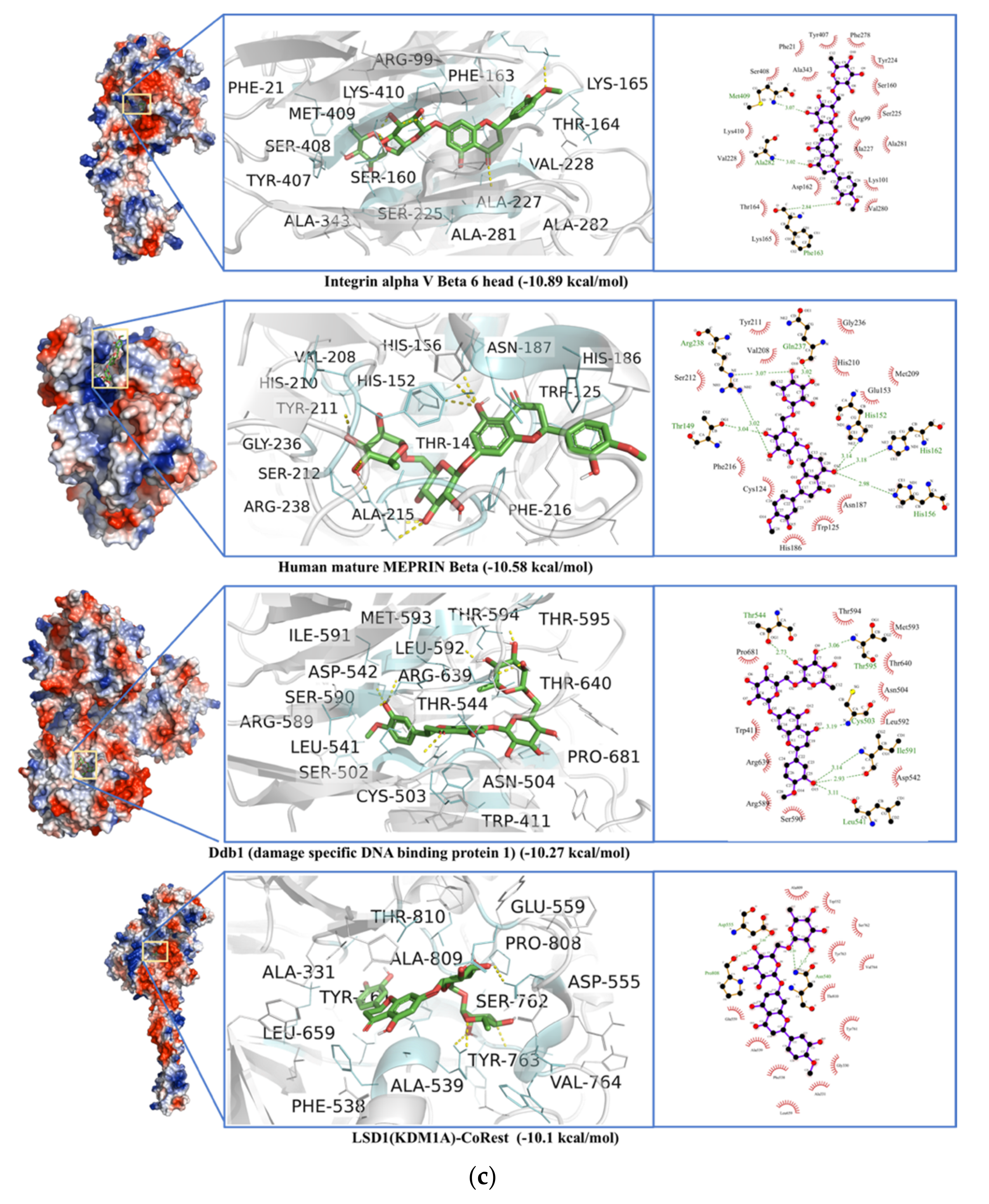
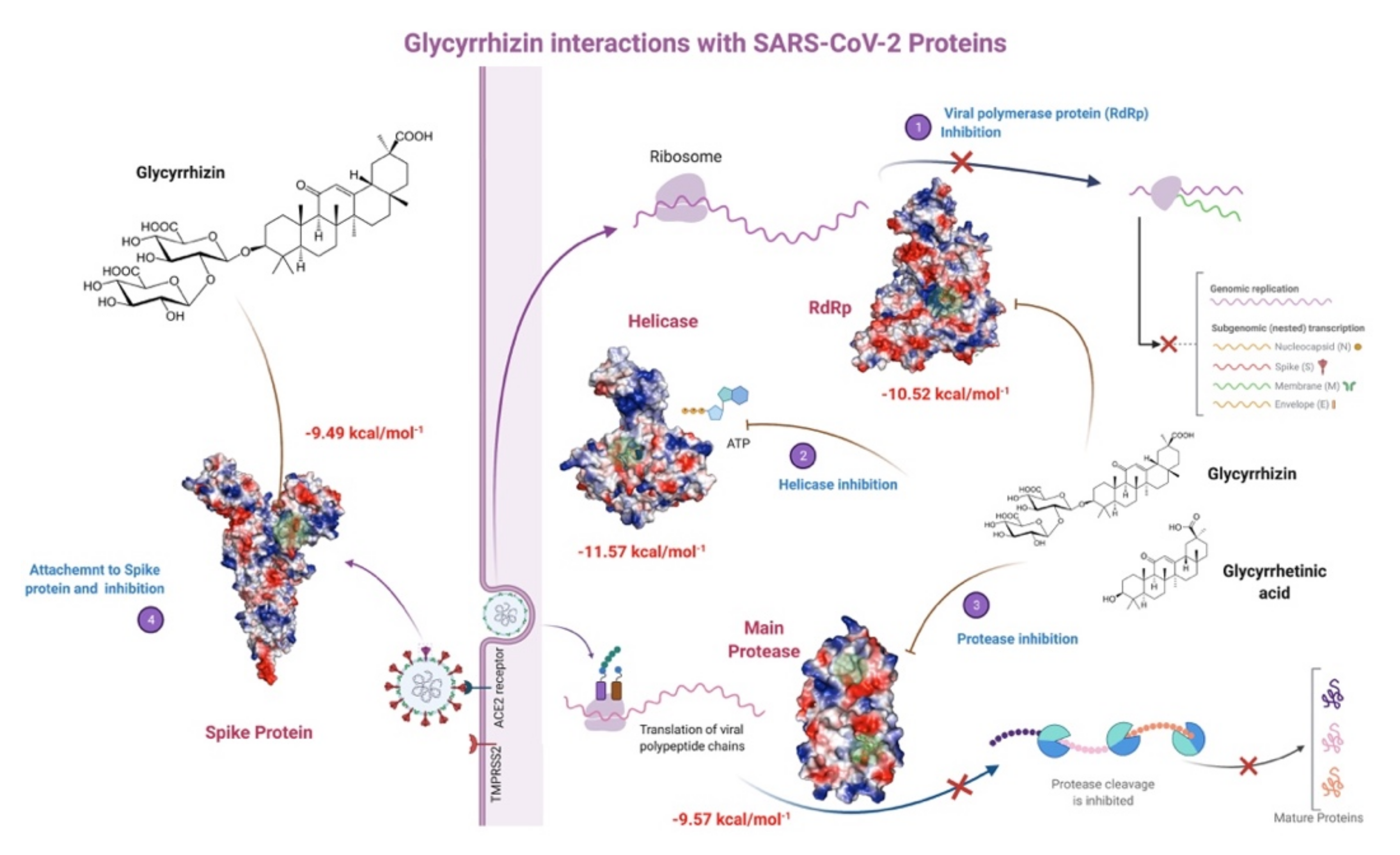
Lipinski’s Rule of Five [46], Ghose filter (Amgen) [47], Veber’s (GSK) [48] rules are used to predict ADME properties. According to the pharmacokinetic properties, all compounds show Gastrointestinal low absorption except 18-β glycyrrhetinic acid (GA), lopinavir, and euchrestaflavanone A, which have high absorption. These compounds have the least BBB permeability, and no CYP inhibition was observed (Table 2).

3. Discussion

Antioxidants including Glycyrrhizin, 18,β Glycyrrhetinic acid, Rhodiolin, Baicalin, and Hesperidin have shown remarkable potential in targeting various SARS-CoV-2 enzymes and proteins. Glycyrrhizin, largely found in licorice root, has already been found active against many viral proteases, including herpesviruses [49,50], flaviviruses [51], and Human Immunodeficiency Virus [52]. Glycyrrhizin was already used to treat patients with hepatitis C [53] and upper respiratory tract infections [54]. Glycyrrhizin, Glycyrrhizic acid, its other derivatives and Baicalin were the first compounds found active against SARS coronavirus (SARS-CoV-1) [55,56]. Flavonoids and their derivatives have been reported to inhibit various SARS-CoV-2 proteins [57]. Hesperidin and quercetin have also been found good antiviral agents [58,59].
The binding site for MPro has already been defined by X-ray crystallographic structure (PDB ID 6WQF) obtained at room temperature [38] where His41 and Cys45 form a catalytic dyad to interact with bound ligand. Other amino acids involved in the stabilization of the active site were Ser46, Leu141, Asn142, Glu166, Pro168, Gln189, Thr190, Ala191. This active site is situated in a cleft between domains I and II [60,61]. In our study, Glycyrrhizin shows two binding sites; one includes the conventional active site of the enzyme, while the second interactions include an allosteric binding site. A previous docking analysis of Glycyrrhizin against MPro has shown a binding energy value −7.81 kcal·mol−1 where it interacts with the proposed active site of the enzyme [62]. In another study, glycyrrhizic acid, Glabridin and Liquiritigenin show strong binding interactions (with a binding energy of −7.0 to −8.0 kcal·mol−1) with MPro conventional active site [63]. The docking analysis of MPro with FDA-approved anti-viral compounds and library of active phytochemicals [64] shows Nelfinavir potent against MPro. Many natural compounds are found to be potential inhibitors of MPro, including Leucoefdin [65], Leupeptin [66], Rutin [67], cannabisin-A, isoacetoside [68], epigallocatechin gallate, and epicatechin gallate [69]. Recently, Glycyrrhizin has been found to indirectly inhibit the SARS-CoV-2 replication by Inhibiting MPro enzyme activity [70]. In our study, many ligands, including Glycyrrhizin, have been found to interact with Mpro allosteric binding sites (Table 1 and Table S1, Figure 2). In a previous study, 2400 FDA-approved drugs have been screened against MPro allosteric binding sites, where selinexor, bromocriptine, Dihydroergotamine, nilotinib, entrectinib, digitoxin, and diosmin have shown promising binding to the enzyme [71].
PLPro consists of an N-terminal ubiquitin-like (Ubl) domain (1–60 a.a) and a catalytic region with a right-handed thumb-palm-fingers architecture. The PLPro binding site is found in the thumb and palm domain and is characterized by the presence of a catalytic triad (Cys111, His272, and Asp286) [72]. In our study, Baicalin and Hesperidin have been found to be potential PLpro inhibitors that interact with their proposed active site (Figure 4). GRL0617 with PLpro shows binding to the same site [73], whereas π-π interactions with Tyr268 have shown definite inhibition of PLpro activity. Natural compounds like Caesalpiniaphenol A, and Sappanone B, also interact with Try268. Corylifol A, chromen, darunavir, sofosbuvir and some other drugs were screened against PLPro. These drugs were found to bind near the proposed catalytic triad [74]. Phytochemicals from Vitex negundo L. are also found active against PLPro [75]. Along with natural compounds, many approved antibacterial and antiviral drugs also have been repurposed [76,77,78].
Here, MD simulations validate the role of Tyr268 in Baicalin, Hesperidin, and Solophenol enzyme complexes (Figure 5). All catalytic residues seem to interact with these ligands in 30 ns MD simulation. In a previous study, MD simulations for PLpro were found stabilized after 12 ns and remained stable until 50 ns [79]. Baicalin can be extracted from the roots of Scutellaria baicalensis Georgi. In another study, six phytochemicals, including Baicalin, rutin, biopterin, licoleafol, luteolin, and quercetin, have shown binding to PLpro [80]. In addition, Baicalin showed antiviral activity against dengue virus (DENV) [81], Influenza A virus (IAV) [82], Zika virus (ZIKV) [83], Chikungunya virus (CHIKV) [84], and Human Immunodeficiency Virus 1 (HIV-1) [85].
Glycyrrhizin, Hesperidin, and Biacalin show strong interactions with RdRp. Glycyrrhizin binds in the enzyme’s potential active site pocket while making H-bonding with Asp760 and other residues (Figure 6). Asp760,761 have been proposed as active site residues, while Tyr619, Cys622, Ser759, Ala762, Glu811, Cys813, and Ser814 are found in potential binding sites for Ribavirin, Remdesivir, and other antivirals interactions [86]. In a screening with flavonoid compounds, Delphinidin 3-O-beta-D-glucoside 5-O-(6-coumaroyl-beta-D-glucoside) complex with RdRp has been found most stable, where Asp760,761 have been found in the ligand-binding site [87]. Lanreotide, Argiprestocin, Demoxytocin, and Polymyxin B1 also interact with Asp760. Previously, polyphenols with binding energy <7.0 have been reported to interact with RdRp, while Remdesivir showed binding energy of 7.9 kcal·mol−1 [88] and interacts with a similar ligand-binding site as for Glycyrrhizin, Hesperidin. This site includes Asp760,761, and Glu811 for Remdesivir [89,90]. In another study, approved antivirals including Ribavirin, Remdesivir, Sofosbuvir, Galidesivir, and Tenofovir have shown strong interactions with RdRp, where Ribavirin have shown binding energy of −8.7 kcal/mol [86]. Many compounds from the ZINC database have been screened against RdRp and 40 ns MD simulations were performed for Rifabutin, ZINC09128258 and ZINC09883305 [91].
Glycyrrhizin strongly binds to the S2 subunit of the spike protein. Although the binding residues are different from the receptor-binding domain (331–524 a.a.), it is expected that this interaction may bring the conformation change in the protein that may affect the receptor binding. This conformational change is also indicated by MD simulation showing large RMSD and RMSF changes in the receptor-binding region and formation of the close complex (low Rg) (Figure 8 and Figure S4). A number of flavonoid compounds were docked to spike protein and naringin has been found as the most potent compound against SARS-CoV-2 spike protein [92]. In our study, Glycyrrhizin showed the best binding energy against the open and close state of spike glycoproteins. Most of the Glycyrrhizin’s interactions have been found with the S2 subunit of the spike protein (Figure 8b). In one study, 66 compounds were found to interact with RBD of spike protein and Glycyrrhizic acid was found to be the most potent antiviral and spike protein inhibitor [93]. Rhodiolin and Hesperidin bind to the N-terminal domain of spike protein and show the second-best binding energies (Figure 8b). Mutations in spike protein lead to new SARS-CoV-2 variants. Glycyrrhizin was docked against the spike protein variants initially reported from Brazil (B.1.1.28), South Africa (B.1.351), and the United Kingdom (B.1.1.17). The variant B.1.1.7 was first reported in the United Kingdom on 14 December 2020 [94,95], B.1.1.28 originated from Brazil [96]. Both mutations, B.1.1.7 and B.1.1.28, carry the N501Y mutation in the RBD of the spike protein. Later, the B.1.351 variant (20H/501Y.V2) was first reported in South Africa [97], was marked by mutation of K417N and E484K in the RBD region of the spike protein. Surprisingly, the affinity of the Glycyrrhizin slightly decreased against the spike protein variants, though it still shows considerable binding energy and dissociation constant (Figure S6). Catechins and tamibarotene have been found to interact strongly with the UK variant and triple SARS-CoV-2 variant [98,99].
In the case of SARS-CoV-2 helicase, the interface between RecA1 and RecA2 domains contains the active site residues for helicase enzyme, including Lys288, Ser289, Asp374, Glu375, Gln404, and Arg567 [100,101]. In this study, Glycyrrhizin binds to an allosteric site near the catalytic cleft, while Glycyrrhetinic acid interacts with residues in stalk and 1B domain. Triphenyamine and Darunavir have been reported to bind the same active site [102]. Rutin, xanthones, and many other polyphenols have been reported to be ATPase inhibitors [59]. A database of 14,000 phytochemicals has been docked against helicase using virtual screening; out of them, 368 compounds have been found to be potent helicase inhibitors [103]. Picrasidine M from a herb Picrasma quassioides shows the best binding to helicase, where it forms 2 H-bonds with Ser289 and one H-bond with Gln404 and Arg567 [103]. E-channel blockers are demonstrated to be potent antivirals by protecting hosts cells from death. Glycyrrhizin, Glycyrrhetinic acid, and Baicalin have also shown binding to E-channel protein. Proanthocyanidins have been reported to inhibit the MPro and E-channel protein of the SARS-CoV-2 [104].
Glycyrrhizin seems to interact with four SARS-CoV-2 key proteins with high affinity, where its binding with helicase and RdRp has been found to be more stable (Figure 14). Glycyrrhizin has also been reported to interact with TMPRSS2, involved in viral penetration into the host cells [105]. It also indicates that licorice root, a potential source of Glycyrrhizin, may be used as a possible cure and a household remedy for COVID-19. The non-specific interactions of Glycyrrhizin show that it may interact with Ddb1 and other non-specific human blood proteins. Glycyrrhizin as Glycyrrhizic acid from Glycyrrhiza glabra and as a derivative in the form of β-Glycyrrhetinic acid are reported to show penetration through the blood-brain barrier and are non-carcinogenic [63] (Table 2). This study shows that natural antioxidant compounds, either partially purified or in crude form, may provide protection against SARS-CoV-2 severity and complications by interacting with its key enzymes. Although we have performed 30 ns MD simulations as in the case of many protein-ligand complexes equilibrium was achieved and complex was found stable, longer MD simulations (>100 ns) in future studies may help to better understand the behavior and stability of the complexes. In vivo studies for the selected compounds are also recommended to probe the computational result and may help in the formulation of natural antivirals with less toxicity and more efficacy.

4. Conclusions

In the current pandemic situation, COVID-19′s battle with humanity is still continued. The availability of various vaccines has eased the aggravating situation, but the rise of SARS-CoV2 variants warns that humans have to live with the virus, evading its devastating effects. Potential natural cures/medications/home remedies without any side effects seems a viable solution in the current circumstances. Our study has examined and screened more than a hundred natural compounds from plants against six SARS CoV-2 proteins by using molecular docking and molecular dynamics simulations to identify potential bioactive compounds. Glycyrrhizin was found as the best ligand showing strong inhibition of five SARS CoV-2 proteins. Glycyrrhizin is present in a large amount in an inexpensive household herb licorice already registered for its magical curative properties against a number of diseases. Other phytochemicals found potent against SARS-CoV2 include Hesperidin and Baicalin present in citrus fruits and many other plants. In our study, Glycyrrhizin, Hesperidin, and Baicalin were docked against non-specific human blood proteins and have shown interactions with DNA binding proteins. Based on our findings, we suggest that further in vivo evaluation of Glycyrrhizin and its sister compounds as potential antivirals will signify their role in the treatment and management of COVID-19.

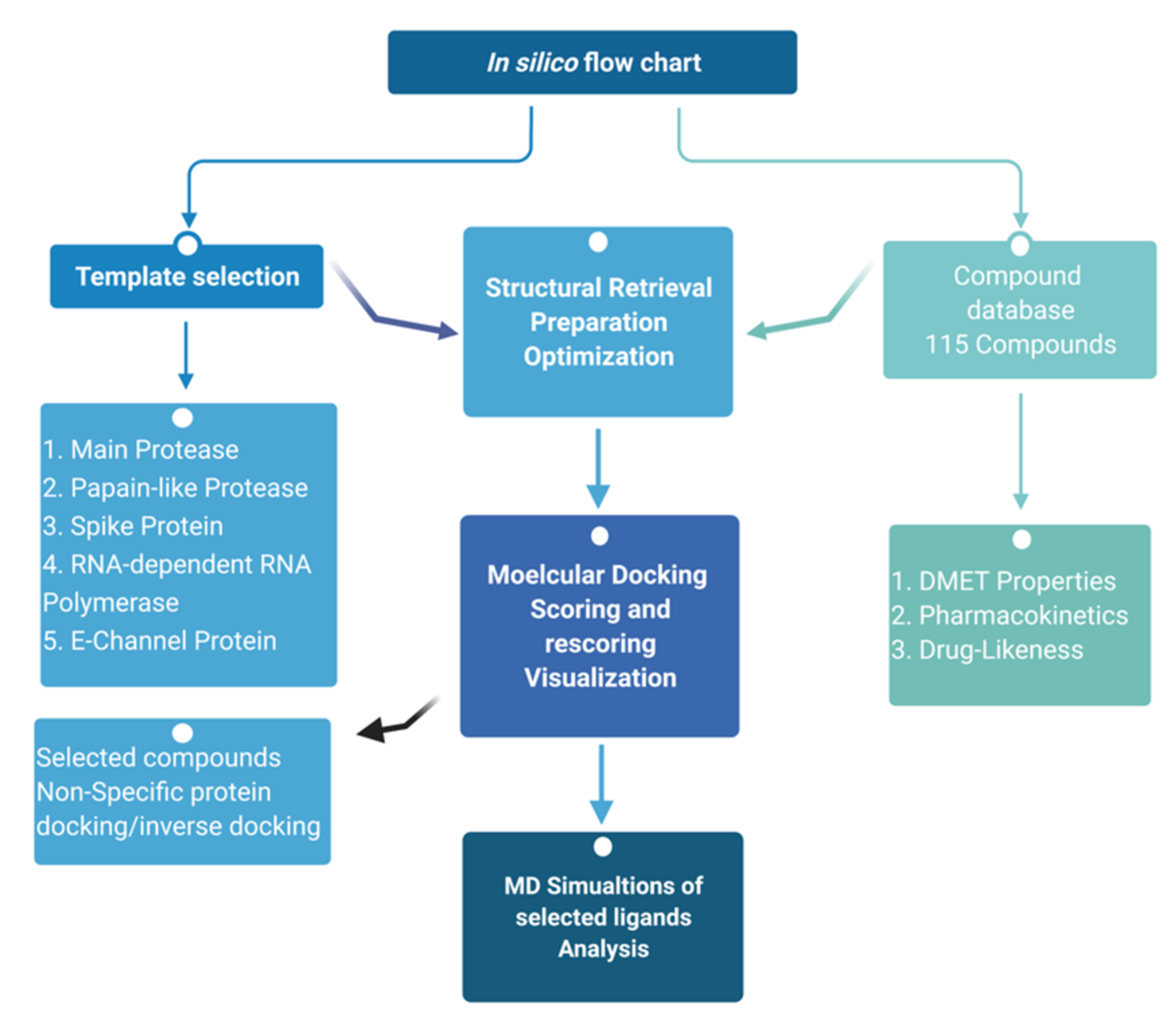
5. Materials and Methods

A total of 115 natural compounds with established therapeutic properties were targeted against six SARS-CoV-2 proteins by molecular docking. The compound structures were obtained from various chemical databases, including PubChem (https://pubchem.ncbi.nlm.nih.gov/, accessed on 25 March 2021), ChemSpider (https://chemspider.com, accessed on 25 March 2021) and MolPort (https://www.molport.com/, accessed on 25 March 2021) (Table S1). The compound structures were obtained in form of The Spatial Data File (SDF) and optimized using the MM1 forcefield in YASARA Structure ver. 20.7.4 [106]. The ligand structures were merged in a single file prepared for virtual screening. Six SARS-CoV-2 proteins were obtained from Protein Data Bank (PDB), including Main Protease (MPro, PDB ID 6Y84), Papain-like protease (PLpro, PDB ID 6WX4), RNA-dependent RNA polymerase (RdRp, PDB ID 6M71), Spike Glycoprotein (S, open and closed state, PDB IDs 6VXX and 6VYB), Helicase (Nsp13) (PDB ID 6ZSL), and E-Channel (Envelop small membrane protein) (PDB ID 5 × 29) (Figure 1). Spike protein variants’ structures as well as structures for all selected non-specific proteins were also obtained from PDB (Table S2a–c). The single-chain structures for all proteins were prepared using YASARA Structure ver. 20.7.4 [106] and heteroatoms were removed.

5.1. Molecular Docking

The ligand-protein interactions and binding energies were calculated by applying a virtual screening module in YASARA software version 20.7.4 [106] that uses a modified AutoDock-Lamarckian Genetic Algorithm. The parameters used for the virtual screening and molecular docking have been described earlier [107], where AMBER03-FF was used with a hundred global docking runs and by keeping the random seed value of 1000. AutoDock local search (LGA-LS) was also used for selected top five ligands to reassure best binding and energy minimization. Protein-ligand interactions were mapped in terms of binding energies and dissociation constants, while the docking scores were calculated by Equation (1)
ΔG = ΔG(van der Waals) + ΔG(H-bonding)+ ΔG(electrostatic)+ ΔG (torsional free energy) + ΔG(desolvation energy)
LigPlus [108] was used to obtain and ligand-protein interactions. The specific ligand was selected in the ligand-protein complex file and interactions including H-bonds were mapped. Non-specific Interactions of selected ligands against human blood proteins were studied by an inverse docking procedure where more than 100 human blood protein targets were screened against the selected ligands (Table S2a–c).

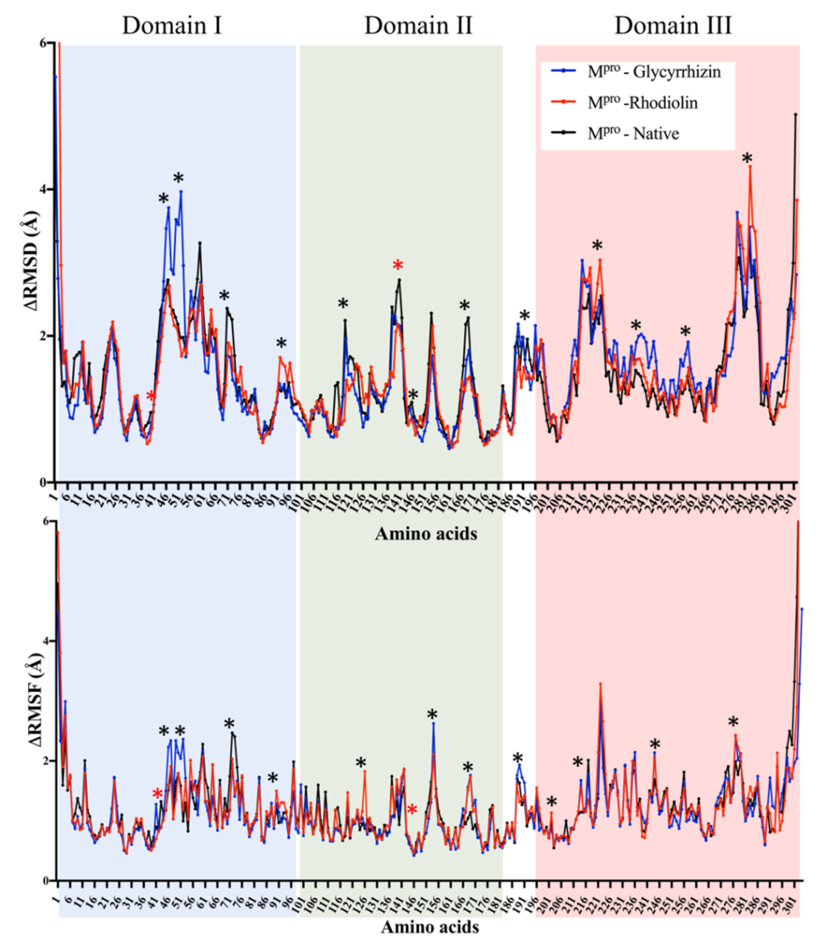
5.2. Molecular Dynamic Simulations

Molecular Dynamic simulations (MDS) were performed using YASARA Structure ver. 20.7.4 [106] with AMBER14 as a force field as described before [109]. The simulation cell was prepared by providing 20 Å water-filled space around the fully mobile protein with a density of 0.997 g/mL. The system was neutralized with 0.9% NaCl while maintaining 298 K temperature, pH 7.4, periodic boundaries, and 7.86 cut-off for long-range coulomb electrostatics forces. After the initial steepest descent minimization, MDS was performed at the rate of 1.25–2.50 fs time steps, and the simulation snapshot was saved every 100 ps. MDS of 10–30 ns were calculated for different proteins depending on the number of atoms in the simulation cell. The raw data were analyzed using GraphPad Prism ver. 7.0. [110]. RMSD and RMSF values were tabulated and analyzed for the fluctuations. A docking and MD simulation flow chart had been given in Figure 15.

5.3. Pharmacokinetics and Drug-Likeness

The pharmacokinetic properties and drug-likeness prediction of the top 10 li were performed by the SwissADME server (http://www.swissadme.ch/, accessed on 20 April 2021). It calculates the topological polar surface area (TPSA), logP (lipophilicity), and logS (solubility). The drug-likeness was predicted by following Lipinski, Ghose, and Veber rules and bioavailability scores [46,47,48]. The Lipinski’s Rule of Five states that the absorption or permeation of a molecule is more likely when the molecular mass is under 500 g/mol, the value of log P is lower than 5, and the molecule has utmost 5 H-donor and 10 H-acceptor atoms [46]. Ghose filter (Amgen) [47] defines drug-likeness based on log P between −0.4–5.6, MW between 160–480, molar refractivity between 40–130, and the total number of atoms between 20–70. Veber (GSK) [48], the rule defines drug-likeness as rotatable bond count ≤10 and polar surface area (PSA) ≤ 140.

Supplementary Materials

The following are available online at https://0-www-mdpi-com.brum.beds.ac.uk/article/10.3390/antibiotics10081011/s1, Figure S1: 30 ns Molecular dynamics simulation of Glycyrrhizin (blue) and Rhodiolin (red) docked with SARS-CoV-2 main protease (MPro), Figure S2: 30 ns Molecular dynamics simulation of Baicalin (red), Hesperidin (green), and Solophenol (blue) docked with SARS-CoV-2 PLpro, Figure S3: 30 ns Molecular dynamics simulation of Glycyrrhizin (red), Hesperidin (green), Baicalin (blue) in complex with SARS-CoV-2 RdRp. Figure S4: 30 ns Molecular dynamics simulation of Glycyrrhizin (red) and Hesperidin (green) in complex with SARS-CoV-2 spike protein (closed state) Figure S5: 30 ns Molecular dynamics simulation of Glycyrrhizin (red), Hesperidin (green), Baicalin (blue) in complex with SARS-CoV-2 helicase Figure S6: Glycyrrhizin interactions with variants of Spike protein of SARS-CoV-2, Table S1: Ligands docked to the SARS-CoV-2 proteins; binding energies, dissociation constants and active sites, Table S2a: Glycyrrhizin docking to nonspecific human blood proteins, Table S2b: Baicalin docking to nonspecific human blood proteins, Table S2c: Hesperidine docking to nonspecific human blood proteins and Table S3.

Author Contributions

Conceptualization, M.F.u.R. and M.A.; methodology, M.F.u.R., A.I.B. and S.A.; software, M.F.u.R., R.E. and S.A.; validation, M.F.u.R., M.S.A., M.S. and F.K.; formal analysis, S.A. and R.E.; investigation, S.A., R.E. and A.I.B.; resources, M.F.u.R., M.S.A. and C.L.; data curation, S.A. and M.S.; writing—original draft preparation, S.A., M.F.u.R., M.S.; writing—review and editing, M.F.u.R., S.A., C.L. and M.S.A.; visualization, C.L., Z.S. and M.F.u.R.; supervision, M.F.u.R.; project administration, M.F.u.R., M.S., C.L., Z.S. and M.M.; funding acquisition, M.F.u.R., M.M. and C.L. All authors have read and agreed to the published version of the manuscript.

Funding

Funding in the C.L. lab was provided by the Fundamental Research Funds for the Central Universities (2232021G-04), Shanghai Science and Technology Committee (19ZR1471100). M.F.u.R. is thankful to the Office and Research and Innovation Centre (ORIC), University of Sargodha, Sargodha, Pakistan, for the award of COVID-19 related research Project awarded via number SU/ORIC/923. M.F.u.R is also thankful for online utilization of computational resources at C.L. lab, Donghua University, China.

Conflicts of Interest

The authors declare no conflict of interest.

References

  1. Yang, Y.; Peng, F.; Wang, R.; Guan, K.; Jiang, T.; Xu, G.; Sun, J.; Chang, C. The deadly coronaviruses: The 2003 SARS pandemic and the 2020 novel coronavirus epidemic in China. J. Autoimmun. 2020, 109, 102434. [Google Scholar] [CrossRef]
  2. Rehman, M.F.; Fariha, C.; Anwar, A.; Shahzad, N.; Ahmad, M.; Mukhtar, S.; Haque, M.F.U. Novel coronavirus disease (COVID-19) pandemic: A recent mini review. Comput. Struct. Biotechnol. J. 2020, 19, 612–623. [Google Scholar] [CrossRef]
  3. Coronavirus-Worldwide-Graphs. Available online: https://www.worldometers.info (accessed on 12 August 2021).
  4. Dinleyici, E.C.; Borrow, R.; Safadi, M.A.P.; Van Damme, P.; Munoz, F.M. Vaccines and routine immunization strategies during the COVID-19 pandemic. Hum. Vaccines Immunother. 2021, 17, 400–407. [Google Scholar] [CrossRef]
  5. Mazzola, A.; Todesco, E.; Drouin, S.; Hazan, F.; Marot, S.; Thabut, D.; Varnous, S.; Soulié, C.; Barrou, B.; Marcelin, A.-G.; et al. Poor Antibody Response After Two Doses of Severe Acute Respiratory Syndrome Coronavirus 2 (SARS-CoV-2) Vaccine in Transplant Recipients. Clin. Infect. Dis. 2021. [Google Scholar] [CrossRef]
  6. Mohammed, F.S.; Akgul, H.; Sevindik, M.; Khaled, B.M.T. Phenolic content and biological activities of Rhus coriaria var. zebaria. Fresenius Environ. Bull. 2018, 27, 5694–5702. [Google Scholar]
  7. Sevindik, M.; Akgul, H.; Selamoglu, Z.; Braidy, N. Antioxidant and antigenotoxic potential of infundibulicybe geotropa mushroom collected from Northwestern Turkey. Oxidative Med. Cell. Longev. 2020, 2020, 5620484. [Google Scholar] [CrossRef] [PubMed] [Green Version]
  8. Sevindik, M.; Akgul, H.; Bal, C.; Selamoglu, Z. Phenolic contents, oxidant/antioxidant potential and heavy metal levels in Cyclocybe cylindracea. Indian J. Pharm. Educ. Res. 2018, 52, 437–441. [Google Scholar] [CrossRef] [Green Version]
  9. Mani, J.S.; Johnson, J.B.; Steel, J.C.; Broszczak, D.A.; Neilsen, P.M.; Walsh, K.B.; Naiker, M. Natural product-derived phytochemicals as potential agents against coronaviruses: A review. Virus Res. 2020, 284, 197989. [Google Scholar] [CrossRef]
  10. Li, S.-y.; Chen, C.; Zhang, H.-q.; Guo, H.-y.; Wang, H.; Wang, L.; Zhang, X.; Hua, S.-n.; Yu, J.; Xiao, P.-g. Identification of natural compounds with antiviral activities against SARS-associated coronavirus. Antivir. Res. 2005, 67, 18–23. [Google Scholar] [CrossRef]
  11. Kim, J.Y.; Kim, Y.I.; Park, S.J.; Kim, I.K.; Choi, Y.K.; Kim, S.-H. Safe, high-throughput screening of natural compounds of MERS-CoV entry inhibitors using a pseudovirus expressing MERS-CoV spike protein. Int. J. Antimicrob. Agents 2018, 52, 730. [Google Scholar] [CrossRef] [PubMed]
  12. Yang, X.; Yu, Y.; Xu, J.; Shu, H.; Liu, H.; Wu, Y.; Zhang, L.; Yu, Z.; Fang, M.; Yu, T. Clinical course and outcomes of critically ill patients with SARS-CoV-2 pneumonia in Wuhan, China: A single-centered, retrospective, observational study. Lancet Respir. Med. 2020, 8, 475–481. [Google Scholar] [CrossRef] [Green Version]
  13. Huang, C.; Wang, Y.; Li, X.; Ren, L.; Zhao, J.; Hu, Y.; Zhang, L.; Fan, G.; Xu, J.; Gu, X. Clinical features of patients infected with 2019 novel coronavirus in Wuhan, China. Lancet 2020, 395, 497–506. [Google Scholar] [CrossRef] [Green Version]
  14. Mao, R.; Qiu, Y.; He, J.-S.; Tan, J.-Y.; Li, X.-H.; Liang, J.; Shen, J.; Zhu, L.-R.; Chen, Y.; Iacucci, M. Manifestations and prognosis of gastrointestinal and liver involvement in patients with COVID-19: A systematic review and meta-analysis. Lancet Gastroenterol. Hepatol. 2020, 5, 667–678. [Google Scholar] [CrossRef]
  15. Van Norman, G.A. Drugs, Devices, and the FDA: Part 1: An Overview of Approval Processes for Drugs. JACC: Basic Transl. Sci. 2016, 1, 170–179. [Google Scholar] [CrossRef] [Green Version]
  16. Li, G.; De Clercq, E. Therapeutic Options for the 2019 Novel Coronavirus (2019-nCoV); Nature Publishing Group: Berlin, Germany, 2020. [Google Scholar]
  17. Savarino, A.; Di Trani, L.; Donatelli, I.; Cauda, R.; Cassone, A. New insights into the antiviral effects of chloroquine. Lancet Infect. Dis. 2006, 6, 67–69. [Google Scholar] [CrossRef]
  18. Yan, Y.; Zou, Z.; Sun, Y.; Li, X.; Xu, K.-F.; Wei, Y.; Jin, N.; Jiang, C. Anti-malaria drug chloroquine is highly effective in treating avian influenza A H5N1 virus infection in an animal model. Cell Res. 2013, 23, 300–302. [Google Scholar] [CrossRef] [Green Version]
  19. Savarino, A.; Boelaert, J.R.; Cassone, A.; Majori, G.; Cauda, R. Effects of chloroquine on viral infections: An old drug against today’s diseases. Lancet Infect. Dis. 2003, 3, 722–727. [Google Scholar] [CrossRef]
  20. Mizui, T.; Yamashina, S.; Tanida, I.; Takei, Y.; Ueno, T.; Sakamoto, N.; Ikejima, K.; Kitamura, T.; Enomoto, N.; Sakai, T. Inhibition of hepatitis C virus replication by chloroquine targeting virus-associated autophagy. J. Gastroenterol. 2010, 45, 195–203. [Google Scholar] [CrossRef]
  21. Farias, K.J.S.; Machado, P.R.L.; De Almeida Junior, R.F.; De Aquino, A.A.; Da Fonseca, B.A.L. Chloroquine interferes with dengue-2 virus replication in U937 cells. Microbiol. Immunol. 2014, 58, 318–326. [Google Scholar] [CrossRef]
  22. Delvecchio, R.; Higa, L.M.; Pezzuto, P.; Valadão, A.L.; Garcez, P.P.; Monteiro, F.L.; Loiola, E.C.; Dias, A.A.; Silva, F.J.; Aliota, M.T. Chloroquine, an endocytosis blocking agent, inhibits Zika virus infection in different cell models. Viruses 2016, 8, 322. [Google Scholar] [CrossRef] [Green Version]
  23. Dowall, S.D.; Bosworth, A.; Watson, R.; Bewley, K.; Taylor, I.; Rayner, E.; Hunter, L.; Pearson, G.; Easterbrook, L.; Pitman, J. Chloroquine inhibited Ebola virus replication in vitro but failed to protect against infection and disease in the in vivo guinea pig model. J. Gen. Virol. 2015, 96, 3484. [Google Scholar] [CrossRef]
  24. Vincent, M.J.; Bergeron, E.; Benjannet, S.; Erickson, B.R.; Rollin, P.E.; Ksiazek, T.G.; Seidah, N.G.; Nichol, S.T. Chloroquine is a potent inhibitor of SARS coronavirus infection and spread. Virol. J. 2005, 2, 69. [Google Scholar] [CrossRef] [Green Version]
  25. Fiolet, T.; Guihur, A.; Rebeaud, M.; Mulot, M.; Peiffer-Smadja, N.; Mahamat-Saleh, Y. Effect of hydroxychloroquine with or without azithromycin on the mortality of COVID-19 patients: A systematic review and meta-analysis. Clin. Microbiol. Infect. 2020, 27, 19–27. [Google Scholar] [CrossRef] [PubMed]
  26. Roustit, M.; Guilhaumou, R.; Molimard, M.; Drici, M.D.; Laporte, S.; Montastruc, J.L. Chloroquine and hydroxychloroquine in the management of COVID-19: Much kerfuffle but little evidence. Therapies 2020, 75, 363–370. [Google Scholar] [CrossRef] [PubMed]
  27. Tang, W.; Cao, Z.; Han, M.; Wang, Z.; Chen, J.; Sun, W.; Wu, Y.; Xiao, W.; Liu, S.; Chen, E.; et al. Hydroxychloroquine in patients mainly with mild to moderate COVID-19: An open-label, randomized, controlled trial. medRxiv 2020. [Google Scholar] [CrossRef] [Green Version]
  28. Borba, M.; De Almeida Val, F.; Sampaio, V.S.; Alexandre, M.A.; Melo, G.C.; Brito, M.; Mourao, M.; Sousa, J.D.B.; Guerra, M.V.F.; Hajjar, L.; et al. Chloroquine diphosphate in two different dosages as adjunctive therapy of hospitalized patients with severe respiratory syndrome in the context of coronavirus (SARS-CoV-2) infection: Preliminary safety results of a randomized, double-blinded, phase IIb clinical trial (CloroCovid-19 Study). medRxiv 2020. [Google Scholar] [CrossRef] [Green Version]
  29. Guaraldi, G.; Meschiari, M.; Cozzi-Lepri, A.; Milic, J.; Tonelli, R.; Menozzi, M.; Franceschini, E.; Cuomo, G.; Orlando, G.; Borghi, V.; et al. Tocilizumab in patients with severe COVID-19: A retrospective cohort study. Lancet Rheumatol. 2020, 2, e474–e484. [Google Scholar] [CrossRef]
  30. Cortegiani, A.; Ippolito, M.; Greco, M.; Granone, V.; Protti, A.; Gregoretti, C.; Giarratano, A.; Einav, S.; Cecconi, M. Rationale and evidence on the use of tocilizumab in COVID-19: A systematic review. Pulmonology 2020, 27, 52–66. [Google Scholar] [CrossRef] [PubMed]
  31. Della-Torre, E.; Campochiaro, C.; Cavalli, G.; De Luca, G.; Napolitano, A.; La Marca, S.; Boffini, N.; Da Prat, V.; Di Terlizzi, G.; Lanzillotta, M.; et al. Interleukin-6 blockade with sarilumab in severe COVID-19 pneumonia with systemic hyperinflammation: An open-label cohort study. Ann. Rheum. Dis. 2020, 79, 1277–1285. [Google Scholar] [CrossRef]
  32. Chen, L.; Xiong, J.; Bao, L.; Shi, Y. Convalescent plasma as a potential therapy for COVID-19. Lancet Infect. Dis. 2020, 20, 398–400. [Google Scholar] [CrossRef]
  33. Bloch, E.M.; Shoham, S.; Casadevall, A.; Sachais, B.S.; Shaz, B.; Winters, J.L.; Van Buskirk, C.; Grossman, B.J.; Joyner, M.; Henderson, J.P. Deployment of convalescent plasma for the prevention and treatment of COVID-19. J. Clin. Investig. 2020, 130, 2757–2765. [Google Scholar] [CrossRef] [Green Version]
  34. Hoffmann, M.; Schroeder, S.; Kleine-Weber, H.; Müller, M.A.; Drosten, C.; Pöhlmann, S. Nafamostat Mesylate Blocks Activation of SARS-CoV-2: New Treatment Option for COVID-19. Antimicrob. Agents Chemother. 2020, 64, e00754-20. [Google Scholar] [CrossRef] [Green Version]
  35. Shalhoub, S. Interferon beta-1b for COVID-19. Lancet 2020, 395, 1670–1671. [Google Scholar] [CrossRef]
  36. Group, T.R.C. Dexamethasone in Hospitalized Patients with Covid-19—Preliminary Report. N. Engl. J. Med. 2020. [Google Scholar] [CrossRef]
  37. Beigel, J.H.; Tomashek, K.M.; Dodd, L.E.; Mehta, A.K.; Zingman, B.S.; Kalil, A.C.; Hohmann, E.; Chu, H.Y.; Luetkemeyer, A.; Kline, S. Remdesivir for the treatment of Covid-19—Preliminary report. N. Engl. J. Med. 2020, 383, 1813–1836. [Google Scholar] [CrossRef]
  38. Kneller, D.W.; Phillips, G.; O’Neill, H.M.; Jedrzejczak, R.; Stols, L.; Langan, P.; Joachimiak, A.; Coates, L.; Kovalevsky, A. Structural plasticity of SARS-CoV-2 3CL M(pro) active site cavity revealed by room temperature X-ray crystallography. Nat. Commun. 2020, 11, 3202. [Google Scholar] [CrossRef] [PubMed]
  39. Jin, Z.; Du, X.; Xu, Y.; Deng, Y.; Liu, M.; Zhao, Y.; Zhang, B.; Li, X.; Zhang, L.; Peng, C.; et al. Structure of Mpro from SARS-CoV-2 and discovery of its inhibitors. Nature 2020, 582, 289–293. [Google Scholar] [CrossRef] [Green Version]
  40. Sztain, T.; Amaro, R.; McCammon, J.A. Elucidation of Cryptic and Allosteric Pockets within the SARS-CoV-2 Main Protease. J. Chem. Inf. Modeling 2021, 61, 3495–3501. [Google Scholar] [CrossRef]
  41. Dubanevics, I.; McLeish, T.C. Computational analysis of dynamic allostery and control in the SARS-CoV-2 main protease. J. R. Soc. Interface 2021, 18, 20200591. [Google Scholar] [CrossRef]
  42. Goyal, B.; Goyal, D. Targeting the Dimerization of the Main Protease of Coronaviruses: A Potential Broad-Spectrum Therapeutic Strategy. ACS Comb. Sci. 2020, 22, 297–305. [Google Scholar] [CrossRef]
  43. Osipiuk, J.; Azizi, S.-A.; Dvorkin, S.; Endres, M.; Jedrzejczak, R.; Jones, K.A.; Kang, S.; Kathayat, R.S.; Kim, Y.; Lisnyak, V.G.; et al. Structure of papain-like protease from SARS-CoV-2 and its complexes with non-covalent inhibitors. Nat. Commun. 2021, 12, 743. [Google Scholar] [CrossRef]
  44. Rut, W.; Lv, Z.; Zmudzinski, M.; Patchett, S.; Nayak, D.; Snipas, S.J.; El Oualid, F.; Bekes, M.; Huang, T.T.; Drag, M. Activity profiling and structures of inhibitor-bound SARS-CoV-2-PLpro protease provides a framework for anti-COVID-19 drug design. bioRxiv 2020. [Google Scholar] [CrossRef]
  45. Gao, Y.; Yan, L.; Huang, Y.; Liu, F.; Zhao, Y.; Cao, L.; Wang, T.; Sun, Q.; Ming, Z.; Zhang, L.; et al. Structure of the RNA-dependent RNA polymerase from COVID-19 virus. Science 2020, 368, 779–782. [Google Scholar] [CrossRef] [Green Version]
  46. Lipinski, C.A.; Lombardo, F.; Dominy, B.W.; Feeney, P.J. Experimental and computational approaches to estimate solubility and permeability in drug discovery and development settings. Adv. Drug Deliv. Rev. 2001, 46, 3–26. [Google Scholar] [CrossRef]
  47. Ghose, A.K.; Viswanadhan, V.N.; Wendoloski, J.J. A knowledge-based approach in designing combinatorial or medicinal chemistry libraries for drug discovery. 1. A qualitative and quantitative characterization of known drug databases. J. Comb. Chem. 1999, 1, 55–68. [Google Scholar] [CrossRef] [PubMed]
  48. Veber, D.F.; Johnson, S.R.; Cheng, H.-Y.; Smith, B.R.; Ward, K.W.; Kopple, K.D. Molecular properties that influence the oral bioavailability of drug candidates. J. Med. Chem. 2002, 45, 2615–2623. [Google Scholar] [CrossRef] [PubMed]
  49. Lampi, G.; Deidda, D.; Pinza, M.; Pompei, R. Enhancement of anti-herpetic activity of glycyrrhizic acid by physiological proteins. Antivir. Chem. Chemother. 2001, 12, 125–131. [Google Scholar] [CrossRef] [PubMed] [Green Version]
  50. Lin, J.C. Mechanism of action of glycyrrhizic acid in inhibition of Epstein-Barr virus replication in vitro. Antivir. Res. 2003, 59, 41–47. [Google Scholar] [CrossRef]
  51. Crance, J.M.; Scaramozzino, N.; Jouan, A.; Garin, D. Interferon, ribavirin, 6-azauridine and glycyrrhizin: Antiviral compounds active against pathogenic flaviviruses. Antivir. Res. 2003, 58, 73–79. [Google Scholar] [CrossRef]
  52. Sasaki, H.; Takei, M.; Kobayashi, M.; Pollard, R.B.; Suzuki, F. Effect of glycyrrhizin, an active component of licorice roots, on HIV replication in cultures of peripheral blood mononuclear cells from HIV-seropositive patients. Pathobiology 2002, 70, 229–236. [Google Scholar] [CrossRef]
  53. Miyake, K.; Tango, T.; Ota, Y.; Mitamura, K.; Yoshiba, M.; Kako, M.; Hayashi, S.; Ikeda, Y.; Hayashida, N.; Iwabuchi, S.; et al. Efficacy of Stronger Neo-Minophagen C compared between two doses administered three times a week on patients with chronic viral hepatitis. J. Gastroenterol. Hepatol. 2002, 17, 1198–1204. [Google Scholar] [CrossRef] [PubMed]
  54. Yanagawa, Y.; Ogura, M.; Fujimoto, E.; Shono, S.; Okuda, E. Effects and cost of glycyrrhizin in the treatment of upper respiratory tract infections in members of the Japanese maritime self-defense force: Preliminary report of a prospective, randomized, double-blind, controlled, parallel-group, alternate-day treatment assignment clinical trial. Curr. Res. Clin. Exp. 2004, 65, 26–33. [Google Scholar] [CrossRef] [Green Version]
  55. Chen, F.; Chan, K.H.; Jiang, Y.; Kao, R.Y.T.; Lu, H.T.; Fan, K.W.; Cheng, V.C.C.; Tsui, W.H.W.; Hung, I.F.N.; Lee, T.S.W. In vitro susceptibility of 10 clinical isolates of SARS coronavirus to selected antiviral compounds. J. Clin. Virol. 2004, 31, 69–75. [Google Scholar] [CrossRef] [PubMed]
  56. Hoever, G.; Baltina, L.; Michaelis, M.; Kondratenko, R.; Baltina, L.; Tolstikov, G.A.; Doerr, H.W.; Cinatl, J. Antiviral activity of glycyrrhizic acid derivatives against SARS−coronavirus. J. Med. Chem. 2005, 48, 1256–1259. [Google Scholar] [CrossRef] [PubMed]
  57. Russo, M.; Moccia, S.; Spagnuolo, C.; Tedesco, I.; Russo, G.L. Roles of flavonoids against coronavirus infection. Chem. Interact. 2020, 328, 109211. [Google Scholar] [CrossRef]
  58. Kandeil, A.; Mostafa, A.; Kutkat, O.; Moatasim, Y.; Al-Karmalawy, A.A.; Rashad, A.A.; Kayed, A.E.; Kayed, A.E.; El-Shesheny, R.; Kayali, G.; et al. Bioactive Polyphenolic Compounds Showing Strong Antiviral Activities against Severe Acute Respiratory Syndrome Coronavirus 2. Pathogens 2021, 10, 758. [Google Scholar] [CrossRef]
  59. Wu, C.; Liu, Y.; Yang, Y.; Zhang, P.; Zhong, W.; Wang, Y.; Wang, Q.; Xu, Y.; Li, M.; Li, X. Analysis of therapeutic targets for SARS-CoV-2 and discovery of potential drugs by computational methods. Acta Pharm. Sin. B 2020, 10, 766–788. [Google Scholar] [CrossRef] [PubMed]
  60. Zhang, L.; Lin, D.; Sun, X.; Curth, U.; Drosten, C.; Sauerhering, L.; Becker, S.; Rox, K.; Hilgenfeld, R. Crystal structure of SARS-CoV-2 main protease provides a basis for design of improved alpha-ketoamide inhibitors. Science 2020, 368, 409–412. [Google Scholar] [CrossRef] [Green Version]
  61. Suarez, D.; Diaz, N. SARS-CoV-2 Main Protease: A Molecular Dynamics Study. J. Chem. Inf. Model. 2020, 60, 5815–5831. [Google Scholar] [CrossRef]
  62. Das, S.; Sarmah, S.; Lyndem, S.; Singha Roy, A. An investigation into the identification of potential inhibitors of SARS-CoV-2 main protease using molecular docking study. J. Biomol. Struct. Dyn. 2020, 39, 3347–3357. [Google Scholar] [CrossRef]
  63. Srivastava, V.; Yadav, A.; Sarkar, P. Molecular docking and ADMET study of bioactive compounds of Glycyrrhiza glabra against main protease of SARS-CoV2. Mater. Today: Proc. 2020. [Google Scholar] [CrossRef] [PubMed]
  64. Chandel, V.; Raj, S.; Rathi, B.; Kumar, D. In Silico Identification of Potent COVID-19 Main Protease Inhibitors from FDA Approved Antiviral Compounds and Active Phytochemicals through Molecular Docking: A Drug Repurposing Approach. Chem. Biol. Lett. 2020. [Google Scholar] [CrossRef]
  65. Singh, A.; Mishra, A. Leucoefdin a potential inhibitor against SARS CoV-2 Mpro. J. Biomol. Struct. Dyn. 2020, 39, 4427–4432. [Google Scholar] [CrossRef] [PubMed]
  66. Mittal, L.; Kumari, A.; Srivastava, M.; Singh, M.; Asthana, S. Identification of potential molecules against COVID-19 main protease through structure-guided virtual screening approach. J. Biomol. Struct. Dyn. 2020, 39, 3662–3680. [Google Scholar] [CrossRef] [PubMed]
  67. Rizzuti, B.; Grande, F.; Conforti, F.; Jimenez-Alesanco, A.; Ceballos-Laita, L.; Ortega-Alarcon, D.; Vega, S.; Reyburn, H.T.; Abian, O.; Velazquez-Campoy, A. Rutin Is a Low Micromolar Inhibitor of SARS-CoV-2 Main Protease 3CLpro: Implications for Drug Design of Quercetin Analogs. Biomedicines 2021, 9, 375. [Google Scholar] [CrossRef]
  68. Ngo, S.T.; Quynh Anh Pham, N.; Thi Le, L.; Pham, D.-H.; Vu, V.V. Computational determination of potential inhibitors of SARS-CoV-2 main protease. J. Chem. Inf. Modeling 2020. [Google Scholar] [CrossRef]
  69. Ghosh, R.; Chakraborty, A.; Biswas, A.; Chowdhuri, S. Evaluation of green tea polyphenols as novel corona virus (SARS CoV-2) main protease (Mpro) inhibitors–an in silico docking and molecular dynamics simulation study. J. Biomol. Struct. Dyn. 2020. [Google Scholar] [CrossRef]
  70. Van de Sand, L.; Bormann, M.; Alt, M.; Schipper, L.; Heilingloh, C.S.; Steinmann, E.; Todt, D.; Dittmer, U.; Elsner, C.; Witzke, O. Glycyrrhizin Effectively Inhibits SARS-CoV-2 Replication by Inhibiting the Viral Main Protease. Viruses 2021, 13, 609. [Google Scholar] [CrossRef]
  71. Yuce, M.; Cicek, E.; Inan, T.; Dag, A.B.; Kurkcuoglu, O.; Sungur, F.A. Repurposing of FDA-approved drugs against active site and potential allosteric drug-binding sites of COVID-19 main protease. Proteins: Struct. Funct. Bioinform. 2021. [Google Scholar] [CrossRef] [PubMed]
  72. Zhao, Y.; Du, X.; Duan, Y.; Pan, X.; Sun, Y.; You, T.; Han, L.; Jin, Z.; Shang, W.; Yu, J.; et al. High-throughput screening identifies established drugs as SARS-CoV-2 PLpro inhibitors. Protein Cell 2021. [Google Scholar] [CrossRef] [PubMed]
  73. Parmar, P.; Rao, P.; Sharma, A.; Shukla, A.; Rawal, R.M.; Saraf, M.; Patel, B.V.; Goswami, D. Meticulous assessment of natural compounds from NPASS database for identifying analogue of GRL0617, the only known inhibitor for SARS-CoV2 papain-like protease (PLpro) using rigorous computational workflow. Mol. Divers. 2021. [Google Scholar] [CrossRef]
  74. Li, D.; Luan, J.; Zhang, L. Molecular docking of potential SARS-CoV-2 papain-like protease inhibitors. Biochem. Biophys. Res. Commun. 2021, 538, 72–79. [Google Scholar] [CrossRef]
  75. Mitra, D.; Verma, D.; Mahakur, B.; Kamboj, A.; Srivastava, R.; Gupta, S.; Pandey, A.; Arora, B.; Pant, K.; Panneerselvam, P.; et al. Molecular docking and simulation studies of natural compounds of Vitex negundo L. against papain-like protease (PLpro) of SARS CoV-2 (coronavirus) to conquer the pandemic situation in the world. J. Biomol. Struct. Dyn. 2021. [Google Scholar] [CrossRef] [PubMed]
  76. Jamalan, M.; Barzegari, E.; Gholami-Borujeni, F. Structure-Based Screening to Discover New Inhibitors for Papain-like Proteinase of SARS-CoV-2: An In Silico Study. J. Proteome Res. 2021, 20, 1015–1026. [Google Scholar] [CrossRef] [PubMed]
  77. Delre, P.; Caporuscio, F.; Saviano, M.; Mangiatordi, G.F. Repurposing Known Drugs as Covalent and Non-covalent Inhibitors of the SARS-CoV-2 Papain-Like Protease. Front. Chem. 2020, 8, 594009. [Google Scholar] [CrossRef] [PubMed]
  78. Kumar, Y.; Singh, H. In silico identification and docking-based drug repurposing against the main protease of SARS-CoV-2, causative agent of COVID-19. Biol. Med. Chem. 2020, 13, 1210–1223. [Google Scholar] [CrossRef]
  79. Surti, M.; Patel, M.; Adnan, M.; Moin, A.; Ashraf, S.A.; Siddiqui, A.J.; Snoussi, M.; Deshpande, S.; Reddy, M.N. Ilimaquinone (marine sponge metabolite) as a novel inhibitor of SARS-CoV-2 key target proteins in comparison with suggested COVID-19 drugs: Designing, docking and molecular dynamics simulation study. RSC Adv. 2020, 10, 37707–37720. [Google Scholar] [CrossRef]
  80. Laskar, M.A.; Choudhury, M.D. Search for therapeutics against COVID 19 targeting SARS-CoV-2 papain-like protease: An in silico study. Res. Square 2020, Preprint. [Google Scholar] [CrossRef]
  81. Moghaddam, E.; Teoh, B.-T.; Sam, S.-S.; Lani, R.; Hassandarvish, P.; Chik, Z.; Yueh, A.; Abubakar, S.; Zandi, K. Baicalin, a metabolite of baicalein with antiviral activity against dengue virus. Sci. Rep. 2014, 4, 5452. [Google Scholar] [CrossRef] [PubMed] [Green Version]
  82. Nayak, M.K.; Agrawal, A.S.; Bose, S.; Naskar, S.; Bhowmick, R.; Chakrabarti, S.; Sarkar, S.; Chawla-Sarkar, M. Antiviral activity of baicalin against influenza virus H1N1-pdm09 is due to modulation of NS1-mediated cellular innate immune responses. J. Antimicrob. Chemother. 2014, 69, 1298–1310. [Google Scholar] [CrossRef] [PubMed] [Green Version]
  83. Oo, A.; Teoh, B.T.; Sam, S.S.; Bakar, S.A.; Zandi, K. Baicalein and baicalin as Zika virus inhibitors. Arch. Virol. 2019, 164, 585–593. [Google Scholar] [CrossRef]
  84. Oo, A.; Rausalu, K.; Merits, A.; Higgs, S.; Vanlandingham, D.; Bakar, S.A.; Zandi, K. Deciphering the potential of baicalin as an antiviral agent for Chikungunya virus infection. Antivir. Res. 2018, 150, 101–111. [Google Scholar] [CrossRef]
  85. Kitamura, K.; Honda, M.; Yoshizaki, H.; Yamamoto, S.; Nakane, H.; Fukushima, M.; Ono, K.; Tokunaga, T. Baicalin, an inhibitor of HIV-1 production in vitro. Antivir. Res. 1998, 37, 131–140. [Google Scholar] [CrossRef]
  86. Elfiky, A.A. Ribavirin, Remdesivir, Sofosbuvir, Galidesivir, and Tenofovir against SARS-CoV-2 RNA dependent RNA polymerase (RdRp): A molecular docking study. Life Sci. 2020, 253, 117592. [Google Scholar] [CrossRef]
  87. Rameshkumar, M.R.; Indu, P.; Arunagirinathan, N.; Venkatadri, B.; El-Serehy, H.A.; Ahmad, A. Computational selection of flavonoid compounds as inhibitors against SARS-CoV-2 main protease, RNA-dependent RNA polymerase and spike proteins: A molecular docking study. Saudi J. Biol. Sci. 2021, 28, 448–458. [Google Scholar] [CrossRef]
  88. Singh, S.; Sk, M.F.; Sonawane, A.; Kar, P.; Sadhukhan, S. Plant-derived natural polyphenols as potential antiviral drugs against SARS-CoV-2 via RNA-dependent RNA polymerase (RdRp) inhibition: An in-silico analysis. J. Biomol. Struct. Dyn. 2020. [Google Scholar] [CrossRef]
  89. Mishra, A.; Rathore, A.S. RNA dependent RNA polymerase (RdRp) as a drug target for SARS-CoV2. J. Biomol. Struct. Dyn. 2021. [Google Scholar] [CrossRef]
  90. Arba, M.; Wahyudi, S.T.; Brunt, D.J.; Paradis, N.; Wu, C. Mechanistic insight on the remdesivir binding to RNA-Dependent RNA polymerase (RdRp) of SARS-cov-2. Comput. Biol. Med. 2021, 129, 104156. [Google Scholar] [CrossRef] [PubMed]
  91. Parvez, M.S.A.; Karim, M.A.; Hasan, M.; Jaman, J.; Karim, Z.; Tahsin, T.; Hasan, M.N.; Hosen, M.J. Prediction of potential inhibitors for RNA-dependent RNA polymerase of SARS-CoV-2 using comprehensive drug repurposing and molecular docking approach. Int. J. Biol. Macromol. 2020, 163, 1787–1797. [Google Scholar] [CrossRef] [PubMed]
  92. Jain, A.S.; Sushma, P.; Dharmashekar, C.; Beelagi, M.S.; Prasad, S.K.; Shivamallu, C.; Prasad, A.; Syed, A.; Marraiki, N.; Prasad, K.S. In silico evaluation of flavonoids as effective antiviral agents on the spike glycoprotein of SARS-CoV-2. Saudi J. Biol. Sci. 2021, 28, 1040–1051. [Google Scholar] [CrossRef] [PubMed]
  93. Yu, S.; Zhu, Y.; Xu, J.; Yao, G.; Zhang, P.; Wang, M.; Zhao, Y.; Lin, G.; Chen, H.; Chen, L.; et al. Glycyrrhizic acid exerts inhibitory activity against the spike protein of SARS-CoV-2. Phytomedicine 2021, 85, 153364. [Google Scholar] [CrossRef]
  94. Leung, K.; Shum, M.H.; Leung, G.M.; Lam, T.T.; Wu, J.T. Early transmissibility assessment of the N501Y mutant strains of SARS-CoV-2 in the United Kingdom, October to November 2020. Eurosurveillance 2021, 26, 2002106. [Google Scholar] [CrossRef]
  95. Galloway, S.E.; Paul, P.; MacCannell, D.R.; Johansson, M.A.; Brooks, J.T.; MacNeil, A.; Slayton, R.B.; Tong, S.; Silk, B.J.; Armstrong, G.L. Emergence of SARS-CoV-2 b. 1.1. 7 lineage—United States, December 29, 2020–January 12, 2021. Morb. Mortal. Wkly. Rep. 2021, 70, 95. [Google Scholar] [CrossRef]
  96. Paiva, M.H.S.; Guedes, D.R.D.; Docena, C.; Bezerra, M.F.; Dezordi, F.Z.; Machado, L.C.; Krokovsky, L.; Helvecio, E.; Da Silva, A.F.; Vasconcelos, L.R.S. Multiple Introductions Followed by Ongoing Community Spread of SARS-CoV-2 at One of the Largest Metropolitan Areas of Northeast Brazil. Viruses 2020, 12, 1414. [Google Scholar] [CrossRef] [PubMed]
  97. Mwenda, M.; Saasa, N.; Sinyange, N.; Busby, G.; Chipimo, P.J.; Hendry, J.; Kapona, O.; Yingst, S.; Hines, J.Z.; Minchella, P.; et al. Detection of B.1.351 SARS-CoV-2 Variant Strain—Zambia, December 2020. MMWR Morb. Mortal. Wkly. Rep. 2021, 70, 280–282. [Google Scholar] [CrossRef] [PubMed]
  98. Mujwar, S. Computational repurposing of tamibarotene against triple mutant variant of SARS-CoV-2. Comput. Biol. Med. 2021, 136, 104748. [Google Scholar] [CrossRef] [PubMed]
  99. Mhatre, S.; Gurav, N.; Shah, M.; Patravale, V. Entry-inhibitory role of catechins against SARS-CoV-2 and its UK variant. Comput. Biol. Med. 2021, 135, 104560. [Google Scholar] [CrossRef] [PubMed]
  100. Mirza, M.U.; Froeyen, M. Structural elucidation of SARS-CoV-2 vital proteins: Computational methods reveal potential drug candidates against main protease, Nsp12 polymerase and Nsp13 helicase. J. Pharm. Anal. 2020, 10, 320–328. [Google Scholar] [CrossRef]
  101. Jia, Z.; Yan, L.; Ren, Z.; Wu, L.; Wang, J.; Guo, J.; Zheng, L.; Ming, Z.; Zhang, L.; Lou, Z. Delicate structural coordination of the Severe Acute Respiratory Syndrome coronavirus Nsp13 upon ATP hydrolysis. Nucleic Acids Res. 2019, 47, 6538–6550. [Google Scholar] [CrossRef] [Green Version]
  102. Rowaiye, A.; Onuh, O.; Asala, T.; Ogu, A.; Bur, D.; Nwankwo, E.; Orji, U.; Ibrahim, Z.; Hamza, J.; Ugorji, A. In silico identification of potential allosteric inhibitors of the SARS-CoV-2 Helicase. ChemRxiv. Prepr. 2020. [Google Scholar] [CrossRef]
  103. Vivek-Ananth, R.P.; Krishnaswamy, S.; Samal, A. Potential phytochemical inhibitors of SARS-CoV-2 helicase Nsp13: A molecular docking and dynamic simulation study. Mol. Divers. 2021, 1–14. [Google Scholar] [CrossRef]
  104. Wang, Y.; Fang, S.; Wu, Y.; Cheng, X.; Zhang, L.-k.; Shen, X.-r.; Li, S.-q.; Xu, J.-r.; Shang, W.-j.; Gao, Z.-b.; et al. Discovery of SARS-CoV-2-E channel inhibitors as antiviral candidates. Acta Pharmacol. Sin. 2021. [Google Scholar] [CrossRef] [PubMed]
  105. Hoffmann, M.; Kleine-Weber, H.; Schroeder, S.; Krüger, N.; Herrler, T.; Erichsen, S.; Schiergens, T.S.; Herrler, G.; Wu, N.-H.; Nitsche, A. SARS-CoV-2 cell entry depends on ACE2 and TMPRSS2 and is blocked by a clinically proven protease inhibitor. Cell 2020, 181, 271–280.e278. [Google Scholar] [CrossRef] [PubMed]
  106. Krieger, E.; Vriend, G. YASARA View—Molecular graphics for all devices—From smartphones to workstations. Bioinformatics 2014, 30, 2981–2982. [Google Scholar] [CrossRef] [Green Version]
  107. Bilal, S.; Hassan, M.M.; Ur Rehman, M.F.; Nasir, M.; Sami, A.J.; Hayat, A. An insect acetylcholinesterase biosensor utilizing WO3/g-C3N4 nanocomposite modified pencil graphite electrode for phosmet detection in stored grains. Food Chem. 2021, 346, 128894. [Google Scholar] [CrossRef]
  108. Laskowski, R.A.; Swindells, M.B. LigPlot+: Multiple Ligand–Protein Interaction Diagrams for Drug Discovery. J. Chem Infor. Modeling. 2011, 51, 2778–2786. [Google Scholar] [CrossRef]
  109. Fernandez-Poza, S.; Padros, A.; Thompson, R.; Butler, L.; Islam, M.; Mosely, J.; Scrivens, J.H.; Rehman, M.F.; Akram, M.S. Tailor-made recombinant prokaryotic lectins for characterisation of glycoproteins. Anal. Chim. Acta 2021, 1155, 338352. [Google Scholar] [CrossRef]
  110. Motulsky, H. Prism 4 Statistics Guide—Statistical Analyses for Laboratory and Clinical Researchers; GraphPad Softw. Inc.: San Diego, CA, USA, 2003; pp. 122–126. [Google Scholar]
Figure 1. SARS-CoV-2 target proteins including Main Protease (MPro, * shows domains in the second subunit of the enzyme), Papain-like Protease (PLPro), RNA-dependent RNA polymerase (RdRNA polymerase), Spike glycoprotein (S), helicase, and E-Channel protein.
Antibiotics 10 01011 g001
Figure 2. Molecular docking of Glycyrrhizin, 18,β-Glycyrrhetinic acid, Rhodiolin, Baicalin, and Silymarin with SARS-CoV-2 main protease (MPro).
Antibiotics 10 01011 g002
Figure 3. RMSD and RMSF calculated from 30 ns molecular dynamics simulation of Glycyrrhizin (blue) and Rhodiolin (red) docked with SARS-CoV-2 main protease (MPro) The residues with dynamic RMSD and RMSF are mentioned with ‘*’ while red ‘*’ shows the active site residues.
Antibiotics 10 01011 g003
Figure 4. Molecular docking of Baicalin, Hesperidin, Solophenol, Naringen, Flemiflavonone D, and Euchrestaflavanone A against SARS-CoV-2 papain-like protease (PLPro).
Antibiotics 10 01011 g004
Figure 5. RMSD and RMSF calculated from a 30 ns molecular dynamics simulation of Baicalin (red), Hesperidin (green), and Solophenol (blue) docked with SARS-CoV-2 PLpro. The residues with dynamic RMSD and RMSF are mentioned with ‘*’ while red ‘*’ shows the active site residues.
Antibiotics 10 01011 g005
Figure 6. Glycyrrhizin, Hesperidin, Baicalin, Naringen, and Oleuropein docking against SARS-CoV-2 RNA-dependent RNA polymerase (RdRp).
Antibiotics 10 01011 g006
Figure 7. RMSD and RMSF calculated from molecular dynamics simulation of Glycyrrhizin (red), Hesperidin (green), Baicalin (blue) in complex with SARS-CoV-2 RdRp. The residues with dynamic RMSD and RMSF are mentioned with ‘*’.
Antibiotics 10 01011 g007
Figure 8. (a) Glycyrrhizin, Rhodiolin, Hesperidin, Ellagic acid, and 18,β-Glycyrrhetinic Acid docking to open state of the spike protein of SARS-CoV-2 Spike protein (S). (b) Glycyrrhizin, Hesperidin, Umbelliprenin, 18, β-Glycyrrhetinic Acid, and Silyhermin docking to the closed state of the spike protein of SARS-CoV-2 Spike protein (S).
Antibiotics 10 01011 g008aAntibiotics 10 01011 g008b
Figure 9. RMSD and RMSF calculated from 30 ns molecular dynamics simulation of Glycyrrhizin (red) and Hesperidin (green) in complex with SARS-CoV-2 spike protein (closed state). The residues with dynamic RMSD and RMSF are mentioned with ‘*’.
Antibiotics 10 01011 g009
Figure 10. Glycyrrhizin, β-Glycyrrhetinic Acid, Solophenol A, Hesperidin, and Baicalin docking to helicase of SARS-CoV-2 Spike protein (S).
Antibiotics 10 01011 g010
Figure 11. RMSD and RMSF calculated from 30 ns molecular dynamics simulation of Glycyrrhizin (red), Hesperidin (green), Baicalin (blue) in complex with SARS-CoV-2 RdRp. The residues with dynamic RMSD and RMSF are mentioned with ‘*’.
Antibiotics 10 01011 g011
Figure 12. Glycyrrhizin, β-Glycyrrhetinic Acid, Solophenol A, Hesperidin, and Baicalin docking to helicase of SARS-CoV-2 Spike protein (S).
Antibiotics 10 01011 g012
Figure 13. (a) Glycyrrhizin non-specific interactions with human proteins including Ddb1 (damage specific DNA binding protein 1), Human RECQ-like helicase, PI3KA (transferase), and Human OSC (an isomerase). (b) Baicalin non-specific interactions with damage specific DNA binding protein 1 (Ddb1), Camp-dependent protein kinase a (catalytic alpha subunit), Human focal adhesion kinase, and Human MTH1 (a hydrolase). (c) Hesperidin non-specific interactions with Integrin alpha V Beta 6 head, Human mature MEPRIN Beta (a hydrolase), Ddb1 (damage specific DNA binding protein 1), and LSD1(KDM1A)-CoRest (a transcription protein).
Antibiotics 10 01011 g013aAntibiotics 10 01011 g013bAntibiotics 10 01011 g013c
Figure 14. Glycyrrhizin interactions with SARS-CoV-2 proteins (prepared with bio renderer).
Antibiotics 10 01011 g014
Figure 15. Flow diagram of Methodology.
Antibiotics 10 01011 g015
Table 1. The best five docked ligands to Main Protease, Papain-like protease, RNA-dependent RNA polymerase, Spike Glycoprotein and Helicase, E-Channel; their binding energies, dissociations constants and active site residues.
TargetPhytochemicalsBinding Energy (kcal/mol)Dissociation Constant (µM)Active Site Residue
Main Protease (MPro)Glycyrrhizin−9.570.11Thr24, Thr25, Thr26, Leu27, Gly29, His41, Cys44, Ser46, Met49, Tyr118, Asn119, Asn142, Gly143, Cys145, His163, His164, Met165, Glu166
−9.460.76Arg131, Asn133, Thr135, Val137, Thr169, Val171, Ala194, Gly195, Thr196, Asp197, Thr198, Thr199, Tyr237, Asn238, Tyr239, Leu272, Leu286, Leu287, Asp289
18,β-Glycyrrhetinic acid−9.190.35Lys5, Arg131, Lys137, Asp197, Thr199, Tyr237, Tyr239, Leu272, Leu286, Leu287, Glu288, Asp289, Glu290
Rhodiolin−9.050.23Thr25, His41, Cys44, Thr45, Ser46, Met49, Leu141, Asn142, Gly143, Ser144, Cys145, His163, Met165, Glu166, Leu167, Pro168, Arg188, Gln189, Thr190, Gln192
Baicalin−8.850.33Val104, Ile106, Gln110, Thr111, Asn151, Ile152, Asp153, Tyr154, Pro252, Thr292, Phe294, Asp295, Val297, Arg298, Val303
Silymarin−8.710.41Thr24, Thr25, Thr26, Leu27, His41, Cys44, Thr45, Ser46, Met49, Gly143, Cys145, His164, Met165, Glu166, Leu167, Pro168, Arg188, Gln189, Thr190, Gln192
Papain like protease (PLPro)Baicalin−10.820.01Cys155, Asn156, Lys157, Glu161, Leu162, Gly163, Asp164, Val165, Arg166, Glu167, Tyr171, Val202, Met206, Met208, Pro248, Tyr264, Tyr268, Gln269, Tyr273
Hesperidin−10.610.02Lys157, Thr158, Glu161, Leu162, Gly163, Asp164, Val165, Glu167, Leu199, Glu203, Tyr207, Met208, Lys232, Pro248, Tyr264, Tyr268, Gln269, Cys270, Tyr273, Thr301
Naringen−10.170.04Cys155, Asn156, Glu161, Leu162, Gly163, Asp164, Arg166, Gln167, Ser170, Leu185, Leu199, Val202, Glu203, Met206, Tyr207, Met208, Ile222, Pro223, Lys232, Tyr268
Flemiflavanone D−10.070.04Lys157, Glu161, Leu162, Gly163, Asp164, Arg166, Glu167, Ser170, Val202, Glu203, Met206, Tyr207, Met208, Tyr264, Tyr268, Gln269, Tyr273
Euchrestaflavanone A−9.950.05Lys157, Glu161, Leu162, Gly163, Asp164, Arg166, Glu167, Ser170, Val202, Glu203, Met206, Tyr207, Met208, Tyr268
RNA-dependent RNA polymerase (RdRP)Glycyrrhizin−10.520.03Asp452, Tyr455, Lys551, Arg553, Ala554, Arg555, Thr556, Trp617, Asp618, Tyr619, Pro620, Lys 621, Cys622, Asp623, Arg624, Ser759, Asp760, Asp761, Ala762, Lys798, Cys799, Trp800, Glu811, Phe812, Cys813 Ser814
−9.960.05Asp452, Tyr455, Arg553, Ala554, Arg555, Trp617, Asp618, Lys621, Cys622, Asp623, Arg624, Ser759, Asp760, Asp761, Lys798, Trp800, Glu811, Cys813, Ser814
Hesperidin−9.530.1Val166, Tyr456, Met542, Arg553, Ala554, Arg555, Thr556, Val557, Ala558, Asp618, Tyr619, Pro620, Lys621, Cys622, Asp623, Arg624, Lys676, Thr680, Ser681, Ser682, Phe793, Ser795, Lys798
Baicalin−9.010.25Val31, Tyr32, Lys47, Tyr129, Ala130, His133, Phe134, Asp135, Asn138, Cys139, Thr141, Asn705, Ala706, Ser709, Thr710, Lys780, Asn781, Ser784
Naringen−8.540.55Tyr32, Lys47, Tyr129, His133, Phe134, Asp135, Asn138, Ser709, Thr710, Asp711, Lys714, Ala771, Ser772, Gln773, Gly774, Ser778, Lys780, Asn781, Ser784
Oleuropein−8.310.81Tyr32, Lys47, Phe48, Tyr129, Ala130, His133, Phe134, Asp135, Asn138, Cys139, Asp140, Thr141, Leu142, Thr710, Asp711, Lys714, Ser778, Lys780, Asn781, Ser784
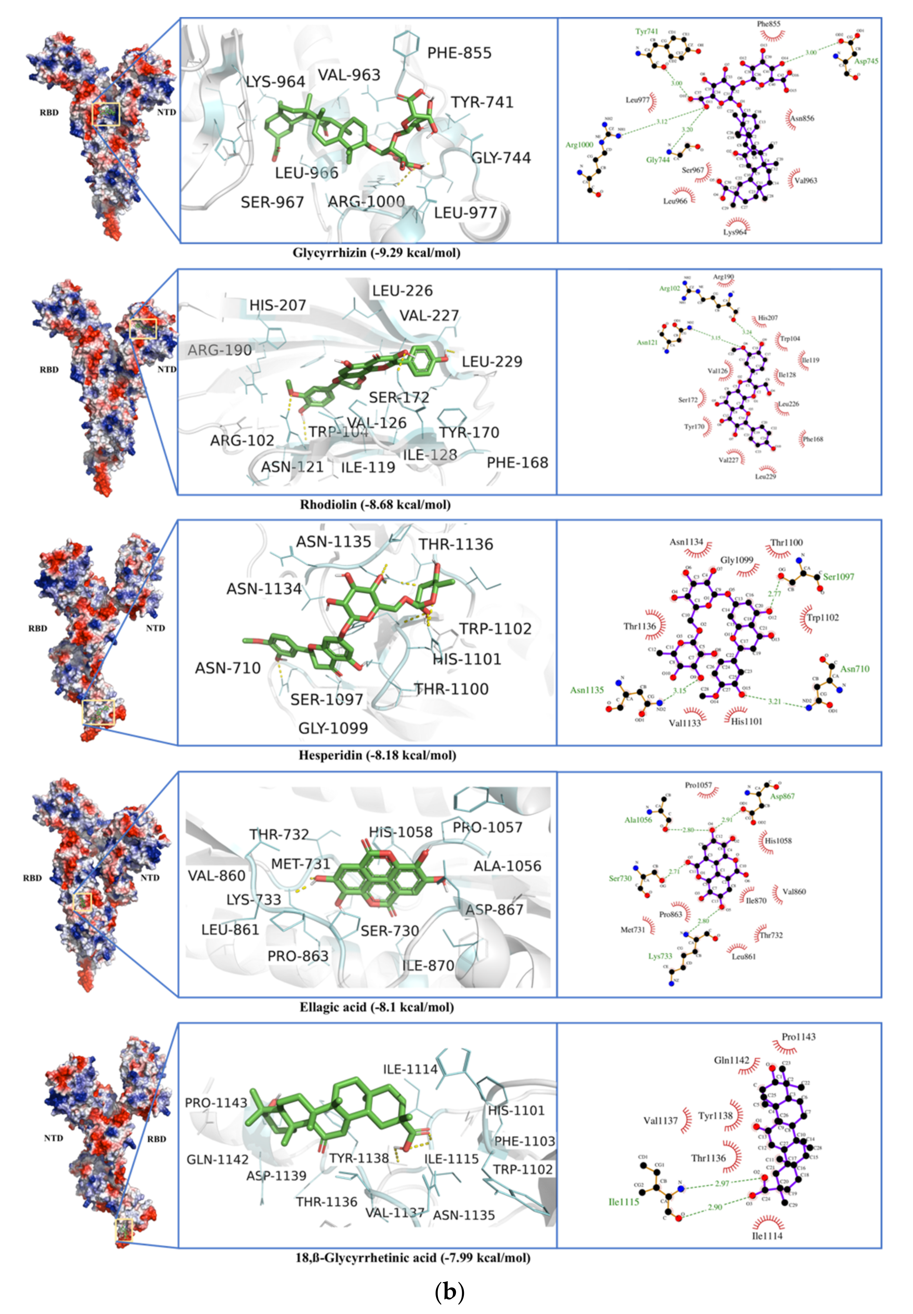
Spike protein (S)Glycyrrhizin−9.490.11Tyr741, Ile742, Cys743, Gly744, Asp745, Phe855, Asn856, Val963, Lys964, Leu966, Ser967, Ser975, Val976, Leu977, Asn978, Arg1000
−9.290.16Val47, His49, Lys304, Met740, Tyr741, Ile742, Cys743, Gly744, Asp745, Phe855, Asn856, Val963, Lys964, Leu966, Ser967, Ser975, Val976, Leu977, Asn978, Arg1000
Rhodiolin−8.680.43Arg102, Gly103, Trp104, Ile119, Asn121, Val126, Ile128, Phe168, Tyr170, Ser172, Arg190, Phe192, Ile203, His207, Leu226, Val227, Asp228, Leu229
Hesperidin−8.530.56Tyr38, Asp40, Lys41, Val42, Phe43, Arg44, Lys206, Phe220, Ser221, Ala222, Glu224, Pro225, Leu226, Tyr279, Gly283, Thr284
−8.181.01Asn710, Thr1076, Ser1097, Gly1099, Thr1100, His1101, Trp1102, Ile1114, Ile1115, Val1133, Asn1134, Asn1135, Thr1136, Tyr1138
Umbelliprenin−8.121.11Trp104, Ile119, Asn121, Val126, Ile128, Phe168, Tyr170, Ser172, Arg190, Phe192, Phe194, Ile203, Ser205, Lys206, His207, Leu226, Val227, Leu229
18,β-Glycyrrhetinic acid−8.081.2Tyr38, Asp40, Lys41, Val42, Phe43, Arg44, Glu224, Pro225, Tyr279, Gly283
−7.991.38His1101, Trp1102, Phe1103, Ile1114, Ile1115, Asn1135, Thr1136, Val1137, Tyr1138, Asp1139, Gln1142, Pro1143
Silyhermin−8.051.25Arg355, Tyr396, Pro426, Asp428, Phe429, Thr430, Lys462, Pro463, Phe464, Ser514, Phe515, Glu516, Leu517, Leu518
Ellagic acid−8.11.16Ser730, Met731, Thr732, Lys733, Gln774, Thr778, Phe823, Val860, Leu861, Pro862, Pro863, Asp867, Ile870, Ala1056, Pro1057, His1058, Gly1059
HelicaseGlycyrrhizin−11.570.003Pro175, Leu176, Asn177, Lys202, Leu405, Pro406, Ala407, Pro408, Arg409, Leu412, Thr413, Gly415, Thr416, Leu417, Phe422, Ser485, Ser486, Pro514, Tyr515, Asn516, Asn519, Thr532, Val533, Asp534, His554, Asn557, Asn559, Arg560
18,β-Glycyrrhetinic acid−9.910.054Ala4, Val6, Arg15, Arg21, Arg22, Pro23, Phe24, Arg129, Leu132, Phe133, Glu136, Pro234, Leu235, Ser236
Solophenol A−9.40.13Pro175, Leu176, Asn177, Arg178, Thr199, Phe200, Glu201, Lys202, Gly203, Asp204, Val210, Tyr211, Arg212, Val484, Ser486, Asn516, Ser517, Asn519, Ala520, Thr530, Thr532, Asp534
Hesperidin−8.930.283Lys139, Glu142, Glu143, Lys146, Arg178, Asn179, Val181, Glu197, Thr228, His230, Cys309, Arg337, Arg339, Met378, Ala379, Thr380, Tyr382, Asp383, Ala407, Pro408, Thr410
Baicalin−8.90.29Asn177, Arg178, Thr199, Phe200, Glu201, Lys202, Arg212, Asn516, Asn519, Ala520, Thr530, Gln531, Thr532, Asp534
E-channel proteinGlycyrrhizin−10.070.04Arg61, Asn64, leu28, val29, leu31, ala32, ile33, ala36, arg38, Leu27, Thr30, Leu31, Leu34, Leu37, Leu39, Tyr42, Cys43, Ile46, Val47, Val49, Ser50, Leu51, Pro54, Tyr57
18,β-Glycyrrhetinic acid−9.720.07Arg61, Leu28, Val29, Leu31, Ala32, Thr35, Leu27, Thr30, Leu31, Ile46, Val47, Leu51, Pro54, Tyr57
Umbelliprenin−8.900.3Ala22, Val25, Phe26, Leu28, Val29, Leu19, Phe20, Ala22, Phe23, Val24, Phe26, Leu27, Asn64, Leu65
Euchrestaflavanone A−8.900.3Arg61, Asn64dval25dphe26dleu28dval29, Ala22, Phe23, Phe26, Leu27, Thr30, Leu34, Cys43, Ile46, Val47, Val49, Ser50, Leu51
Baicalin−8.890.3Ala22, Val25, Phe26, Val29, Phe20, Ala22, Phe23, Val24, Phe26, Leu27, Thr30, Arg61, Asn64, Leu65
Silibinin A−8.131.1Leu28, Leu31, Ala32, Thr35, Cys40, Ile46, Ser50, Leu51, Lys53, Pro54, Phe56, Tyr57, Tyr59, Ser60
Table 2. ADMET properties of selected ligands.
CompoundsMolecular FormulaMolecular WeightHBAHBDTPSALog P−0.7–5Log S 0–6GI AbsorptionBBB PermeantCYPP1A2 InhibitorCYP2D6 InhibitorLog Kp (Skin Permeation), cm/sLipinski ViolationsGhose ViolationsVeber ViolationsBioavailability Score
GlycyrrhizinC42H62O16822.93168267.041.49−6.24LowNoNoNo−9.333 MW > 500, NorO > 10, NHorOH > 53 MW > 480, MR > 130, atoms > 701 TPSA > 1400.11
18,β Glycyrrhetinic acidC30H46O5470.684274.65.13−6.15HighNoNoNo−4.271 MLOGP.4.163 WLOGP > 5.6, MR > 130.Yes1.56
RhodiolinC25H20O10480.42105159.052.3−4.99LowNoNoNo−7.0201 MW > 4801 TPSA > 1400.55
BaicalinC21H18O11446.36116187.120.25−3.41LowNoNoNo−8.233 NorO.10, NHorOH > 5Yes1 TPSA > 1400.11
HesperidinC28H34O15610.56158234.29−1.06−3.28LowNoNoNo−10.123 MW > 500, NorO > 10, NHorOH > 54 MW > 480, WLOGP < 0.4, MR > 130, #atoms > 701 TPSA > 1400.17
Solophenol AC30H36O6492.664107.225.69−7.5LowNoNoNo−3.80 4 MW > 480, WLOGP < 0.4, MR > 130, #atoms > 711 TPSA > 1400.55
NaringinC26H30O14566.51148225.06−0.99−2.68LowNoNoNo−10.393 MW > 500, NorO > 10, NHorOH > 53MW > 480, WLOGP < 0.4 MR > 130,1 TPSA > 1400.17
LopinavirC37H48N4O5628.8541204.37−6.64HighNoNoNo−5.931 MW > 5003 MW > 480, MR > 130, #atoms > 701 Rotors > 100.55
HBA: Number of Hydrogen bond acceptors HBD: Number of hydrogen bond donor, TPSA: Tropological polar surface area.
Publisher’s Note: MDPI stays neutral with regard to jurisdictional claims in published maps and institutional affiliations.

