
Bacterial Resistance to Antibiotics and Clonal Spread in COVID-19-Positive Patients on a Tertiary Hospital Intensive Care Unit, Czech Republic

 , ,
, , 
Abstract
:1. Introduction
2. Materials and Methods
3. Results
4. Discussion
5. Conclusions
Author Contributions
Funding
Institutional Review Board Statement
Informed Consent Statement
Conflicts of Interest
References
- Situation by Region, Country, Territory & Area—WHO Coronavirus (COVID-19) Dashboard. Available online: https://covid19.who.int/table (accessed on 30 May 2022).
- O’Neill, J. Review on Antimicrobial Resistance Antimicrobial Resistance: Tackling a Crisis for the Health and Wealth of Nations; The Review on Antimicrobial Resistance: London, UK, 2014; Available online: https://amr-review.org/sites/default/files/AMR%20Review%20Paper%20-%20Tackling%20a%20crisis%20for%20the%20health%20and%20wealth%20of%20nations_1.pdf (accessed on 20 July 2021).
- Beadling, C.; Slifka, M.K. How do viral infections predispose patients to bacterial infections? Curr. Opin. Infect. Dis. 2004, 17, 185–191. [Google Scholar] [CrossRef] [PubMed]
- Metzger, D.W.; Sun, K. Immune dysfunction and bacterial coinfections following influenza. J. Immunol. 2013, 191, 2047–2052. [Google Scholar] [CrossRef] [PubMed] [Green Version]
- Jia, L.; Xie, J.; Zhao, J.; Cao, D.; Liang, Y.; Hou, X.; Ligui, W.; Li, Z. Mechanisms of severe mortality-associated bacterial co-infections following influenza virus infection. Front. Cell Infect. Microbiol. 2017, 7, 338. [Google Scholar] [CrossRef]
- Katsurada, N.; Suzuki, M.; Aoshima, M.; Yaegashi, M.; Ishifuji, T.; Asoh, N.; Hamashige, N.; Abe, M.; Ariyoshi, K.; Morimoto, K. on behalf of the Adult Pneumonia Study Group-Japan. The impact of virus infections on pneumonia mortality is complex in adults: A prospective multicentre observational study. BMC Infect. Dis. 2017, 17, 755. [Google Scholar] [CrossRef] [PubMed] [Green Version]
- Quah, J.; Jiang, B.; Tan, P.C.; Siau, C.; Tan, T.Y. Impact of microbial aetiology on mortality in severe community-acquired pneumonia. BMC Infect. Dis. 2018, 18, 451. [Google Scholar] [CrossRef]
- Morris, D.E.; Cleary, D.W.; Clarke, S.C. Secondary bacterial infections associated with influenza pandemics. Front. Microbiol. 2017, 8, 1041. [Google Scholar] [CrossRef] [Green Version]
- Rawson, T.M.; Moore, L.S.P.; Zhu, N.; Ranganathan, N.; Skolimowska, K.; Gilchrist, M.; Satta, G.; Cooke, G.; Holmes, A. Bacterial and Fungal Coinfection in Individuals With Coronavirus: A Rapid Review To Support COVID-19 Antimicrobial Prescribing. Clin. Infect. Dis. 2020, 71, 2459–2468. [Google Scholar] [CrossRef]
- Langford, B.J.; So, M.; Raybardhan, S.; Leung, V.; Westwood, D.; MacFadden, D.R.; Soucy, J.R.; Daneman, N. Bacterial co-infection and secondary infection in patients with COVID-19: A living rapid review and meta-analysis. Clin. Microbiol. Infect. 2020, 26, 1622–1629. [Google Scholar] [CrossRef]
- Lansbury, L.; Lim, B.; Baskaran, V.; Lim, W.S. Co-infections in people with COVID-19: A systematic review and meta-analysis. J. Infect. 2020, 81, 266–275. [Google Scholar] [CrossRef]
- Hughes, S.; Troise, O.; Donaldson, H.; Mughal, N.; Moore, L.S.P. Bacterial and fungal coinfection among hospitalized patients with COVID-19: A retrospective cohort study in a UK secondary-care setting. Clin. Microbiol. Infect. 2020, 26, 1395–1399. [Google Scholar] [CrossRef]
- Garcia-Vidal, C.; Sanjuan, G.; Moreno-García, E.; Puerta-Alcalde, P.; Garcia-Pouton, N.; Chumbita, M.; Fernandez-Pittol, M.; Pitart, C.; Inciarte, A.; Bodro, M.; et al. Incidence of co-infections and superinfections in hospitalised patients with COVID-19: A retrospective cohort study. Clin. Microbiol. Infect. 2020, 27, 83–88. [Google Scholar] [CrossRef] [PubMed]
- Vaughn, V.M.; Gandhi, T.N.; Petty, L.A.; Patel, P.K.; Prescott, H.C.; Malani, A.N.; Ratz, D.; McLaughlin, E.; Chopra, V.; Flanders, S.A. Empiric antibacterial therapy and community-onset bacterial co-infection in patients hospitalized with COVID-19: A multi-hospital cohort study. Clin. Infect. Dis. 2020, 72, e533–e541. [Google Scholar] [CrossRef] [PubMed]
- Youngs, J.; Wyncoll, D.; Hopkins, P.; Arnold, A.; Ball, J.; Bicanic, T. Improving antibiotic stewardship in COVID-19: Bacterial co-infection is less common than with influenza. J. Infect. 2020, 81, e55–e57. [Google Scholar] [CrossRef] [PubMed]
- Clancy, C.J.; Nguyen, M.H. COVID-19, superinfections and antimicrobial development: What can we expect? Clin. Infect. Dis. 2020, 71, 2736–2743. [Google Scholar] [CrossRef] [PubMed]
- World Health Organization. Clinical management of COVID-19: Interim Guidance, 27 May 2020. Available online: https://apps.who.int/iris/handle/10665/332196 (accessed on 20 July 2021).
- Raoof, S.; Nava, S.; Carpati, C.; Hill, N.S. High-Flow, Noninvasive Ventilation and Awake (Nonintubation) Proning in Patients With Coronavirus Disease 2019 With Respiratory Failure. Chest 2020, 158, 1992–2002. [Google Scholar] [CrossRef]
- Alhazzani, W.; Møller, M.H.; Arabi, Y.M.; Loeb, M.; Gong, M.N.; Fan, E.; Oczkowski, S.; Levy, M.M.; Derde, L.; Dzierba, A.; et al. Surviving Sepsis Campaign: Guidelines on the management of critically ill adults with Coronavirus Disease 2019 (COVID-19). Intensive Care Med. 2020, 46, 854–887. [Google Scholar] [CrossRef] [Green Version]
- Zhang, W.; Du, R.H.; Li, B.; Zheng, X.S.; Yang, X.L.; Hu, B.; Wang, Y.Y.; Xiao, G.F.; Yan, B.; Shi, Z.L.; et al. Molecular and serological investigation of 2019-nCoV infected patients: Implication of multiple shedding routes. Emerg. Microbes Infect. 2020, 9, 386–389. [Google Scholar] [CrossRef] [Green Version]
- European Committee on Antimicrobial Susceptibility Testing. Breakpoint Tables for Interpretation of MICs and Zone Diameters. Available online: http://www.eucast.org/clinical_breakpoints (accessed on 12 July 2021).
- Htoutou Sedlakova, M.; Hanulik, V.; Chroma, M.; Hricova, K.; Kolar, M.; Latal, T.; Schaumann, R.; Rodloff, A.C. Phenotypic detection of broad-spectrum beta-lactamases in microbiological practice. Med. Sci. Monit. 2011, 17, BR147–BR152. [Google Scholar] [CrossRef] [Green Version]
- Tamma, P.D.; Simner, P.J. Phenotypic detection of carbapenemase producing organisms from clinical isolates. J. Clin. Microbiol. 2018, 56, e01140-18. [Google Scholar] [CrossRef] [Green Version]
- Oliveira, D.C.; de Lencastre, H. Multiplex PCR strategy for rapid identification of structural types and variants of the mec element in methicillin-resistant Staphylococcus aureus. Antimicrob. Agents Chemother. 2002, 46, 2155–2161. [Google Scholar] [CrossRef] [Green Version]
- Dutka-Malen, S.; Evers, S.; Courvalin, P. Detection of glycopeptide resistance genotypes and identification to the species level of clinically relevant enterococci by PCR. J. Clin. Microbiol. 1995, 33, 24–27. [Google Scholar] [CrossRef] [PubMed] [Green Version]
- Standard Operating Procedure for PulseNet PFGE of Escherichia coli O157:H7, Escherichia coli non-O157 (STEC), Salmonella serotypes, Shigella sonnei and Shigella flexneri. Available online: https://www.cdc.gov/pulsenet/pdf/ecoli-shigella-salmonella-pfge-protocol-508c.pdf (accessed on 12 July 2021).
- Zakaria, A.M.; Hassuna, N.A. Modified PFGE protocol for improving typeability of DNA degradation susceptible nosocomial Klebsiella pneumoniae. J. Med. Microbiol. 2019, 68, 1787–1792. [Google Scholar] [CrossRef] [PubMed]
- Mahenthiralingam, E.; Campbell, M.E.; Henry, D.A.; Speert, D.P. Epidemiology of Burkholderia cepacia infection in patients with cystic fibrosis: Analysis by randomly amplified polymorphic DNA fingerprinting. J. Clin. Microbiol. 1996, 34, 2914–2920. [Google Scholar] [CrossRef] [PubMed] [Green Version]
- WHO Collaborating Centre for Drug Statistics Methodology. ATC/DDD Index. 2020. Available online: https://www.whocc.no/atc_ddd_index/ (accessed on 15 July 2021).
- van Berkel, M.; Kox, M.; Frenzel, T.; Pickkers, P.; Schouten, J.; On behalf of the RCI-COVID-19 Study Group. Biomarkers for antimicrobial stewardship: A reappraisal in COVID-19 times? Crit. Care 2020, 24, 600. [Google Scholar] [CrossRef] [PubMed]
- Pink, I.; Raupach, D.; Fuge, J.; Vonberg, R.P.; Hoeper, M.M.; Welte, T.; Rademacher, J. C-reactive protein and procalcitonin for antimicrobial stewardship in COVID-19. Infection 2021, 49, 935–943. [Google Scholar] [CrossRef] [PubMed]
- Klein, E.Y.; Monteforte, B.; Gupta, A.; Jiang, W.; May, L.; Hsieh, Y.H.; Dugas, A. The frequency of influenza and bacterial coinfection: A systematic review and meta-analysis. Influenza Other Respir. Viruses 2016, 10, 394–403. [Google Scholar] [CrossRef] [Green Version]
- Saini, V.; Jain, C.; Singh, N.P.; Alsulimani, A.; Gupta, C.; Dar, S.A.; Haque, S.; Das, S. Paradigm Shift in Antimicrobial Resistance Pattern of Bacterial Isolates during the COVID-19 Pandemic. Antibiotics 2021, 10, 954. [Google Scholar] [CrossRef]
- Li, J.; Wang, J.; Yang, Y.; Cai, P.; Cao, J.; Cai, X. Etiology and antimicrobial resistance of secondary bacterial infections in patients hospitalized with COVID-19 in Wuhan, China: A retrospective analysis. Antimicrob. Resist. Infect. Control 2020, 9, 153. [Google Scholar] [CrossRef]
- Porretta, A.D.; Baggiani, A.; Arzilli, G.; Casigliani, V.; Mariotti, T.; Mariottini, F.; Scardina, G.; Sironi, D.; Totaro, M.; Barnini, S.; et al. Increased risk of acquisition of New Delhi metallo-beta-lactamase-producing carbapenem-resistant Enterobacterales (NDM-CRE) among a cohort of COVID-19 patients in a teaching hospital in Tuscany, Italy. Pathogens 2020, 9, 635. [Google Scholar] [CrossRef]
- Tiri, B.; Sensi, E.; Marsiliani, V.; Cantarini, M.; Priante, G.; Vernelli, C.; Martella, L.A.; Costantini, M.; Mariottini, A.; Andreani, P.; et al. Antimicrobial stewardship program, COVID-19, and infection control: Spread of carbapenem-resistant Klebsiella pneumoniae colonization in ICU COVID-19 patients. What did not work? J. Clin. Med. 2020, 9, 2744. [Google Scholar] [CrossRef]
- Lai, C.C.; Chen, S.Y.; Ko, W.C.; Hsueh, P.R. Increased antimicrobial resistance during the COVID-19 pandemic. Int. J. Antimicrob. Agents 2021, 57, 106324. [Google Scholar] [CrossRef]
- Pascale, R.; Bussini, L.; Gaibani, P.; Bovo, F.; Fornaro, G.; Lombardo, D.; Giannella, M. Carbapenem-resistant bacteria in an intensive care unit during the coronavirus disease 2019 (COVID-19) pandemic: A multicenter before-and-after cross-sectional study. Infect. Control. Hosp. Epidemiol. 2021, 43, 461–466. [Google Scholar] [CrossRef]
- Bentivegna, E.; Luciani, M.; Arcari, L.; Santino, I.; Simmaco, M.; Martelletti, P. Reduction of multidrug-resistant (MDR) bacterial infections during the COVID-19 pandemic: A retrospective study. Int. J. Environ. Res. Public Health 2021, 18, 1003. [Google Scholar] [CrossRef]
- Kolar, M.; Htoutou Sedlakova, M.; Urbanek, K.; Mlynarcik, P.; Roderova, M.; Hricova, K.; Mezerova, K.; Kucova, P.; Zapletalova, J.; Fiserova, K.; et al. Implementation of Antibiotic Stewardship in a University Hospital Setting. Antibiotics 2021, 10, 93. [Google Scholar] [CrossRef] [PubMed]
- Martak, D.; Meunier, A.; Sauget, M.; Cholley, P.; Thouverez, M.; Bertrand, X.; Valot, B.; Hocquet, D. Comparison of pulsed-field gel electrophoresis and whole-genome-sequencing-based typing confirms the accuracy of pulsed-field gel electrophoresis for the investigation of local Pseudomonas aeruginosa outbreaks. J. Hosp. Infect. 2020, 105, 643–647. [Google Scholar] [CrossRef]
- Papajk, J.; Mezerová, K.; Uvízl, R.; Štosová, T.; Kolář, M. Clonal Diversity of Klebsiella spp. and Escherichia spp. Strains Isolated from Patients with Ventilator-Associated Pneumonia. Antibiotics 2021, 10, 674. [Google Scholar] [CrossRef] [PubMed]
- Pudová, V.; Htoutou Sedláková, M.; Kolář, M.; working group. Clonality of Bacterial Pathogens Causing Hospital-Acquired Pneumonia. Curr. Microbiol. 2016, 73, 312–316. [Google Scholar] [CrossRef]
- Hanulík, V.; Uvízl, R.; Husičková, V.; Htoutou Sedláková, M.; Kolář, M. Pneumonia-causing bacterial pathogens in intensive care patients. Klin. Mikrobiol. Infekc. Lek. 2011, 17, 135–140. [Google Scholar]
- García-Meniño, I.; Forcelledo, L.; Rosete, Y.; García-Prieto, E.; Escudero, D.; Fernández, J. Spread of OXA-48-producing Klebsiella pneumoniae among COVID-19-infected patients: The storm after the storm. J. Infect. Public Health 2021, 14, 50–52. [Google Scholar] [CrossRef] [PubMed]
- Durán-Manuel, E.M.; Cruz-Cruz, C.; Ibáñez-Cervantes, G.; Bravata-Alcantará, J.C.; Sosa-Hernández, O.; Delgado-Balbuena, L.; León-García, G.; Cortés-Ortíz, I.A.; Cureño-Díaz, M.A.; Castro-Escarpulli, G.; et al. Clonal dispersion of Acinetobacter baumannii in an intensive care unit designed to patients COVID-19. J. Infect. Dev. Ctries. 2021, 15, 58–68. [Google Scholar] [CrossRef]
- Patel, A.; Emerick, M.; Cabunoc, M.K.; Williams, M.H.; Preas, M.A.; Schrank, G.; Rabinowitz, R.; Luethy, P.; Johnson, J.K.; Leekha, S. Rapid Spread and Control of Multidrug-Resistant Gram-Negative Bacteria in COVID-19 Patient Care Units. Emerg. Infect. Dis. 2021, 27, 1234–1237. [Google Scholar] [CrossRef] [PubMed]
- Bogdanová, K.; Doubravská, L.; Vágnerová, I.; Hricová, K.; Pudová, V.; Röderová, M.; Papajk, J.; Uvízl, R.; Langová, K.; Kolář, M. Clostridioides difficile and Vancomycin-Resistant Enterococci in COVID-19 Patients with Severe Pneumonia. Life 2021, 11, 1127. [Google Scholar] [CrossRef] [PubMed]
- Patel, M.; Gangemi, A.; Marron, R.; Chowdhury, J.; Yousef, I.; Zheng, M.; Mills, N.; Tragesser, L.; Giurintano, J.; Gupta, R.; et al. Retrospective analysis of high flow nasal therapy in COVID-19-related moderate-to-severe hypoxaemic respiratory failure. BMJ Open Respir. Res. 2020, 7, e000650. [Google Scholar] [CrossRef] [PubMed]
- World Health Organization Interim Guidance for the Clinical Management of COVID-19. Available online: https://www.who.int/publications/i/item/clinical-management-of-severe-acute-respiratory-infection-when-novel-coronavirus-(ncov)-infection-is-suspected/ (accessed on 18 July 2021).
- Coronavirus Disease 2019 (COVID-19) Treatment Guidelines. National Institutes of Health. Available online: https://www.covid19treatmentguidelines.nih.gov/ (accessed on 18 July 2021).
- Australian Guidelines for the Clinical Care of People with COVID-19. Available online: https://covid19evidence.net.au/ (accessed on 18 July 2021).
- Hui, D.S.; Hall, S.D.; Chan, M.T.V.; Chow, B.; Ng, S.S.; Gin, T.; Sung, J.J.Y. Exhaled Air Dispersion During Oxygen Delivery Via a Simple Oxygen Mask. Chest 2007, 132, 540–546. [Google Scholar] [CrossRef]
- Ip, M.; Tang, J.W.; Hui, D.S.; Wong, A.L.N.; Chan, M.T.V.; Joynt, G.M.; So, A.T.P.; Hall, S.D.; Chan, P.K.S.; Sung, J.J.Y. Airflow and droplet spreading around oxygen masks: A simulation model for infection control research. Am. J. Infect. Control 2007, 35, 684–689. [Google Scholar] [CrossRef]
- Leung, C.C.H.; Joynt, G.M.; Gomersall, C.D.; Wong, W.T.; Lee, A.; Ling, L.; Chan, P.K.S.; Lui, P.C.W.; Tsoi, P.C.Y.; Ling, C.M.; et al. Comparison of high-flow nasal cannula versus oxygen face mask for environmental bacterial contamination in critically ill pneumonia patients: A randomized controlled crossover trial. J. Hosp. Infect. 2019, 101, 84–87. [Google Scholar] [CrossRef] [Green Version]
- Kotoda, M.; Hishiyama, S.; Mitsui, K.; Tanikawa, T.; Morikawa, S.; Takamino, A.; Matsukawa, T. Assessment of the potential for pathogen dispersal during high-flow nasal therapy. J. Hosp. Infect. 2020, 104, 534–537. [Google Scholar] [CrossRef]
- Jermy, M.C.; Spence, C.J.T.; Kirton, R.; O’Donnell, J.F.; Kabaliuk, N.; Gaw, S.; Hockey, H.; Jiang, Y.; Zulkhairi Abidin, Z.; Dougherty, R.L.; et al. Assessment of dispersion of airborne particles of oral/nasal fluid by high flow nasal cannula therapy. PLoS ONE 2021, 16, e0246123. [Google Scholar] [CrossRef]
- Li, J.; Fink, J.B.; Ehrmann, S. High-flow nasal cannula for COVID-19 patients: Risk of bio-aerosol dispersion. Eur. Respir. J. 2020, 55, 2000892. [Google Scholar] [CrossRef]
- Elshof, J.; Hebbink, R.; Duiverman, M.L.; Hagmeijer, R. High-flow nasal cannula for COVID-19 patients: Risk of bio-aerosol dispersion. Eur. Respir. J. 2020, 56, 2003004. [Google Scholar] [CrossRef]
- Li, J.; Fink, J.B.; Ehrmann, S. High-flow nasal cannula for COVID-19 patients: Risk of bio-aerosol dispersion. Eur. Respir. J. 2020, 56, 2003136. [Google Scholar] [CrossRef] [PubMed]
- Somsen, G.A.; van Rijn, C.; Kooij, S.; Bem, R.A.; Bonn, D. Small droplet aerosols in poorly ventilated spaces and SARS-CoV-2 transmission. Lancet Respir. Med. 2020, 8, 658–659. [Google Scholar] [CrossRef]
- Macias, A.E.; Huertas, M.; de Leon, S.P.; Munoz, J.M.; Chavez, A.R.; Sifuentes-Osornio, J.; Romero, C.; Bobadilla, M. Contamination of intravenous fluids: A continuing cause of hospital bacteremia. Am. J. Inf. Control 2010, 38, 217–221. [Google Scholar] [CrossRef] [PubMed]
- Yuen, J.W.M.; Chung, T.W.K.; Loke, A.Y. Methicillin-Resistant Staphylococcus aureus (MRSA) Contamination in Bedside Surfaces of a Hospital Ward and the Potential Effectiveness of Enhanced Disinfection with an Antimicrobial Polymer Surfactant. Int. J. Environ. Res. Public Health 2015, 12, 3026–3041. [Google Scholar] [CrossRef] [PubMed]
- Shaban, R.Z.; Maloney, S.; Gerrard, J.; Collignon, P.; Macbeth, D.; Cruickshank, M.; Hume, A.; Jennison, A.V.; Graham, R.; Bergh, H.; et al. Outbreak of health care-associated Burkholderia cenocepacia bacteremia and infection attributed to contaminated sterile gel used for central line insertion under ultrasound guidance and other procedures. Am. J. Inf. Control 2017, 45, 954–958. [Google Scholar] [CrossRef]


| Patients during the COVID-19 Pandemic (N = 372) | Patients before the COVID-19 Pandemic (N = 189) | p-Value | |
|---|---|---|---|
| No. of male patients (%) | 241 (64) | 136 (72) | 0.087 |
| Age (median, range IQR) | 67 (22–90) (16) | 65 (18–97) (20) | 0.753 |
| Admissions up to 48 h after admission to hospital (%) | 186 (50) | 82 (43) | 0.138 |
| Admissions from other hospital or department (%) | 186 (50) | 107 (57) | 0.138 |
| No. of patients on mechanical ventilation (%) | 249 (67) | 184 (97) | <0.0001 |
| No. of patients only on mechanical ventilation (%) | 78 (21) | 180 (95) | <0.0001 |
| No. of patients on HFNOT (%) | 283 (76) | 10 (5) | <0.0001 |
| No. of patients only on HFNOT (%) | 112 (30) | 3 (2) | <0.0001 |
| No. of patient on mechanical ventilation and HFNOT (%) | 182 (49) | 6 (3) | <0.0001 |
| No. of patients on ECMO (%) | 24 (7) | 0 (0) | 0.004 |
| Mortality on ICU (%) | 158 (43) | 74 (39) | 0.450 |
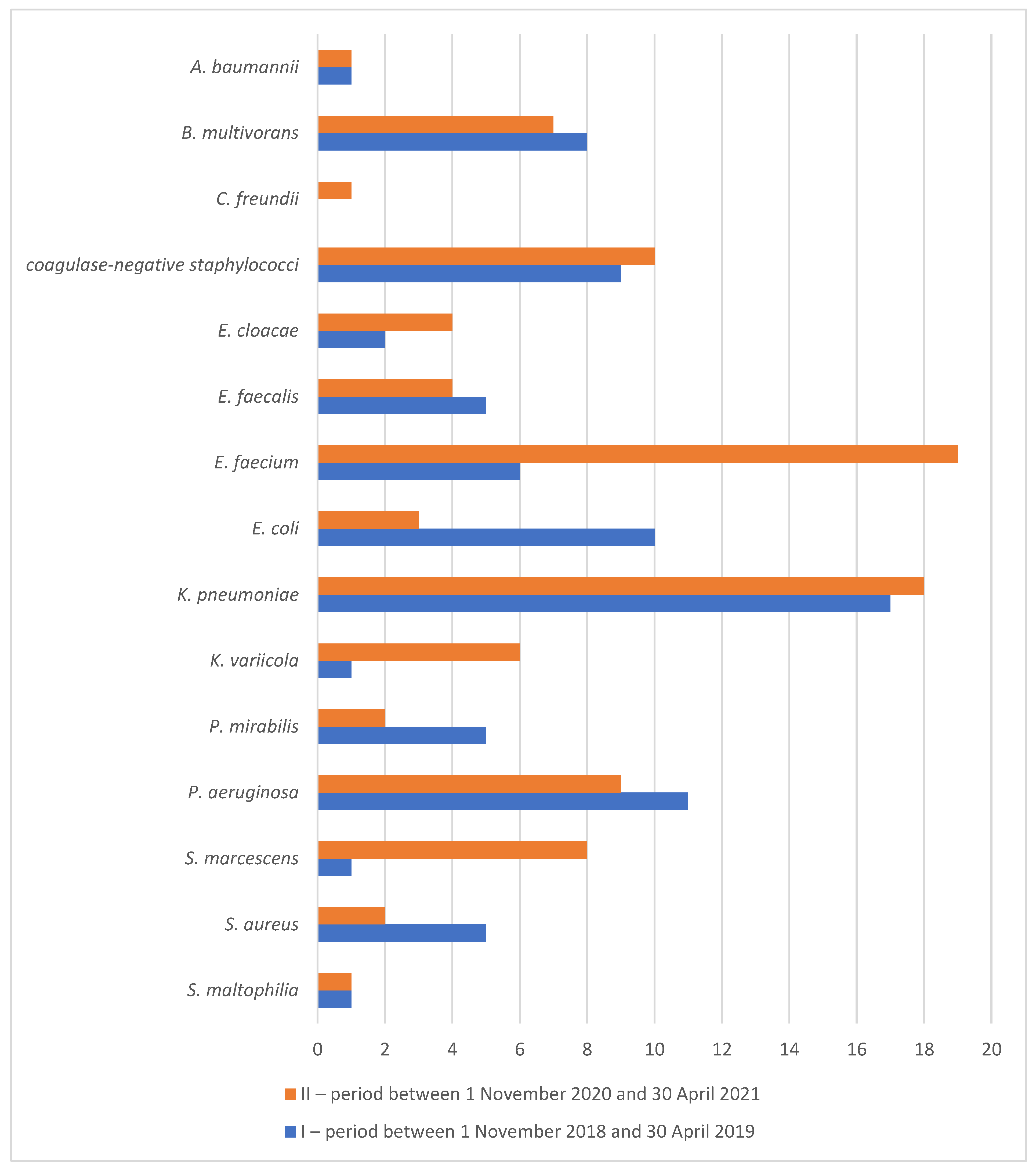
| Material from Lower Respiratory Tract | Material from Upper Respiratory Tract | Blood Cultures and Vascular Catethers | Urine | Material from Wounds, Drains, Punctuations, Pus | Stool | Others | ||||||||
|---|---|---|---|---|---|---|---|---|---|---|---|---|---|---|
| I. | II. | I. | II. | I. | II. | I. | II. | I. | II. | I. | II. | I. | II. | |
| S. maltophilia | 5 | 12 | 4 | 4 | 0 | 0 | 0 | 0 | 0 | 0 | 0 | 0 | 1 | 0 |
| S. aureus | 14 | 16 | 11 | 22 | 2 | 0 | 2 | 0 | 2 | 6 | 0 | 0 | 0 | 1 |
| S. marcescens | 5 | 55 | 2 | 11 | 0 | 8 | 0 | 8 | 0 | 0 | 0 | 0 | 0 | 0 |
| P. aeruginosa | 33 | 50 | 16 | 31 | 2 | 4 | 7 | 23 | 8 | 2 | 0 | 2 | 14 | 3 |
| P. mirabilis | 11 | 8 | 11 | 5 | 0 | 0 | 6 | 6 | 8 | 4 | 0 | 0 | 3 | 1 |
| K. variicola | 3 | 40 | 8 | 71 | 0 | 0 | 2 | 10 | 4 | 0 | 0 | 0 | 0 | 2 |
| K. pneumoniae | 44 | 126 | 50 | 142 | 5 | 6 | 13 | 30 | 14 | 5 | 0 | 1 | 10 | 7 |
| E. coli | 23 | 12 | 14 | 18 | 1 | 0 | 11 | 17 | 16 | 3 | 0 | 0 | 3 | 2 |
| E. faecium | 8 | 65 | 5 | 22 | 1 | 23 | 13 | 75 | 22 | 5 | 0 | 9 | 5 | 11 |
| E. faecalis | 10 | 19 | 0 | 2 | 1 | 4 | 7 | 11 | 10 | 0 | 0 | 0 | 5 | 2 |
| E. cloacae | 8 | 29 | 14 | 29 | 0 | 3 | 1 | 3 | 3 | 0 | 0 | 0 | 1 | 1 |
| coagulase-negative staphylococci | 0 | 0 | 0 | 0 | 30 | 80 | 4 | 7 | 31 | 3 | 0 | 0 | 20 | 14 |
| C. freundii | 1 | 3 | 2 | 6 | 0 | 0 | 0 | 2 | 2 | 1 | 0 | 0 | 1 | 0 |
| B. multivorans | 27 | 60 | 26 | 77 | 1 | 1 | 1 | 0 | 4 | 1 | 0 | 0 | 2 | 1 |
| A. baumannii | 4 | 11 | 2 | 29 | 0 | 0 | 1 | 0 | 1 | 0 | 0 | 0 | 0 | 0 |
| Others | 47 | 36 | 41 | 29 | 6 | 10 | 6 | 8 | 15 | 7 | 3 | 26 | 9 | 1 |
| Total | 243 | 542 | 206 | 498 | 49 | 139 | 74 | 200 | 140 | 37 | 3 | 38 | 74 | 46 |
| I. Time-Frame 1 November 2018–30 April 2019 | I. | II. | p | I. | II. | p | I. | II. | p | I. | II. | p | I. | II. | p | I. | II. | p | I. | II. | p |
|---|---|---|---|---|---|---|---|---|---|---|---|---|---|---|---|---|---|---|---|---|---|
| II. Time-Frame 1 November 2020–30 April 2021 | |||||||||||||||||||||
| Species (No. of isolates in I. and II. time-frame) | AMS | CRX | CTX | CTZ | CPM | PPT | MER | ||||||||||||||
| E. coli (53; 38) | 50 | 50 | 1.000 | 45 | 39 | 0.6693 | 42 | 39 | 1.000 | 42 | 39 | 1.000 | 42 | 39 | 1.000 | 40 | 32 | 0.5099 | 0 | 0 | 1.000 |
| K. pneumoniae (135; 316) | 84 | 87 | 0.375 | 80 | 81 | 0.7957 | 79 | 79 | 1.000 | 79 | 78 | 0.804 | 79 | 78 | 0.804 | 80 | 81 | 0.7957 | 2 | 0 | 0.0264 |
| E. cloacae (27; 65) | 100 | 100 | 1.000 | 100 | 100 | 1.000 | 33 | 69 | 0.0002 | 33 | 68 | 0.005 | 22 | 42 | 0.0974 | 33 | 68 | 0.0049 | 0 | 0 | 1.000 |
| C. freundii (6; 12) | 100 | 100 | 1.000 | 100 | 100 | 1.000 | 50 | 25 | 0.3441 | 50 | 25 | 0.3441 | 0 | 0 | 1.000 | 50 | 25 | 0.3441 | 0 | 0 | 1.000 |
| S. marcescens (7; 82) | 100 | 100 | 1.000 | 100 | 100 | 1.000 | 14 | 70 | 0.0066 | 14 | 70 | 0.007 | 0 | 70 | 0.0005 | 14 | 68 | 0.008 | 0 | 0 | 1.000 |
| P. aeruginosa (80; 115) | 100 | 100 | 1.000 | 100 | 100 | 1.000 | 100 | 100 | 1.000 | 16 | 21 | 0.462 | 19 | 25 | 0.3027 | 28 | 31 | 0.634 | 51 | 22 | 0.0001 |
| B. mutivorans (59; 139) | 100 | 100 | 1.000 | 100 | 100 | 1.000 | 100 | 100 | 1.000 | 5 | 1 | 0.080 | 5 | 3 | 0.4273 | 100 | 100 | 1.000 | 29 | 2 | 0.0001 |
| S. maltophilia (10; 16) | 100 | 100 | 1.000 | 100 | 100 | 1.000 | 100 | 100 | 1.000 | 40 | 56 | 0.688 | 100 | 100 | 1.000 | 100 | 100 | 1.000 | 100 | 100 | 1.000 |
| A. baumannii (7; 40) | 71 | 0 | 0.0001 | 100 | 100 | 1.000 | 100 | 100 | 1.000 | 71 | 0 | 0.0001 | 71 | 0 | 0.0001 | 71 | 0 | 0.0001 | 71 | 0 | 0.0001 |
| I. time-frame 1 November 2018–30 April 2019 | I. | II. | p | I. | II. | p | I. | II. | p | I. | II. | p | I. | II. | p | I. | II. | p | |||
| II. time-frame 1 November 2020–30 April 2021 | |||||||||||||||||||||
| Species (No. of isolates in I. and II. time-frame) | TIG | COT | GEN | AMI | CIP | COL | |||||||||||||||
| E. coli (53; 38) | 9 | 0 | 0.073 | 51 | 29 | 0.052 | 19 | 0 | 0.004 | 2 | 8 | 0.304 | 51 | 39 | 0.296 | 0 | 0 | 1.000 | |||
| K. pneumoniae (135; 316) | 7 | 7 | 1.000 | 83 | 87 | 0.302 | 75 | 74 | 0.907 | 1 | 1 | 1.000 | 82 | 76 | 0.173 | 4 | 4 | 1.000 | |||
| E. cloacae (27; 65) | 7 | 2 | 0.205 | 33 | 34 | 1.000 | 15 | 29 | 0.190 | 0 | 0 | 1.000 | 30 | 40 | 0.477 | 0 | 3 | 1.000 | |||
| C. freundii (6; 12) | 0 | 0 | 1.000 | 0 | 0 | 1.000 | 0 | 17 | 0.529 | 0 | 0 | 1.000 | 17 | 0 | 0.333 | 0 | 0 | 1.000 | |||
| S. marcescens (7; 82) | 14 | 100 | <0.0001 | 0 | 71 | 0.0004 | 0 | 69 | 0.0005 | 0 | 9 | 1.000 | 0 | 67 | 0.001 | 100 | 100 | 1.000 | |||
| P. aeruginosa (80; 115) | 100 | 100 | 1.000 | 100 | 100 | 1.000 | 23 | 23 | 1.000 | 9 | 0 | 0.002 | 30 | 26 | 0.626 | 3 | 2 | 1.000 | |||
| B. mutivorans (59; 139) | 7 | 4 | 0.488 | 0 | 0 | 1.000 | 100 | 100 | 1.000 | 100 | 100 | 1.000 | 100 | 100 | 1.000 | 100 | 100 | 1.000 | |||
| S. maltophilia (10; 16) | 0 | 0 | 1.000 | 22 | 0 | 0.138 | 100 | 100 | 1.000 | 100 | 100 | 1.000 | 70 | 100 | 0.046 | 50 | 88 | 0.069 | |||
| A. baumannii (7; 40) | 0 | 0 | 1.000 | 71 | 0 | <0.0001 | 71 | 0 | <0.0001 | 71 | 0 | <0.0001 | 71 | 8 | 0.001 | 0 | 0 | 1.000 | |||
| I. time-frame 1 November 2018–30 April 2019, | I. | II. | p | I. | II. | p | I. | II. | p | I. | II. | p | I. | II. | p | ||||||
| II. time-frame 1 November 2020–30 April 2021 | |||||||||||||||||||||
| Species (No. of isolates in I. and II. time-frame) | OXA | COT | ERY | CLI | CIP | ||||||||||||||||
| S. aureus (31; 45) | 0 | 7 | 0.266 | 0 | 5 | 0.511 | 42 | 24 | 0.135 | 42 | 27 | 0.216 | 0 | 9 | 0.141 | ||||||
| GEN | TIG | TEI | VAN | TET | |||||||||||||||||
| S. aureus (31; 45) | 13 | 7 | 0.434 | 0 | 0 | 1.000 | 0 | 0 | 1.000 | 0 | 0 | 1.000 | 3 | 7 | 0.641 | ||||||
| I. time-frame 1 November 2018–30 April 2019, | I. | II. | p | I. | II. | p | I. | II. | p | I. | II. | p | |||||||||
| II. time-frame 1 November 2020–30 April 2021 | |||||||||||||||||||||
| Species (No. of isolates in I. and II. time-frame) | AMP | TIG | TEI | VAN | |||||||||||||||||
| E. faecalis (33; 38) | 0 | 0 | 1.000 | 0 | 0 | 1.000 | 0 | 0 | 1.000 | 0 | 0 | 1.000 | |||||||||
| E. faecium (54; 210) | 100 | 100 | 1.000 | 13 | 0 | <0.0001 | 19 | 17 | 0.841 | 17 | 18 | 1.000 | |||||||||
| Antibiotics | I. | I. | II. | II. | p |
|---|---|---|---|---|---|
| DDD | Percentage | DDD | Percentage | ||
| meropenem | 703 | 19.6 | 1427 | 19.3 | 0.861 |
| piperacillin/tazobactam | 301 | 8.4 | 570 | 7.7 | 0.792 |
| amoxicillin/clavulanic acid | 299 | 8.3 | 262 | 3.5 | 0.020 |
| clarithromycin | 294 | 8.2 | 641 | 8.7 | 0.803 |
| tigecyclin | 270 | 7.5 | 565 | 7.6 | 1.000 |
| gentamicin | 250 | 7 | 33 | 0.4 | 0.243 |
| metronidazol | 237 | 6.6 | 402 | 5.4 | 0.495 |
| sulfamethoxazol/trimethoprim | 217 | 6 | 370 | 5 | 0.708 |
| ampicilin/sulbactam | 130 | 3.6 | 53 | 0.7 | 0.323 |
| ciprofloxacin | 124 | 3.5 | 150 | 2 | 0.705 |
| amikacin | 115 | 3.2 | 376 | 5.1 | 0.618 |
| vancomycin | 103 | 2.9 | 169 | 2.3 | 1.000 |
| ceftazidime | 101 | 2.8 | 206 | 2.8 | 1.000 |
| linezolid | 28 | 0.8 | 179 | 2.4 | 1.000 |
| kolistin | 62 | 1.7 | 229 | 3.1 | 1.000 |
| cefotaxime | 60 | 1.7 | 1487 | 20.1 | <0.0001 |
| cefuroxime | 60 | 1.7 | 10 | 0.1 | 1.000 |
| other | 234 | 6.5 | 267 | 3.8 | 0.218 |
Publisher’s Note: MDPI stays neutral with regard to jurisdictional claims in published maps and institutional affiliations. |
© 2022 by the authors. Licensee MDPI, Basel, Switzerland. This article is an open access article distributed under the terms and conditions of the Creative Commons Attribution (CC BY) license (https://creativecommons.org/licenses/by/4.0/).
Share and Cite
Doubravská, L.; Htoutou Sedláková, M.; Fišerová, K.; Pudová, V.; Urbánek, K.; Petrželová, J.; Röderová, M.; Langová, K.; Mezerová, K.; Kučová, P.; et al. Bacterial Resistance to Antibiotics and Clonal Spread in COVID-19-Positive Patients on a Tertiary Hospital Intensive Care Unit, Czech Republic. Antibiotics 2022, 11, 783. https://0-doi-org.brum.beds.ac.uk/10.3390/antibiotics11060783
Doubravská L, Htoutou Sedláková M, Fišerová K, Pudová V, Urbánek K, Petrželová J, Röderová M, Langová K, Mezerová K, Kučová P, et al. Bacterial Resistance to Antibiotics and Clonal Spread in COVID-19-Positive Patients on a Tertiary Hospital Intensive Care Unit, Czech Republic. Antibiotics. 2022; 11(6):783. https://0-doi-org.brum.beds.ac.uk/10.3390/antibiotics11060783
Chicago/Turabian StyleDoubravská, Lenka, Miroslava Htoutou Sedláková, Kateřina Fišerová, Vendula Pudová, Karel Urbánek, Jana Petrželová, Magdalena Röderová, Kateřina Langová, Kristýna Mezerová, Pavla Kučová, and et al. 2022. "Bacterial Resistance to Antibiotics and Clonal Spread in COVID-19-Positive Patients on a Tertiary Hospital Intensive Care Unit, Czech Republic" Antibiotics 11, no. 6: 783. https://0-doi-org.brum.beds.ac.uk/10.3390/antibiotics11060783