Review

Adapting Clofazimine for Treatment of Cutaneous Tuberculosis by Using Self-Double-Emulsifying Drug Delivery Systems

by
Daniélle van Staden
,
Richard K. Haynes
and
Joe M. Viljoen
*
Centre of Excellence for Pharmaceutical Sciences (Pharmacen™), Building G16, Faculty of Health Sciences, North-West University, Potchefstroom 2520, South Africa
*
Author to whom correspondence should be addressed.
Submission received: 3 May 2022 / Revised: 10 June 2022 / Accepted: 12 June 2022 / Published: 15 June 2022
(This article belongs to the Special Issue Antibacterial Topical Treatment - New Perspectives)

Abstract

:
Although chemotherapeutic treatment regimens are currently available, and considerable effort has been lavished on the development of new drugs for the treatment of tuberculosis (TB), the disease remains deeply intractable and widespread. This is due not only to the nature of the life cycle and extraordinarily disseminated habitat of the causative pathogen, principally Mycobacterium tuberculosis (Mtb), in humans and the multi-drug resistance of Mtb to current drugs, but especially also to the difficulty of enabling universal treatment of individuals, immunocompromised or otherwise, in widely differing socio-economic environments. For the purpose of globally eliminating TB by 2035, the World Health Organization (WHO) introduced the “End-TB” initiative by employing interventions focusing on high impact, integrated and patient-centered approaches, such as individualized therapy. However, the extraordinary shortfall in stipulated aims, for example in actual treatment and in TB preventative treatments during the period 2018–2022, latterly and greatly exacerbated by the COVID-19 pandemic, means that even greater pressure is now placed on enhancing our scientific understanding of the disease, repurposing or repositioning old drugs and developing new drugs as well as evolving innovative treatment methods. In the specific context of multidrug resistant Mtb, it is furthermore noted that the incidence of extra-pulmonary TB (EPTB) has significantly increased. This review focusses on the potential of utilizing self-double-emulsifying drug delivery systems (SDEDDSs) as topical drug delivery systems for the dermal route of administration to aid in treatment of cutaneous TB (CTB) and other mycobacterial infections as a prelude to evaluating related systems for more effective treatment of CTB and other mycobacterial infections at large. As a starting point, we consider here the possibility of adapting the highly lipophilic riminophenazine clofazimine, with its potential for treatment of multi-drug resistant TB, for this purpose. Additionally, recently reported synergism achieved by adding clofazimine to first-line TB regimens signifies the need to consider clofazimine. Thus, the biological effects and pharmacology of clofazimine are reviewed. The potential of plant-based oils acting as emulsifiers, skin penetration enhancers as well as these materials behaving as anti-microbial components for transporting the incorporated drug are also discussed.

1. Introduction

Control of the global tuberculosis (TB) burden has become a considerable concern in both developing as well as developed countries [1,2]. The countries with the highest TB incidence are India, Indonesia, Nigeria, the Philippines, Pakistan and South Africa [3]. It has been recently noted that there has been an increase in TB cases in Germany and Switzerland [4,5,6], a phenomenon that may be linked to the flow of African refugees to Europe [4,6,7,8]. In general, however, there has been an overall decrease of approximately 10% per annum in TB incidence in Europe since 2010. Nonetheless, it does appear that as a proportion of all individuals who have TB, Europe has the highest percentage of infections due to drug-resistant TB [9].
However, the drive to eliminate TB is not only limited to the aforementioned countries [3]. A recent publication describes the daunting challenge of maintaining control of TB in Australia [10]. Statistics indicate that 90% of TB cases reported in Australia during 2013 were of infected individuals that originated from other countries [11]. This occurrence is linked to the high mobility of modern populations, in this case, disease carriers coming in from neighboring countries such as Papua New Guinea [12]. On the other hand, the United States (US) experienced 22 years of declining TB incidence until 2015 when the number of TB cases increased for the first time since 1983. The incidence of multi-drug resistant TB (MDR-TB) in the US is described as stable, but special attention has to be paid to the vulnerability of infected children as MDR-TB affects both adults and minors alike [13].
Largely through difficulties in diagnoses and a generally active physical demeanor, TB in children is frequently misdiagnosed or overlooked. There is nonetheless a vast number of children infected with TB and MDR-TB, and more concerning is the fact that this number is increasing [14]. In TB endemic areas, it furthermore appears to be one of the ten main causes of death in children younger than 5 years of age [15]. Additionally, TB deaths in children are often misdiagnosed as being due to meningitis, pneumonia, malnutrition and/or HIV/Acquired Immunodeficiency Syndrome (AIDS). If these aspects are taken into consideration, deaths due to TB in children are considerably higher than as based on the initial correct diagnoses [14,16]. In addition, accurate estimates of the true MDR-TB burden among children are unavailable as TB displays a paucibacillary nature in children, which complicates confirmation of the disease [13]. Therefore, this disease is now receiving much closer attention in both developing and developed countries; this is mandatory for acquiring control over, and eventual elimination of TB [17,18].
For these reasons, it is recommended that the focus of TB treatment must shift to the global antimicrobial resistance crisis as less than 50% of patients treated for drug-resistant TB infections are efficiently cured. It is furthermore advocated that individualized therapy should be implemented [9,19]. Currently, poor adherence to anti-tubercular treatment regimens is attributed to prolonged treatment schedules and adverse reactions accompanied by extended administration [3]. In turn, poor adherence to first-line anti-tubercular regimens leads to the development of resistant TB isolates [3]. Therefore, the potential of including existing anti-tubercular agents, such as clofazimine, to first-line regimens might shorten treatment time and improve patient adherence [3].
The World Health Organization (WHO) implemented the “End-TB” campaign in September 2018 that had as its central aim the elimination of TB by the year 2035. To achieve this objective, three pillars of the WHO End TB Strategy were developed, where “intensified research and innovation” was included as one of the pillars. The two key components of this pillar are “discovery, development and rapid uptake of new tools, interventions and strategies” and “research to optimize implementation and impact, and promote innovations”. Moreover, at the first ever United Nations high level meeting on TB held in 2018, the commitment was made to increase research and innovation, with the development of new tools for TB treatment [20,21,22,23,24,25]. Therein, emphasis has now been placed on finding unique drug delivery systems, while utilizing “familiar drugs”, in addition to using more traditional approaches of discovering new and active compounds.
However, a drastic shortfall in aims of the End-TB Strategy has lately and starkly become evident, as is described in the WHO 2021 TB report [26]. The report clearly states that the global progress of the End-TB Strategy has been reversed to a 18% decline in newly diagnosed TB cases [26], thus representing a stark regression back to the situation as was noted in 2012 [26]. This drastic decline is attributed to the COVID-19 pandemic as the lock-down response greatly revoked the provision of essential TB services [26]. Therefore, it is clear that research into TB as outlined in the 2018 United Nations report must now be greatly intensified in order to now fulfil the aims of the WHO End-TB strategy. Therefore, this review aims to discuss the potential of adapting current anti-tubercular drug delivery by presenting clofazimine in Self-Double-Emulsifying drug delivery systems (SDEDDSs). Dermal drug delivery is suggested for the purpose of limiting adverse reactions associated with clofazimine while improving patient compliance as an additional therapy to provide topical treatment options for cutaneous TB (CTB). The possibility of plant-based oils acting as emulsifiers, skin penetration enhancers as well as these materials behaving as anti-microbial components for transporting the included drug(s) are also considered.

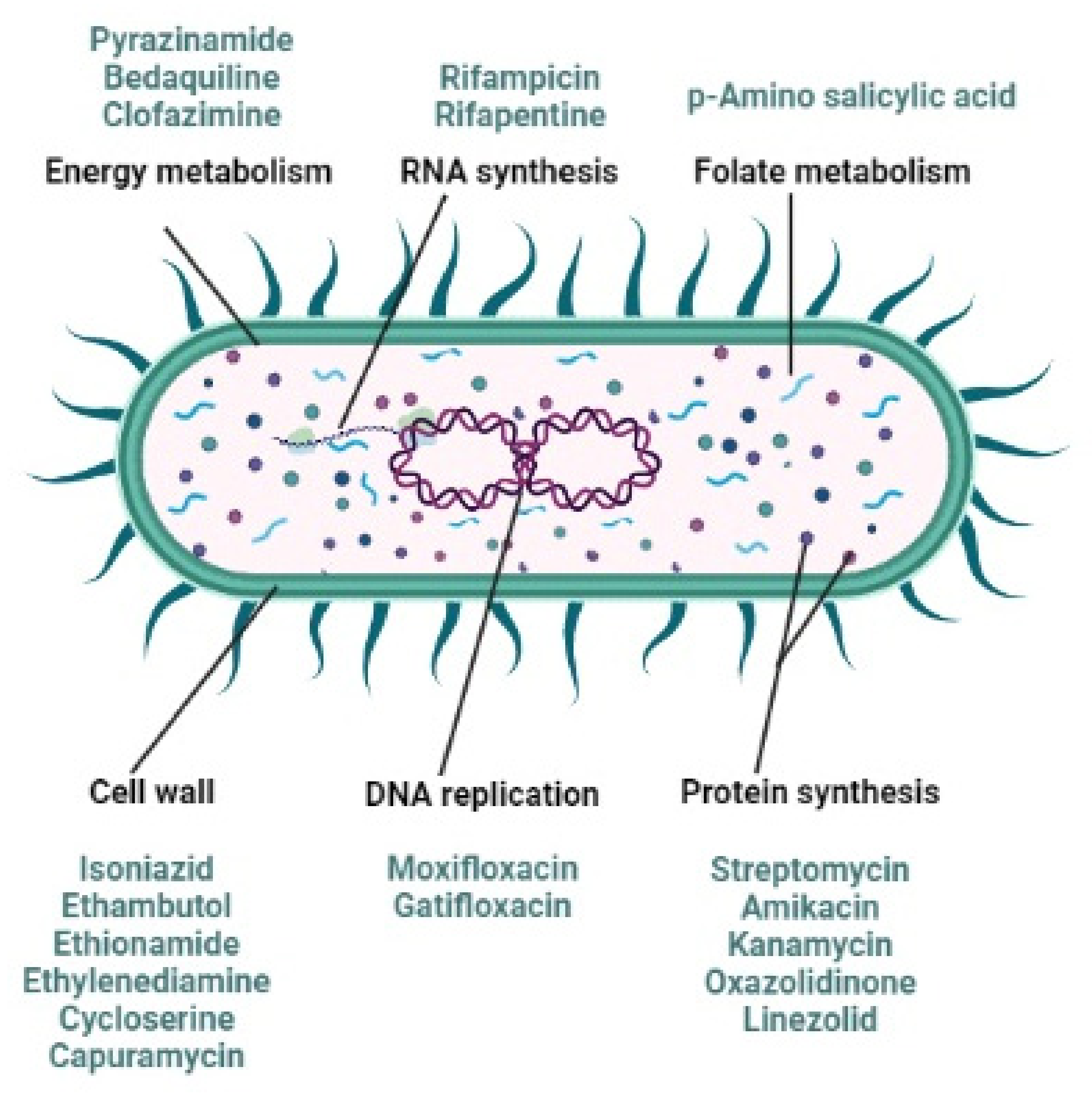
2. Anti-Tubercular Drug Resistance

TB is one of the principal epidemics that cause global morbidity and mortality. It is a fatal airborne infection, which is generated by Mycobacterium tuberculosis (Mtb) [26]. Mtb is categorized as an acid-fast bacillus with a slow growing rate [1]. Furthermore, this organism is able to survive in a quiescent state whilst within the human body without eliciting any TB symptoms [27]. This inactive TB state is referred to as latent TB infection (LTBI) [14,27]. Current research indicates that at least one in four people worldwide have latent TB. In almost 10% of the population infected with latent TB, this will develop into active TB. However, numerous individuals, including children, who may be living with TB patients in poor, overcrowded conditions, as well as immune-compromised patients, are especially vulnerable to LTBI. Subsequently, upon development of an immunocompromised situation such as through infection with HIV, development of diabetes mellitus, through an organ transplant, by becoming pregnant, or living in overcrowded and unhygienic environments coupled with poor nutritional intake, the inactive TB state may transform into an active TB infection. Persons, for example, infected with HIV, are 20 to 30 times more prone to contract active TB from LTBI [14,28,29,30,31].
In short, active TB is currently systemically treated and consists of two phases. The first being termed the intensive phase, which is about 8 weeks long. During this stage, first-line anti-tubercular agents namely, isoniazid, rifampicin, ethambutol and pyrazinamide, are concurrently administered. Following, once patients are no longer deemed infectious, they still require more treatment to successfully eliminate the infection, therefore, a maintenance phase is employed that entails concurrent administration of isoniazid and rifampicin. This stage is continued for at least 16–28 weeks, depending on the HIV status of a patient. It can even last up to 12 months. Importantly, patients need to adhere strictly to this treatment regimen to avoid drug resistance [32,33,34].
What is more concerning however is that the WHO indicated that 458,000 of the 10 million active TB cases reported in 2017 were triggered by Mtb with specific resistance towards rifampicin and isoniazid, which are two of the four first-line drugs normally used against active TB [25,26,35]. MDR-TB is defined as resistance to at least two of the front line anti-tubercular agents and is more prevalent in patients not adhering to anti-TB therapy. Multi-drug resistance is furthermore predictive of a poor outcome since these drugs are the key constituents of anti-TB treatment [25,35,36,37,38,39,40]. Estimates from the WHO 2021 TB report indicated a decline in the number or patients that were enrolled for treatment of MDR-TB from 2018 to 2020. However, this is by no means a good report as it only indicated that fewer patients registered for MDR-TB treatment and adhered to the treatment guidelines. This was probably due to the global COVID-19 pandemic lock-down responses, which in turn, significantly prevented essential TB services from being performed [24]. Thus, this extremely infectious disease is rendered even more complex due to the increasing occurrence of antimicrobial drug resistance.
Nevertheless, how does drug resistance occur given that TB is both preventable and treatable? As stated, TB drug resistance principally means that Mtb is resistant to first line anti-tubercular agents including isoniazid, rifampicin, pyrazinamide, streptomycin and ethambutol [36,41]. In order for a TB regimen to be effective, the simultaneous administration of various bactericidal and sterilizing anti-tubercular agents must be maintained for a sufficient period of time [1,42] in order to guarantee anti-microbial effectiveness together with the prevention of Mtb mutations that might lead to drug resistance [40]. Resistance generally emerges because of inadequate TB management when anti-tubercular drugs are misused or mismanaged. Examples include:
  • patient noncompliance (failure to complete the correct full course of treatment),
  • reinfection with Mtb post treatment,
  • the inappropriate utilization of medicines such as when health-care providers prescribe the incorrect treatment regime (incorrect employment of antibiotics),
  • prescribing an incorrect dosage, or length of time for treatment (incorrect treatment regime),
  • an inconsistent treatment (drug availability),
  • restricted access to treatment, or
  • when the requisite drugs to be used are of poor quality [31,43,44].
It has even been found that clinical and genetic risk factors may also encourage the development of MDR-TB in non-HIV infected patients [45].
Even so, it has been established that the risk of development of drug-resistant TB is higher among HIV-infected individuals compared to HIV-seronegative patients. A study conducted in 1993–1994 in the USA found that HIV-infected patients with TB who had not previously been treated for TB were already infected by bacterial isolates with an 11.3% incidence of isoniazid resistance and an 8.9% incidence frequency of rifampicin resistance. These numbers were almost double those recorded from an HIV-negative population [46]. Although the mechanisms involved in the development of rifampicin resistance are not yet clearly understood, the poor absorption of rifampicin and ethambutol in HIV-infected patients may be associated with acquired drug resistance leading to treatment failure [45]. As portrayed in Figure 1, Mtb is combated with multiple anti-tubercular agents attacking this deadly infection with different mechanisms of action.
MDR-TB is generally treated with fluoroquinolones (moxifloxacin and ofloxacin) and injectable agents (amikacin, capreomycin, kanamycin or streptomycin), normally employed as last line anti-tubercular agents, in combination with p-aminosalicylic acid, cycloserine or ethionamide for at least 18 months [37,44]. Table 1 indicates the latest classification system provided by the WHO to initiate a prolonged, but effective MDR-TB treatment regimen. Clearly, this represents a complex treatment regimen that is costly and significantly extended, which furthermore tends to enhance lack of compliance. Moreover, in 8.5% of all patients that contracted MDR-TB, Mtb is also found to be resistant to at least one fluoroquinolone and/or one of the second-line injectable aminoglycosides. Resistance to both fluoroquinolones and aminoglycosides is described as extensively drug resistant TB (XDR-TB), whereas pre-XDR refers to resistance either to fluoroquinolones or aminoglycosides, respectively [25,35,40,48,49,50].
The emergence and transmission of these types of TB are again partly driven by the association between TB and HIV (TB/HIV coinfection). Evidence shows that populations infected with HIV are more prone to contract XDR-TB, which is fundamentally untreatable with normal TB treatment regimens. XDR-TB is a critical health hazard, predominantly in communities with a high incidence of HIV [44]. According to the 2018 WHO global TB statement, treatment is only effective in 55% of individuals that contracted MDR-TB, and is only effective in 34% of XDR-TB patients [25,35]. The presence of MDR-TB and XDR-TB furthermore enhance the occurrence of extra-pulmonary TB infections [52]. Therefore, treatment is even more complicated as 5% of all new TB cases are considered multi-drug resistant, with the global estimate being that 3.3% of first time TB cases and 20.5% of previously treated TB patients display MDR-TB [32,38].

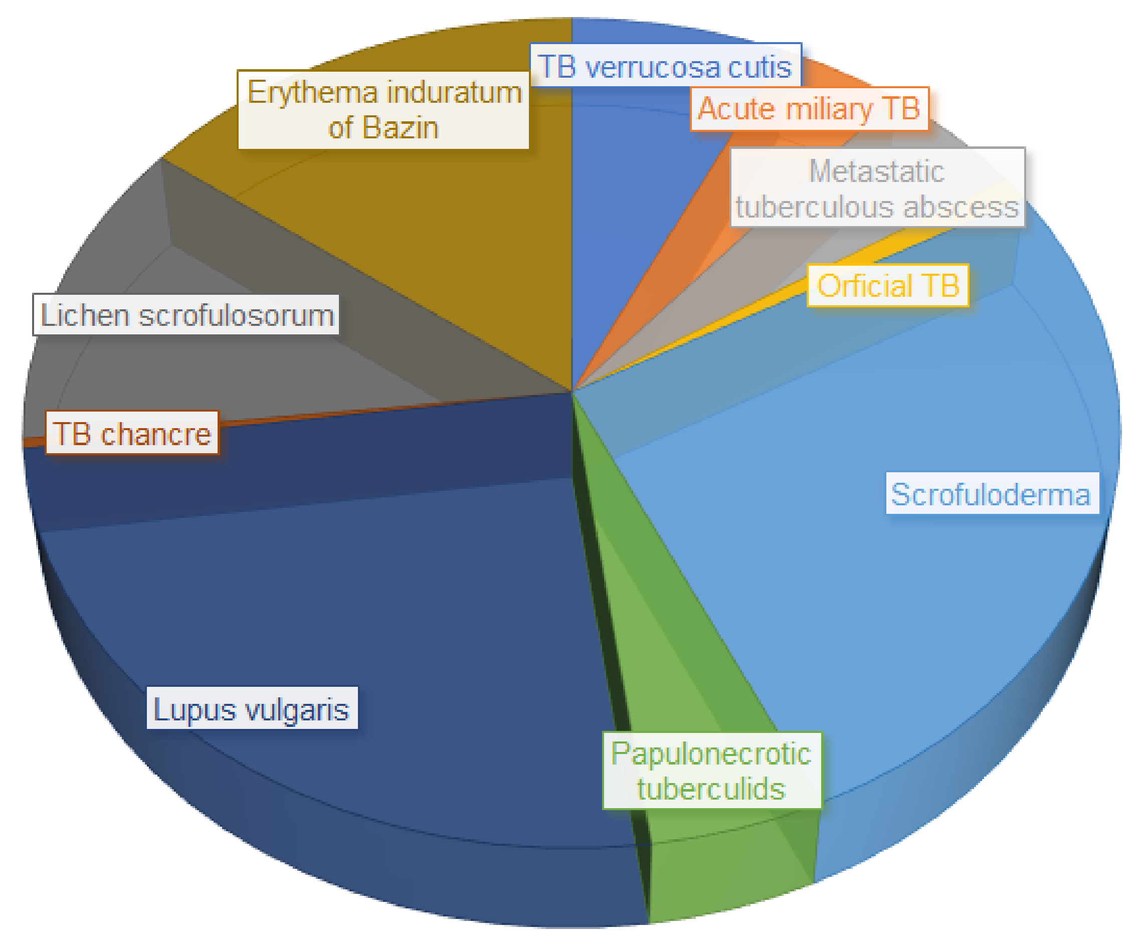
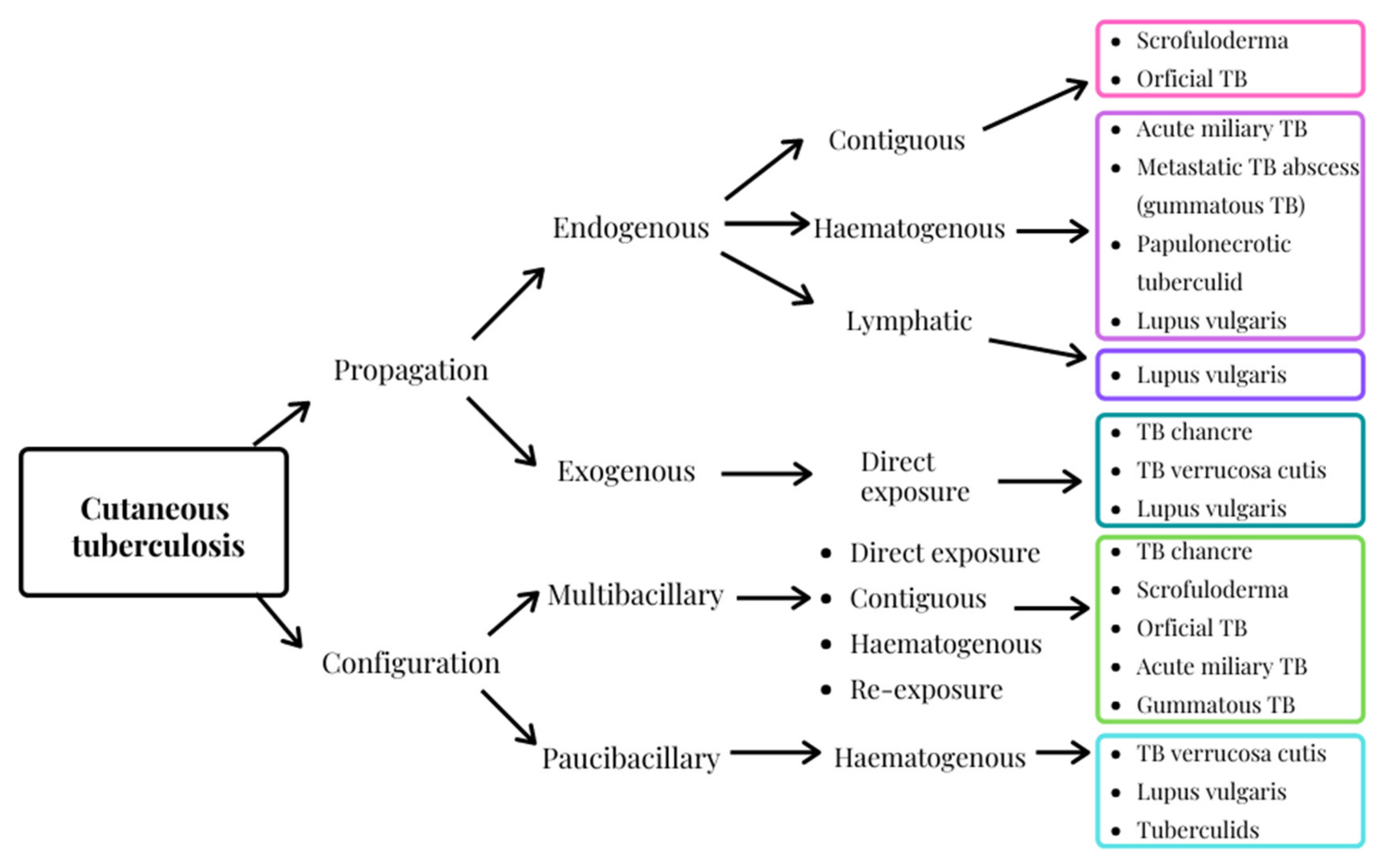
3. Cutaneous Tuberculosis, an Extra-Pulmonary Infection

TB presents normally as a pulmonary infection. However, approximately 20% of TB cases are extrapulmonary (EPTB) and this number is rising; consequently, it cannot be ignored any longer. EPTB includes ocular TB, hepatobiliary and splenic TB, TB spondylodiscitis, pleural TB, TB encephalitis, breast TB, TB pericarditis, TB lymphadenitis, gastrointestinal and peritoneal TB, genitourinary TB, osteoarticular TB, TB meningitis and cutaneous TB (CTB).
The incidence of EPTB increases with associated HIV infection, an increase in MDR-TB and immunosuppressive treatment, specifically utilizing inhibitors of tumor necrosis factor-alpha TNFα [1,32,33,53,54,55,56,57,58,59,60,61,62].
CTB is a relatively uncommon disease where merely 1–2% of all EPTB infections involve cutaneous association and of these cases only about 0.1–1% of all cutaneous disorders can be related to CTB [32,54,59,63]. CTB was first described in 1826 by Théophile Laennec, inventor of the stethoscope, who reported a lesion on his hand that was evidently triggered through mycobacterial penetration through his skin. The causative organism was however only identified in 1882 when Robert Koch first identified the bacterium Mtb as the TB pathogen [32,33,54,55,59,63,64,65].
Skin manifestations due to EPTB are normally caused by Mtb and are known as true CTB. However, other atypical Mycobacterium species may also account for cutaneous manifestations. These Mycobacteria can be divided into two sub-categories, namely: slow growers (M. haemophilum, M. leprae, M. marinum, M. ulcerans, and less regularly the M. avium complex, M. kansasii, M. malmoense, M. scrofulaceum) and rapid/fast growers (M. chelonae, M. fortuitum, and infrequently M. abscessus). Rapidly growing organisms have a 7 day or less incubation period, whereas slow growers have a longer incubation time [32,66,67,68,69,70,71]. M. bovis and the Bacille Calmette-Guérin vaccine, an attenuated strain of M. bovis, have moreover been reported to cause CTB [70]. Infections by means of atypical mycobacteria appear primarily in immunocompromised populations and there is a tendency for dissemination, whereas CTB infection in immunocompetent hosts follows skin penetration. This type of CTB is normally described as localized [55].
True CTB is more prevalent among women and young adults. The most frequently affected area of the skin is the face, although it commonly appears on the neck and torso as well [71]. Factors contributing to the diverse manifestations of the different CTB sub-categories include age and sex. For example, scrofuloderma is more common amongst pediatric patients, whereas erythema induratum of Bazin presents more frequently in females, and TB verrucosa cutis is predominantly observed in males [72,73,74]. The incidence of divergent CTB manifestation is displayed in Figure 2.
Individuals exposed to malnutrition or unhygienic conditions, or suffering from diabetes mellitus, end-stage renal disease, malignancies, HIV co-infection, or through submission to immunosuppressive therapy, and infants and pregnant women, amongst others, are at risk of contracting active CTB [26,76,77]. Overall, a diverse collection of individuals is vulnerable, especially due to the global increase in MDR-TB and XDR-TB [78].
Currently three criteria are used to classify CTB, namely, pathogenesis of the causative organism, clinical presentation, and histology. CTB is therefore generally categorized as either an endogenous or exogenous propagation, considering the bacterial basis and the infection means, as well as a multibacillary or paucibacillary configuration, depending on the bacterial load in the lesion. The exogenous mechanism refers to direct inoculation into the skin, whereas the endogenous mechanism is secondary to a previous infection where transmission arises by means of contiguous, lymphatic or hematogenous dissemination [79,80,81,82,83]. This variety of clinical CTB manifestations severely complicates diagnoses. Moreover, CTB is an elusive dermal condition as it resembles a number of visually similar dermal disorders [32,63,76,79]. In addition, the microbial basis of CTB infection (Figure 3) may be difficult to define, even though improved detection techniques including culture smears, polymerase chain reaction examinations and enzyme-linked-immunosorbent serologic assays are available [33,34,63,79,83,84].
Currently, treatment options for CTB remain the same as for general systemic TB due to the involvement of systemic infections in most cases. Furthermore, surgical interventions involving excision of lesions and correction of deformities in certain indications are sometimes necessary [32,55,85]. CTB treatment also comprises two phases. The first phase is the intensive phase and lasts approximately 8 weeks. This phase consists of simultaneous administration of first line anti-tubercular agents. Following this regimen, patients are no longer considered infectious, but do require more treatment in order to eradicate the infection. Subsequently, a maintenance phase is implemented consisting of simultaneous administration of isoniazid and rifampicin. This phase is sustained for a period of at least 16 weeks for HIV negative patients and 28 weeks for HIV positive patients, but nonetheless may even last for 9–12 months. For the treatment to be successful, patients need to adhere stringently to this treatment regimen [32,33,34]. However, due to these long-term treatment phases, patient compliance may be problematic, and in order not only to improve patient compliance, but also enhance positive treatment outcomes, alternative solutions are required. Firstly, anti-microbial agents other than conventional TB drugs must be explored in order to shorten the treatments required to cure MDR-TB and XDR-TB, and to minimize the adverse effects that are normally associated with first-line anti-tubercular compounds. Likewise, novel therapeutic combinations of existing antibiotics and first line TB agents may be more beneficial as recommended by the WHO and United Nations [2,19,20,21,22,23].
Recently, interest in so-called “orphan drugs”, i.e., drugs intended for rare diseases that for example affect less than 200,000 people per annum in the USA, has increased despite the fact that these drugs are considered orphans both therapeutically and commercially due to the minor patient population in need of these compounds [80]. Interestingly, orphan drugs are attracting attention from researchers aiming to treat dermal conditions since many side effects can be circumvented by utilizing dermal applications [80].
Clofazimine is one such compound that notably displayed a significant efficacy against resistant TB strains in the ‘Bangladesh regimen’ described below [2,25,81]. Individualized anti-tubercular therapy is furthermore increasingly recommended, as it is seen as the most effective and safe manner of targeting TB resistance in individual patients without inducing further resistance [82,83]. In order to turn the tide against TB and CTB, reintroduction of agents such as clofazimine should not be disregarded as these can assist in both individualized therapy where drugs are only employed if effective against the infection as indicated by predictive tests, as well as contributing to shortening of the treatment phases [2,48,84].

4. Clofazimine, an Orphan Drug

Clofazimine, initially known as “B663”, was originally synthesized in 1954 by Vincent Barry and co-workers to treat TB [85]. It is a highly lipophilic riminophenazine dye, which is significantly active against Mtb. However, despite high bactericidal and sterilizing activity in vitro as well as in a mouse model of MDR-TB, and its good oral absorption in micronized form along with its deposition and persistence in adipose tissue and in cells of the reticuloendothelial system, this compound did not initially progress as an anti-tubercular agent as it was found to be ineffective against TB in humans [14,86,87,88,89,90,91]. Nonetheless, it was concluded that due to the accumulation of clofazimine inside macrophages, it should be effective against intracellular diseases [92]. Efficacy against leprosy was noted at the end of 1959, and after clinical trials during the 1960s in Nigeria and elsewhere, the Swiss pharmaceutical company Novartis marketed clofazimine in 1969 under the brand name Lamprene® as an anti-leprosy agent [25,93,94].
In 1982, a WHO Study Group recommended utilizing clofazimine as a constituent of a triple drug combination with dapsone and rifampicin for treatment of multibacillary leprosy [88,94]. However, Novartis was only granted FDA approval of clofazimine at the end of 1986 and its use as anti-tubercular agent lapsed for several decades [93,95,96]. Moreover, clofazimine was classified as a Group 5 drug by the WHO, indicating that it is a drug with unclear efficacy that is not recommended for routine use [97].
Later, interest in clofazimine for the treatment of TB was awakened following its inclusion in the so-called “Bangladesh regimen” where it was established that approximately 90% of patients that contracted MDR-TB were healed within a 9–11 month period [2,81,98]. This was a substantial improvement over the standard WHO-proposed 24 months regimen that cured less than 50% of MDR-TB patients. Results from a larger clinical trial study in Bangladesh and from other clinical trials in Cameroon and Niger confirmed these conclusions [89,99,100,101]. The Bangladesh regimen according to the WHO 2016 guidelines comprises an initial treatment phase consisting of 4–6 months’ treatment with kanamycin, moxifloxacin, prothionamide, clofazimine, pyrazinamide, isoniazid (in high dosages) and ethambutol. This initial phase is followed by a 5 months treatment period with moxifloxacin, clofazimine, pyrazinamide and ethambutol [2,100]. The specific bactericidal and treatment-shortening action of clofazimine was furthermore noted in MDR-TB mouse models, and it was likewise apparent in a controlled clinical trial performed in China [102,103,104].
In contrast, the randomized controlled STREAM trial indicated non-inferiority of the short-course-containing regimen, but not superiority [105]. For these reasons, the full potential of clofazimine as an anti-tubercular agent has not yet been established. Nonetheless, clofazimine is listed in the WHO Model List of Essential Medicines as a leprostatic-, anti-tubercular- and anti-inflammatory agent [106]. It is also categorized by the WHO as a Group C agent, that is, other core second line anti-tubercular agents [48]. Therefore, clofazimine may have the capacity to significantly advance the treatment of MDR-TB and CTB.
Oral absorption of clofazimine is variable, fluctuating from 45–62% when taken on an empty stomach; however, absorption is increased when ingested with food. This drug is metabolized by glucuronidation in the liver, incompletely expelled by the biliary route, and only negligibly excreted in urine, sputum, sebum, and sweat. Clofazimine has been reported to cross the placenta and is excreted in breast milk [90,95,107,108]. Second line anti-tubercular drugs are known to be more toxic [109] and clofazimine is no exception as the highly lipophilic nature of this drug leads to accumulation in macrophages [92]. However, it must be noted that human macrophages are the primary host cells for Mtb, and thus, such a property will be of decisive clinical advantage when using clofazimine [92]. Moreover, accumulation of clofazimine in adipose cells such as in the heart, liver, breasts, adrenal glands, pancreas, spleen, bone marrow and lamina propria of the jejunum, extends the half-life to 65–70 days [88,89,90,95,107,108,110,111,112,113,114]. However, such accumulation leads to adverse reactions including reversible skin, hair and corneal dyschromia to brownish-black, dark red, or even to orange pink. Approximately 46% of patients receiving clofazimine treatment display conjunctival deposition, whereas nearly 53% experience crystal deposition in the cornea and 32% of patients present with clofazimine crystals in their tears [114,115]. Deposition of a dark-brown pigmentation is furthermore sometimes visible on the fingernails. Discoloration is more evident in fair-skinned individuals exposed to sunlight and even though it is reversible, it may take years to be completely resolved. Breast milk, sweat, urine, and feces may also discolor. In addition, dry skin and ichthyosis, defined as an increase in skin thickness due to lack of shedding of dead skin cells, often transpires. Severe gastrointestinal reactions (nausea, vomiting, abdominal pain, diarrhea) are rather common (reported in >10% of patients) and deemed most concerning, as accumulation, as well as precipitation of clofazimine in the wall of the small intestine, ensue after sustained administration [88,95,102,107,108,110,111,116,117]. In 1–10% of patients, weight loss, pruritus, diminished visual perception, and/or eye irritation/dryness occur. Less commonly, more severe adverse effects may be observed, including enteropathy possibly complicated by intestinal obstruction, gastrointestinal bleeding, and splenic infarction [107,108,118,119].
Normally, side effects accompanied by clofazimine treatment are dependent on the dose administered and the duration of administration [87]. Clofazimine up to 300 mg daily is often well tolerated; however, 100 mg per day is normally recommended due to gastrointestinal intolerance. Higher dosage regimens given over longer periods, i.e., beyond 4 months, may lead to crystal enteropathy and malabsorption, which is accompanied by effusive vague and/or severe abdominal distress. Coincidentally, discontinuation of treatment normally leads to a reasonably quick clearing of symptoms. Granting the fact that gastrointestinal side effects are not infrequent and have been observed in roughly one third of patients treated with clofazimine, the severity is usually not substantial enough to evoke termination of the treatment [14,95,102,107,108,113,118,119,120].
Fortunately, clofazimine is predominantly well-tolerated in children as observed from pediatric leprosy treatment [14]. It is classified as Pregnancy Category C, though little research has been conducted regarding its use to treat MDR-TB during pregnancy and limited data exist for later life teratogenicity. From data obtained from case reports, it was found that only 5 MDR-TB pregnant patients received clofazimine as part of the treatment regimen. All infants born were considered normal. Moreover, no significant adverse reactions due to clofazimine as part of the leprosy treatment regimen during pregnancy have been reported [88,107,108,121,122]. Clofazimine is furthermore safe to co-administer with antiretroviral therapy [123] and no known significant drug-drug interactions have been reported [88,121]. Thus, overall, insight into the physicochemical characteristics of clofazimine enables the design of novel drug delivery systems, and it should be possible to circumvent the most serious adverse effects of severe gastrointestinal reactions and accumulation of clofazimine in the gastro-intestinal tract. Clearly, the topical administration route will be advantageous as gastrointestinal accumulation can be bypassed and patient compliance not compromised [124]. Thus, an alternative route of administration together with a unique clofazimine dosage form will fortify this orphan drug with a more user-friendly profile.

5. An Alternative Strategy to Deliver Clofazimine More Effectively

5.1. Contemplating Topical Delivery

As stated, oral delivery of clofazimine is not only erratic, but also evokes numerous systemic adverse effects. On the other hand, topical drug delivery represents “the application of a drug onto the surface of the body to a localized area, such as the nose, eyes, rectum, vagina or skin, in order to establish relief of a condition restricted at the area of application, without creating any systemic effect” [125,126].
Over the past three decades, the trend to deliver drugs via the skin has been markedly increasing. It is becoming a considerably more popular alternative administration route, especially when aiming to avoid complications that accompany oral drug delivery [124]. Through implementation of this route to treat various topical ailments, patient compliance is increased, the risk of drug and food interactions is decreased, and hepatic metabolism is circumvented amongst others [127,128], thus rendering the dermal application route conspicuously beneficial [129]. However, the skin is designed to fulfil a barrier function, and subsequently permeation of various drugs, in concentrations that are of clinical relevance, is limited [130,131]. The barrier function of the skin is linked to the lipophilic nature of the outermost layer of the skin, namely the stratum corneum (SC) [131]. Therefore, the main challenge when formulating a topical drug delivery system is to attain an optimum concentration of the encapsulated drug at the site of action for a sufficient period of time [125,132].
The skin can be described as a multi-layered organ [133]. Each layer fulfils its own role in protecting the body, maintaining hydration and allowing sufficient blood perfusion to the skin [134,135,136]. The skin consists of three layers, namely the epidermis, dermis and hypodermis [137]. Epidermis thickness varies according to the cell layer thickness at the different skin regions of the body [135]. This layer is divided into two distinct fragments, the SC and viable epidermis [134]. The SC forms the outermost layer of the epidermis and is the lipophilic, protective layer of the skin that complicates drug delivery of a vast range of substances [136]. This protecting function of the SC is enforced by its lipophilic nature [134]. The SC consists of lifeless, anucleated cells comprising corneocytes linked by corneodesmosomes surrounded by a lipid matrix containing a mixture of cholesterol, triglycerides, free fatty acids and ceramides [134]. The second skin layer, the dermis is the most biochemically active layer of the skin [134]. It is responsible for body temperature regulation, cutaneous sensation, skin elasticity, blood circulation in addition to skin nutrition, and the removal of toxic substances [134,135]. Below the dermis is the third skin layer known as the hypodermis [137]. Subcutaneous fat is encased within the hypodermis [135,138]. Thereby, it acts as a supportive membrane for both the dermis and epidermis [135,139]. In order for a drug to be delivered through the skin and into the systemic circulation, it must be able to move through the lipophilic SC and the underlying hydrophilic viable epidermis and dermis, respectively. Consequently, a drug must have both hydrophilic and lipophilic properties in order to facilitate successful transdermal drug delivery [134,136].
There are three pathways that enable drug diffusion through the skin to occur [138]. First, a drug enters the skin through the intracellular or intercellular pathway [137]. Highly hydrated keratins are part of the corneocytes that are the main building blocks of the epidermis, where some components are able to traverse these cells. Moreover, hydrophilic drugs predominantly pass through the skin via the intracellular pathway [134]. Secondly, intercellular permeation may occur via diffusion of a compound through the lipid matrix of the skin as mostly observed for lipophilic drugs [134,137]. The third route, the transappendageal pathway, is restricted as a mere 0.1% of the skin surface is occupied by hair follicles and sweat glands that present an opportunity for modest transappendageal permeation of drugs [136,137]. Here, an additional limiting step for permeation of hydrophilic composites is the lipid-rich nature of the sebum that fills the sebaceous glands [140]. On the other hand, however, delivery of lipophilic drugs may be enhanced through this route [134]. Sweat, on the other hand, provides a pathway for the delivery of hydrophilic drugs via the transappendageal pathway [137], although drug delivery in the presence of sweat is limited as diffusion must occur against the diffusion gradient [140]. Hence, the physicochemical properties of a drug are highly important for the development of a drug delivery system that may enable topical and/or transdermal drug delivery [141]. The skin anatomy and dermal drug penetration pathways are displayed in Figure 4.

5.2. Clofazimine Characteristics Challenging the Topical Route

The physicochemical characteristics of clofazimine are presented in Table 2. Molecules with molecular weights less than 500 Da, such as clofazimine, with a molecular weight of 473.40 Da [142], are capable of relatively facile diffusion through the skin [141,143]. As skin is a non-homogenous medium, smaller particles will display increased skin permeation [144]. Clofazimine furthermore exhibits an exceptionally long half-life, as noted [94], which is beneficial since an extended half-life may require less frequent dosing. In turn, this characteristic might even reduce the adverse skin discoloration normally associated with the oral absorption of clofazimine [145].
Generally, the rate of diffusion through the skin will only occur at a rapid transfer rate if the concentration of clofazimine present on the skin surface is saturated [145]. It has been established that supersaturation on the skin can increase skin penetration and permeation of lipophilic drugs [147]. The diffusion and partition parameters assumed for lipophilic compounds indicate that supersaturation relates to increased thermodynamic activity without apparent effect on the barrier function of the skin. Hence, the clofazimine concentration incorporated into formulations, as well as its solubility in these formulations will significantly contribute toward improving the transfer rate through the skin [146]. Elsewhere, it has been established that the melting point of a compound may be used for evaluation of compound purity, as well as being used as a predictor of compound solubility [148]. In general, a melting point of less than 200 °C is recommended to ensure adequate solubility. Clofazimine has a slightly higher melting point (210–212 °C) than recommended, and although this difference is only marginal, drug solubility might possibly present some challenges [94,141]. The solubility parameter of a drug destined for dermal delivery should be similar to that of the skin (approximately 10 cal/cm3) [149]. An aqueous solubility of more than 1 mg/mL is deemed most favorable for a drug that needs to diffuse through the skin [94].
Clofazimine possesses a Log P value of 7.66, reflecting its highly lipophilic nature [150,151]. The Log P value is the n-octanol-water partition coefficient, which will reflect the partitioning of a drug between the lipophilic SC and the hydrophilic viable epidermis and dermis [152]. Thus, the high Log P value of clofazimine negatively affects skin permeation as the ideal Log P range, identified for topically delivered drugs, is approximately between 1 and 3 [142]. Overall, the high lipophilicity and very low aqueous solubility add to the challenges of formulating clofazimine into a topical/transdermal drug delivery system [95].
Another property to consider is the pH of the skin where the SC is able to tolerate a pH range from 5–9 [141]. For this reason, the pH of a dermal formulation is an important factor that must be considered in order to avoid skin irritation [153]. Moreover, the pH of a formulation will determine the fraction of unionized molecules available at the skin surface [154]. Drug molecules presented at the skin surface in an ionic form will cross the SC via the transappendageal route [155]. Conversely, unionized drug molecules will enter the skin via the intercellular (lipid) route [156]. The acidic or basic properties of a drug can determine which physicochemical properties are more important during topical or transdermal drug delivery [157]. For weakly acidic drugs, the order of importance is molecular weight > Log P > solubility; whereas the order of importance for weakly basic drugs, such as clofazimine is molecular weight > solubility > Log P.
Overall, although the effective topical delivery of highly hydrophobic compounds for example clofazimine faces various challenges, these may be overcome by utilizing excipients as well as unconventional drug delivery systems. For example, natural oils initiate reversible fluidization and rearrangement of the lipid matrix within the SC that facilitates penetration enhancement of drugs. Thus, the inclusion of natural oils may assist topical drug delivery [158]. In addition, natural oils are frequently employed during the formulation of dermal drug delivery systems as stabilizers, solubility enhancers and emulsifiers. Furthermore, the application of natural oils onto the skin is considered safe and generally acceptable to the public [159,160].

5.3. Auxiliary Natural Excipients to Enhance Topical Clofazimine Delivery

As stated by the WHO [160], approximately 80% of communities located in developing countries deploy ethno therapy, including topical application of natural oils as a more affordable health care and cosmetic alternative where skin pathologies are problematic. Insights provided by current research indicate that the topical application of natural oils may be accompanied by mutually beneficial properties influenced by the composition of the individual natural oil together with its method of extraction from usually a plant, or less commonly, an animal source. These properties may include enhanced skin barrier homeostasis, anti-oxidative activity, anti-inflammatory action, anti-microbial properties, promotion of wound healing, and possible anti-carcinogenic characteristics [161,162,163,164,165,166].
Unrefined plant-based oils, cold-pressed or cold extracted, are not exposed to heat or chemical processes during manufacturing, and thus the components remain unaltered [166,167,168,169,170]. Naturally occurring sediments, such as wax, in unrefined plant-based oils may improve dermal occlusive effects that inhibit skin dehydration whilst providing prolonged contact time with the skin to improve drug delivery. Unrefined natural oils retain the highest nutrient concentration together with unaltered fatty acids, and display a decreased tendency towards skin irritation [163,169,170]. On the other hand, cutaneous lipid disruption induced by fatty acids present within natural oils is required for skin penetration for delivery of the drug [167]. Thus, the extent of skin penetration may vary due to the unique composition of the plant oil selected in a topical formulation [158]. Further, the amount of drug which dissolves will be dependent on the nature of the oil, that is, different concentrations of dissolved drug are obtained for different oils, as has been observed for ibuprofen [171].
Free fatty acids are released when natural oils are metabolized within the skin [158]. They are of considerable importance for dermal drug delivery. The fatty acids present in plant-based oils disrupt the lipid structure of the SC enabling entry of the drug into the skin. The chemical structure, degree of unsaturation of the fatty acid and alkyl chain length influence the extent of skin penetration [172]. Interestingly, it has been found that unsaturated fatty acids have improved cutaneous penetration compared to saturated fatty acids with identical alkyl chain lengths [172]. This is attributed to improved capacity to disrupt the dermal lipid structure due to the more rigid chain structure of unsaturated fatty acids [172]. Not unexpectedly, combining different plant-based oils can provide a synergistic enhancement of dermal penetration [172].
Fatty acids can also contribute towards the barrier repair function of the skin. The best-known fatty acid for effecting skin penetration enhancement by means of disrupting the lipid structure of the SC is the essential mono-unsaturated fatty acid oleic acid. In contrast, the essential di-unsaturated fatty acid linoleic acid repairs the barrier function of the skin as it restores the lamellar phase of the lipid matrix within the SC [158,162,163]. In general, oleic and linoleic acids predominate in individual plant-based oils [163]. However, the optimum ratio of oleic acid to linoleic acid for favoring dermal drug delivery is still unknown [163].
Overall, designing and optimising dermal drug delivery systems for drugs as lipophilic as clofazimine present unique challenges. We next consider the potential of SDEDDSs compared to other lipid-based dermal drug delivery vehicles for topical delivery of clofazimine.

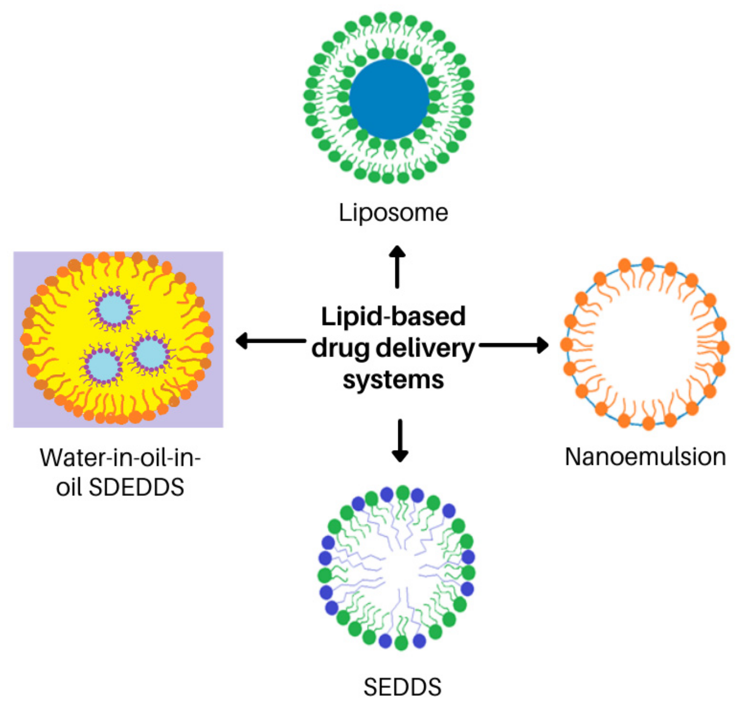
6. The Prospect of Self-Double-Emulsifying Drug Delivery Systems for Enhancing Topical Delivery of Clofazimine

6.1. Lipid-Based Carrier Systems Ideal for Lipophilic Drugs

It is estimated that approximately 30% of current commercial drugs including clofazimine and up to 50% of newly discovered drugs are highly hydrophobic [152,153]. Thus, lipid-based carrier systems are receiving special attention in the pharmaceutical industry in order to improve delivery of these drugs, especially those displaying poor aqueous solubility [173,174,175,176]. Encapsulation of the lipophilic drugs into inert lipid vehicles is shown to provide reproducible drug concentration profiles [173,177]. Lipid carrier systems include oils, surfactant dispersions, emulsions, nanoemulsions, liposomes, solid lipid carrier systems, nanostructured lipid carrier systems, self-emulsifying drug delivery systems (SEDDSs) and SDEDDSs [173,177,178,179]. Overall, lipid carrier systems can facilitate targeted drug delivery, thereby improving therapeutic effects and decreasing side effects, which will be important for anti-tubercular treatment [178].

6.2. Why SDEDDSs Are Considered More Appropriate Than Other Lipid-Based Carrier Systems

Liposomes, emulsions and nanoemulsions are frequently employed as drug delivery systems. These lipid-based formulations are also attracting increasing attention for enhancing topical and transdermal drug delivery [178,179,180,181]. Liposomes are spherical, bilayer structures of 100–200 nm in diameter. They are generally prepared from phospholipids and are able to encapsulate a wide range of diverse drugs, either in their aqueous central compartment or within the lipid bilayer. Their size, number of bilayers and various other components that may be included bestow their distinct characteristics. For example, inclusion of a surfactant results in the formation of transferosomes that are able to cross the SC. Inclusion of ethanol produces more flexible liposomes termed ethosomes. Liposomes were originally developed for parenteral administration of anticancer and antifungal agents. They are not as widely utilized for topical or transdermal drug delivery and are mainly included in cosmetics and long-acting sunscreen formulations, although their membrane structure is similar to that of natural membranes found in skin. Liposomes are expected to remain at the surface of the skin where they fuse with the skin lipids and slowly release the encapsulated drug [179,182,183,184].
Conversely, emulsions are heterogeneous, thermolabile biphasic liquids consisting of two or more immiscible fluids, which are made miscible through the addition of surfactants or emulsifying agents. They can be further categorized into micro- and nano-emulsions, as well as SEDDSs, depending on the formulation methods that are used. However, the instability of conventional emulsions causes difficulty in drug delivery [180,185]. Microemulsions technically are not emulsions, but are thermodynamically stable, single-phase systems that form spontaneously once the correct oil to water ratios are achieved. They possess a number of different microstructures depending on the nature and concentrations of the components. Alternatively, nanoemulsions are thermodynamically unstable dispersions that contain individual small droplets with a mean diameter of less than 200 nm. However, these dispersions possess reasonably good kinetic stability, as the droplets do not collide as frequently as in ordinary emulsions. The small droplet size furthermore allows for deep penetration into tissues as well as through fine capillaries. Positively charged (cationic) nanoemulsions increase skin permeation of poorly soluble drugs due to their interaction with the negatively charged skin epithelial cells. The main advantages of nanoemulsions include increased drug loading, enhanced drug solubility and bioavailability, reduced variability of drug uptake between different patients, controlled drug release, and protection from enzymatic degradation [178,180,182].
SEDDSs were originally developed from emulsions in order to generate a more physically stable formulation that is easier to prepare and can be used for oral drug delivery, as exemplified by their development specifically for oral administration of the immunosuppressant cyclosporine [178,185,186]. More recent activities focus on developing SEDDSs for other drug delivery routes of administration such as via the ocular-, vaginal- and sublingual routes [186,187,188,189,190]. Additionally, use of SEDDSs for treatment of dermal pathologies have been described [191,192]. SEDDSs are isotropic and thermodynamically stable emulsions containing an oil, surfactant, co-surfactant and drug(s) that spontaneously transform into oil-in-water emulsions upon the addition of aqueous media under gentle agitation [193]. SEDDSs are best suited for the incorporation of lipophilic drugs [194] as they are solubilized in the oily concentrate and then introduced into the aqueous media with mild agitation [178,185,186,194,195]. Interestingly, it has been demonstrated that SEDDSs encapsulating poorly water-soluble drugs elicited higher oral bioavailability in comparison with SEDDSs incorporating hydrophilic drugs; this clearly indicates the potential of SEDDSs to improve lipophilic drug delivery [196,197]. Nonetheless, for topical delivery of a drug such as clofazimine, the amount of clofazimine incorporated into the SEDDSs should be greater than the saturated concentration established in the oil phase in order to maximize clofazimine flux through the skin, as has also been noted for other poorly aqueous soluble drugs [197].
SEDDSs can be formulated in either nano- or micro droplets [198], where nanodroplet formation may occur in the presence of surface-active agents that cause spontaneous emulsification of finely dispersed droplets [194,199]. Formation of these finely dispersed nano-sized droplets follows from the intrinsic physicochemical properties of the constituents. Stability of these droplets depend upon the ratio of oil to surfactant [200]. However, it has been noted that high surfactant concentrations can cause tissue irritation [201]. Thus, spontaneous emulsification has to occur with decreased surfactant concentrations without reducing the stability of the formulations [202].
In view of the advantages of SEDDSs in terms of ease of formulation and solubilizing capacity for lipophilic drugs, SDEDDSs should provide even more benefits given the inclusion of multiple phases to optimize solubility and drug delivery via the dermal route of administration [203]. Drug delivery can be optimized by selecting immiscible oil phases in order to develop oil-in-oil-in water (O/O/W) or water-in-oil-in-oil (W/O/O) emulsions [203]. The employment of O/O/W systems will be beneficial if immiscible oil phases are used to create oil droplet formation in an inner oil phase within an outer oil phase [203]. This will provide a lipid matrix for enhancing solubility of lipophilic drugs while the water phase provides dermal hydration, which in turn establishes increased dermal drug delivery [204]. Conversely, W/O/O systems can provide a matrix for solubilization of both hydrophilic and lipophilic drugs with the added benefit of increased skin penetration as achieved by the outer oil phase that leads to enhanced dermal drug delivery [203,204,205].
As TB treatment requires combinations of different drugs administered over long periods of time as discussed above [37], the use of SDEDDSs can potentially provide an attractive dermal drug delivery vehicle by enabling simultaneous inclusion of several drugs within the hydrophilic and lipophilic phases of these multi-emulsions. Thereby, use of SDEDDSs represents a novel dermal drug delivery approach when compared to known dermal drug delivery vehicles. The lipid-based drug delivery systems discussed here are presented in Figure 5.
SDEDDSs are polydispersed systems (Figure 5) [206] that for oral drug delivery have the promise of increasing the oral bioavailability of drugs known to have low water solubility and high permeability, that is of the Biopharmaceutical classification system (BCS) Class II drugs. By transporting drugs dispersed in oil droplets while passing through the gastrointestinal tract (GIT) [206], they enable prolonged drug release within the GIT—the drugs incorporated in the inner-oil phase must partition across multiple phases before reaching the site of action where drug release is desired [206]. For topical application, the formulation in principle does not require prolonged release since the formulation is directly applied to the affected area to establish a localised therapeutic effect [207]. Nevertheless, the capacity for prolonged drug release provided by SDEDDSs can establish the intended therapeutic effect by controlling drug release as well as enabling the release of multiple drugs [206,207]. Conrolled drug release into the skin will obviate erratic drug release which can result in unwanted systemic uptake of the applied drug [207]. Use of conventional topical formulations that do not have controlled release properties place focus on establishing direct skin penetration of the encapsulated drugs [207]. In general, this is understandable as drugs must cross the protective barrier provided by the skin. This barrier reduces entry of drugs into the deeper skin layers thereby limiting dermal bioavailability which also compromises therapeutic effectiveness [207]. Therefore, optimal selection of excipients accompanied by a refined pre-formulation approach for preparing the SDEDDSs should overcome the challenges posed by drug solubilization and skin lipid disruption, and enable sustained release required to achieve dermal drug delivery [204,205,207].
SEDDSs and SDEDDSs additionally distribute drugs into the lymphatic system [178,180,196,208]. Due to absorption of SEDDSs and SDEDDSs through lymphatic vessels found in the dermis, drug delivery may be further enhanced [209,210]. Lymphatic absorption of clofazimine will provide an advantage as lymphadenitis is the most commonly observed manifestation of EPTB in both adults and children [68]. The lipid oil component is important for effecting lymphatic absorption through the dermis. Note that for oral administration, the oil has to solubilize a minimum of 50 mg/mL drug in order for lymphatic uptake to occur. In this respect, oils containing long chain triglycerides demonstrated increased lymphatic uptake, and the quantity of oil significantly contributes towards successful lymphatic uptake [171,186]. It was established that that a concentration of ≥25% oil content provides sufficient lymphatic absorption [131]. Unfortunately, lipids that are highly unsaturated may undergo autoxidation, and thus the addition of anti-oxidants must also be considered for topical drug delivery [208].
Not only is the evolution of SEDDSs moving towards alternative drug administration routes, but these systems are also being modified to advance muco-adhesion in order to extend contact time at the site of application [211,212,213]. Moreover, SEDDSs are being reformed to possess zeta-potential changing properties to improve drug delivery across mucus membranes [214]. Thus, combining mucolytic enzyme carrier systems with SEDDSs in order to enable local cleavability of mucus membranes resulted in increased drug permeation [215]. Consequently, SEDDSs have the potential to revolutionize drug delivery [187,194].

7. Conclusions

To summarize, the potential advantages of SEDDSs and SDEDDSs are enhanced drug entrapment capacity within physically more stable formulations, especially when compared to liposomes and emulsions. In addition, the formation of submicron droplets leads to an increase in the absorption surface area. SEDDSs elicit increased rates and amounts of drug absorption, thus increasing drug bioavailability. They moreover are able to deliver BCS Class II drugs efficiently. SEDDSs similarly have the potential for effective delivery of BCS class III drugs that display high solubility and decreased permeability properties, BCS class IV drugs that exhibit both low solubility and permeability characteristics, and hydrolytically susceptible drugs. Additionally, they offer consistent progressive diffusion profiles with reduced dosing and dosing frequencies. Moreover, upscaling techniques to prepare SEDDSs and SDEDDSs are simplified and cost effective compared to preparation of other lipid-based drug delivery systems.
Overall, the development of a topical SDEDDSs to aid in CTB treatment is potentially of considerable value as a reduced drug dose will be required to establish bioequivalence with oral drug delivery [201]. Due to the intractable nature of TB, high fixed doses of anti-tubercular drugs are normally recommended as inter-individual variability can lead to drug concentrations below the effective therapeutic dosage. Consequently, utilizing SDEDDSs for dermal administration of clofazimine will require decreased drug concentrations, will provide consistent drug delivery profiles that will be cytotoxic towards Mtb and thereby suppress drug resistance [216]. Importantly, such a route of administration should reduce the unpleasant discoloration associated with oral administration of clofazimine [87,210]. The improved drug loading capacity of SEDDSs can further support anti-tubercular treatment by either including higher concentrations of clofazimine or incorporating fixed-dose drug combinations with other antitubercular drugs [217,218]. In the latter respect, incorporation of both artemether and lumefantrine in a solid SEDDS double fixed dosage form was used successfully for treatment of malaria [217]. For TB, it should be possible to delay the development of anti-microbial resistance by co-administration of two drugs known to act synergistically [216]. For this reason, clofazimine and pyrazinamide, for example, can be combined into a SDEDDS formulation as synergism between the two drugs has already been established [71,219]. Moreover, recent synergism was reported from evaluating a combination of clofazimine, isoniazid and rifampicin against actively replicating planktonic organisms and slow replicating organisms [220]. Interestingly, first-line anti-tubercular drugs are not the only drugs that acts synergistically when used in combination with clofazimine. In a model study involving the use of macrophages infected with Mtb, synergism was detected between clofazimine and the novel prenylated amino-artemisinin WHN296 [221]. These significant findings support the contention that clofazimine will be a key component to most anti-tubercular drug regimens intended to treat active TB, resistant TB infections, and also CTB.

Author Contributions

D.v.S., J.M.V. and R.K.H. contributed to the conceptualization; J.M.V. and D.v.S. wrote the original draft preparation; J.M.V., R.K.H. and D.v.S. contributed to the writing—review and editing; J.M.V. and R.K.H. were responsible for the supervision of the study; J.M.V. handled the project administration and J.M.V. and R.K.H. the funding acquisition. J.M.V. saw to the revision of the paper. All authors have read and agreed to the published version of the manuscript.

Funding

This work is funded by the the South African National Research Foundation (SA NRF) grant (UID 129135) (J.M.V.).

Acknowledgments

The authors would like to thank the Centre of Excellence for Pharmaceutical Sciences, Faculty of Health Sciences, North-West University, South Africa for the financial contribution to this project.

Conflicts of Interest

The authors have no conflict of interest to declare.

Disclaimer

Any opinion, finding, conclusion, or recommendation expressed in this material is that of the authors alone, and the South African National Research Foundation does not accept any liability in this regard.

References

  1. Ankrah, A.O.; Glaudemans, A.W.J.M.; Maes, A.; van der Wiele, C.; Dierckx, R.A.J.O.; Vorster, M.; Sathekge, M.M. Tuberculosis. Semin. Nucl. Med. 2018, 48, 108–130. [Google Scholar] [CrossRef] [PubMed]
  2. Sotgiu, G.; Tiberi, S.; Centis, R.; D’Ambrosio, L.; Fuentes, Z.; Zumla, A.; Migliori, G.B. Applicability of the shorter ‘Bangladesh regimen’ in high multidrug-resistant tuberculosis settings. Int. J. Infect. Dis. 2017, 56, 190–193. [Google Scholar] [CrossRef] [PubMed] [Green Version]
  3. Grace, A.G.; Mittal, A.; Jain, S.; Tripathy, J.P.; Satyanarayana, S.; Tharyan, P.; Kirubakaran, R. Shortened treatment regimens versus the standard regimen for drug-sensitive pulmonary tuberculosis. Cochrane Database Syst. Rev. 2018, 1, CD012918. [Google Scholar] [CrossRef] [Green Version]
  4. Nellums, L.B.; Thompson, H.; Holmes, A.; Castro-Sánchez, E.; Otter, J.A.; Norredam, M.; Friedland, J.S.; Hargreaves, S. Antimicrobial resistance among migrants in Europe: A systematic review and meta-analysis. Lancet Infect. Dis. 2018, 18, 796–811. [Google Scholar] [CrossRef] [Green Version]
  5. Walker, T.M.; Merker, M.; Knoblauch, A.M.; Helbling, P.; Schoch, O.D.; van der Werf, M.J.; Kranzer, K.; Fiebig, L.; Kröger, S.; Haas, W.; et al. A cluster of multidrug-resistant Mycobacterium tuberculosis among patients arriving in Europe from the Horn of Africa: A molecular epidemiological study. Lancet Infect. Dis. 2018, 18, 431–440. [Google Scholar] [CrossRef] [Green Version]
  6. Dicko, A.; Faye, O.; Fofana, Y.; Soumoutera, M.; Berthé, S.; Touré, S.; Traoré, B.; Guindo, B.; Tall, K.; Keita, A.; et al. Cutaneous tuberculosis in Bamako, Mali. Pan. Afr. Med. J. 2017, 27, 102. [Google Scholar] [CrossRef]
  7. Boudville, D.A.; Joshi, R.; Rijkers, G.T. Migration and tuberculosis in Europe. J. Clin. Tuberc. Other Mycobact. Dis. 2020, 18, 100143. [Google Scholar] [CrossRef]
  8. Olaru, I.D.; Albert, H.; Zallet, J.; Werner, U.E.; Ahmed, N.; Rieder, H.L.; Salfinger, M.; Kranzer, K. Impact of quality improvement in tuberculosis laboratories in low-and lower-middle-income countries: A systematic review. Int. J. Tuberc. Lung Dis. 2018, 22, 309–320. [Google Scholar] [CrossRef]
  9. Monedero-Recuero, I. Drug-Resistant Tuberculosis in Europe. What Are We Waiting For? Am. J. Respir. Crit. Care Med. 2018, 198, 302–304. [Google Scholar] [CrossRef]
  10. Bainomugisa, A.; Pande, S.; Donnan, E.; Simpson, G.; Foster, J.B.; Lavu, E.; Hiasihri, S.; McBryde, E.S.; Moke, R.; Vincent, S.; et al. Cross-border movement of highly drug-resistant mycobacterium tuberculosis from Papua New Guinea to Australia through Torres Strait Protected Zone, 2010–2015. Emerg. Infect. Dis. 2019, 25, 406. [Google Scholar] [CrossRef] [Green Version]
  11. Toms, C.; Stapledon, R.; Waring, J.; Douglas, P. Tuberculosis notifications in Australia, 2012 and 2013. Commun. Dis. Intell. Q. Rep. 2015, 39, E217–E235. [Google Scholar]
  12. Lönnroth, K.; Mor, Z.; Erkens, C.; Bruchfeld, J.; Nathavitharana, R.R.; van der Werf, M.J.; Lange, C. Tuberculosis in migrants in low-incidence countries: Epidemiology and intervention entry points. Int. J. Tuberc. Lung Dis. 2017, 21, 624–636. [Google Scholar] [CrossRef]
  13. Smith, S.E.; Pratt, R.; Trieu, L.; Barry, P.M.; Thai, D.T.; Ahuja, S.D.; Shah, S. Epidemiology of pediatric multidrug-resistant tuberculosis in the United States, 1993–2014. Clin. Infect. Dis 2017, 65, 437–1443. [Google Scholar] [CrossRef]
  14. Marais, B.J. Improving access to tuberculosis preventive therapy and treatment for children. Int. J. Infect. Dis. 2017, 56, 122–125. [Google Scholar] [CrossRef] [Green Version]
  15. Dodd, P.J.; Yuen, C.M.; Sismanidis, C.; Seddon, J.A.; Jenkins, H.E. The global burden of tuberculosis mortality in children: A mathematical modelling study. Lancet Glob. Health 2017, 5, e898–e906. [Google Scholar] [CrossRef] [Green Version]
  16. Graham, S.M.; Sismanidis, C.; Menzies, H.J.; Marais, B.J.; Detjen, A.K. Black RE. Importance of tuberculosis control to address child survival. Lancet 2014, 383, 1605–1607. [Google Scholar] [CrossRef] [Green Version]
  17. Tacconelli, E.; Pezzani, M.D. Public health burden of antimicrobial resistance in Europe. Lancet Infect. Dis. 2019, 19, 4–6. [Google Scholar] [CrossRef] [Green Version]
  18. Baker, S.J.; Payne, D.J.; Rappuoli, R.; De Gregorio, E. Technologies to address antimicrobial resistance. Proc. Natl. Acad. Sci. USA 2018, 115, 12887–12895. [Google Scholar] [CrossRef] [Green Version]
  19. World Health Organization (WHO). The End TB Strategy. 2016. Available online: http://www.who.int/tb/post2015_TBstrategy.pdf?ua=1 (accessed on 18 March 2022).
  20. World Health Organization (WHO). UN General Assembly High-Level Meeting on the Fight Against Tuberculosis. 2018. Available online: https://www.who.int/news-room/events/un-general-assembly-high-level-meeting-on-ending-tb (accessed on 10 April 2021).
  21. Stop TB Partnership. 2019. Available online: http://www.stoptb.org/global/advocacy/unhlm_whatis.asp (accessed on 20 May 2021).
  22. General Assembly of the United Nations. United Nations High-Level Meeting on the Fight to End Tuberculosis 26 September 2018, UNHQ, New York. 2018. Available online: https://www.un.org/pga/73/event/fight-to-end-tuberculosis/ (accessed on 20 March 2022).
  23. United Nations: Meetings Coverage and Press Releases. World Leaders Reaffirm Commitment to End Tuberculosis by 2030, as General Assembly Adopts Declaration Outlining Actions for Increased Financing, Treatment Access. 2018. Available online: https://www.un.org/press/en/2018/ga12067.doc.htm (accessed on 20 May 2021).
  24. World Health Organization (WHO). Global Tuberculosis Report 2021; World Health Organization (WHO): Geneva, Switzerland, 2021; ISBN 9789240037021. (Electronic Version). [Google Scholar]
  25. Lange, C.; Chesov, D.; Heyckendorf, J. Clofazimine for the treatment of multidrug-resistant tuberculosis. Clin. Microbiol. Infect. 2019, 25, 128–130. [Google Scholar] [CrossRef] [Green Version]
  26. van Zyl, L.; Viljoen, J.M.; Haynes, R.K.; Aucamp, M.; Ngwane, A.H.; du Plessis, J. Topical delivery of artemisone, clofazimine and decoquinate encapsulated in vesicles and their in vitro efficacy against Mycobacterium tuberculosis. AAPS Pharm. Sci. Tech. 2019, 20, 33–44. [Google Scholar] [CrossRef]
  27. Osuchukwu, O.; Nuῆez, M.; Packard, S.; Ehiri, J.; Rosales, C.; Hawkins, E.; Gerardo Avilés, J.G.; Gonzalez-Salazar, F.; Oren, E. Latent Tuberculosis Infection Screening Acceptability among Migrant Farmworkers. Int. Migr. 2017, 55, 62–74. [Google Scholar] [CrossRef]
  28. Cardona, P.J. Pathogenesis of tuberculosis and other mycobacteriosis. Enferm. Infecc. Microbiol. Clin. 2018, 36, 38–46. [Google Scholar] [CrossRef] [PubMed]
  29. Centis, R.; D’Ambrosio, L.; Zumla, A.; Migliori, G.B. Shifting from tuberculosis control to elimination: Where are we? What are the variables and limitations? Is it achievable? Int. J. Infect. Dis. 2017, 56, 30–33. [Google Scholar] [CrossRef] [PubMed] [Green Version]
  30. dos Santos, J.B.D.; Figueiredo, A.R.; Ferraz, C.E.; Oliveira, M.H.D.; Silva, P.G.D.; Medeiros, V.L.S.D. Cutaneous tuberculosis: Epidemiologic, etiopathogenic and clinical aspects-part I. An. Bras. Dermatol. 2014, 89, 219–228. [Google Scholar] [CrossRef] [PubMed]
  31. Churchyard, G. This Kills More South Africans Than Any Other Disease. There’s a New Way to Stop it. BHEKISISA Centre for Health Journalism. 2019. Available online: https://bhekisisa.org/article/2019-03-28-00-3hp-latent-tb-treatment-short-course-guidelines-regimen-isoniazid-rifapentine (accessed on 19 February 2022).
  32. van Zyl, L.; du Plessis, J.; Viljoen, J. Cutaneous tuberculosis overview and current treatment regimens. Tuberculosis 2015, 95, 629–638. [Google Scholar] [CrossRef]
  33. Chen, S.T.; Cahalane, A.M.; Ryan, E.T.; Foreman, R.K. Case 2-2019: A 36-year-old man with rash, abdominal pain, and lymphadenopathy. N. Engl. J. Med. 2019, 380, 275–283. [Google Scholar] [CrossRef] [Green Version]
  34. Mohanty, N.; Nayak, B.B. Cutaneous tuberculosis, tuberculosis verrucosa cutis. Med. J. Dr. D. Y. Patil. Univ. 2014, 7, 53–55. [Google Scholar] [CrossRef]
  35. World Health Organization (WHO). Global Tuberculosis Report 2018; World Health Organization (WHO): Geneva, Switzerland, 2018; ISBN 9789241565646. [Google Scholar]
  36. Yao, L.; LiangLiang, C.; JinYue, L.; WanMei, S.; Lili, S.; YiFan, L.; HuaiChen, L. Ambient air pollution exposures and risk of drug-resistant tuberculosis. Environ. Int. 2019, 124, 161–169. [Google Scholar] [CrossRef]
  37. Venkatesh, U.; Srivastava, D.K.; Srivastava, A.K.; Tiwari, H.C. Epidemiological profile of multidrug-resistant tuberculosis patients in Gorakhpur Division, Uttar Pradesh, India. J. Fam. Med. Prim. Care 2018, 7, 589–595. [Google Scholar] [CrossRef]
  38. Velayati, A.A.; Farnia, P.; Hoffner, S. Drug-resistant Mycobacterium tuberculosis: Epidemiology and role of morphological alterations. J. Glob. Antimicrob. Resist. 2018, 12, 192–196. [Google Scholar] [CrossRef]
  39. McBryde, E.S.; Meehan, M.T.; Doan, T.N.; Ragonnet, R.; Marais, B.J.; Guernier, V.; Trauer, J.M. The risk of global epidemic replacement with drug-resistant Mycobacterium tuberculosis strains. Int. J. Infect. Dis. 2017, 56, 14–20. [Google Scholar] [CrossRef] [PubMed] [Green Version]
  40. Aaron, L.; Saadoun, D.; Calatroni, I.; Launay, O.; Memain, N.; Vincent, V.; Marchal, G.; Dupont, B.; Bouchaud, O.; Valeyre, D.; et al. Tuberculosis in HIV-infected patients: A comprehensive review. Clin. Microbiol. Infect. 2004, 10, 388–398. [Google Scholar] [CrossRef] [PubMed] [Green Version]
  41. Pourakbari, B.; Sadeghi, R.H.; Mahmoudi, S.; Parvaneh, N.; Valian, S.K.; Mamishi, S. Evaluation of interleukin-12 receptor β1 and interferon gamma receptor 1 deficiency in patients with disseminated BCG infection. Allergol. Immunopathol. 2019, 47, 38–42. [Google Scholar] [CrossRef] [PubMed]
  42. Caminero, J.A.; Cayla, J.A.; García-García, J.M.; García-Pérez, F.J.; Palacios, J.J.; Ruiz-Manzano, J. Diagnosis and treatment of drug-resistant tuberculosis. Arch. Bronconeumol. 2017, 53, 501–509. [Google Scholar] [CrossRef] [PubMed]
  43. Tuberculosis (TB). Division of Tuberculosis Elimination. Centers for Disease Control and Prevention. CDC Twenty Four Seven. Saving Lives, Protecting People. 2016. Available online: https://www.cdc.gov/tb/publications/factsheets/drtb/mdrtb.htm (accessed on 15 February 2022).
  44. Alexander, P.E.; De, P. The emergence of extensively drug-resistant tuberculosis (TB): TB/HIV coinfection, multidrug-resistant TB and the resulting public health threat from extensively drug-resistant TB, globally and in Canada. Can. J. Infect. Dis. Med. Microbiol. 2007, 18, 289–291. [Google Scholar] [CrossRef]
  45. Sharma, S.K.; Turaga, K.K.; Balamurugan, A.; Saha, P.K.; Pandey, R.M.; Jain, N.K.; Katoch, V.M.; Mehrab, N.K. Clinical and genetic risk factors for the development of multi-drug resistant tuberculosis in non-HIV infected patients at a tertiary care center in India: A case-control study. Infect. Genet. Evol. 2003, 3, 183–188. [Google Scholar] [CrossRef]
  46. Munsiff, S.S.; Joseph, S.; Ebrahimzadeh, A.; Frieden, T.R. Rifampin monoresistant tuberculosis in New York City, 1993–1994. Clin. Infect. Dis. 1997, 25, 1465–1467. [Google Scholar] [CrossRef] [Green Version]
  47. Bhat, Z.S.; Rather, M.A.; Maqbool, M.; Ahmad, Z. Drug targets exploited in Mycobacterium tuberculosis: Pitfalls and promis-es on the horizon. Biomed Pharm. 2018, 103, 1733–1747. [Google Scholar] [CrossRef]
  48. Tiberi, S.; Muñoz-Torrico, M.; Duarte, R.; Dalcolmo, M.; D’Ambrosio, L.; Migliori, G.B. New drugs and perspectives for new anti-tuberculosis regimens. Pulmonology 2018, 24, 86–98. [Google Scholar] [CrossRef]
  49. Peña, M.J.M.; García, B.S.; Baquero-Artigao, F.; Pérez, D.M.; Pérez, R.P.; Echevarría, A.M.; Amador, J.T.R.; Durán, D.G.P.; Julian, A.N. Tuberculosis treatment for children: An update. An. de Pediatría 2018, 88, 52.e1–52.e12. [Google Scholar] [CrossRef]
  50. Maitre, T.; Petitjean, G.; Chauffour, A.; Bernard, C.; El Helali, N.; Jarlier, V.; Reibel, F.; Chavanet, P.; Aubry, A.; Veziris, N. Are moxifloxacin and levofloxacin equally effective to treat XDR tuberculosis? J. Antimicrob. Chemother. 2017, 72, 2326–2333. [Google Scholar] [CrossRef] [PubMed] [Green Version]
  51. Tiberi, S.; Utjesanovic, N.; Galvin, J.; Centis, R.; D’Ambrosio, L.; van den Boom, M.; Zumla, A.; Migliori, G.B. Drug resistant TB–latest developments in epidemiology, diagnostics and management. IJID 2022, (in press). [Google Scholar] [CrossRef] [PubMed]
  52. Jiang, H.; Jin, Y.; Vissa, V.; Zhang, L.; Liu, W.; Qin, L.; Wan, K.; Wu, X.; Wang, H.; Liu, W.; et al. Molecular characteristics of mycobacterium tuberculosis strains isolated from cutaneous tuberculosis patients in China. Acta Derm.-Venereol. 2017, 97, 472–477. [Google Scholar] [CrossRef] [PubMed] [Green Version]
  53. Desai, U.; Joshi, J.M. Extrapulmonary drug-resistant tu.uberculosis at a drug-resistant tuberculosis center, Mumbai: Our experience–Hope in the midst of despair! Lung India 2019, 36, 3–7. [Google Scholar] [CrossRef]
  54. Haitz, K.; Ly, A.; Smith, G. Idiopathic granulomatous mastitis. Cutis 2019, 103, 38–42. [Google Scholar]
  55. Mimesh, S.A.; Memish, Z.A. Cutaneous Tuberculosis. In Extrapulmonary Tuberculosis, 1st ed.; Sener, A., Erdem, H., Eds.; Springer: Cham, Switzerland, 2019; pp. 175–180. [Google Scholar]
  56. de los Santos Moreno, A.; Gómez, A.S.; Blasco, E.R.; Cuevas, M.C.; Saborido, D.G. Infecciones bacterianas crónicas (I). Tuberculosis. Med.-Programa de Form. Médica Contin. Acreditado 2018, 12, 3115–3123. [Google Scholar] [CrossRef]
  57. Peirse, M.; Houston, A. Extrapulmonary tuberculosis. Medicine 2017, 45, 747–752. [Google Scholar] [CrossRef]
  58. Dusthackeer, A.; Sekar, G.; Chidambaram, S.; Kumar, V.; Mehta, P.; Swaminathan, S. Drug resistance among extrapulmonary TB patients: Six years’ experience from a supranational reference laboratory. Indian J. Med. Res. 2015, 142, 568–574. [Google Scholar] [CrossRef]
  59. Lee, J.Y. Diagnosis and treatment of extrapulmonary tuberculosis. Tuberc. Respir. Dis. 2015, 78, 47–55. [Google Scholar] [CrossRef] [Green Version]
  60. Mehta, P.K.; Raj, A.; Singh, N.; Khuller, G.K. Diagnosis of extrapulmonary tuberculosis by PCR. FEMS Immunol. Med. Microbiol. 2012, 66, 20–36. [Google Scholar] [CrossRef] [Green Version]
  61. Joshi, J.M. Tuberculosis chemotherapy in the 21st century: Back to the basics. Lung India 2011, 28, 193–200. [Google Scholar] [CrossRef] [PubMed]
  62. Mohan, A.; Sharma, S.K. Medical schools and tuberculosis control: Bridging the discordance between what is preached and what is practiced. Indian J. Chest Dis. Allied Sci. 2004, 46, 5–7. [Google Scholar] [PubMed]
  63. Frankel, A.; Penrose, C.; Emer, J. Cutaneous tuberculosis: A practical case report and review for the dermatologist. J. Clin. Aesthet. Dermatol. 2009, 2, 19–27. [Google Scholar] [PubMed]
  64. de Mello, R.B.; do Vale, E.C.S.; Baeta, I.G.R. Scrofuloderma: A diagnostic challenge. An. Bras. Dermatol. 2019, 94, 102–104. [Google Scholar] [CrossRef] [Green Version]
  65. Rullán, J.; Seijo-Montes, R.E.; Vaillant, A.; Sánchez, N.P. Cutaneous manifestations of pulmonary disease. In Atlas of Dermatology in Internal Medicine, 14th ed.; Sánchez, N.P., Ed.; Springer: New York, NY, USA, 2012; pp. 17–30. [Google Scholar]
  66. Ghosh, S.; Aggarwal, K.; Jain, V.K.; Chaudhuri, S.; Ghosh, E. Tuberculosis verrucosa cutis presenting as diffuse plantar keratoderma: An unusual sight. Indian J. Dermatol. 2014, 59, 80–81. [Google Scholar] [CrossRef]
  67. Walter, N.; Daley, C.L. Tuberculosis and nontuberculous mycobacterial infections. In Clinical Respiratory Medicine, 4th ed.; Spiro, S.G., Silvestri, G.A., Agustí, A., Eds.; Elsevier Ltd.: Philadelphia, PA, USA, 2012; pp. 383–405. [Google Scholar]
  68. Jones-Lopez, E.C.; Ellner, J.J. Tuberculosis and atypical mycobacterial infections. In Tropical Infectious Diseases, 3rd ed.; Guerrant, R.L., Walker, D.H., Weller, P.F., Eds.; Elsevier Inc.: London, UK, 2011; pp. 228–247. [Google Scholar]
  69. Yates, V.M. Mycobacterial infections. In Rook’s Textbook of Dermatology, 8th ed.; Burns, T., Breathnach, S., Cox, N., Griffiths, C., Eds.; Blackwell Publishing Ltd.: West Sussex, UK, 2010; Volume 2, pp. 31.1–31.41. [Google Scholar]
  70. Gharabaghi, M.A. Cutaneous tuberculosis caused by isoniazid-resistant Mycobacterium tuberculosis. BMJ Case Rep. 2012, 2012, bcr2012006253. [Google Scholar] [CrossRef] [Green Version]
  71. Hernández Solis, A.; Herrera González, N.E.; Cazarez, F.; Mercadillo Pérez, P.; Olivera Diaz, H.O.; Escobar-Gutierrez, A.; Cortés Ortíz, I.; González González, H.; Reding-Bernal, A.; Sabido, R.C. Skin biopsy: A pillar in the identification of cutaneous Mycobacterium tuberculosis infection. J. Infect. Dev. Ctries. 2012, 6, 626–631. [Google Scholar] [CrossRef] [Green Version]
  72. Zhang, J.; Fan, Y.K.; Wang, P.; Chen, Q.Q.; Wang, G.; Xu, A.E.; Chen, L.Q.; Hu, R.; Chen, W.; Song, Z.Q.; et al. Cutaneous tuberculosis in China—A multicentre retrospective study of cases diagnosed between 1957 and 2013. J. Eur. Acad. Dermatol. Venereol. 2018, 32, 632–638. [Google Scholar] [CrossRef]
  73. Ho, C.K.; Ho, M.H.; Chong, L.Y. Cutaneous tuberculosis in Hong Kong: An update. Hong Kong Med. J. 2006, 12, 272–277. [Google Scholar]
  74. Gopinathan, R.; Pandit, D.; Joshi, J.; Jerajani, H.; Mathur, M. Clinical and morphological variants of cutaneous tuberculosis and its relation to Mycobacterium species. Indian J. Med. Microbl. 2001, 19, 193–196. [Google Scholar]
  75. Kaul, S.; Kaur, I.; Mehta, S.; Singal, A. Cutaneous tuberculosis. Part I: Pathogenesis, classification, and clinical features. JAAD 2022, (in press). [Google Scholar] [CrossRef] [PubMed]
  76. De Maio, F.; Trecarichi, E.M.; Visconti, E.; Sanguinetti, M.; Delogu, G.; Sali, M. Understanding cutaneous tuberculosis: Two clinical cases. JMM Case Rep. 2016, 3, e005070–e005076. [Google Scholar] [CrossRef] [PubMed]
  77. Teixeira, H.C.; Abramo, C.; Munk, M.E. Immunological diagnosis of tuberculosis: Problems and strategies for success. J. Bras. Pneumol. 2007, 33, 323–334. [Google Scholar] [CrossRef] [PubMed] [Green Version]
  78. Sloan, D.J.; Lewis, J.M. Management of multidrug-resistant TB: Novel treatments and their expansion to low resource settings. Trans. R Soc. Trop Med. Hyg. 2016, 110, 163–172. [Google Scholar] [CrossRef] [PubMed] [Green Version]
  79. Chen, Q.; Chen, W.; Hao, F. Cutaneous Tuberculosis: A Great Imitator. Clin. Dermatol. 2019, 37, 192–199. [Google Scholar] [CrossRef]
  80. Karas, L.; Lu, C.Y.; Agrawal, P.B.; Asgari, M.M. The impact of the Orphan Drug Act on Food and Drug Administration-approved therapies for rare skin diseases and skin-related cancers. J. Am. Acad. Dermatol. 2019, 81, 867–877. [Google Scholar] [CrossRef]
  81. van Deun, A.; Salim, H.; Kumar Das, A.P.; Bastian, I.; Portaels, F. Results of a standardised regimen for multidrug-resistant tuberculosis in Bangladesh. Int. J. Tuberc. Lung Dis. 2010, 8, 560–567. [Google Scholar]
  82. Günther, G.; Van Leth, F.; Alexandru, S.; Altet, N.; Avsar, K.; Bang, D.; Barbuta, R.; Bothamley, G.; Ciobanu, A.; Crudu, V.; et al. Clinical management of multidrug-resistant tuberculosis in 16 European countries. Am. J. Respir. Crit. Care Med. 2018, 198, 379–386. [Google Scholar] [CrossRef]
  83. Shah, M.A.; Shah, I. Increasing Prevalence of Pediatric Drug-resistant Tuberculosis in Mumbai, India, and Its Outcome. Pediatr. Infect. Dis. J. 2018, 37, 1261–1263. [Google Scholar] [CrossRef]
  84. Cox, H.; Hughes, J.; Black, J.; Nicol, M.P. Precision medicine for drug-resistant tuberculosis in high-burden countries: Is individualised treatment desirable and feasible? Lancet Infect. Dis. 2018, 18, e282–e287. [Google Scholar] [CrossRef]
  85. Barry, V.C.; Belton, J.G.; Conalty, M.L.; Den-Steny, J.M.; Edward, D.W.; O’Sullivan, J.F.; Twomey, D.; Winder, F. A new series of phenazines (rimino-compounds) with high antituberculosis activity. Nature 1957, 179, 1013–1015. [Google Scholar] [CrossRef] [PubMed]
  86. Drugbank. Clofazimine. 2017. Available online: https://www.drugbank.ca/drugs/DB00845 (accessed on 3 June 2021).
  87. Ammerman, N.C.; Swanson, R.V.; Bautista, E.M.; Almeida, D.V.; Saini, V.; Omansen, T.F.; Guo, H.; Chang, Y.S.; Li, S.-Y.; Tapley, A.; et al. Impact of clofazimine dosing on treatment-shortening of the first-line regimen in a mouse model of tuberculosis. Antimicrob. Agents Chemother. 2018, 62, e00636-18. [Google Scholar] [CrossRef] [PubMed] [Green Version]
  88. McGuffin, S.A.; Pottinger, P.S.; Harnisch, J.P. Clofazimine in nontuberculous mycobacterial infections: A growing niche. Open Forum Infect. Dis. 2017, 4, ofx147. [Google Scholar] [CrossRef] [PubMed]
  89. Swanson, R.V.; Adamson, J.; Moodley, C.; Ngcobo, B.; Ammerman, N.C.; Dorasamy, A.; Moodley, S.; Mgaga, Z.; Tapley, A.; Bester, L.A.; et al. Pharmacokinetics and pharmacodynamics of clofazimine in a mouse model of tuberculosis. Antimicrob. Agents Chemother. 2015, 59, 3042–3051. [Google Scholar] [CrossRef] [PubMed] [Green Version]
  90. Cholo, M.C.; Steel, H.C.; Fourie, P.B.; Germishuizen, W.A.; Anderson, R. Clofazimine: Current status and future prospects. J. Antimicrob. Chemother. 2012, 67, 290–298. [Google Scholar] [CrossRef]
  91. Zhu, H.; Fu, L.; Wang, B.; Chen, X.; Zhao, J.; Huang, H.; Lu, Y. Activity of Clofazimine and TBI-166 against Mycobacterium tuberculosis in Different Administration Intervals in Mouse Tuberculosis Models. Antimicrob. Agents Chemother. 2021, 11, e02164-20. [Google Scholar] [CrossRef]
  92. Browne, S.G.; Hogerzeil, L.M. “B 663” in the treatment of leprosy. Preliminary report of a pilot trial. Lepr. Rev. 1962, 33, 6–10. [Google Scholar] [CrossRef]
  93. Attwood, M.M.; Rask-Andersen, M.; Schiöth, H.B. Orphan drugs and their impact on pharmaceutical development. Trends Pharmacol. Sci. 2018, 39, 525–535. [Google Scholar] [CrossRef]
  94. Brennan, P.J.; Young, D.B. Tuberculosis. In Handbook of Anti-Tuberculosis Agents; Elsevier: Amsterdam, The Netherlands, 2008; Volume 88, pp. 85–170. [Google Scholar]
  95. Cholo, M.C.; Mothiba, M.T.; Fourie, B.; Anderson, R. Mechanisms of action and therapeutic efficacies of the lipophilic antimycobacterial agents clofazimine and bedaquiline. J. Antimicrob. Chemother. 2017, 72, 338–353. [Google Scholar] [CrossRef] [Green Version]
  96. Zhang, Z.; Li, T.; Qu, G.; Pang, Y.; Zhao, Y. In vitro synergistic activity of clofazimine and other antituberculous drugs against multidrug-resistant Mycobacterium tuberculosis isolates. Int. J. Antimicrob. Agents 2015, 45, 71–75. [Google Scholar] [CrossRef]
  97. Dooley, K.E.; Obuku, E.A.; Durakovic, N.; Belitsky, V.; Mitnick, C.; Nuermberger, E.L. Efficacy Subgroup, RESIST-TB. World Health Organization group 5 drugs for the treatment of drug-resistant tuberculosis: Unclear efficacy or untapped potential? J. Infect. Dis. 2013, 207, 1352–1358. [Google Scholar] [CrossRef] [PubMed]
  98. Mirnejad, R.; Asadi, A.; Khoshnood, S.; Mirzaei, H.; Heidary, M.; Fattorini, L.; Ghodousi, A.; Darban-Sarokhalil, D. Clofazimine: A useful antibiotic for drug-resistant tuberculosis. Biomed. Pharmacother. 2018, 105, 1353–1359. [Google Scholar] [CrossRef] [PubMed]
  99. Kuaban, C.; Noeske, J.; Rieder, H.L.; Ait-Khaled, N.; Abena Foe, J.L.; Trébucq, A. High effectiveness of a 12-month regimen for MDR-TB patients in Cameroon. Int. J. Tuberc. Lung Dis. 2015, 19, 517–524. [Google Scholar] [CrossRef] [PubMed]
  100. Aung, K.J.M.; Van Deun, A.; Declercq, E.; Sarker, M.R.; Das, P.K.; Hossain, M.A.; Rieder, H.L. Successful ‘9-month Bangladesh regimen’ for multidrug-resistant tuberculosis among over 500 consecutive patients. Int. J. Tuberc. Lung Dis. 2014, 18, 1180–1187. [Google Scholar] [CrossRef]
  101. Piubello, A.; Harouna, S.H.; Souleymane, M.B.; Boukary, I.; Morou, S.; Daouda, M.; Hanki, Y.; Van Deun, A. High cure rate with standardized short-course multidrug-resistant tuberculosis treatment in Niger: No relapses. Int. J. Tuberc. Lung Dis. 2014, 18, 1188–1194. [Google Scholar] [CrossRef]
  102. Tang, S.; Yao, L.; Hao, X.; Liu, Y.; Zeng, L.; Liu, G.; Li, M.; Li, F.; Wu, M.; Zhu, Y.; et al. Clofazimine for the treatment of multidrug-resistant tuberculosis: Prospective, multicenter, randomized controlled study in China. Clin. Infect. Dis. 2015, 60, 1361–1367. [Google Scholar] [CrossRef] [Green Version]
  103. Tyagi, S.; Ammerman, N.C.; Li, S.Y.; Adamson, J.; Converse, P.J.; Swanson, R.V.; Almeida, D.V.; Grosset, J.H. Clofazimine shortens the duration of the first-line treatment regimen for experimental chemotherapy of tuberculosis. Proc. Natl. Acad. Sci. USA 2015, 112, 869–874. [Google Scholar] [CrossRef] [Green Version]
  104. Grosset, J.H.; Tyagi, S.; Almeida, D.V.; Converse, P.J.; Li, S.Y.; Ammerman, N.C.; Bishai, W.R.; Enarson, D.; Trébucq, A. Assessment of clofazimine activity in a second-line regimen for tuberculosis in mice. Am. J. Respir. Crit. Care Med. 2013, 188, 608–612. [Google Scholar] [CrossRef] [Green Version]
  105. Seung, K.J.; Hewison, C. Now is the time for shorter all-oral regimens for multidrug-resistant tuberculosis. Lancet Glob Health 2019, 7, e706. [Google Scholar] [CrossRef] [Green Version]
  106. Durusu, İ.Z.; Hüsnügil, H.H.; Ataş, H.; Biber, A.; Gerekçi, S.; Güleç, E.A.; Özen, C. Anti-cancer effect of clofazimine as a single agent and in combination with cisplatin on U266 multiple myeloma cell line. Leuk Res. 2017, 55, 33–40. [Google Scholar] [CrossRef]
  107. Food and Drug Administration. LAMPRENE® (clofazimine) Capsules, Full Prescribing Information; U.S. Food and Drug Administration; Reference ID: 3956651; Silver Spring, MD, USA, 2016. Available online: https://www.accessdata.fda.gov/drugsatfda_docs/label/2016/019500s013lbl.pdf (accessed on 5 January 2022).
  108. Novartis. Lamprene® Prescribing Information; NDA 19-500/S-010; Novartis: East Hanover, NJ, USA, 2002. Available online: https://www.accessdata.fda.gov/drugsatfda_docs/label/2003/19500slr010_lamprene_lbl.pdf (accessed on 5 January 2022).
  109. Chen, Y.; Yuan, Z.; Shen, X.; Wu, J.; Wu, Z.; Xu, B. Resistance to second-line antituberculosis drugs and delay in drug susceptibility testing among multidrug-resistant tuberculosis patients in Shanghai. BioMed. Res. Int. 2016, 2016, 2628913. [Google Scholar] [CrossRef] [PubMed] [Green Version]
  110. Dalcolmo, M.; Gayoso, R.; Sotgiu, G.; D’Ambrosio, L.; Rocha, J.L.; Borga, L.; Fandinho, F.; Braga, J.U.; Galesi, V.M.N.; Barreira, D.; et al. Effectiveness and safety of clofazimine in multidrug-resistant tuberculosis: A nationwide report from Brazil. Eur. Respir. J. 2017, 49, 1602445. [Google Scholar] [CrossRef] [PubMed]
  111. Szeto, W.; Garcia-Buitrago, M.T.; Abbo, L.; Rosenblatt, J.D.; Moshiree, B.; Morris, M.I. Clofazimine enteropathy: A rare and under recognized complication of mycobacterial therapy. Open Forum. Infect. Dis. 2016, 2, ofw004. [Google Scholar] [CrossRef] [PubMed] [Green Version]
  112. Yoon, G.S.; Keswani, R.K.; Sud, S.; Rzeczycki, P.M.; Murashov, M.D.; Koehn, T.A.; Standiford, T.J.; Stringer, K.A.; Rosania, G.R. Clofazimine biocrystal accumulation in macrophages upregulates interleukin 1 receptor antagonist production to induce a systemic anti-inflammatory state. Antimicrobl. Agents Chemothe. 2016, 60, 3470–3479. [Google Scholar] [CrossRef] [PubMed] [Green Version]
  113. Gopal, M.; Padayatchi, N.; Metcalfe, J.Z.; O’Donnell, M.R. Systematic review of clofazimine for the treatment of drug-resistant tuberculosis. Int. J. Tuberc. Lung Dis. 2013, 17, 1001–1007. [Google Scholar] [CrossRef] [Green Version]
  114. Barot, R.K.; Viswanath, V.; Pattiwar, M.S.; Torsekar, R.G. Crystalline deposition in the cornea and conjunctiva secondary to long-term clofazimine therapy in a leprosy patient. Indian J. Ophthalmol. 2011, 59, 328–329. [Google Scholar] [CrossRef]
  115. Kaur, I.; Ram, J.; Kumar, B.; Kaur, S.; Sharma, V.K. Effect of clofazimine on eye in multibacillary leprosy. Indian J. Lepr. 1990, 62, 87–90. [Google Scholar]
  116. Zaccagnino, A.; Managò, A.; Leanza, L.; Gontarewitz, A.; Linder, B.; Azzolini, M.; Biasutto, L.; Zoratti, M.; Peruzzo, R.; Legler, K.; et al. Tumor-reducing effect of the clinically used drug clofazimine in a SCID mouse model of pancreatic ductal adenocarcinoma. Oncotarget 2017, 8, 38276–38293. [Google Scholar] [CrossRef]
  117. Dixit, V.B.; Chaudhary, S.D.; Jain, V.K. Clofazimine induced nail changes. Indian J. Lepr. 1989, 61, 476–478. [Google Scholar]
  118. World Health Organization. WHO Model Prescribing Information: Drugs Used in Leprosy; World Health Organization: Geneva, Switzerland, 1998; Available online: http://apps.who.int/medicinedocs/pdf/h2988e/h2988e.pdf (accessed on 5 January 2022).
  119. National Hansen’s Disease (Leprosy) Program. Recommended Treatment Regimens; US Department of Health and Human Services: Washington, DC, USA, 2018. Available online: https://www.hrsa.gov/hansensdisease/diagnosis/recommendedtreatment.html (accessed on 5 January 2022).
  120. Hwang, T.J.; Dotsenko, S.; Jafarov, A.; Weyer, K.; Falzon, D.; Lunte, K.; Nunn, P.; Jaramillo, E.; Keshavjee, S.; Wares, D.F. Safety and availability of clofazimine in the treatment of multidrug and extensively drug-resistant tuberculosis: Analysis of published guidance and meta-analysis of cohort studies. BMJ Open 2014, 4, e004143. [Google Scholar] [CrossRef]
  121. Ozturk, Z.; Tatliparmak, A. Leprosy treatment during pregnancy and breastfeeding: A case report and brief review of literature. Dermatol. Ther. 2017, 30, e12414. [Google Scholar] [CrossRef] [PubMed] [Green Version]
  122. Drobac, P.C.; del Castillo, H.; Sweetland, A.; Anca, G.; Joseph, J.K.; Furin, J.; Shin, S. Treatment of multidrug-resistant tuberculosis during pregnancy: Long-term follow-up of 6 children with intrauterine exposure to second-line agents. Clin. Infect. Dis. 2005, 40, 1689–1692. [Google Scholar] [CrossRef] [PubMed]
  123. Levis, W.; Rendini, T. Clofazimine Mechanisms of Action in Mycobacteria, HIV, and Cancer. J. Infect. Dis. 2017, 215, 1488. [Google Scholar] [CrossRef] [PubMed]
  124. Machado, T.C.; Gelain, A.B.; Rosa, J.; Cardoso, S.G.; Caon, T. Cocrystallization as a novel approach to enhance the transdermal administration of meloxicam. Eur. J. Pharm. Sci. 2018, 123, 184–190. [Google Scholar] [CrossRef] [PubMed]
  125. Singh Malik, D.; Mital, N.; Kaur, G. Topical drug delivery systems: A patent review. Expert. Opin. Ther. Pat. 2016, 26, 213–228. [Google Scholar] [CrossRef] [PubMed]
  126. Bhowmik, D.; Gopinath, H.; Kumar, B.P.; Duraivel, S.; Kumar, K.S. Recent advances in novel topical drug delivery system. Pharm. Innov. 2012, 1, 12–31. [Google Scholar]
  127. Gratieri, T.; Alberti, I.; Lapteva, M.; Kalia, Y.N. Next generation intra-and transdermal therapeutic systems: Using non-and minimally-invasive technologies to increase drug delivery into and across the skin. Eur. J. Pharm. Sci. 2013, 50, 609–622. [Google Scholar] [CrossRef]
  128. Prausnitz, M.R.; Mitragotri, S.; Langer, R. Current status and future potential of transdermal drug delivery. Nat. Rev. Drug Discov. 2004, 3, 115–124. [Google Scholar] [CrossRef]
  129. Zhang, Y.; Zhang, N.; Song, H.; Li, H.; Wen, J.; Tan, Y.; Zheng, W. Design, characterization and comparison of transdermal delivery of colchicine via borneol-chemically-modified and borneol-physically-modified ethosome. Drug Deliv. 2019, 26, 70–77. [Google Scholar] [CrossRef] [Green Version]
  130. Nan, L.; Liu, C.; Li, Q.; Wan, X.; Guo, J.; Quan, P.; Fang, L. Investigation of the enhancement effect of the natural transdermal permeation enhancers from Ledum palustre L. var. angustum N. Busch: Mechanistic insight based on interaction among drug, enhancers and skin. Eur. J. Pharm Sci. 2018, 124, 105–113. [Google Scholar] [CrossRef]
  131. Ita, K. Transdermal delivery of drugs with microneedles—potential and challenges. Pharmaceutics 2015, 7, 90–105. [Google Scholar] [CrossRef] [PubMed] [Green Version]
  132. Roberts, M.S.; Mohammed, Y.; Pastore, M.N.; Namjoshi, S.; Yousef, S.; Alinaghi, A.; Haridass, I.N.; Abd, E.; Leite-Silva, V.R.; Benson, H.A.E.; et al. Topical and cutaneous delivery using nanosystems. J. Control. Release 2017, 247, 86–105. [Google Scholar] [CrossRef] [PubMed] [Green Version]
  133. Slominski, A.T.; Manna, P.R.; Tuckey, R.C. On the role of skin in the regulation of local and systemic steroidogenic activities. Steroids 2015, 103, 72–88. [Google Scholar] [CrossRef] [PubMed] [Green Version]
  134. Bellefroid, C.; Lechanteur, A.; Evrard, B.; Piel, G. Lipid gene nanocarriers for the treatment of skin diseases: Current state-of-the-art. Eur. J. Pharm. Biopharm. 2019, 137, 95–111. [Google Scholar] [CrossRef] [PubMed]
  135. Marwah, H.; Garg, T.; Goyal, A.K.; Rath, G. Permeation enhancer strategies in transdermal drug delivery. Drug Deliv. 2016, 23, 564–578. [Google Scholar] [CrossRef]
  136. Burger, C.; Gerber, M.; Du Preez, J.L.; Du Plessis, J. Optimised transdermal delivery of pravastatin. Int. J. Pharm. 2015, 496, 518–525. [Google Scholar] [CrossRef]
  137. Das, A.; Ahmed, A.B. Natural permeation enhancer for transdermal drug delivery system and permeation evaluation: A review. Asian J. Pharm. Cli. Res. 2017, 10, 5–9. [Google Scholar] [CrossRef] [Green Version]
  138. Li, J.; Xu, W.; Liang, Y.; Wang, H. The application of skin metabolomics in the context of transdermal drug delivery. Pharmacol. Rep. 2017, 69, 252–259. [Google Scholar] [CrossRef]
  139. Sun, R.; Celli, A.; Crumrine, D.; Hupe, M.; Adame, L.C.; Pennypacker, S.D.; Park, K.; Uchida, Y.; Feingold, K.R.; Elias, P.M.; et al. Lowered humidity produces human epidermal equivalents with enhanced barrier properties. Tissue Eng. Part. C Methods 2014, 21, 15–22. [Google Scholar] [CrossRef] [Green Version]
  140. Ramani, R.; Pandya, S.; Motka, U.; Lakhani, D.; Ramaney, D.; Sheth, D. Drug penetration enhancement in transdermal drug delivery system by chemical penetration enhancers. Int. J. Preclin. Pharm. Res. 2013, 4, 10–17. [Google Scholar]
  141. Naik, A.; Kalia, Y.N.; Guy, R.H. Transdermal drug delivery: Overcoming the skin’s barrier function. Pharm. Sci. Technolo. Today 2000, 3, 318–326. [Google Scholar] [CrossRef]
  142. British Pharmacopoeia (BP). Clofazomine. 2018. Available online: https://www-pharmacopoeia-com.nwulib.nwu.ac.za/bp-2018/monographs/clofazimine.html?date=2018-01-01&page=21 (accessed on 22 March 2022).
  143. Alkilani, A.; McCrudden, M.T.; Donnelly, R. Transdermal drug delivery: Innovative pharmaceutical developments based on disruption of the barrier properties of the stratum corneum. Pharmaceutic 2015, 7, 438–470. [Google Scholar] [CrossRef] [PubMed] [Green Version]
  144. Su, R.; Fan, W.; Yu, Q.; Dong, X.; Qi, J.; Zhu, Q.; Zhao, W.; Wu, W.; Chen, Z.; Li, Y.; et al. Size-dependent penetration of nanoemulsions into epidermis and hair follicles: Implications for transdermal delivery and immunization. Oncotarget 2017, 8, 38214. [Google Scholar] [CrossRef] [PubMed] [Green Version]
  145. Gidal, B.E.; Clark, A.M.; Anders, B.; Gilliam, F. The application of half-life in clinical decision making: Comparison of the pharmacokinetics of extended-release topiramate (USL255) and immediate-release topiramate. Epilepsy Res. 2017, 129, 26–32. [Google Scholar] [CrossRef] [PubMed] [Green Version]
  146. Kathe, K.; Kathpalia, H. Film forming systems for topical and transdermal drug delivery. Asian J. Pharm. Sci. 2017, 12, 487–497. [Google Scholar] [CrossRef]
  147. Moser, K.; Kriwet, K.; Froehlich, C.; Kalia, Y.N.; Guy, R.H. Supersaturation: Enhancement of skin penetration and permeation of a lipophilic drug. Pharm. Res. 2001, 18, 1006–1011. [Google Scholar] [CrossRef]
  148. Mao, F.; Kong, Q.; Ni, W.; Xu, X.; Ling, D.; Lu, Z.; Li, J. Melting point distribution analysis of globally approved and discontinued drugs: A research for improving the chance of success of drug design and discovery. Chem. Open 2016, 5, 357–368. [Google Scholar] [CrossRef] [Green Version]
  149. Das, M.K.; Bhattacharya, A.; Ghosal, S.K. Effect of different terpene-containing essential oils on percutaneous absorption of trazodone hydrochloride through mouse epidermis. Drug Deliv. 2006, 13, 425–431. [Google Scholar] [CrossRef]
  150. Sangana, R.; Gu, H.; Chun, D.Y.; Einolf, H.J. Evaluation of clinical drug interaction potential of clofazimine using static and dynamic modelling approaches. Drug Metab. Dispos. 2018, 46, 26–32. [Google Scholar] [CrossRef] [Green Version]
  151. Zhang, Y.; Feng, J.; McManus, S.A.; Lu, H.D.; Ristroph, K.D.; Cho, E.J.; Dobrijevic, E.L.; Chan, H.K.; Prud’homme, R.K. Design and solidification of fast-releasing clofazimine nanoparticles for treatment of cryptosporidiosis. Mol. Pharm. 2017, 14, 3480–3488. [Google Scholar] [CrossRef] [Green Version]
  152. N’Da, D. Prodrug strategies for enhancing the percutaneous absorption of drugs. Molecules 2014, 19, 20780–20807. [Google Scholar] [CrossRef] [PubMed] [Green Version]
  153. Schmid-Wendtner, M.H.; Korting, H.C. The pH of the skin surface and its impact on the barrier function. Skin Pharmacol. Physiol. 2006, 19, 296–302. [Google Scholar] [CrossRef] [PubMed] [Green Version]
  154. Nair, A.; Jacob, S.; Al-Dhubiab, B.; Attimarad, M.; Harsha, S. Basic considerations in the dermatokinetics of topical formulations. Braz. J. Pharm. Sci. 2013, 49, 423–434. [Google Scholar] [CrossRef] [Green Version]
  155. Dhote, V.; Bhatnagar, P.; Mishra, P.K.; Mahajan, S.C.; Mishra, D.K. Iontophoresis: A Potential Emergence of a Transdermal Drug Delivery System. Sci. Pharm. 2012, 8, 1–28. [Google Scholar] [CrossRef] [Green Version]
  156. Chaulagain, B.; Jain, A.; Tiwari, A.; Verma, A.; Jain, S.K. Passive delivery of protein drugs through transdermal route. Artif. Cells Nanomed. Biotechnol. 2018, 46, 472–487. [Google Scholar] [CrossRef] [Green Version]
  157. Pranitha, A.; Lakshmi, P.K. Effect of pH on weakly acidic and basic model drugs and determination of their ex vivo transdermal permeation routes. Braz. J. Pharm. Sci. 2018, 54, e00070. [Google Scholar] [CrossRef]
  158. Viljoen, J.M.; Cowley, A.; Du Preez, J.; Gerber, M.; du Plessis, J. Penetration enhancing effects of selected natural oils utilized in topical dosage forms. Drug Dev. Ind. Pharm. 2015, 41, 2045–2054. [Google Scholar] [CrossRef]
  159. Čižinauskas, V.; Elie, N.; Brunelle, A.; Briedis, V. Skin Penetration Enhancement by Natural Oils for Dihydroquercetin Delivery. Molecules 2017, 22, 1536. [Google Scholar] [CrossRef] [Green Version]
  160. Zaid, A.N.; Jaradat, N.A.; Eid, A.M.; Al Zabadi, H.; Alkaiyat, A.; Darwish, S.A. Ethnopharmacological survey of home remedies used for treatment of hair and scalp and their methods of preparation in the West Bank-Palestine. BMC Complement. Altern Med. 2017, 17, 355–370. [Google Scholar] [CrossRef] [Green Version]
  161. Standard for Olive Oils and Olive Pomace Oils CODEX STAN 33–1981. In International Food Standards: World Health Organization Food and Agriculture Organization of the United Nations; WHO: Geneva, Switzerland, 2015; Available online: https://www.fao.org/fao-who-codexalimentarius/sh-proxy/en/?lnk=1&url=https%253A%252F%252Fworkspace.fao.org%252Fsites%252Fcodex%252FStandards%252FCXS%2B33-1981%252FCXS_033e.pdf (accessed on 2 February 2022).
  162. Lin, T.K.; Zhong, L.; Santiago, J. Anti-inflammatory and skin barrier repair effects of topical application of some plant oils. Int. J. Mol. Sci. 2018, 19, 70. [Google Scholar] [CrossRef] [PubMed] [Green Version]
  163. Vaughn, A.R.; Clark, A.K.; Sivamani, R.K.; Shi, V.Y. Natural oils for skin-barrier repair: Ancient compounds now backed by modern science. Am. J. Clin. Dermatol. 2018, 19, 103–117. [Google Scholar] [CrossRef]
  164. Cooke, A.; Cork, M.; Danby, S. A national survey of UK maternity and neonatal units regarding the use of oil for baby skincare. Br. J. Midwifery 2011, 19, 354–362. [Google Scholar] [CrossRef]
  165. Kulkarni, A.; Kaushik, J.S.; Gupta, P.; Sharma, H.; Agrawal, R.K. Massage and touch therapy in neonates: The current evidence. Indian Pediatr. 2010, 47, 771–776. [Google Scholar] [CrossRef]
  166. Loden, M. Role of topical emollients and moisturizers in the treatment of dry skin barrier disorders. Am. J. Clin. Dermatol. 2003, 4, 771–788. [Google Scholar] [CrossRef]
  167. Fox, L.T.; Gerber, M.; Plessis, J.D.; Hamman, J.H. Transdermal drug delivery enhancement by compounds of natural origin. Molecules 2011, 16, 10507–10540. [Google Scholar] [CrossRef]
  168. Yang, J.Y.; Li, J.; Wang, M.; Zou, X.G.; Peng, B.; Yin, Y.L.; Deng, Z.Y. A novel aqueous extraction for camellia oil by emulsified oil: A frozen/thawed method. Eur. J. Lipid Sci. Tech. 2019, 121, 1800431. [Google Scholar] [CrossRef]
  169. Sparr, E.; Millecamps, D.; Isoir, M.; Burnier, V.; Larsson, Å.; Cabane, B. Controlling the hydration of the skin though the application of occluding barrier creams. J. Royal Soc. Interface 2013, 10, 20120788. [Google Scholar] [CrossRef]
  170. Carranco, N.; Farrés-Cebriá, M.; Saurina, J.; Núñez, O. Authentication and quantitation of fraud in extra virgin olive oils based on HPLC-UV fingerprinting and multivariate calibration. Foods 2018, 7, 44. [Google Scholar] [CrossRef] [Green Version]
  171. Roni, M.A.; Jalil, R.U. Comparative study of ibuprofen solubility in synthetic and natural lipid vehicles. Dhaka Univ. J. Pharml. Sci. 2011, 10, 65–66. [Google Scholar] [CrossRef]
  172. Elmowafy, M. Skin penetration/permeation success determinants of nanocarriers: Pursuit of a perfect formulation. Colloids Surf. B Biointerfaces 2021, 203, 111748. [Google Scholar] [CrossRef]
  173. Rahman, M.A.; Hussain, A.; Hussain, M.S.; Mirza, M.A.; Iqbal, Z. Role of excipients in successful development of self-emulsifying/microemulsifying drug delivery system (SEDDS/SMEDDS). Drug Dev. Ind. Pharm. 2013, 39, 1–19. [Google Scholar] [CrossRef]
  174. Bernkop-Schnürch, A.; Jalil, A. Do drug release studies from SEDDS make any sense? J. Control. Releas 2018, 271, 55–59. [Google Scholar] [CrossRef]
  175. Morgen, M.; Saxena, A.; Chen, X.-Q.; Miller, W.; Nkansah, R.; Goodwin, A.; Cape, J.; Haskell, R.; Su, C.; Gudmundsson, O.; et al. Lipophilic salts of poorly soluble compounds to enable high-dose lipidic SEDDS formulations in drug discovery. Eur. J. Pharm. Biopharm. 2017, 117, 212–223. [Google Scholar] [CrossRef]
  176. Balganesh, T.S.; Alzari, P.M.; Cole, S.T. Rising standards for tuberculosis drug development. Trends Pharmacol. Sci. 2008, 29, 576–581. [Google Scholar] [CrossRef]
  177. Chatterjee, B.; Hamed Almurisi, S.; Ahmed Mahdi Dukhan, A.; Mandal, U.K.; Sengupta, P. Controversies with self-emulsifying drug delivery system from pharmacokinetic point of view. Drug Deliv. 2016, 23, 3639–3652. [Google Scholar] [CrossRef] [Green Version]
  178. Gonçalves, A.; Nikmaram, N.; Roohinejad, S.; Estevinho, B.N.; Rocha, F.; Greiner, R.; Mc Clements, D.J. Production, properties, and applications of solid self-emulsifying delivery systems (S-SEDS) in the food and pharmaceutical industries. Colloids Surf. A Physicochem. Eng. Asp. 2018, 538, 108–126. [Google Scholar] [CrossRef]
  179. Watanabe, A.; Murayama, S.; Karasawa, K.; Yamamoto, E.; Morikawa, S.; Takita, R.; Murata, S.; Kato, M. A simple and easy method of monitoring doxorubicin release from a liposomal drug formulation in the serum using fluorescence spectroscopy. Chem. Pharm. Bull. 2019, 67, 367–371. [Google Scholar] [CrossRef] [Green Version]
  180. Gupta, P.K.; Bhandari, N.; Shah, H.N.; Khanchandani, V.; Keerthana, R.; Nagarajan, V.; Hiremath, L. An update on nanoemulsions using nanosized liquid in liquid colloidal systems. In Nanoemulsions—Properties, Fabrications and Applications; Online First; IntechOpen: London, UK, 2019. [Google Scholar] [CrossRef] [Green Version]
  181. Chime, S.A.; Kenechukwu, F.C.; Attama, A.A. Nanoemulsions—Advances in formulation, characterization and applications in drug delivery. In Application of Nanotechnology in Drug Delivery; IntechOpen: London, UK, 2014. [Google Scholar]
  182. Williams, A.C. Topical and transdermal drug delivery. In Aulton’s Pharmaceutics: The Design and Manufacture of Medicines, 5th ed.; Aulton, M.E., Taylor, K.M.G., Eds.; Churchill Livingstone: Edinburgh, Scotland, 2018; pp. 675–697. [Google Scholar]
  183. Barry, B.W. Novel mechanisms and devices to enable successful transdermal drug delivery. Eur. J. Pharm. Sci. 2001, 14, 101–114. [Google Scholar] [CrossRef]
  184. El Maghraby, G.M.M.; Williams, A.C.; Barry, B.W. Oestradiol skin delivery from ultradeformable liposomes: Refinement of surfactant concentration. Int. J. Pharm. 2000, 196, 63–74. [Google Scholar] [CrossRef]
  185. Patil, J.M.; Yadava, S.K.; Mokale, V.J.; Naik, J. Development of surfactant free nanoparticles by a single emulsion high pressure homogenization technique and effect of formulation parameters on the drug entrapment and release. Int. J. Pharm. 2013, 3, 843–852. [Google Scholar]
  186. Rohrer, J.; Zupančič, O.; Hetenyi, G.; Kurpiers, M.; Bernkop-Schnürch, A. Design and evaluation of SEDDS exhibiting high emulsifying properties. J. Drug Deliv. Sci. Technol. 2018, 44, 366–372. [Google Scholar] [CrossRef]
  187. Köllner, S.; Nardin, I.; Markt, R.; Griesser, J.; Prüfert, F.; Bernkop-Schnürch, A. Self-emulsifying drug delivery systems: Design of a novel vaginal delivery system for curcumin. Eur. J. Pharm. Biopharm. 2017, 115, 268–275. [Google Scholar] [CrossRef]
  188. Pattewar, S.V.; Kasture, S.B.; Pande, V.V.; Sharma, S.K. A new self microemulsifying mouth dissolving film. Indian J. Pharm. Educ. Res. 2016, 50, S191–S199. [Google Scholar] [CrossRef] [Green Version]
  189. ElKasabgy, N.A. Ocular supersaturated self-nanoemulsifying drug delivery systems (S-SNEDDS) to enhance econazole nitrate bioavailability. Int. J. Pharm. 2014, 460, 33–44. [Google Scholar] [CrossRef]
  190. Xiao, L.; Yi, T.; Liu, Y. A new self-microemulsifying mouth dissolving film to improve the oral bioavailability of poorly watersoluble drugs. Drug Dev. Ind. Pharm. 2013, 39, 1284–1290. [Google Scholar] [CrossRef]
  191. Khan, M.; Nadhman, A.; Sehgal, S.A.; Siraj, S.; Yasinzai, M.M. Formulation and characterization of a self-emulsifying drug delivery system (sedds) of curcumin for the topical application in cutaneous and mucocutaneous leishmaniasis. Curr. Top. Med. Chem. 2018, 18, 1603–1609. [Google Scholar] [CrossRef]
  192. Pratiwi, L.; Fudholi, A.; Martien, R.; Pramono, S. Self-nanoemulsifying drug delivery system (Snedds) for topical delivery of mangosteen peels (Garcinia mangostana L.): Formulation Design and In vitro Studies. J. Young Pharm. 2017, 9, 341–346. [Google Scholar] [CrossRef] [Green Version]
  193. Vasconcelos, T.; Marques, S.; Sarmento, B. Measuring the emulsification dynamics and stability of self-emulsifying drug delivery systems. Eur. J. Pharm. Biopharm. 2018, 123, 1–8. [Google Scholar] [CrossRef]
  194. Rani, S.; Rana, R.; Saraogi, G.K.; Kumar, V.; Gupta, U. Self-emulsifying oral lipid drug delivery systems: Advances and challenges. AAPS Pharm. Sci. Tech. 2019, 20, 129. [Google Scholar] [CrossRef]
  195. Nikolakakis, I.; Partheniadis, I. Self-emulsifying granules and pellets: Composition and formation mechanisms for instant or controlled release. Pharmaceutics 2017, 9, 50. [Google Scholar] [CrossRef] [Green Version]
  196. Gurram, A.K.; Deshpande, P.B.; Kar, S.S.; Nayak, U.Y.; Udupa, N.; Reddy, M.S. Role of components in the formation of self-microemulsifying drug delivery systems. Indian J. Pharm. Sci. 2015, 77, 249–257. [Google Scholar] [CrossRef] [Green Version]
  197. Smith, K.L. Penetrant characteristics influencing skin absorption. In Methods for Skin Absorption, 1st ed.; Kemppainen, B.W., Reifenrath, W.G., Eds.; CRC Press: Boca Raton, FL, USA, 1990; pp. 23–34. [Google Scholar]
  198. Rajeshwar, V.; Shrivastava, B. Self-emulsifying drug delivery system (SEDDS): A conventional alternative and approach to improve oral bioavailability. Int. J. Pharm. Sci. Res. 2018, 9, 3114–3127. [Google Scholar] [CrossRef]
  199. Zhang, L.; Zhang, I.; Zhang, M.; Pang, Y.; Li, Z.; Zhao, A.; Feng, J. Self-emulsifying drug delivery system and the applications in herbal drugs. Drug Deliv. 2015, 22, 475–486. [Google Scholar] [CrossRef] [Green Version]
  200. Anton, N.; Vandamme, T.F. Nano-emulsions and micro-emulsions: Clarifications of the critical differences. Pharm. Res. 2011, 28, 978–985. [Google Scholar] [CrossRef]
  201. Thakare, P.; Mogal, V.; Borase, P.; Dusane, J.; Kshirsagar, S. A review on self-emulsified drug delivery system. Pharm. Biol. Eval. 2016, 3, 140–153. [Google Scholar]
  202. Senapati, P.C.; Sahoo, S.K.; Sahu, A.N. Mixed surfactant based (SNEDDS) self-nanoemulsifying drug delivery system presenting efavirenz for enhancement of oral bioavailability. Biomed. Pharmacother. 2016, 80, 42–51. [Google Scholar] [CrossRef]
  203. Hu, C.; Wang, Q.; Ma, C.; Xia, Q. Non-aqueous self-double-emulsifying drug delivery system: A new approach to enhance resveratrol solubility for effective transdermal delivery. Colloids Surf. A Physicochem. 2016, 489, 360–369. [Google Scholar] [CrossRef]
  204. van Staden, D.; du Plessis, J.; Viljoen, J. Development of topical/transdermal self-emulsifying drug delivery systems, not as simple as expected. Sci. Pharm. 2020, 88, 17. [Google Scholar] [CrossRef] [Green Version]
  205. van Staden, D.; du Plessis, J.; Viljoen, J. Development of a self-emulsifying drug delivery system for optimized topical delivery of clofazimine. Pharmaceutics 2020, 12, 523. [Google Scholar] [CrossRef]
  206. Soares, T.B.; Loureiro, L.; Carvalho, A.; Oliveira, M.E.C.R.; Dias, A.; Sarmento, B.; Lúcio, M. Lipid nanocarriers loaded with natural compounds: Potential new therapies for age related neurodegenerative diseases? Prog. Neurobiol. 2018, 168, 21–41. [Google Scholar] [CrossRef]
  207. Paiva-Santos, A.C.; Gama, M.; Peixoto, D.; Sousa-Oliveira, I.; Ferreira-Faria, I.; Zeinali, M.; Abbaspour-Ravasjani, S.; Mascarenhas-Melo, F.; Hamishehkar, H.; Veiga, F. Nanocarrier-based dermopharmaceutical formulations for the topical management of atopic dermatitis. Int. J. Pharm. 2022, 618, 121656. [Google Scholar] [CrossRef] [PubMed]
  208. Akula, S.; Gurram, A.K.; Devireddy, S.R. Self-microemulsifying drug delivery systems: An attractive strategy for enhanced therapeutic profile. Int. Sch. Res. Notices 2014, 2014, 964051. [Google Scholar] [CrossRef] [PubMed]
  209. Jepps, O.G.; Dancik, Y.; Anissimov, Y.G.; Roberts, M.S. Modelling the human skin barrier—Towards a better understanding of dermal absorption. Adv. Drug Deliv. Rev. 2013, 65, 152–168. [Google Scholar] [CrossRef] [PubMed]
  210. King, M.J.; Michel, D.; Foldvari, M. Evidence for lymphatic transport of insulin by topically applied biphasic vesicles. J. Pharm. Pharmacol. 2003, 55, 1339–1344. [Google Scholar] [CrossRef] [PubMed]
  211. Elbahwy, I.A.; Lupo, N.; Ibrahim, H.M.; Ismael, H.R.; Kasem, A.A.; Caliskan, C.; Matuszczak, B.; Bernkop-Schnürch, A. Mucoadhesive self-emulsifying delivery systems for ocular administration of econazole. Int. J. Pharm. 2018, 541, 72–80. [Google Scholar] [CrossRef] [PubMed]
  212. Efiana, N.A.; Mahmood, A.; Lam, H.T.; Zupančič, O.; Leonaviciute, G.; Bernkop-Schnürch, A. Improved mucoadhesive properties of self-nanoemulsifying drug delivery systems (SNEDDS) by introducing acyl chitosan. Int. J. Pharm. 2018, 519, 206–212. [Google Scholar] [CrossRef]
  213. Leonaviciute, G.; Adamovic, N.T.; Lam, H.T.; Rohrer, J.; Partenhauser, A.; Bernkop-Schnürch, A. Self-emulsifying drug delivery systems (SEDDS): Proof-of-concept how to make them mucoadhesive. Eur. J. Pharm. Biopharm. 2017, 112, 51–57. [Google Scholar] [CrossRef]
  214. Salimi, E.; Le-Vinh, B.; Zahir-Jouzdani, F.; Matuszczak, B.; Ghaee, A.; Bernkop-Schnürch, A. Self-emulsifying drug delivery systems changing their zeta potential via a flip-flop mechanism. Int. J. Pharm. 2018, 550, 200–206. [Google Scholar] [CrossRef]
  215. Menzel, C.; Bernkop- Schnürch, A. Enzyme decorated drug carriers: Targeted swords to cleave and overcome the mucus barrier. Adv. Drug Deliv. Rev. 2018, 124, 164–174. [Google Scholar] [CrossRef]
  216. Xu, Y.; Wu, J.; Liao, S.; Sun, Z. Treating tuberculosis with high doses of anti-TB drugs: Mechanisms and outcomes. Ann. Clin. Microbiol. Antimicrob. 2017, 16, 67–80. [Google Scholar] [CrossRef]
  217. Bhandari, S.; Rana, V.; Tiwary, A.K. Antimalarial solid self-emulsifying system for oral use: In vitro investigation. Ther. Deliv. 2017, 8, 201–213. [Google Scholar] [CrossRef] [PubMed]
  218. Nigade, P.M.; Patil, S.L.; Tiwari, S.S. Self-emulsifying drug delivery system (SEDDS): A review. Int. J. Pharm. Biol. 2012, 2, 42–52. [Google Scholar]
  219. Gaur, R.K. Antibiotic resistance: Alternative approaches. Indian J. Pharmacol. 2017, 49, 208–210. [Google Scholar] [CrossRef]
  220. Mashele, S.A.; Steel, H.C.; Matjokotja, M.T.; Rasehlo, S.S.M.; Anderson, R.; Cholo, M.C. Assessment of the efficacy of clofazimine alone and in combination with primary agents against Mycobacterium tuberculosis in vitro. J. Glob. Antimicrob. Resist. 2022, 29, 343–352. [Google Scholar] [CrossRef] [PubMed]
  221. Tanner, L.; Mashabela, G.T.; Omollo, C.C.; de Wet, T.J.; Parkinson, C.J.; Warner, D.F.; Haynes, R.K.; Wiesner, L. Intracellular Accumulation of Novel and Clinically Used TB Drugs Potentiates Intracellular Synergy. Microbiol. Spectr. 2021, 9, e00434-21. [Google Scholar] [CrossRef]
Figure 1. Mechanism of action of numerous anti-tubercular drugs (adapted from [47]).
Antibiotics 11 00806 g001
Figure 2. Incidence of different CTB manifestations (adapted from [75]).
Antibiotics 11 00806 g002
Figure 3. Classification system of CTB (adapted from [71,83]).
Antibiotics 11 00806 g003
Figure 4. Dermal drug penetration pathways (adapted from [134]).
Antibiotics 11 00806 g004
Figure 5. Lipid-based drug delivery systems [204,206].
Antibiotics 11 00806 g005
Table 1. Recent MDR-TB drug classification system for promoting an extensive MDR-TB treatment regimen as established by the WHO, adapted from [51].
Priority Drug GroupsTB Drug
Group A:
Incorporate all three drugs as part of regimen
Levofloxacin OR Moxifloxacin
Bedaquiline
Linezolid
Group B:
Add one or both drugs to regimen
Clofazimine
Cycloserine OR Terizidone
Group C:
Add to complete regimen and include when drugs from Group A and B cannot be included as influenced by resistance, toxicity, or tolerability
Ethambutol
Delamanid
Pyrazinamide
Imipenem–cilastatin OR
Meropenem AND Amoxicillin/Clavulanate
Amikacin OR Streptomycin
Ethionamide OR Prothionamide
Aminosalicylic acid
Table 2. Physicochemical Characteristics of Clofazimine a.
Physicochemical Characteristics
Chemical formulaC27H22Cl2N4
Molecular weight473.40 Da
Chemical structure Antibiotics 11 00806 i001
Melting point210–212 °C
AppearanceReddish-brown, fine powder
Log P(Octanol-water partition coefficient)7.66
UV Detection wavelength254 nm
Elimination half life70 days
pKa value8.51
SolubilitySoluble in methylene chloride, very slightly soluble in ethanol, aqueous solubility < 0.001 mg/L
a from [92,142,146].
Publisher’s Note: MDPI stays neutral with regard to jurisdictional claims in published maps and institutional affiliations.

