Article

Effect of the Symbiosis with Mycoplasma hominis and Candidatus Mycoplasma Girerdii on Trichomonas vaginalis Metronidazole Susceptibility

1
Department of Biomedical Sciences, University of Sassari, Viale San Pietro 43/B, 07100 Sassari, Italy
2
Department of Parasitology, Hue University of Medicine and Pharmacy, 06 Ngo Quyen Street, Hue 49000, Vietnam
3
Biosciences Institute, Faculty of Medical Sciences, Newcastle University, Newcastle upon Tyne NE2 4HH, UK
4
Mediterranean Centre for Disease Control (MCDC), 07110 Sassari, Italy
*
Author to whom correspondence should be addressed.
These authors contributed equally to this work.
Submission received: 12 May 2022 / Revised: 14 June 2022 / Accepted: 15 June 2022 / Published: 16 June 2022

Abstract

:
Trichomoniasis, the most common non-viral sexually transmitted infection worldwide, is caused by the protozoon Trichomonas vaginalis. The 5- nitroimidazole drugs, of which metronidazole is the most prescribed, are the only effective drugs to treat trichomoniasis. Resistance against metronidazole is increasingly reported among T. vaginalis isolates. T. vaginalis can establish an endosymbiosis with two Mycoplasma species, Mycoplasma hominis and Candidatus Mycoplasma girerdii, whose presence has been demonstrated to influence several aspects of the protozoan pathobiology. The role of M. hominis in T. vaginalis resistance to metronidazole is controversial, while the influence of Ca. M. girerdii has never been investigated. In this work, we investigate the possible correlation between the presence of Ca. M. girerdii and/or M. hominis and the in vitro drug susceptibility in a large group of T. vaginalis isolated in Italy and in Vietnam. We also evaluated, via RNA-seq analysis, the expression of protozoan genes involved in metronidazole resistance in a set of syngenic T. vaginalis strains, differing only for the presence/absence of the two Mycoplasmas. Our results show that the presence of M. hominis significantly increases the sensitivity to metronidazole in T. vaginalis and affects gene expression. On the contrary, the symbiosis with Candidatus Mycoplasma girerdii seems to have no effect on metronidazole resistance in T. vaginalis.

1. Introduction

Trichomoniasis is the most common non-viral human sexually transmitted disease in the world with 156 million new cases per year [1]. It is caused by Trichomonas vaginalis, a flagellated protist lacking a cyst stage. In women, the infection is often asymptomatic and the usual symptoms include vaginal discharge, spotting, and bleeding [2], while in men it usually presents as a transient asymptomatic infection, but can be associated with urethritis and prostatitis [3]. The presence of T. vaginalis in the urogenital tract has been also associated with the persistence of most carcinogenic HPV types, cervical and prostate cancer, and a higher risk of HIV and HPV infection [4,5].
The treatment of trichomoniasis was introduced only in 1959 through the use of metronidazole (MTZ), a 5-nitroimidazole derivative that, together with tinidazole, represents the only drugs known so far to be effective against infection [6]. Nitroimidazole resistance was reported for the first time in 1962 [7], and since then an increment of rates of resistance among Trichomonas vaginalis isolates has been reported [8].
Metronidazole enters the trichomonad cell in an inactive form through passive diffusion, reaching the hydrogenosomes [9]. Activation occurs through the reduction of MTZ in its toxic nitro-radical form and through competition with the enzyme hydrogenase as an electron acceptor from ferredoxin. In the presence of oxygen, toxic radicals are converted back to the inactive prodrug. The aerobic resistance observed in clinical T. vaginalis isolates can be caused by a reduced oxygen scavenging capacity that leads to higher oxygen concentrations inside the hydrogenosomes, thus decreasing drug effects. A reduced expression of the enzyme flavin reductase (FR1), which is part of the protozoan oxygen defense system, has been observed in metronidazole resistant T. vaginalis [10]. Resistance has also been associated with a downregulation of hydrogenosomal genes coding for metronidazole-activating enzymes such as pyruvate:ferredoxin oxidoreductase (PFOR) [11].
The percentage of T. vaginalis isolates resistant to MTZ varies from 2 to almost 10% depending on their geographic origin [11,12,13,14]. In fact, the true proportion is unknown since the susceptibility to metronidazole of T. vaginalis isolated strains is performed only in specialized laboratories mainly for research purposes.
An intriguing feature in T. vaginalis biology is its ability to establish stable relationships with other microorganisms of the vaginal microenvironment. In fact, in 1998 a symbiotic relationship between T. vaginalis and the bacterium Mycoplasma hominis was demonstrated. The relationship is highly species-specific. In fact, neither Mycoplasma genitalium nor Ureaplasma spp. have been detected to be in association with T. vaginalis [15]. Since then, several groups have investigated the association rate between the two microorganisms, showing that the percentage of T.vaginalis hosting M. hominis greatly varies depending on the geographical setting of the studies [16]. M. hominis is an obligate parasite of the human urogenital tract belonging to the class Mollicutes, the smallest organisms capable of independent replication. The infection is, in most cases, asymptomatic, and it can be associated with alterations of the vaginal microbiota and bacterial vaginosis [17].
The biological association between T. vaginalis and M. hominis has been shown to have a relevant impact on several aspects of the pathogenesis of both microorganisms. In fact, M. hominis receives protection from antibiotics and host immune response through intracellular localization in trichomonad cells, while T. vaginalis, in turn, increases its cytopathic activity, host cell damage, phagocytosis, and proinflammatory response [18,19].
M. hominis can colonize uterus and placental membranes in pregnant women, causing severe complications, such as preterm birth and chorioamnionitis [20]. The same adverse pregnancy outcomes have also been correlated with the presence of T. vaginalis, that, differing from M. hominis, is unable to reach the amniotic fluid [21,22]. Interestingly, T. vaginalis may function as a protected niche and act as a “Trojan horse” for M. hominis. Metronidazole treatment during pregnancy could induce a massive release of M. hominis from trichomonad cells that can reach placental membranes and amniotic fluid, leading to severe pregnancy sequelae [23].
Through metagenomic analyses a novel Mycoplasma species, initially named “Mnola” and later renamed Candidatus Mycoplasma girerdii, has been described in association with T. vaginalis [24]. Interestingly, the DNA of Ca. M. girerdii was found almost exclusively in the vaginal discharge of T. vaginalis-infected women [25]. The relevance of this new unculturable Mycoplasma species regarding the pathobiology of T. vaginalis is still largely unknown.
The possible role of the symbionts in the development of metronidazole resistance of T. vaginalis has been, so far, poorly investigated. In fact, the effect of M. hominis presence in association with T. vaginalis is controversial: while some studies suggest a positive association between the presence of bacteria in T. vaginalis and resistance to metronidazole [26,27], other works report a lack of correlation between M. hominis presence and drug resistance [28,29,30]. Moreover, the consequences of the presence of the new mycoplasma species Ca. M. girerdii on metronidazole sensibility in T. vaginalis strains have never been studied. Thus, for the first time, we investigated the prevalence of Ca. M. girerdii and M. hominis in T. vaginalis strains isolated in two distant geographical areas—Italy and Vietnam—and tested their sensitivity to MTZ to shed light on the possible correlation between the presence of one or both species of Mycoplasma and the in vitro drug susceptibility. Then, to overcome any influence of trichomonad strain-to-strain variability, we produced a set of syngenic T. vaginalis differing only for the presence/absence of one or both Mycoplasma, and we evaluated the expression of protozoan genes involved in MTZ resistance via RNA-seq analysis.

2. Results

2.1. Mycoplasma Hominis and Candidatus Mycoplasma Girerdii Detection in T. vaginalis Isolates

In this study, 47 strains of T. vaginalis isolated in Vietnam and 17 T. vaginalis strains isolated in Italy were screened by PCR for the presence of M. hominis (Mh) and Ca. M. girerdii (Mg). We found M. hominis in 32% of T. vaginalis strains isolated in Vietnam and in 76.5% of strains from Italy, while Ca. M. girerdii was present in 19% of the Vietnamese and 59% of the Italian isolates. Interestingly, a high rate of contemporary presence of the two Mycoplasma species was observed, especially in Italian strains (47%). The 62% of Vietnamese strains were totally Mycoplasma-free, while, among Italian strains, the percentage decreased to 12%. (Table 1).

2.2. Metronidazole Susceptibility of T. vaginalis Isolates

All 64 T. vaginalis isolated were tested for their in vitro susceptibility to MTZ. The minimum lethal concentration (MLC) was assessed in aerobic conditions after 48 h of incubation at 37 °C. The overall MLC mean value was 4.6 µg/mL; higher in Vietnamese strains (mean 5.0 µg/mL ± 4.5) than in the Italian ones (mean 3.2 µg/mL ± 4.18). Five strains (8%) showed a reduced susceptibility to metronidazole, with an MLC value of 17.1 µg/mL. Interestingly, 4 out of 5 T. vaginalis strains showing lower sensitivity to metronidazole were isolated from Vietnam (Table 2).

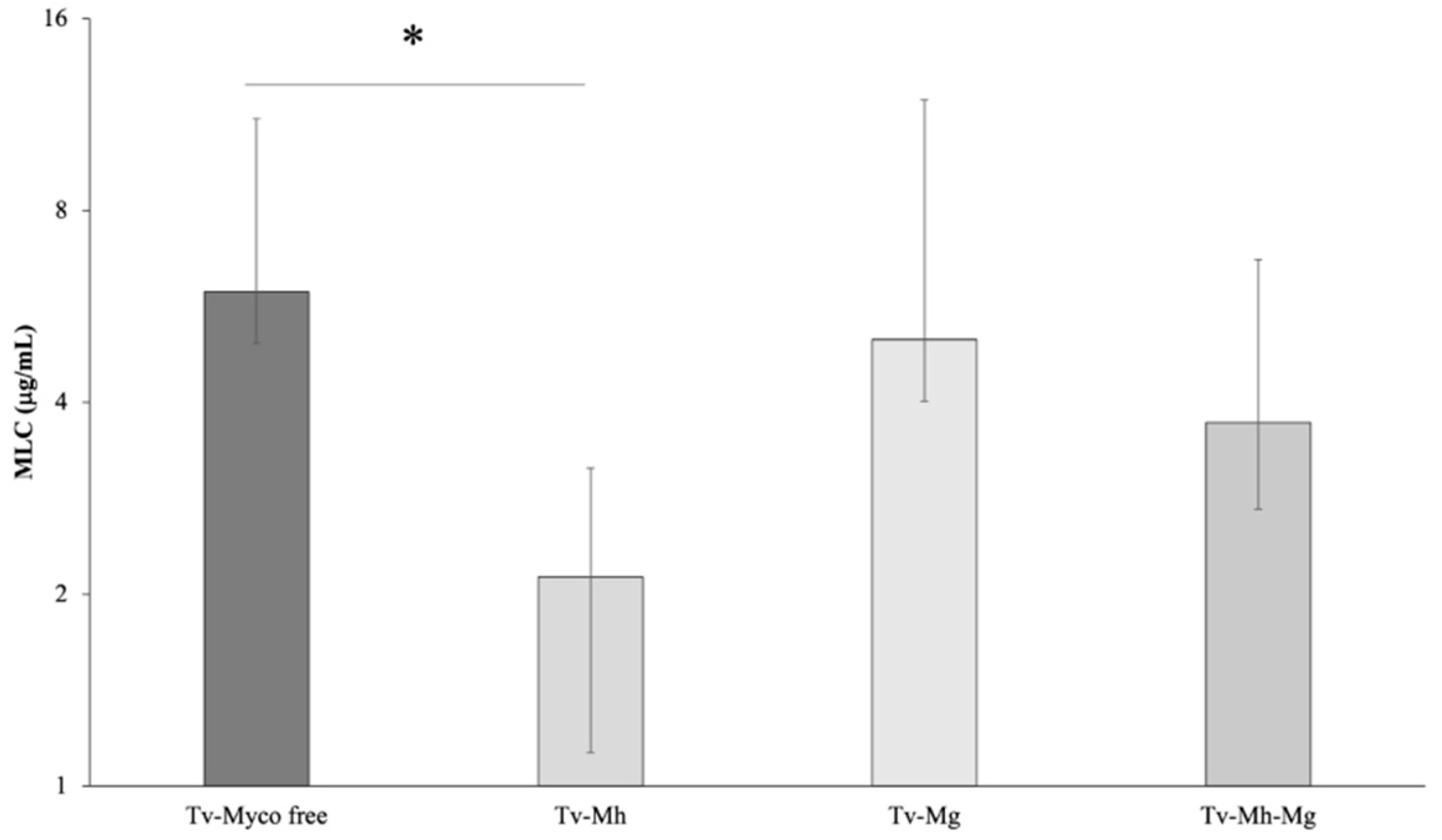
2.3. Association between Symbionts Presence and Metronidazole Sensitivity in T. vaginalis

The association between the presence of M. hominis and/or Ca. M. girerdii and the sensitivity to metronidazole in the 64 T. vaginalis isolates was evaluated (Figure 1). A statistically significant difference in the mean MLC values of M. hominis-infected T. vaginalis compared with non-infected strains was observed (p < 0.01), with the MLC mean value of Mh-free isolates two-fold higher than the MLC average of T. vaginalis strains harbouring M. hominis. On the contrary, the presence of Ca. M. girerdii seemed to not influence the sensitivity to MTZ of T. vaginalis strains (p = 0.55).
These results suggest a possible correlation between M. hominis symbiosis and metronidazole susceptibility in T. vaginalis strains. Interestingly, 4 out of 5 T. vaginalis strains showing a reduced sensitivity to metronidazole (MLC value ≥ 17 µg/mL) are totally Mycoplasma free.

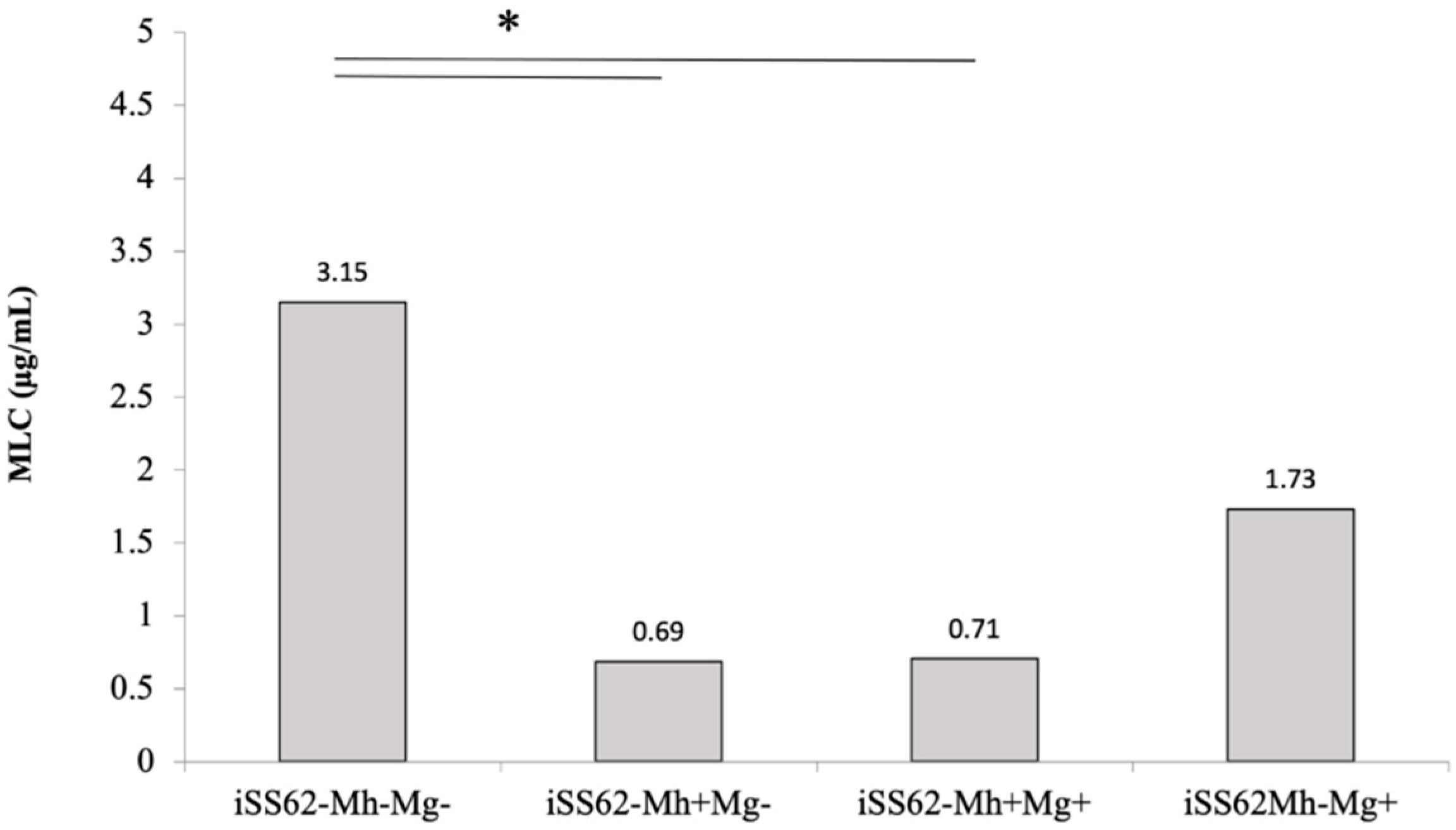
2.4. Sensitivity of Isogenic T. vaginalis Strains to Metronidazole in Aerobic Conditions

In order to eliminate any strain-to-strain variability among trichomonad isolates, we generated a set of isogenic T. vaginalis strains differing only in the presence/absence of one or both Mycoplasma species, as assessed by PCR: iSS62-Mh+Mg+, iSS62-Mh+Mg, iSS62-MhMg+and iSS62-MhMg. A metronidazole sensitivity assay was then performed on the four T. vaginalis isogenic strains. As shown in Figure 2, the presence of M. hominis in both iSS62-Mh+Mg+, iSS62-Mh+Mg caused a 4.5-fold reduction of MLC values compared with iSS62-MhMg (p < 0.05). On the contrary, although the iSS62-MhMg+ strain showed a slightly lower sensitivity to the drug compared with iSS62-MhMg (p = 0.1061), the presence of Ca. M. girerdii in T. vaginalis does not significantly influence the sensitivity to MTZ.

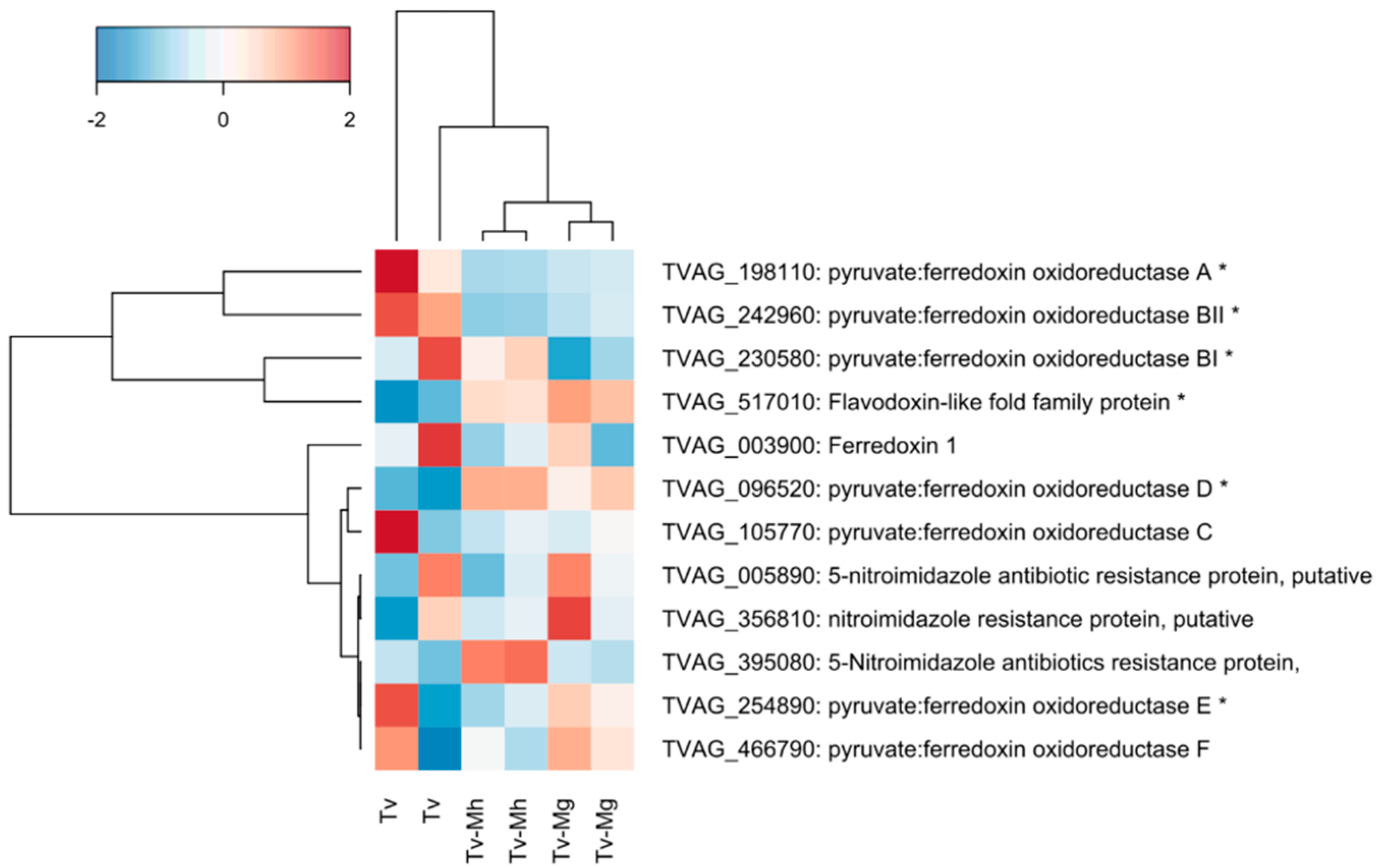
2.5. Expression of Genes Associated with Drug Resistance in T. vaginalis Isogenic Strains

Through gene ontology (GO) enrichment analysis, we investigated selected genes associated with metronidazole resistance in T. vaginalis (Supplementary Table S1) and observed a slightly significant decrease in the expression of two pyruvate: ferredoxin oxidoreductase (PFOR) genes; namely, PFOR A (TVAG_198110) and PFOR BII (TVAG_242960), in T. vaginalis associated with M. hominis (iSS62-Mh+Mg) compared with Mycoplasma-free T. vaginalis iSS62-MhMg. The average log2 fold change value for the two genes was, respectively, −1.61 (p-value 0.0002) and −1.39 (p-value 0.00001) (presence/absence of M. hominis). PFOR A and PFOR BII were also significantly downregulated in the presence of Ca. M. girerdii compared with the control (respective log2 fold changes were −1.26 and −1.02, with p-values of 0.001 and 0.0002). Intriguingly, T. vaginalis PFOR D (TVAG_096520) showed the opposite regulatory profile and was significantly upregulated during co-culture with Ca. M. girerdii compared with the control (log2 fold change 2.29; p-value 0.00001). The expression of flavin reductase 1 (TVAG_517010) was also upregulated in iSS62- Mh+Mg (log2 fold 0.86, p-value 0.0007) and iSS62- MhMg+ (log2 fold 1.02, p-value 0.0001). Finally, we did not detect any large differences in the expression of ferredoxin 1 (TVAG_003900, log2 fold −0.4, p-value 0.16) when T. vaginalis was infected by M. hominis, as previously reported [27], (Figure 3).

3. Discussion

Metronidazole and tinidazole are currently the only drugs officially used for the treatment of trichomoniasis, the most common non-viral sexually transmitted infection worldwide. A recent review reported that the global prevalence of T. vaginalis is resistant to MTZ ranges between 2.2−9.6% [11]. In recent years, several research groups investigated T. vaginalis MTZ resistance, but the underlying molecular mechanisms are not yet fully understood. Metronidazole enters the trichomonad cell in an inactive form through passive diffusion and needs to be reduced to its active nitro radical anion form in order to damage T. vaginalis. MTZ enters the hydrogenosome and competes with hydrogenase for electrons from ferredoxin, turning into the reduced active drug. Under aerobic conditions, oxygen converts MTZ back into its inactive form. Impaired oxygen scavenging and energy-production pathways have been linked to aerobic metronidazole resistance [9,30].
Interestingly, T. vaginalis is able to establish a stable symbiosis with two Mycoplasma species. The presence of the bacterium Mycoplasma hominis, an obligate human parasite colonizing the lower urogenital tract, has been demonstrated since 1998 in trichomonad isolates from different geographical areas, and, more recently, a new Mycoplasma species known as Ca. Mycoplasma girerdii, has been observed via metagenomic analysis almost exclusively in the vaginas of women infected by T. vaginalis. Several studies have demonstrated that the presence of the symbiont M. hominis within trichomonad cell has important consequences for the pathobiology of the protozoon [22], while the possible effect of the presence of Ca. Mycoplasma girerdii on trichomonad features has only recently been studied [31]. The possibility that the symbiosis could interfere with the resistance to MTZ of T. vaginalis isolates has only been investigated for M. hominis, leading to contradictory results [27,28,29,30].
In the present work, we investigated the possible correlation between the presence of the two Mycoplasma symbionts and the in vitro drug susceptibility in a large group of T.vaginalis isolated in Italy and Vietnam.
In our study 31.9% and 76.5% of T. vaginalis strains, respectively, isolated in Vietnam and Italy, were infected by M. hominis, confirming the high infection rate variability in different geographic areas. In fact, since the first observations of Rappelli and colleagues [15], several groups used PCR to demonstrate the presence of M. hominis in the trichomonad isolates of different geographic origins, with infection rates ranging from a minimum of 5% observed in Cuba [32] to over 89% detected in Italy [15]. We have also verified the presence of Ca. M. girerdii in our protozoan isolates, with and without M. hominis. Until now, the new uncultured Mycoplasma species had been mostly observed in the vaginal secretions of women affected by trichomoniasis through 16S rRNA microbial surveys and metagenomic analyses, but not in pure T. vaginalis cultures [24,25]. In fact, Ioannidis et al. [33] observed Ca. M. girerdii in 32 out of 100 cultured T. vaginalis strains from Greece, but the contemporary presence of both symbionts in a single T. vaginalis isolate is not described. We found that 19.8% of Vietnamese and 58.8% of Italian trichomonad strains were positive for Ca. M. girerdii. Interestingly, 13% of Vietnamese strains and 47% of Italian strains are infected by both Mycoplasma. Fettweis et al. observed, via metagenomic analysis, the contemporary presence of Ca. M. girerdii and M. hominis in 67% of the vaginal samples from African American women affected by acute trichomoniasis [25]. Our results confirmed for the first time the contemporary presence of both Mycoplasma species in a single trichomonad isolate. The percentage of T. vaginalis, totally free of Mycoplasma, greatly differs in the two groups, being 62% among the Vietnamese strains and only 12% among the Italian ones.
To establish if the presence of the two symbionts could influence the sensibility to metronidazole, T. vaginalis isolates were tested for in vitro metronidazole resistance in aerobic conditions. All 64 strains tested had a result of sensitivity to metronidazole, with 75% of isolates showing MLC values ≤ 4.3 µg/mL. Altogether, Vietnamese strains show a higher mean MLC value compared to the Italian ones (5.0 µg/mL versus 3.2 µg/mL). Moreover, among the five T. vaginalis strains showing a reduced sensitivity (MLC value 17.1 µg/mL), four were isolated in Vietnam. To our knowledge, this is the first report on the metronidazole susceptibility of T. vaginalis isolated in Vietnam.
In our study, the presence of the symbiont M. hominis is positively related to a higher sensitivity to MTZ in T. vaginalis: the mean MLC was 2.9 µg/mL in Mh-positive and 5.9 µg/mL in Mh-negative strains (p < 0.05). Moreover, we observed that 4 out of 5 isolates showing a reduced sensitivity to MTZ are Mycoplasma free. Our results contrast with those obtained by Xiao et al. in 2006, who reported an increased resistance in protozoa infected by M. hominis [26]. More recently, other groups reported the absence of an association between metronidazole susceptibility and the presence of M. hominis in T. vaginalis. Butler et al. tested 55 isolates from the USA, reporting a slight, insignificant increase of resistance in the group of Mh negative T. vaginalis [28], and da Luz Becker et al. analyzed 30 trichomonad isolated in Brazil, obtaining similar results [30]. Interestingly, considering only MTZ resistant strains, 20 out of 24 isolates from the USA and 3 out of 4 from Brazil were M. hominis free. Notably, in our study 4 out of 5 strains with reduced sensitivity to MTZ were Mycoplasma free. Moreover, in a previous study we observed that the only T. vaginalis strain resistant to metronidazole (MLC > 50 µg/mL) was M. hominis free [34].
We investigated, for the first time, a possible relationship between infection by Ca. M. girerdii and trichomonad sensitivity to MTZ. Unlike M. hominis, the presence of Ca. M. girerdii seems to not influence drug resistance: MLC mean values were the same in T. vaginalis positive and negative for Ca. M. girerdii (4.6 µg/mL).
In order to reduce the strain-to-strain variability characterizing T. vaginalis isolates, we artificially infected a naturally Mycoplasma-free T. vaginalis with one or both Mycoplasma species, obtaining four isogenic strains that were subjected to a metronidazole susceptibility assay. Results confirm data obtained with T. vaginalis clinical isolates: Mycoplasma-free T. vaginalis are more tolerant to MTZ than T. vaginalis associated with M. hominis or with both Mycoplasmas (p < 0.05). On the contrary, as observed with our clinical isolates, the presence of Ca. M. girerdii does not influence MTZ resistance, although strains artificially infected with Ca. M. girerdii showed a slightly reduced sensitivity to the drug compared with negative T. vaginalis (p = 0.1061). These findings suggest a correlation between M. hominis symbiosis and metronidazole susceptibility in T. vaginalis strains, supporting previous data where the presence of bacteria was associated with a lower MIC value of trichomonad cells compared with Mycoplasma-free isolates.
The metronidazole resistance in T. vaginalis strains has been associated to a decrease of the expression of hydrogenosomal proteins, including flavin reductase 1 (FR1), ferredoxin (Fdx), and pyruvate ferredoxin oxidoreductase (PFOR) [35].
In order to investigate if the presence of Mycoplasma species in trichomonad cells can modulate the expression of genes involved in MTZ resistance, we analyzed their mRNA levels in our isogenic strains by RNAseq analysis.
We observed a significant increase of flavin reductase1 expression in T. vaginalis infected with M. homins and with Ca. M. girerdii compared to the mycoplasma-free isogenic strain. In T. vaginalis, the enzyme plays a pivotal role in oxygen scavenging mechanisms, lowering intracellular oxygen concentrations and, consequently, reducing drug inactivation [10]. The increase of Fd1 expression can therefore explain, at least in part, the higher sensitivity to metronidazole observed in T. vaginalis infected by M. hominis in aerobic conditions.
A decrease in the expression of two out of seven pyruvate:ferredoxin oxidoreductase (PFOR) genes, namely PFOR A and PFOR BII, was observed in M. hominis and in Ca. M. girerdii positive T. vaginalis compared with Mycoplasma-free T. vaginalis. In our experimental model, the presence of M. hominis is associated with an increased susceptibility to MTZ, suggesting that a down-regulation of PFOR is not necessarily associated with drug resistance. Our results are in agreement with previous studies, showing the absence of association between the down-regulation of PFOR and MTZ resistance in T. vaginalis [27,30], supporting the hypothesis that PFOR RNA expression is not necessarily linked to a resistance to metronidazole. The slight down-regulation of PFOR could simply be due to the presence of Mycoplasmas, as suggested by Fürnkranz and colleagues [27].

4. Materials and Methods

4.1. Culture Conditions of Trichomonas vaginalis Isolates

A total of 64 T. vaginalis clinical isolates from women affected by acute trichomoniasis were analysed. Forty-seven isolates were collected by the Department of Parasitology of Hue University of Medicine and Pharmacy University and Reproductive Health Centre (Vietnam), and 17 isolates by the laboratory of Microbiology of University of Sassari (Italy). Trichomonad isolates were cultured in Diamond’s TYM medium supplemented with 10% FBS at 37 °C in a 5% CO2 atmosphere [36] for at least 15 days, and then preserved via freezing at −80 °C with FBS 10%, and adding 5% dimethylsulfoxide (DMSO) until use. Protozoa in an exponential growth phase, exhibiting a viability of >95%, were used in all experiments.

4.2. Screening for Mycoplasma Species in T. vaginalis Isolates

Genomic DNA was extracted from all 64 T. vaginalis clinical isolates with DNeasy Blood & Tissue Kit (Qiagen Ltd., West Sussex, UK) according to the manufacturer’s protocols. The presence of Mycoplasma species in association with each T. vaginalis strain was assessed by PCR using the following 16S rRNA specific primers: for M. hominis has been used RNA H1 (5-CAATGGCTAATGCCGGATACGC-3) and RNA H2 (5-GGTACCGTCAGTCTGCAAT-3) primers [37], while for Ca. M. girerdii Forward has been used OTU_M1 forward (5-CATTTCCTCTTAGTGCCGTTCG-3) and OTU_M1 reverse CGGAGGTAGCAATACCTTAGC-3) primers [25]. The PCR cycle conditions for both mycoplasma gene amplification were 30 cycles each at 95 °C for 30 s, 62 °C for 1 min (for M. hominis) or 58 °C for 1 min (for Ca. M. girerdii), 72 °C for 30 s and a final extension at 72 °C for 10 min. PCR products were analysed in 10 or 1 min, and 72 °C for 1 min. PCR products were analysed by 1% agarose gel and viewed under a UV transilluminator: the presence of specific bands of 344 bp and 310 bp confirmed the presence of M. hominis and Ca. M. girerdii, respectively.

4.3. Generation of Isogenic T. vaginalis Strains

Using isolate T. vaginalis SS62, we generated a set of isogenic strains differing only for the presence/absence of the two Mycoplasma species: iSS62-Mh+Mg+, iSS62-Mh+Mg, iSS62-MhMg+and iSS62-MhMg.
Being SS62 naturally Ca. M. girerdii-infected (named thereafter as iSS62-MhMg+), to generate iSS62-MhMg we eliminated the bacteria by cultivating trichomonad cells for 7 days in a medium supplemented with Plasmocin™ (Invivogen, San Diego, CA, USA) at a final concentration of 25 μg/mL [27]. Then, the cells were cultivated in complete Diamond’s TYM medium without Plasmocin™ for a further 15 days. At the end of the treatment, the absence of bacteria was confirmed by PCR.
To obtain isogenic T. vaginalis iSS62-Mh+Mg+ and iSS62-Mh+Mg, strains iSS62-MhMg+ and iSS62-MhMg were stably infected with M. hominis isolate MhMPM2 as previously described [38,39]. One ml of an overnight culture of M. hominis (strain MPM2), corresponding to approximately 109 colour-changing units (CCU), was added to a 10-mL mid-log phase culture of the M. hominis-free iSS62-MhMg+ and iSS62-MhMg for 5 days in a TYM complete medium. Parasites were then cultivated for a further 10 days with 1:16 daily passages in TYM complete medium. At the end of the treatment, 100 µL of each supernatant was plated on a solid BEA medium to verify the presence of viable M. hominis. The presence of single or double infections in T. vaginalis isogenic strains was confirmed by PCR.

4.4. Metronidazole Susceptibility Assay in Aerobic Conditions

The 64 T. vaginalis clinical isolates and the four isogenic T. vaginalis strains (iSS62-Mh+Mg+, iSS62-Mh+Mg, iSS62-MhMg+ and iSS62-MhMg), obtained as described above, were tested to assess their metronidazole susceptibility in aerobic conditions, according to the methods previously described by other authors [27,28,29]. Assays were performed in 96-well flat-bottomed microtiter plates and 1 × 10⁴ exponentially growing cells were seeded in each well with increasing levels of metronidazole ranging from 0.2 to 200 μg/mL. After 48 h of incubation in 5% CO2, the plates were microscopically observed and the minimum lethal concentration (MLC), defined as the lowest drug concentration at which no motile trichomonads were observed at the end of the incubation period, was detected.
All experiments were performed twice in triplicate for each isolated test. Untreated cultures of each trichomonad strain were used as controls. T. vaginalis with minimal lethal concentrations ≤25 μg/mL are considered MTZ-sensitive [28].

4.5. RNA Preparation

RNA was extracted from T. vaginalis isogenic strains. For each condition, two aliquots of 2 × 10⁶ exponentially growing cells have been harvested, washed and resuspended in 700 μL of RNAlater (ThermoFisher Scientific, Waltham, MA, USA), then stored at −80 °C until use.
Samples were then thawed on ice, diluted with 0.7 mL nuclease-free PBS, and pelleted by centrifugation at 6 k× g for 5 min at 4 °C. RNA was extracted from the resulting pellet using TRIzol (ThermoFisher Scientific, Waltham, MA, USA) according to the manufacturer’s instructions, with some modifications. Briefly, TRIzol and chloroform were used to lyse cells and solubilise cell components, then RNA was precipitated, washed, and resuspended in 30 µL nuclease-free water.
RNA concentration was assessed by Qubit RNA High Sensitivity kit (ThermoFisher Scientific, Waltham, MA, USA), according to the manufacturer’s instructions. UV absorbance at 230 nm, 260 nm, and 280 nm was measured using a nanodrop 2000 c spectrophotometer as an indicator of purity. RNA integrity and absence of genomic DNA contamination were confirmed using a TapeStation System (Agilent, Santa Clara, CA, USA) with the resulting gel images and electropherograms manually examined.

4.6. RNA Sequencing and Data Analysis

Library preparation and Illumina sequencing was performed by Novogene UK. A standard protocol was used to prepare libraries and the depletion of prokaryotic and eukaryotic ribosomal RNA (rRNA) was obtained via Ribo-Zero kit (Illumina). Approximately 50 million paired end reads per sample were generated using an Illumina NovaSeq 6000 platform. The read length was 150 bp and the insert size was from 250 bp to 300 bp. The sequences with low quality reads (reads with greater than 10% N (undetermined base) or over 50% of bases at or below 5 Phred quality score) were deleted. All the reads can be accessed via the BioProject entry PRJNA674783 at the NCBI [31].
The T. vaginalis G3 genome (Accession ASM282v1) [40] was used as the parasite reference sequence, and genes with an expression level of at least 1 transcript per million (TPM) were considered to be expressed.
The edgeR package [41] was used to test for differential gene expression using the negative binomial generalised linear model with a quasi-likelihood test, considering only genes with a log2 fold change of greater than 1.2 for testing.
The gene ontology (GO) enrichment analysis PANTHER [42] was used, a set of T. vaginalis genes potentially associated to metronidazole sensibility was used as a reference database, and uninformative and redundant enriched functions were removed manually. KEGG enrichment analysis was performed using edgeR [41]. For significance tests of differential gene expression and functionally enriched KEGG pathways and GO functions, p-values were adjusted using the false discovery rate/Benjamini–Hochberg (FDR/BH) method.

4.7. Statistical Analysis

A statistical analysis was performed using MS Excel for a chi-square test and for a Student’s t-test. A p < 0.05 was considered significant.

5. Conclusions

Our findings demonstrate the contemporary presence of both the symbionts Ca. Mycoplasma girerdii and M. hominis in T. vaginalis isolates and confirm the high differences in prevalence existing between different geographical areas. Moreover, our data strongly suggest that the presence of M. hominis is associated with a reduced resistance to metronidazole in T. vaginalis and affects gene expression. We also observed, for the first time, that, conversely, the symbiosis with Candidatus Mycoplasma girerdii seems to have no effect on metronidazole resistance in T. vaginalis. Further investigation of a larger group of isolates is needed to shed light on the factors underlying the capability of M. hominis to interfere with antimicrobial resistance in T. vaginalis.

Supplementary Materials

The following supporting information can be downloaded at: https://0-www-mdpi-com.brum.beds.ac.uk/article/10.3390/antibiotics11060812/s1, Table S1: resistance gene expression in T. vaginalis with and without Mycoplasma symbionts.

Author Contributions

Conceptualization, V.M., L.C.C. and P.R.; Funding acquisition, P.L.F. and P.R.; Investigation, V.M. and L.C.C.; Methodology, V.M., L.C.C., T.M.C.N. and N.D.; Software, N.P.B.; Supervision, P.A.T.N. and P.L.F.; Validation, T.H.T.N.; Visualization, D.D.; Writing—original draft, V.M. and P.R.; Writing—review & editing, R.P.H. and P.R. All authors have read and agreed to the published version of the manuscript.

Funding

This research was funded by FAR 2019 Rappelli, University of Sassari; FAR 2020 Fiori, University of Sassari; FAR 2020 Rappelli, University of Sassari. It also benefited from a PhD studentship from the Biotechnology and Bioscience Research Council Doctoral Training Partnership for Newcastle, Liverpool and Durham (N.P.B, R.P.H., grant number: BB/M011186/1).

Institutional Review Board Statement

Not applicable.

Informed Consent Statement

Not applicable.

Data Availability Statement

The data presented in this study are available in the article and in supplementary material.

Conflicts of Interest

The authors declare no conflict of interest.

References

  1. Newman, L.; Rowley, J.; Vander, H.S.; Wijesooriya, N.S.; Unemo, M.; Low, N.; Stevens, G.; Gottlieb, S.; Kiarie, J.; Temmerman, M. Global Estimates of the Prevalence and Incidence of Four Curable Sexually Transmitted Infections in 2012 Based on Systematic Review and Global Reporting. PLoS ONE 2015, 10, e0143304. [Google Scholar] [CrossRef] [Green Version]
  2. Petrin, D.; Delgaty, K.; Bhatt, R.; Garber, G. Clinical and Microbiological Aspects of Trichomonas vaginalis. 1998, 11, 300–317. Clin. Microbiol. Rev. 1998, 11, 300–317. [Google Scholar] [CrossRef] [Green Version]
  3. Hirt, R.P.; Sherrard, J. Trichomonas vaginalis Origins, Molecular Pathobiology and Clinical Considerations. Curr. Opin. Infect. Dis. 2015, 28, 72–79. [Google Scholar] [CrossRef] [PubMed]
  4. Stark, J.R.; Judson, G.; Alderete, J.F.; Mundodi, V.; Kucknoor, A.S.; Giovannucci, E.L.; Platz, E.A.; Sutcliffe, S.; Fall, K.; Kurth, T.; et al. Prospective Study of Trichomonas vaginalis Infection and Prostate Cancer Incidence and Mortality: Physicians’ Health Study. J. Natl. Cancer Inst. 2009, 101, 1406–1411. [Google Scholar] [CrossRef]
  5. Yang, M.; Li, L.; Jiang, C.; Qin, X.; Zhou, M.; Mao, X.; Xing, H. Co-Infection with Trichomonas vaginalis Increases the Risk of Cervical Intraepithelial Neoplasia Grade 2-3 among HPV16 Positive Female: A Large Population-Based Study. BMC Infect. Dis. 2020, 20, 642. [Google Scholar] [CrossRef] [PubMed]
  6. Leitsch, D. A Review on Metronidazole: An Old Warhorse in Antimicrobial Chemotherapy. Parasitology 2019, 146, 1167–1178. [Google Scholar] [CrossRef]
  7. Robinson, S.C. Trichomonal Vaginitis Resistant to Metranidazole. Can. Med. Assoc. J. 1962, 86, 665. [Google Scholar] [PubMed]
  8. Snipes, L.J.; Gamard, P.M.; Narcisi, E.M.; Beard, C.B.; Lehmann, T.; Secor, W.E. Molecular Epidemiology of Metronidazole Resistance in a Population of Trichomonas vaginalis Clinical Isolates. J. Clin. Microbiol. 2000, 38, 3004–3009. [Google Scholar] [CrossRef] [PubMed] [Green Version]
  9. Johnson, P.J. Metronidazole and Drug Resistance. Parasitol. Today 1993, 9, 183–186. [Google Scholar] [CrossRef]
  10. Leitsch, D.; Janssen, B.D.; Kolarich, D.; Johnson, P.J.; Duchêne, M. Trichomonas vaginalis Flavin Reductase 1 and Its Role in Metronidazole Resistance. Mol. Microbiol. 2014, 91, 198–208. [Google Scholar] [CrossRef] [Green Version]
  11. Graves, K.J.; Novak, J.; Secor, W.E.; Kissinger, P.J.; Schwebke, J.R. A Systematic Review of the Literature on Mechanisms of 5- Nitroimidazole Resistance in Trichomonas vaginalis Keonte. Parasitology 2020, 147, 1383–1391. [Google Scholar] [CrossRef] [PubMed]
  12. Seña, A.C.; Bachmann, L.H.; Hobbs, M.M. Persistent and Recurrent Trichomonas vaginalis Infections: Epidemiology, Treatment and Management Considerations. Expert Rev. Anti. Infect. Ther. 2014, 12, 673–685. [Google Scholar] [CrossRef] [PubMed]
  13. Kirkcaldy, R.D.; Augostini, P.; Asbel, L.E.; Bernstein, K.T.; Kerani, R.P.; Mettenbrink, C.J.; Pathela, P.; Schwebke, J.R.; Evan Secor, W.; Workowski, K.A.; et al. Trichomonas vaginalis Antimicrobial Drug Resistance in 6 US Cities, STD Surveillance Network, 2009–2010. Emerg. Infect. Dis. 2012, 18, 939–943. [Google Scholar] [CrossRef] [PubMed] [Green Version]
  14. Mabaso, N.; Abbai, N. Distribution of Genotypes in Relation to Metronidazole Susceptibility Patterns in Trichomonas vaginalis Isolated from South African Pregnant Women. Parasitol. Res. 2021, 120, 2233–2241. [Google Scholar] [CrossRef] [PubMed]
  15. Rappelli, P.; Addis, M.F.; Carta, F.; Fiori, P.L. Mycoplasma hominis Parasitism of Trichomonas vaginalis. Lancet 1998, 352, 1286. [Google Scholar] [CrossRef]
  16. Fichorova, R.N.; Fraga, J.; Rappelli, P.; Fiori, P.L. Trichomonas vaginalis Infection in Symbiosis with Trichomonasvirus and Mycoplasma. Res. Microbiol. 2017, 168, 882–891. [Google Scholar] [CrossRef]
  17. Taylor-Robinson, D. Mollicutes in Vaginal Microbiology: Mycoplasma hominis, Ureaplasma urealyticum, Ureaplasma parvum and Mycoplasma genitalium. Res. Microbiol. 2017, 168, 875–881. [Google Scholar] [CrossRef] [PubMed]
  18. Margarita, V.; Rappelli, P.; DessÌ, D.; Pintus, G.; Hirt, R.P.; Fiori, P.L. Symbiotic Association with Mycoplasma hominis Can Influence Growth Rate, ATP Production, Cytolysis and Inflammatory Response of Trichomonas vaginalis. Front. Microbiol. 2016, 7, 953. [Google Scholar] [CrossRef] [Green Version]
  19. Henriquez, F.L.; Mooney, R.; Bandel, T.; Giammarini, E.; Zeroual, M.; Fiori, P.L.; Margarita, V.; Rappelli, P.; Dessì, D. Paradigms of Protist/Bacteria Symbioses Affecting Human Health: Acanthamoeba Species and Trichomonas vaginalis. Front. Microbiol. 2021, 11, 3380. [Google Scholar] [CrossRef]
  20. Margarita, V.; Fiori, P.L.; Rappelli, P. Impact of Symbiosis Between Trichomonas vaginalis and Mycoplasma hominis on Vaginal Dysbiosis: A Mini Review. Front. Cell. Infect. Microbiol. 2020, 10, 179. [Google Scholar] [CrossRef]
  21. Fichorova, R.N. Impact of T. vaginalis Infection on Innate Immune Responses and Reproductive Outcome. J. Reprod. Immunol. 2009, 83, 185–189. [Google Scholar] [CrossRef] [PubMed] [Green Version]
  22. Dessì, D.; Margarita, V.; Cocco, A.R.; Marongiu, A.; Fiori, P.L.; Rappelli, P. Trichomonas vaginalis and Mycoplasma hominis: New Tales of Two Old Friends. Parasitology 2019, 146, 1150–1155. [Google Scholar] [CrossRef] [PubMed]
  23. Thu, T.T.T.; Margarita, V.; Cocco, A.R.; Marongiu, A.; Dessì, D.; Rappelli, P.; Fiori, P.L. Trichomonas vaginalis Transports Virulent Mycoplasma hominis and Transmits the Infection to Human Cells after Metronidazole Treatment: A Potential Role in Bacterial Invasion of Fetal Membranes and Amniotic Fluid. J. Pregnancy 2018, 2018, 5037181. [Google Scholar] [CrossRef] [Green Version]
  24. Martin, D.H.; Zozaya, M.; Lillis, R.A.; Myers, L.; Nsuami, M.J.; Ferris, M.J. Unique Vaginal Microbiota That Includes an Unknown Mycoplasma -Like Organism Is Associated With Trichomonas vaginalis Infection. J. Infect. Dis. 2013, 207, 1922–1931. [Google Scholar] [CrossRef]
  25. Fettweis, J.M.; Serrano, M.G.; Huang, B.; Brooks, J.P.; Glascock, A.L.; Sheth, N.U.; Strauss, J.F.; Jefferson, K.K.; Buck, G.A. An Emerging Mycoplasma Associated with Trichomoniasis, Vaginal Infection and Disease. PLoS ONE 2014, 9, e110943. [Google Scholar] [CrossRef] [PubMed] [Green Version]
  26. Xiao, J.C.; Xie, L.F.; Fang, S.L.; Gao, M.Y.; Zhu, Y.; Song, L.Y.; Zhong, H.M.; Lun, Z.R. Symbiosis of Mycoplasma hominis in Trichomonas vaginalis May Link Metronidazole Resistance in Vitro. Parasitol. Res. 2006, 100, 123–130. [Google Scholar] [CrossRef] [PubMed]
  27. Fürnkranz, U.; Henrich, B.; Walochnik, J. Mycoplasma hominis Impacts Gene Expression in Trichomonas vaginalis. Parasitol. Res. 2018, 117, 841–847. [Google Scholar] [CrossRef] [Green Version]
  28. Butler, S.E.; Augostini, P.; Secor, W.E. Mycoplasma hominis Infection of Trichomonas vaginalis Is Not Associated with Metronidazole-Resistant Trichomoniasis in Clinical Isolates from the United States. Parasitol. Res. 2010, 107, 1023–1027. [Google Scholar] [CrossRef]
  29. Mabaso, N.; Tinarwo, P.; Abbai, N. Lack of Association between Mycoplasma hominis and Trichomonas vaginalis Symbiosis in Relation to Metronidazole Resistance. Parasitol. Res. 2020, 119, 4197–4204. [Google Scholar] [CrossRef]
  30. da Luz Becker, D.; dos Santos, O.; Frasson, A.P.; de Vargas Rigo, G.; Macedo, A.J.; Tasca, T. High Rates of Double-Stranded RNA Viruses and Mycoplasma hominis in Trichomonas vaginalis Clinical Isolates in South Brazil. Infect. Genet. Evol. 2015, 34, 181–187. [Google Scholar] [CrossRef] [Green Version]
  31. Margarita, V.; Bailey, N.P.; Rappelli, P.; Diaz, N.; Dessì, D.; Fettweis, J.M.; Hirt, R.P.; Fiori, P.L. Two Different Species of Mycoplasma Endosymbionts Can Influence Trichomonas vaginalis Pathophysiology. mBio 2022, 24, e0091822. [Google Scholar] [CrossRef]
  32. Fraga, J.; Rodríguez, N.; Fernández, C.; Mondeja, B.; Sariego, I.; Fernández-Calienes, A.; Rojas, L. Mycoplasma hominis in Cuban Trichomonas vaginalis Isolates: Association with Parasite Genetic Polymorphism. Exp. Parasitol. 2012, 131, 393–398. [Google Scholar] [CrossRef] [PubMed]
  33. Ioannidis, A.; Papaioannou, P.; Magiorkinis, E.; Magana, M.; Ioannidou, V.; Tzanetou, K.; Burriel, A.R.; Tsironi, M.; Chatzipanagiotou, S. Detecting the Diversity of Mycoplasma and Ureaplasma Endosymbionts Hosted by Trichomonas vaginalis Isolates. Front. Microbiol. 2017, 8, 1188. [Google Scholar] [CrossRef]
  34. Margarita, V.; Marongiu, A.; Diaz, N.; Dessì, D.; Fiori, P.L.; Rappelli, P. Prevalence of Double-Stranded RNA Virus in Trichomonas vaginalis Isolated in Italy and Association with the Symbiont Mycoplasma hominis. Parasitol. Res. 2019, 118, 3565–3570. [Google Scholar] [CrossRef]
  35. Land, K.M.; Delgadillo-Correa, M.G.; Tachezy, J.; Vanacova, S.; Hsieh, C.L.; Sutak, R.; Johnson, P.J. Targeted Gene Replacement of a Ferredoxin Gene in Trichomonas vaginalis Does Not Lead to Metronidazole Resistance. Mol. Microbiol. 2004, 51, 115–122. [Google Scholar] [CrossRef]
  36. Dessì, D.; Delogu, G.; Emonte, E.; Catania, R.; Fiori, P.L.; Rappelli, P.; Dessı, D.; Catania, M.R. Long-Term Survival and Intracellular Replication of Mycoplasma hominis in Trichomonas vaginalis cells: Potential Role of the protozoan in transmetting bacterial infection. Infect. Immun. 2005, 73, 1180–1186. [Google Scholar] [CrossRef] [Green Version]
  37. Blanchard, A.; Yanez, A.; Dybvig, K.; Watson, H.L.; Griffiths, G.; Cassell, G.H. Evaluation of Intraspecies Genetic Variation within the 16S RRNA Gene of Mycoplasma hominis and Detection by Polymerase Chain Reaction. J. Clin. Microbiol. 1993, 31, 1358–1361. [Google Scholar] [CrossRef] [Green Version]
  38. Rappelli, P.; Carta, F.; Delogu, G.; Addis, M.F.; Dessì, D.; Cappuccinelli, P.; Fiori, P.L. Mycoplasma hominis and Trichomonas vaginalis Symbiosis: Multiplicity of Infection and Transmissibility of M. hominis to Human Cells. Arch. Microbiol. 2001, 175, 70–74. [Google Scholar] [CrossRef]
  39. Morada, M.; Manzur, M.; Lam, B.; Tan, C.; Tachezy, J.; Rappelli, P.; Dessì, D.; Fiori, P.L.; Yarlett, N. Arginine Metabolism in Trichomonas vaginalis Infected with Mycoplasma hominis. Microbiology 2010, 156, 3734–3743. [Google Scholar] [CrossRef] [Green Version]
  40. Carlton, J.M.; Hirt, R.P.; Silva, J.C.; Delcher, A.L.; Schatz, M.; Zhao, Q.; Wortman, J.R.; Bidwell, S.L.; Alsmark, U.C.M.; Besteiro, S.; et al. Draft Genome Sequence of the Sexually Transmitted Pathogen Trichomonas vaginalis. Science 2007, 315, 207–212. [Google Scholar] [CrossRef] [Green Version]
  41. Robinson, M.D.; Oshlack, A. A Scaling Normalization Method for Differential Expression Analysis of RNA-Seq Data. Genome Biol. 2010, 11, R25. [Google Scholar] [CrossRef] [PubMed] [Green Version]
  42. Thomas, P.D.; Campbell, M.J.; Kejariwal, A.; Mi, H.; Karlak, B.; Daverman, R.; Diemer, K.; Muruganujan, A.; Narechania, A. PANTHER: A Library of Protein Families and Subfamilies Indexed by Function. Genome Res. 2003, 13, 2129–2141. [Google Scholar] [CrossRef] [PubMed] [Green Version]
Figure 1. The impact of M. hominis and ‘Ca. M. girerdii’ presence on sensibility of metronidazole of T. vaginalis isolates. Statistical significance was tested via Student’s t-test. Tv = T. vaginalis, Mh = M. hominis, Mg = Ca. M. girerdii. M. hominis positive strains are associated with an increase of sensitivity to metronidazole compared to M.hominis free strains, independently from the presence of Mg (* p < 0.01).
Antibiotics 11 00812 g001
Figure 2. The metronidazole susceptibility of T. vaginalis isogenic strains. The mean values of metronidazole MLC for T. vaginalis experimentally (iSS62-Mh+Mg, iSS62-Mh+Mg+) and naturally Mycoplasma-infected (iSS62-MhMg+), and for Mycoplasma-free T. vaginalis (iSS62-MhMg), were compared. The presence of M. hominis in trichomonad cells is associated with an increase of sensitivity to metronidazole compared to iTv, independently from the presence of Mg (* p < 0.05). Statistical significance was tested by Student’s t-test.
Antibiotics 11 00812 g002
Figure 3. The differential expression of selected T. vaginalis genes related to metronidazole resistance alone and in association with M. hominis or with Ca. M. girerdii. Expression units are z-scaled trimmed mean of M-values (TMM). * Indicates genes with a statistically significant difference of expression between Mycoplasma-free T. vaginalis iSS62-MhMg (Tv) and T. vaginalis associated with M. hominis iSS62-Mh+Mg (Tv-Mh).
Antibiotics 11 00812 g003
Table 1. The occurrence of M. hominis and Ca. M. girerdii among T. vaginalis strains isolated in Vietnam and Italy.
Microbial AssociationT. vaginalis (Vietnam)
N (%)
T. vaginalis (Italy)
N (%)
T.vaginalis Mhneg Mgneg29 (62%) 2 (12%)
T.vaginalis Mhpos Mgneg9 (19%) 5 (29%)
T.vaginalis Mhneg Mgpos3 (6%) 2 (12%)
T.vaginalis Mhpos Mgpos6 (13%) 8 (47%)
Table 2. The MLC value of metronidazole among T. vaginalis strains isolated from Vietnam and Italy. In vitro antimicrobial susceptibility of 64 T. vaginalis isolated strains was evaluated, and minimal lethal concentration was calculated.
MLC (µg/mL)T. vaginalis Total
N (%)
T. vaginalis (Vietnam)
N (%)
T. vaginalis (Italy)
N (%)
≤4.3 µg/mL48 (75 %)34 (72%)14 (82%)
8.6 µg/mL11 (17%)9 (19%)2 (12%)
≥17.1 µg/mL5 (8%)4 (9%)1 (6%)
Publisher’s Note: MDPI stays neutral with regard to jurisdictional claims in published maps and institutional affiliations.

