Article

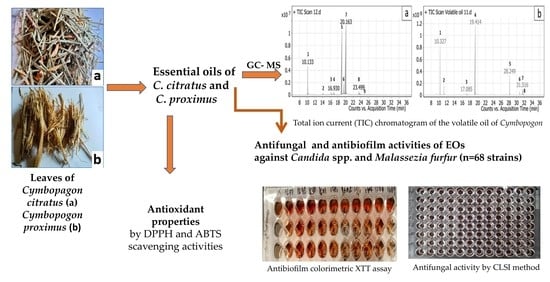
Antifungal, Antioxidant and Antibiofilm Activities of Essential Oils of Cymbopogon spp.

1
Dipartimento di Medicina Veterinaria, Università degli Studi di Bari, 70010 Valenzano, Italy
2
Medicinal and Aromatic Plants Research Department, Pharmaceutical and Drug Industries Institute, National Research Centre (NRC), Dokki, Giza 12622, Egypt
3
Department of Microbiology and Immunology, Veterinary Research Institute, National Research Centre (NRC), Dokki, Giza 12622, Egypt
4
Faculty of Veterinary Sciences, Bu-Ali Sina University, Hamedan 65175, Iran
*
Author to whom correspondence should be addressed.
Submission received: 15 May 2022 / Revised: 8 June 2022 / Accepted: 16 June 2022 / Published: 20 June 2022
(This article belongs to the Special Issue Antimicrobial Activities of Plant Extracts)

Abstract

:
Essential oils (EOs) of Cymbopogon citratus and Cymbopogon proximus are known as sources of monoterpenes and sesquiterpenoids, although their biological activities have not been well investigated. In this study, the compositions of C. citratus and C. proximus EOs of Egyptian origin and their antifungal and antibiofilm properties against Candida spp. and Malassezia furfur were investigated. Antioxidant activities were also evaluated. GC-MS showed the presence of nine and eight constituents in C. citratus and C. proximus EOs, respectively, with geranial and neral as the major compounds of C. citratus EO and piperitone and α-terpinolene as the major compounds of C. proximus EO. Both EOs showed antifungal (MIC values ranging from 1.25 to 20 µL/ mL) and antibiofilm activities (% of reduction ranging from 27.65 ± 11.7 to 96.39 ± 2.8) against all yeast species. The antifungal and antibiofilm activities of C. citratus EO were significantly higher than those observed for C. proximus EO. M. furfur was more susceptible to both EOs than Candida spp. Both EOs exhibited the highest antioxidant activity. This study suggests that C. citratus and C. proximus EOs might be an excellent source of antifungal, antibiofilm and antioxidant drugs and might be useful for preventing Malassezia infections in both medical and veterinary medicine.

Graphical Abstract

1. Introduction

Essential oils (EOs) are secondary metabolites and organic compounds with a low molecular weight produced by plants [1]. They play a role as regulators of cell metabolism under environmental stress and pathogenic attacks and are considered to be relevant therapeutic drugs for the treatment of animals and human infectious diseases [2,3]. In particular, EOs may represent sources of bioactive agents with a large spectrum of pharmacologic applications (i.e., antiphlogistic, spasmolytic, antinociceptive and antioxidant activities) [4]. In addition, their antimicrobial and antibiofilm activities stimulated the interest of the scientific community in proposing a solution for alarming multidrug resistance phenomena [5].
Several EOs displayed fungicidal and antibiofilm effects against different fungal pathogens, namely Aspergillus, Candida, Cryptococcus and Fusarium, which represent the major causes for human and animal diseases with high mortality, mainly in immunocompromised patients [6,7,8]. Recently, Malassezia yeasts have emerged as a threat to both human and veterinary medicine. These yeast species are known to cause skin disorders and fungemia in immunocompromised patients [9]. Despite attempts to control such yeast infections with topical and systemic antifungals, recurrence of clinical signs of skin infections, as well as treatment failure in preventing or treating Malassezia furfur fungemia, have been reported, most likely due to the occurrence of resistant phenomena. Interestingly, essential oils are proposed as promising candidates to control or to prevent Malassezia-associated skin diseases both in humans (i.e., atopic dermatitis, dermatitis, pityriasis versicolor and Malassezia folliculitis) and in animals [9,10,11,12].
In particular, biofilm formation is one of the mechanisms related to multidrug resistance phenomena associated with the highest lethality of infected patients [13]. Several antifungal agents (e.g., amphotericin B, fluconazole, flucytosine, itraconazole and ketoconazole) fail to treat infections caused by yeasts forming biofilm [13], thus raising scientific efforts for the selection of new molecules. EOs have proven to be more effective against fungal biofilm than conventional drugs due to their high content of monoterpenes and sesquiterpenes [14]. Monoterpenes and sesquiterpenes have the potential to affect membrane integrity and suppress genes related to biofilm formation [15]. Earlier studies have shown a large variability in the monoterpenes and sesquiterpenoids composition of Mediterranean medicinal plants EOs (e.g., Origanum vulgare L. (oregano), Salvia officinalis L. (sage) and Thymus vulgaris L. (thyme) (Lamiaceae)). Among Mediterranean plants, the genus Cymbopogon has been largely recommended for its high monoterpene and sesquiterpenoid content [16,17]. In particular, EOs of Cymbopogon citratus and Cymbopogon proximus, commonly named lemongrass and halfabar and largely diffused in Egypt, are used in traditional medicines as anti-diabetic, antihypertensive, antioxidant and anti-inflammatory drugs [18]. Although some investigations on the chemical and biological profiles of these species proved the presence of monoterpenes and sesquiterpenoids [19,20,21], there is a lack of scientific evidence regarding the chemical composition of Egyptian C. citratus and C. proximus EOs and their usefulness as antifungal, antibiofilm and antioxidant drugs. Thus, the present study was designed to characterize the composition of the EOs from C. citratus and C. proximux and to evaluate their antifungal, antibiofilm and antiradical properties.

2. Results

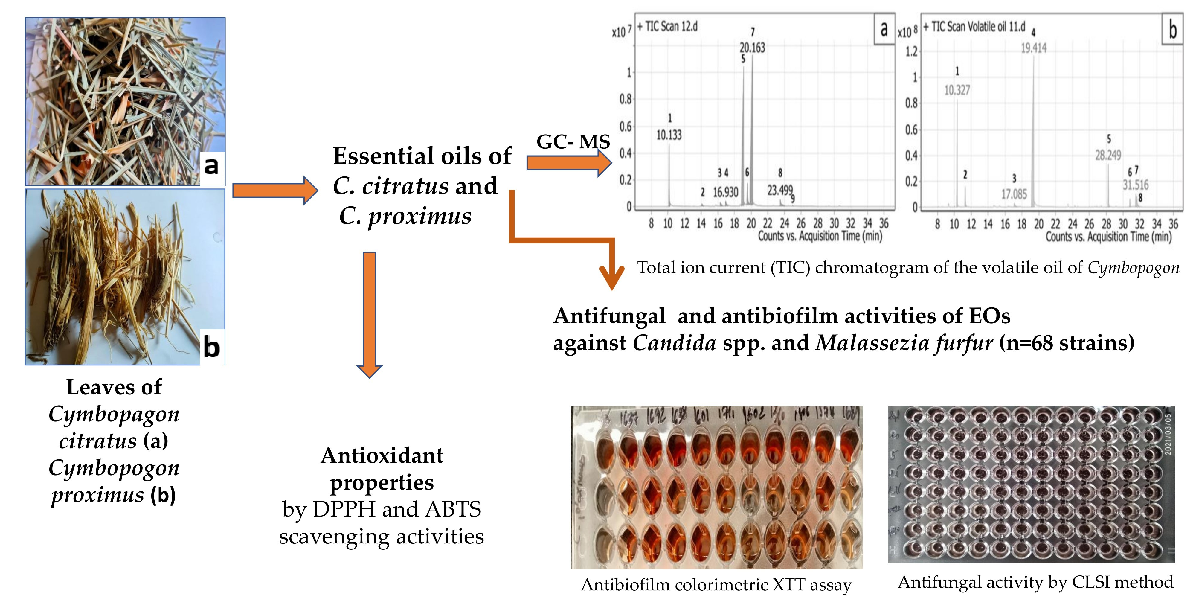
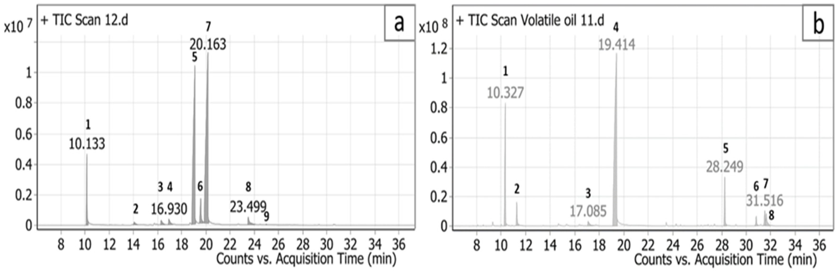
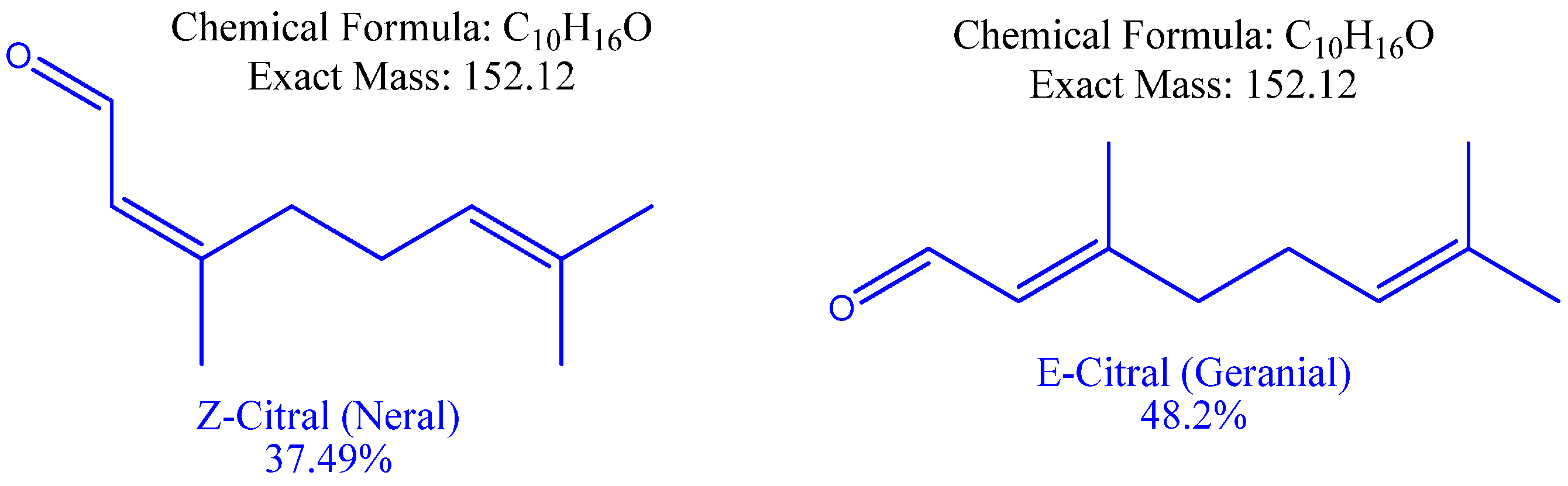
2.1. Chemical Composition of C. citratus and C. proximus Essential Oils

Extraction of C. citratus and C. proximus leaves by hydro-distillation produced EOs with yields of 3 and 3.75% (v/w), respectively. Their GC/MS profiles and chemical compositions (i.e., mass fragmentation and retention indexes) are presented in Figure 1 and Table 1 and Table 2.
Monoterpenes were the most abundant compounds of both EOs representing 87.0 and 99.9% of the total oil composition. A total of nine compounds representing the whole bulk of C. citratus EO were identified, with geranial (48.2%) and neral (37.49%) being the major compounds (Figure 2). Eight compounds, representing 100% of C. proximus EO, were identified, with piperitone (66.99%) and α-terpinolene (15.7%) being the major compounds.

2.2. Antifungal Activity

The antifungal activities of C. citratus and C. proximus EOs obtained by the broth microdilution method are reported in Table 3. The minimal inhibitory concentration (MIC) and the minimal fungicidal concentration (MFC) values vary according to the EO’s origin. The MIC and MFC values of C. citratus EO were lower (MIC values from 1.25 to 5 µL/mL) than those registered for C. proximus EO (MIC values from 2.5 to 20 µL/mL). The M. furfur strains were the most sensitive species to both EOs. Among Candida spp., C. catenulata and C. guilliermondii were less sensitive to C. citratus EO.

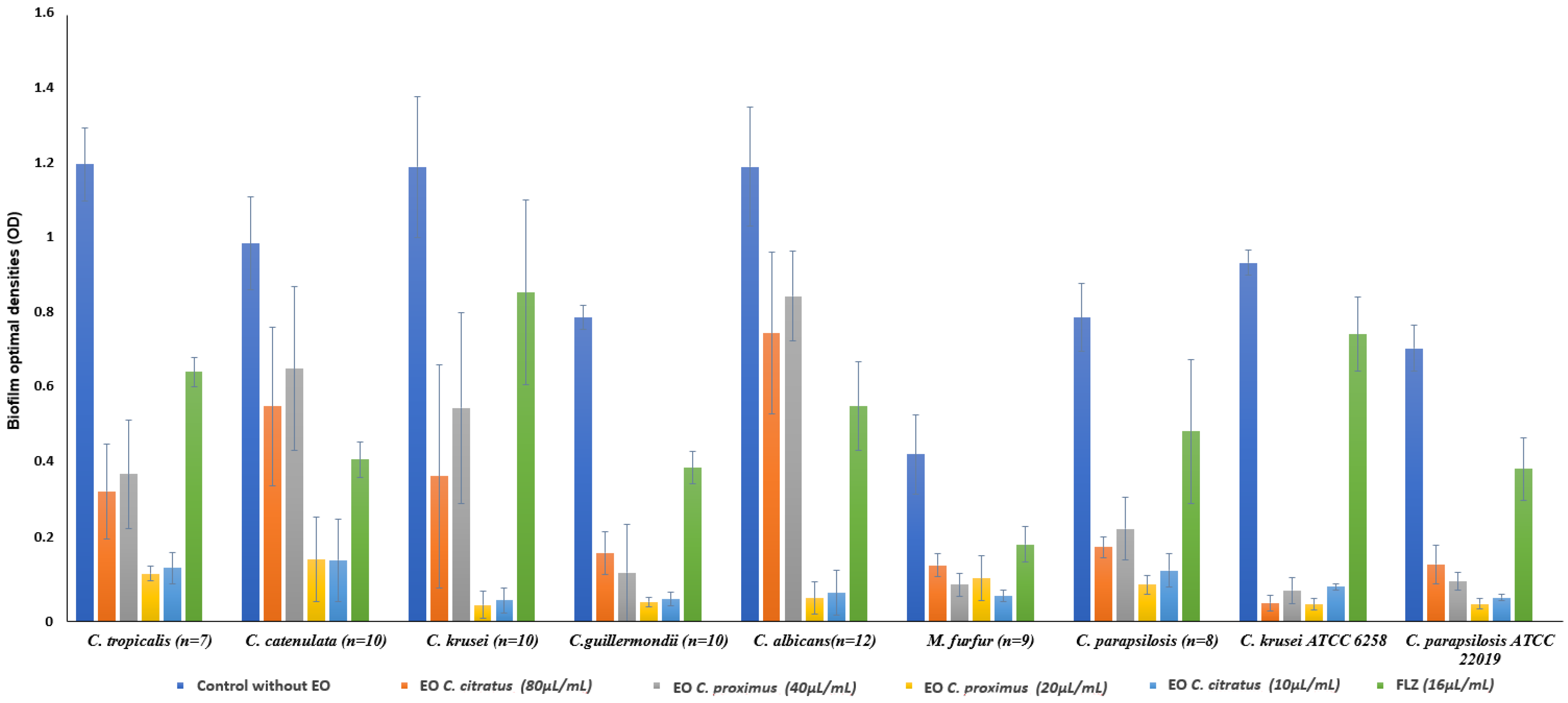
2.3. Inhibitory Effects of Cymbopogon citratus and Cymbopogon proximus EOs on Candida spp. and Malassezia furfur Biofilms

The XTT [2,3-bis(2-methoxy-4-nitro-5-sulfo-phenyl)-2H-tetra-zolium-5-carboxanilide] reduction assay showed that all tested yeasts were able to form biofilm within 24 to 48 h. Among the tested yeast species, C. tropicalis strains were the highest biofilm producers, whereas M. furfur strains were the lowest. All Candida spp. strains were higher biofilm producers (p < 0.05) than M. furfur strains. A significant decrease in biofilm formation compared to the control was observed in the tested yeast strains when grown in the presence of C. citratus or C. proximus EOs (Figure 3), showing an inhibition percentage ranging from 27.65 ± 11.7 to 96.39 ± 2.8%. The antibiofilm properties of both C. citratus and C. proximus EOs were significantly higher than those registered with FLZ (percentage of biofilm inhibition ranging from 19.68 ± 13.1% to 57.22 ± 5.3; Table 4).
The antibiofilm effects of both EOs were not related to their concentrations. The C. citratus EO exhibited significantly higher anti-biofilm activity than C. proximus EO (74.01 ± 11.5 to 96.39 ± 2.8% vs. 27.65 ± 11.7 to 96.39 ± 2%) against all tested yeast species except C. parapsilosis, M. furfur and C. krusei ATCC 6258 strains.

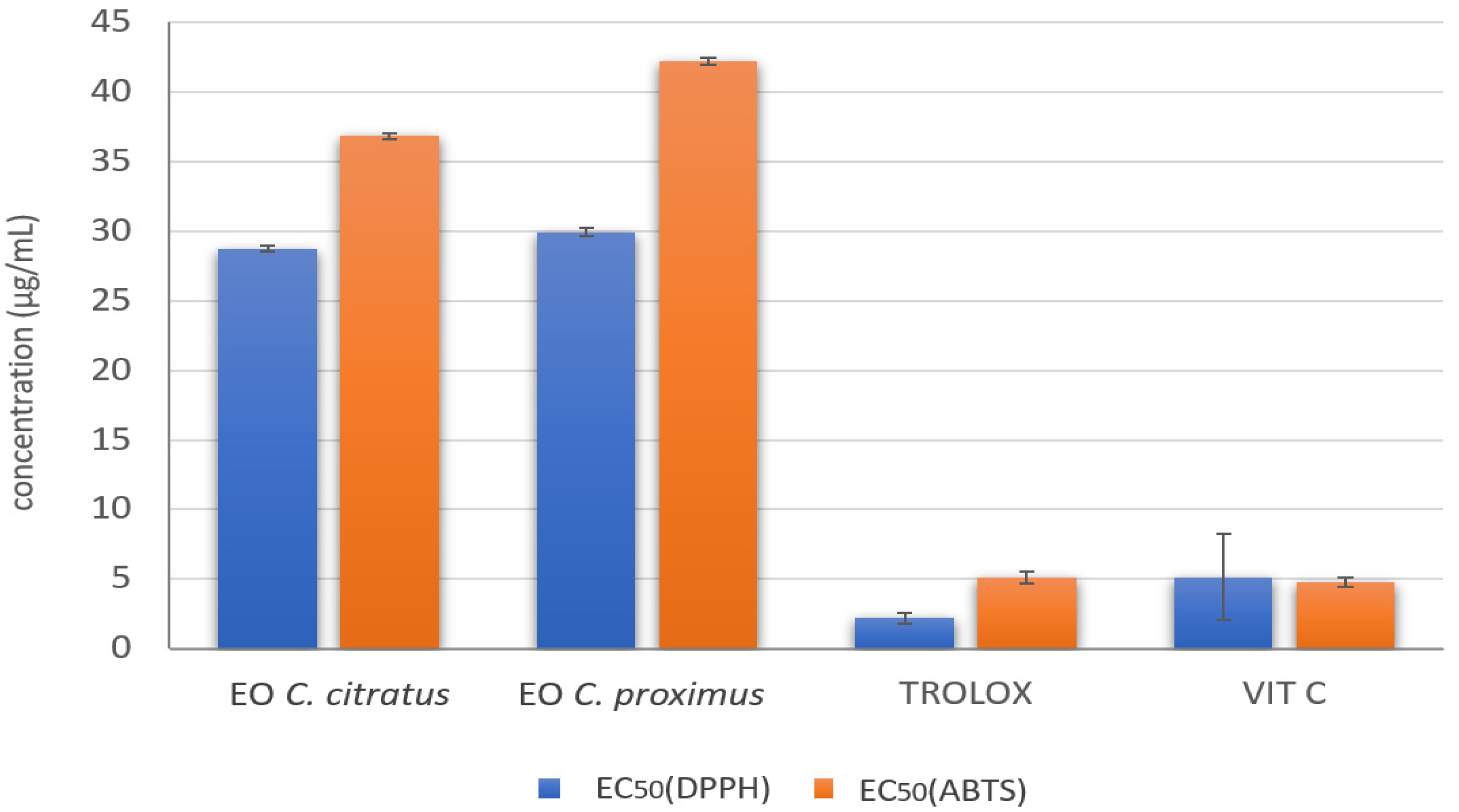
2.4. In Vitro Antioxidant Activity of C. citratus and C. proximus EOs

DPPH and ABTS radicals can accept an electron or hydrogen radical to become stable radicals. They lose absorption when accepting an electron or hydrogen radical [22], which results in a visually noticeable discoloration and indicates the ability of the EOs to act as free radical scavengers or hydrogen donors [22]. C. citratus and C. proximus EOs showed high radical scavenging abilities for DPPH and ABTS. The effective concentration at which 50% of the DPPH or ABTS radicals were scavenged (EC50) ranged from 28.73 to 42.18 µg/mL (Figure 4). No statistically significant differences were registered between C. citratus and C. proximus EOs in scavenging DPPH and ABTS. Trolox and vitamin C demonstrated higher scavenging activity for DPPH and ABTS than those registered for EOs.

3. Discussion

The results of this study confirm that C. citratus and C. proximus are sources of terpenes and demonstrate that these EOs may represent excellent sources of antifungal, antioxidant and antibiofilm drugs. Interestingly, this study revealed for the first time the antifungal and antibiofilm activities against new and emerging yeast pathogens such as C. catenulata, C. guilliermondii and M. furfur. In particular, the chemical profiles of the EOs reveal the usefulness of both plants as sources of terpenes, as previously suggested [14]. In addition, since the yield of the EOs varies according to the plants, the results of this study suggest that C. citratus represents the better source for these compounds, thus confirming previous studies in which the yield of EOs of C. citratus and C. proximus of different origins (Burkina Faso, México, Algeria, and Egypt) were evaluated [23,24,25,26].
Since the EO content and composition can be considerably affected by the geographical origin, in this study, the yield of EO of C. citratus was higher than those previously retrieved in the same plants of different origins with the geranial and neral as major compounds [19,23,24,26].
Both C. citratus and C. proximus EOs displayed growth inhibition activity against yeasts. This finding is in line with previous studies investigating different medicinal plants presenting a richness of terpenes (i.e., Origanum vulgare, Coriandrum sativum L., Juniperus communis L., Lavandula angustifolia Mill, Mentha arvensis L., Mentha pulegium L., Ocimum basilicum L.) [27].
Compared to C. proximus EO, the highest antifungal activity displayed by C. citratus EO could be related to its higher monoterpene content, including geraniol. Furthermore, the richness of geraniol in C. citratus EO might also cause destabilization of fungal cell membranes. In this sense, earlier studies have revealed the potent antifungal activity of geraniol at concentrations ranging from 30 to 130 µg/mL against Candida spp. due to its ability to disrupt cell membrane integrity by interfering with ergosterol biosynthesis and inhibiting the very crucial PM-ATPase [28,29]. Moreover, the high MIC of C. proximus EO herein observed is in accordance with the moderate activity of piperitone against Candida spp. [30]. These results confirm the studies previously performed on some yeast species using the same plants of different origins. Particularly, the results of this study confirm previous findings about the inhibitory effect of C. citratus EOs from France and Brazil against some clinical Candida spp. (i.e., C. albicans, C. krusei, C. tropicalis, C. glabrata, C. parapsilosis and C. tropicalis) [31] and extend the broad spectrum of antifungal activities to other rare opportunistic fungal pathogens, such as C. catenulata, C. guilliermondii and M. furfur. However, the MIC values herein registered for C. citratus EO against Candida spp. were slightly higher than those registered for C. citratus EO from Asia and lower than those for C. citratus EO from Brazil [32,33], suggesting that the chemo-geographical variation in C. citratus EO might also affect its antifungal activities [34,35]. On the contrary, the antifungal activity of C. proximus EO in this study is in contrast with previous studies in which only antibacterial activities were detected [19] and might be due to the low dose of EOs previously employed (i.e., 0.25 to 1 μL/mL vs. 2.5 to 20 μL/mL in our study).
Interestingly, M. furfur strains seem more susceptible than Candida spp. to both EOs and this might be due to the lipid capsule composition of Malassezia spp. that might favor EO solubilization, thus affecting their efficacy [36]. These findings propose that these EOs could be considered an effective alternative approach for the treatment of M. furfur skin infections, which are usually characterized by recurrences.
In particular, new guidelines for the treatment of these infections in animals propose the use of EOs as prophylactic procedures to decrease the risk of recalcitrant Malassezia spp. infection [37,38]. In addition, since these yeast species are considered emerging threats for immunocompromised patients (i.e., preterm infants), accurate hygiene of medical operators’ hands and incubators was usually required to prevent fungemia [39]. However, the chemical substances used for hygiene have very low efficacy against these yeast species; thus, EOs might be considered sources of active drugs for preventing strategies of Malassezia spp. systemic infections [40].
At present, this study demonstrated for the first time that C. citratus and C. proximus EOs are effective agents against biofilm formation. Anti-biofilm activities were also previously demonstrated for other EOs, including citronella, cinnamon, cascarilla bark and helichrysum [41], but the number of compounds with anti-biofilm effects are still scant and new molecules are requested. The excellent ability of C. citratus and C. proximus EOs to interfere with the mature biofilm of yeasts might be due to the hydrophobic interactions of monoterpenes with attachment forces such as lifshitz-Van der Waals, Brownian, sedimentation and electrostatic interaction forces, which are useful for yeast attachment to different surface types [42].
Interestingly, in this study, the antibiofilm activity should also be related to the antioxidant activities of C. citratus and C. proximus EOs. Indeed, both EOs, at very low concentrations, showed radical activities scavenging DPPH and ABTS in vitro (50%) comparable to those of synthetic antioxidants (i.e., butylated hydroxytoluene -BHT), possibly due to the high content of monoterpenes activities [43]. In particular, monoterpenes are able to absorb or neutralize free radicals due to their phenolic structure and redox properties [44,45]. In fungal cells, monoterpenes might act as pro-oxidants by disturbing the healthy redox cycle that might lead to an accumulation of reactive oxygen species (ROS) (i.e., hydrogen peroxide, superoxide and hydroxyl radicals) [46]. Usually, a healthy redox cycle promotes microbial attachment, thus favoring biofilm formation [39]. Inversely, in the presence of pro-oxidant compounds, a high level of ROS might favor a reduction of the extracellular polymeric substance (EPS) production, thus affecting the homogeneous structure, yeast numbers, and community composition of biofilm [46,47,48]. Recently, a strong association between oxidative stress and biofilm formation of bacteria and some yeast species has been demonstrated (C. albicans, C. glabrata, C. krusei, and C. parapsilosis) [47,49]. In detail, in C. albicans cells, the polyphenols from plants (i.e., magnolol and honokiol) induce ROS accumulation, causing decreased expression levels of specific genes (i.e., Ras-like protein 1-RAS1, enhanced filamentous growth protein -EFG1, Ty-transcription activator-TEC1, and ATP pyrophosphate-lyase-CDC35) involved in adhesion, yeast hyphal transition and biofilm formation [50]. Similarly, compounds that could target oxidative stress regulators, including antioxidants, could potentially be exploited as novel strategies for biofilm control [46]. However, the significantly higher antibiofilm activity of C. citratus EO compared to C. proximus EO might be attributable to the occurrence of specific components, mainly geranial and neral or to their synergistic activity, thus suggesting that the antibiofilm activities of EO might be due to different factors acting synergistically and/or additionally. In particular, it has been shown that geraniol is involved in the deterioration of the mature biofilm by affecting chitin and β-glucan synthesis, which are the major fungal cell wall components [51]. In addition, geranial and neral might act in synergy by decreasing intracellular adenosine triphosphate (ATP), pH and cell membrane integrity [52].

4. Materials and Methods

4.1. Plant Material and Essential Oil Isolation

C. citratus and C. proximus were collected from Siwa Oasis, governorate Nubian and Aswan governorate, Egypt, respectively, during September 2020. The plant species were identified by Dr. Monier Abd El-Ghani, Department of Taxonomy, Faculty of Science, Cairo University. Leaves of C. citratus and C. proximus were washed, dried in the shade, crushed into small pieces and 100 g were subjected to hydro distillation for 4 h. EO extraction was repeated 4 times. C. citratus and C. proximus EOs were extracted by steam distillation using a Karlsruhe apparatus. The resulting EOs were dried over anhydrous sodium sulfate and stored at −20 °C until their use. The EO concentrations tested for antifungal and antibiofilm activities ranged from 0.015 to 80 µL/mL being lower than those causing acute toxicity phenomena causing acute toxicity phenomena [24,53,54].

4.2. Identification of the Chemical Composition of EOs by Gas Chromatography–Mass Spectrometry Analysis (GC-MS)

The GC-MS system (Agilent Technologies) was equipped with a gas chromatograph (7890B) and mass spectrometer detector (5977A) at the Central Laboratories Network, National Research Centre (NRC), Cairo, Egypt. EOs were diluted with hexane (1:19, v/v). The GC-MS was equipped with an HP-5MS column (30 m × 0.25 mm internal diameter and 0.25 μm film thickness). Analyses were carried out using helium as the carrier gas at a flow rate of 1.0 mL/min at a split 1:30, injection volume of 1 µL at the following temperature program: 40 °C for 1 min; rising at 4 °C /min to 150 °C and held for 6 min; rising at 4 °C/min to 210 °C and held for 1 min. The injector and detector were held at 280 °C and 220 °C, respectively. Mass spectra were obtained by electron ionization (EI) at 70 eV, using a spectral range of m/z 50–900 and a solvent delay of 5 min. The identification of different constituents was determined by comparing the spectrum fragmentation pattern with those stored in the Wiley and NIST Mass Spectral Library.

4.3. Antifungal Activities

4.3.1. Yeast Strains

A total of 68 strains isolated from the cloaca of domestic and wild animals or from the skin of hospitalized human patients with M. furfur fungemia were employed for antifungal testing (Table 5). The strains were identified biochemically and molecularly, as previously reported [55]. All strains were obtained from the fungal collection of the Department of Veterinary Medicine at the University of Bari Aldo Moro, Italy.

4.3.2. Antifungal Activity

The minimal inhibitory concentration (MIC) and the minimal fungicidal concentration (MFC) of EOs were determined by broth microdilution methods according to the CLSI protocol for Candida and the CLSI modified protocol for Malassezia, as previously reported [56,57]. Stock inoculum suspensions of Candida spp. and M. furfur were adjusted to an optical density of 0.5–2.4 McFarland, respectively, equivalent to 5 × 10⁶ colony forming units (CFU)/mL. Two serial dilutions of Candida spp. (1:10 v/v) and M. furfur (1:5 v/v) were performed in specific media (i.e., Roswell Park Memorial Institute-RPMI for Candida spp. and Sabouraud Dextrose broth—SAB + 1% Tween 80 for M. furfur). One hundred microliters of the final dilution were transferred into a 96-well microtiter plate. Serial 1:2 dilutions of EOs ranging from 0.015 to 20 µL/mL were added to the wells of a 96-well plate (100 μL/well). The MIC end point was defined as the lowest concentration that produced a prominent decrease in turbidity (100%) relative to that of the drug-free control.
The MFC was measured by taking 100 μL of cell suspension from each well after 48 h (for Candida spp.) or 72 h (for M. furfur) of incubation at 32 °C, and then they were centrifuged, washed three times with fresh medium and vortexed for 10s. The solution was cultured on a specific medium (SDA for Candida spp. and SDA + 1% Tween 80 for M. furfur) at 32 °C for 72 h. The MFC value was defined as the MIC values of drugs at which no visible growth was detected. The MIC and MFC values of fluconazole (FLZ) were also detected as positive controls.
The negative control was yeast in broth without any antifungal. The experiment was repeated in duplicate three times on different days. Data obtained were reported as MIC ranges and MIC90 which indicate EO or drug concentration that inhibits the growth of 90% of the isolates.

4.4. Inhibitory Effects of Cymbopogon citratus and Cymbopogon proximus EOs on Candida spp. and Malassezia furfur Biofilms

The biofilm reduction of Candida spp. and M. furfur by C. citratus and C. proximus EOs was evaluated according to a previously reported method [58]. Candida spp. and M. furfur biofilms were performed in microtiter plates by adding 100 µL of cell suspension (1 × 10⁶ cells/mL) suspended in RPMI 1640 medium (Candida spp.) and in SAB supplemented with 1% Tween 80 (M. furfur) and incubated at 37 °C for 24 h for Candida spp. and 48 h for M. furfur. The wells were then washed twice with sterile phosphate buffered saline (PBS) and 100 µL of RPMI or/and SAB tween 1% containing C. citratus (10 and 20 µL/mL) or C. proximus EOs (80 and 40 µL/mL), or FLZ (16 and 8 µg/mL) were added. A medium (100 µL) without EOs was used as a negative control for biofilm growth. Microtiter plates were incubated at 37 °C for an additional 24 h for Candida spp. and 48 h for M. furfur. Then, the medium was removed, and the wells were washed twice with sterile PBS (200 mL per well). Semi-quantification of the fungal cell viability in wells of microtiter plates was calculated using a colorimetric XTT [2,3-bis (2-methoxy-4-nitro-5-sulfo-phenyl)-2H-tetrazolium-5-carboxanilide reduction assay. XTT (Sigma-Aldrich, Milan, Italy) was prepared in a saturated solution at 0.5 g/L in PBS. The solution was filter sterilized with a filter with a pore size of 0.22 mm, aliquoted and stored at −80 °C. Prior to each assay, an aliquot of stock XTT was supplemented with menadione (10 mM stock to a final concentration of 1 µM; Sigma-Aldrich, Milan, Italy). 100 µL of XTT–menadione solution was added to each pre-washed biofilm and control well. The microtiter plates were incubated in the dark at 37 °C for 3 h. Following incubation, 80 µL of the resulting-colored supernatant was transferred to a new microtiter plate and the colorimetric change from XTT reduction was read in at 490 nm using a microtiter plate reader (Benchmark Microplate Reader; Bio-Rad, Hercules, CA, USA). The results were reported as a percentage of biofilm inhibition using the following formula: % inhibition = [(control OD490 nm − Test OD490 nm)/control OD490 nm] × 100.

4.5. In Vitro Antioxidant Activity of C. citratus and C. proximus EOs

The radical scavenging activity of the EOs against 2,2-Diphenyl-1-(2,4,6-trinitrophenyl) hydrazy (DPPH) was determined as previously reported [59]. Briefly, 1 mL of EO solution in methanol (Sigma-Aldrich, Milan, Italy) ranging from 0.5 to 70 μg/mL was combined with 2 mL of methanol DPPH solution (0.1 mM). The obtained samples were mixed vigorously and kept in the dark for 60 min. The absorbance was measured at 517 nm using a double beam UV-VIS spectrophotometer (Shimadzu UV-1601, Kyoto, Japan). Methanol was used as a negative control. Ascorbic acid and Trolox were used as positive controls.
The percentage inhibition of the DPPH radical was calculated according to the following formula: % Inhibition = [(control OD − sample OD)/control OD] × 100, where A is absorbance at 517 nm. The results were expressed as of EC50 (μg EO /mL), which is the concentration necessary to obtain a 50% reduction of DPPH radical.
The 2,2′-Azino-Bis-3-Ethylbenzothiazoline-6-Sulfonic Acid (ABTS) radical scavenging activity of EOs was determined as previously reported [60]. ABTS+ was generated by the reaction of a 7 mM aqueous solution of ABTS with 2.45 mM aqueous solution of K2S2O8 which was conducted in the dark at room temperature for 16 h before use. The ABTS+ solution was diluted with ethanol to an absorbance of 0.70 (±0.02) at 734 nm. About 0.15mL of different concentrations of EOs was mixed with 2.85 mL of ethanolic solution of ABTS+, and the absorbance was read at 734 nm using a spectrophotometer after 15 min. Ethanol was used as a negative control. Ascorbic acid and Trolox were used as positive controls. The ABTS+ inhibition radical was calculated according to the following formula: % inhibition = [(control OD − sample OD)/control OD] × 100, where A is the absorbance at 734 nm. The results were expressed in terms of EC50 (μg EO/mL), which is the concentration necessary for 50% reduction of ABTS+ radical. EC50 was calculated from the graph plotting the percentage of radical scavenging activity (DPPH or ABTS) against EO concentration (1.5, 2.5, 5, 10, 20, 30, 50 and 70 µg/mL).

4.6. Statistical Analysis

The statistical analyses were performed using the Statistical Package for the Social Sciences (SPSS) for Windows program (version 13.0, SPSS Inc., San Rafael, CA, USA). One-way analysis of variance (one-way ANOVA) with post-hoc Tukey HSD (Honestly Significant Difference) was used to compare the differences among the MIC, EC50, biofilm optical densities of different yeast species, and the percentage of biofilm inhibition of C. citratus and C. proximus EOs. Differences were considered statistically significant when p < 0.05.

5. Conclusions

Conclusively, this study suggests that C. citratus and C. proximus EOs could be considered an excellent source of pharmacology ingredients to treat aging-associated diseases caused by free radicals for their antioxidant activities and to treat or prevent fungal infections and in particular might be considered as a drug source for preventing long treating Malassezia spp. skin infections in both medical and veterinary medicine. This study confirms the potential benefits of the use of natural antioxidants as antibiofilm compounds. Further investigations on the mechanism of action of antioxidant agents in treating, preventing and eradicating fungal biofilm are required.

Author Contributions

Conceptualization, W.R. and C.C.; Data curation, C.C. and D.O.; Formal analysis, W.R.; Investigation, W.R., M.A.M. and A.A.K.Z.; Methodology, W.R.; Supervision, C.C.; Visualization, G.G., M.T. and D.O.; Writing—original draft, W.R.; Writing—review & editing, D.O. and C.C. All authors have read and agreed to the published version of the manuscript.

Funding

This research received no external funding.

Institutional Review Board Statement

Not applicable.

Informed Consent Statement

Not applicable.

Data Availability Statement

Data are contained within the article.

Conflicts of Interest

The authors declare no conflict of interest.

References

  1. Erb, M.; Kliebenstein, D.J. Plant Secondary Metabolites as Defenses, Regulators, and Primary Metabolites: The Blurred Functional Trichotomy. Plant Physiol. 2020, 184, 39–52. [Google Scholar] [CrossRef]
  2. Iqbal, Z.; Iqbal, M.S.; Hashem, A.; Abd Allah, E.F.; Ansari, M.I. Plant defense responses to biotic stress and its interplay with fluctuating dark/light conditions. Front. Plant Sci. 2021, 12, 297. [Google Scholar] [CrossRef] [PubMed]
  3. Abegaz, B.M.; Kinfe, H.H. Secondary metabolites, their structural diversity, bioactivity, and ecological functions: An overview. Fundam. Concepts. 2020, 1, 7–54. [Google Scholar] [CrossRef]
  4. Lang, G.; Buchbauer, G. A review on recent research results (2008–2010) on essential oils as antimicrobials and antifungals. A review. Flavour. Fragr. J. 2012, 27, 13–39. [Google Scholar] [CrossRef]
  5. Evangelista, A.G.; Corrêa, J.A.F.; Pinto, A.C.S.M.; Luciano, F.B. The impact of essential oils on antibiotic use in animal production regarding antimicrobial resistance–a review. Crit. Rev. Food. Sci. Nutr. 2021, 8, 1–17. [Google Scholar] [CrossRef]
  6. Cafarchia, C.; De Laurentis, N.; Milillo, M.A.; Losacco, V.; Puccini, V. Antifungal activity of essential oils from leaves and flowers of Inula viscosa (Asteraceae) by Apulian region. Parassitologia 2002, 44, 153–156. [Google Scholar] [PubMed]
  7. Peixoto, L.R.; Rosalen, P.L.; Ferreira, G.L.S.; Freires, I.A.; de Carvalho, F.G.; Castellano, L.R.; De Castro, R.D. Antifungal activity, mode of action and anti-biofilm effects of Laurus nobilis Linnaeus essential oil against Candida spp. Arch. Oral. Biol. 2017, 73, 179–185. [Google Scholar] [CrossRef]
  8. Abd Rashed, A.; Rathi, D.N.G.; Ahmad Nasir, N.A.H.; Abd Rahman, A.Z. Antifungal properties of essential oils and their compounds for application in skin fungal infections: Conventional and nonconventional approaches. Molecules 2021, 26, 1093. [Google Scholar] [CrossRef]
  9. Rhimi, W.; Theelen, B.; Boekhout, T.; Aneke, C.I.; Otranto, D.; Cafarchia, C. Conventional therapy and new antifungal drugs against Malassezia infections. Med. Mycol. 2021, 59, 215–234. [Google Scholar] [CrossRef]
  10. Bismarck, D.; Dusold, A.; Heusinger, A.; Müller, E. Antifungal in vitro Activity of Essential Oils against Clinical Isolates of Malassezia pachydermatis from Canine Ears: A Report from a Practice Laboratory. Complement. Med. Res. 2020, 27, 143–154. [Google Scholar] [CrossRef]
  11. Siva Sai, C.; Mathur, N. Inhibitory Potential of Essential Oils on Malassezia strains by Various Plants. Biol. Life Sci. Forum 2020, 4, 46. [Google Scholar] [CrossRef]
  12. Saunte, D.; Gaitanis, G.; Hay, R.J. Malassezia-Associated Skin Diseases, the Use of Diagnostics and Treatment. Front. Cell. Infect. Microbiol. 2020, 10, 112. [Google Scholar] [CrossRef] [PubMed]
  13. Eix, E.F.; Nett, J.E. How biofilm growth affects Candida-host interactions. Front Microbiol. 2020, 11, 1437. [Google Scholar] [CrossRef] [PubMed]
  14. Stephane, F.F.Y.; Juleshttps, B.K.J. Terpenoids as important bioactive constituents of essential oils. In Essential Oils-Bioactive Compounds, New Perspectives and Applications; IntechOpen: London, UK, 2020. [Google Scholar] [CrossRef]
  15. Nazzaro, F.; Fratianni, F.; Coppola, R.; Feo, V.D. Essential oils and antifungal activity. Pharmaceuticals 2017, 10, 86. [Google Scholar] [CrossRef] [PubMed] [Green Version]
  16. Avoseh, O.; Oyedeji, O.; Rungqu, P.; Nkeh-Chungag, B.; Oyedeji, A. Cymbopogon species; ethnopharmacology, phytochemistry and the pharmacological importance. Molecules 2015, 20, 7438. [Google Scholar] [CrossRef]
  17. Anggraeni, N.I.; Hidayat, I.W.; Rachman, S.D.; Ersanda. Bioactivity of essential oil from lemongrass (Cymbopogon citratus Stapf) as antioxidant agent. AIP Conf. Proc. 2018, 1927, 030007. [Google Scholar] [CrossRef]
  18. Al-Taweel, A.; Fawzy, G.; Perveen, S.; El Tahir, K. Gas chromatographic mass analysis and further pharmacological actions of Cymbopogon proximus essential oil. Drug. Res. 2013, 63, 484–488. [Google Scholar] [CrossRef]
  19. Selim, S.A. Chemical composition, antioxidant and antimicrobial activity of the essential oil and methanol extract of the Egyptian lemongrass Cymbopogon proximus Stapf. Grasas Aceites 2011, 62, 55–61. [Google Scholar] [CrossRef]
  20. Madi, Y.F.; Choucry, M.A.; Meselhy, M.R.; El-Kashoury, E.S.A. Essential oil of Cymbopogon citratus cultivated in Egypt: Seasonal variation in chemical composition and anticholinesterase activity. Nat. Prod. Res. 2020, 35, 4063–4067. [Google Scholar] [CrossRef]
  21. Mahmoud, E.A.M.; Al-Askalany, S.A.; Hanafy, E.A. Antioxidant, antibacterial and cytotoxic effect of Cymbopogon citratus, Mentha longifolia, and Artemisia absinthium essential oils. Egypt. J. Chem. 2022, 65, 287–296. [Google Scholar] [CrossRef]
  22. Shaikh, S.A.M.; Singh, B.G.; Barik, A.; Balaji, N.V.; Subbaraju, G.V.; Naik, D.B.; Pri-yadarsini, K.I. Unravelling the effect of β-diketo group modification on the antioxidant mech-anism of curcumin derivatives: A combined experimental and DFT approach. J. Mol. Struct. 2019, 1193, 166–176. [Google Scholar] [CrossRef]
  23. Menut, C.; Bessiere, J.M.; Samate, D.; Djibo, A.K.; Buchbauer, G.; Schopper, B. Aromatic plants of tropical west Africa. XI. chemical composition, antioxidant and antiradical properties of the essential oils of three Cymbopogon species from Burkina Faso. J. Essent. Oil Res. 2000, 12, 207–212. [Google Scholar] [CrossRef]
  24. El Tahir, K.E.H.; Abdel-Kader, M.S. Chemical and pharmacological study of Cymbopogon proximus volatile oil. Res. J. Med. Plant. 2008, 2, 53–60. [Google Scholar] [CrossRef] [Green Version]
  25. Boukhatem, M.N.; Ferhat, M.A.; Kameli, A.; Saidi, F.; Kebir, H.T. Lemon grass (Cymbopogon citratus) essential oil as a potent anti-inflammatory and antifungal drugs. Libyan. J. Med. 2014, 19, 25431. [Google Scholar] [CrossRef] [PubMed]
  26. Del Carmen Vázquez-Briones, M.; Hernández, L.R.; Guerrero-Beltrán, J.Á. Physicochemical and antioxidant properties of Cymbopogon citratus essential oil. J. Food Res. 2015, 4, 36. [Google Scholar] [CrossRef]
  27. Zabka, M.; Pavela, R.; Prokinova, E. Antifungal activity and chemical composition of twenty essential oils against significant indoor and outdoor toxigenic and aeroallergenic fungi. Chemosphere 2014, 112, 443–448. [Google Scholar] [CrossRef]
  28. Miron, D.; Battisti, F.; Silva, F.K.; Lana, A.D.; Pippi, B.; Casanova, B.; Simone, G.; Fuentefria, A.; Mayorga, P.; Schapoval, E.E. Antifungal activity and mechanism of action of monoterpenes against dermatophytes and yeasts. Rev. Bras. Farmacogn. 2014, 24, 660–667. [Google Scholar] [CrossRef]
  29. Sharma, Y.; Khan, L.A.; Manzoor, N. Anti-Candida activity of geraniol involves disruption of cell membrane integrity and function. J. Mycol. Med. 2016, 26, 244–254. [Google Scholar] [CrossRef]
  30. Iraji, A.; Yazdanpanah, S.; Alizadeh, F.; Mirza Mohammadi, S.; Ghasemi, Y.; Pakshir, K.; Yang, Y.; Zomorodian, K. Screening the antifungal activities of monoterpenes and their isomers against Candida species. J. Appl. Microbiol. 2020, 129, 1541–1551. [Google Scholar] [CrossRef]
  31. Pedroso, R.D.S.; Balbino, B.L.; Andrade, G.; Dias, M.C.P.S.; Alvarenga, T.A.; Pedroso, R.C.N.; Pimenta, L.P.; Lucarini, R.; Pauletti, P.M.; Januário, A.H.; et al. In vitro and in vivo anti-Candida spp. activity of plant-derived products. Plants 2019, 8, 494. [Google Scholar] [CrossRef] [Green Version]
  32. Silva, C.D.B.D.; Guterres, S.S.; Weisheimer, V.; Schapoval, E.E. Antifungal activity of the lemongrass oil and citral against Candida spp. Braz. J. Infect. Dis. 2008, 12, 63–66. [Google Scholar] [CrossRef] [PubMed] [Green Version]
  33. Taweechaisupapong, S.; Aieamsaard, J.; Chitropas, P.; Khunkitti, W. Inhibitory effect of lemongrass oil and ts major constituents on Candida biofilm and germ tube formation. S. Afr. J. Bot. 2012, 81, 95–102. [Google Scholar] [CrossRef] [Green Version]
  34. Viljoen, A.M.; Petkar, S.; Van Vuuren, S.F.; Figueiredo, A.C.; Pedro, L.G.; Barroso, J.G. The chemo-geographical variation in essential oil composition and the antimicrobial properties of “wild mint”–Mentha longifolia subsp. polyadena (Lamiaceae) in southern Africa. J. Essent. Oil. Res. 2006, 18, 60–65. [Google Scholar] [CrossRef]
  35. Jaradat, N.A.; Al Zabadi, H.; Rahhal, B.; Hussein, A.M.A.; Mahmoud, J.S.; Mansour, B.; Issa, A. The effect of inhalation of Citrus sinensis flowers and Mentha spicata leave essential oils on lung function and exercise performance: A quasi-experimental uncontrolled before and after study. J. Int. Soc. Sports. Nutr. 2016, 13, 36. [Google Scholar] [CrossRef] [PubMed] [Green Version]
  36. Chee, H.Y.; Lee, M.H. In vitro activity of celery essential oil against Malassezia furfur. Mycobiology 2009, 37, 67–68. [Google Scholar] [CrossRef] [Green Version]
  37. Ebani, V.V.; Mancianti, F. Use of Essential Oils in Veterinary Medicine to Combat Bacterial and Fungal Infections. Vet. Sci. 2020, 7, 193. [Google Scholar] [CrossRef]
  38. Guillot, J.; Bond, R. Malassezia yeasts in veterinary dermatology: An updated overview. Front. Cell. Infect. Microbiol. 2020, 10, 79. [Google Scholar] [CrossRef]
  39. Rhimi, W.; Theelen, B.; Boekhout, T.; Otranto, D.; Cafarchia, C. Malassezia spp. yeasts of emerging concern in fungemia. Front. Cell. Infect. Microbiol. 2020, 10, 370. [Google Scholar] [CrossRef]
  40. Peano, A.; Gallo, M.G. Management of Malassezia-related diseases in the dog. Parassitologia 2008, 50, 85–88. [Google Scholar] [PubMed]
  41. Almeida, L.D.F.; Paula, J.F.; Almeida, R.V.; Williams, D.W.; Hebling, J.; Cavalcanti, Y.W. Efficacy of citronella and cinnamon essential oils on Candida albicans biofilms. Acta Odontol. Scand. 2016, 74, 393–398. [Google Scholar] [CrossRef]
  42. Porfírio, E.M.; Melo, H.M.; Pereira, A.M.G.; Cavalcante, T.T.A.; Gomes, G.A.; Carvalho, M.G.D.; Júnior, F.E.A.C. In vitro antibacterial and antibiofilm activity of Lippia alba essential oil, citral, and carvone against Staphylococcus aureus. Sci. World J. 2017, 2017, 4962707. [Google Scholar] [CrossRef] [PubMed] [Green Version]
  43. Zieniuk, B.; Groborz, K.; Wołoszynowska, M.; Ratusz, K.; Białecka-Florjańczyk, E.; Fabiszewska, A. Enzymatic Synthesis of Lipophilic Esters of Phenolic Compounds, Evaluation of their Antioxidant Activity and Effect on the Oxidative Stability of Selected Oils. Biomolecules 2021, 11, 314. [Google Scholar] [CrossRef] [PubMed]
  44. Zielińska-Błajet, M.; Feder-Kubis, J. Monoterpenes and their derivatives—Recent development in biological and medical applications. Int. J. Mol. Sci. 2020, 21, 7078. [Google Scholar] [CrossRef] [PubMed]
  45. Hellali, N.; Mahammed, M.H.; Ramdane, F.; Talli, A. Antimicrobial and antioxidant activities of Cymbopogon schoenanthus (L.) spreng. essential oil, growing in Illizi-Algeria. J. Med. Plant Res. 2016, 10, 188–194. [Google Scholar] [CrossRef]
  46. Ong, K.S.; Mawang, C.I.; Daniel-Jambun, D.; Lim, Y.Y.; Lee, S.M. Current anti-biofilm strategies and potential of antioxidants in biofilm control. Expert. Rev. Anti. Infect. Ther. 2018, 16, 855–864. [Google Scholar] [CrossRef] [PubMed]
  47. Gambino, M.; Cappitelli, F. Mini-review: Biofilm responses to oxidative stress. Biofouling 2016, 32, 167–178. [Google Scholar] [CrossRef]
  48. Gabrani, R.; Sharma, G.; Dang, S.; Gupta, S. Interplay among bacterial resistance, biofilm formation and oxidative stress for nosocomial infections. In Free Radicals in Human Health and Disease; Springer: New Delhi, India, 2015; pp. 369–379. [Google Scholar] [CrossRef]
  49. Seneviratne, C.J.; Wang, Y.; Jin, L.; Abiko, Y.; Samaranayake, L.P. Candida albicans biofilm formation is associated with increased anti-oxidative capacities. Proteomics 2008, 8, 2936–2947. [Google Scholar] [CrossRef]
  50. Gupta, P.; Gupta, H.; Poluri, K.M. Geraniol eradicates Candida glabrata biofilm by targeting multiple cellular pathways. Appl. Microbiol. Biotechnol. 2021, 105, 5589–5605. [Google Scholar] [CrossRef]
  51. Sun, L.; Liao, K.; Wang, D. Effects of magnolol and honokiol on adhesion, yeast-hyphal transition, and formation of biofilm by Candida albicans. PLoS ONE 2015, 10, e0117695. [Google Scholar] [CrossRef] [Green Version]
  52. Qian, W.; Liu, M.; Fu, Y.; Wang, T.; Zhang, J.; Yang, M.; Sun, Z.; Li, X.; Li, Y. Antimicrobial and Antibiofilm Activities of Citral Against Carbapenem-Resistant Enterobacter cloacae. Foodborne Pathog. Dis. 2020, 17, 459–465. [Google Scholar] [CrossRef]
  53. Lalko, J.; Api, A.M. Investigation of the Dermal Sensitization Potential of Various Essential oils in the Local Lymph Node Assay. Food Chem. Toxicol. 2006, 44, 739–746. [Google Scholar] [CrossRef] [PubMed]
  54. Althurwi, H.N.; Abdel-Kader, M.S.; Alharthy, K.M.; Salkini, M.A.; Albaqami, F.F. Cymbopogon proximus essential oil protects rats against isoproterenol-induced cardiac hypertrophy and fibrosis. Molecules 2020, 25, 1786. [Google Scholar] [CrossRef] [PubMed] [Green Version]
  55. Rhimi, W.; Aneke, C.I.; Annoscia, G.; Camarda, A.; Mosca, A.; Cantacessi, C.; Ottranto, D.; Cafarchia, C. Virulence and in vitro antifungal susceptibility of Candida albicans and Candida catenulata from laying hens. Int. Microbiol. 2021, 24, 57–63. [Google Scholar] [CrossRef]
  56. Iatta, R.; Figueredo, L.A.; Montagna, M.T.; Otranto, D.; Cafarchia, C. In vitro antifungal susceptibility of Malassezia furfur from bloodstream infections. J. Med. Microbiol. 2014, 63, 1467–1473. [Google Scholar] [CrossRef] [PubMed] [Green Version]
  57. Rhimi, W.; Salem, I.B.; Iatta, R.; Chaabane, H.; Saidi, M.; Boulila, A.; Cafarchia, C. Dittrichia viscosa L. leaves lipid extract: An unexploited source of essential fatty acids and tocopherols with antifungal and anti-inflammatory properties. Ind. Crops Prod. 2018, 113, 196–201. [Google Scholar] [CrossRef]
  58. Pierce, C.G.; Uppuluri, P.; Tristan, A.R.; Wormley, F.L.; Mowat, E.; Ramage, G.; Lopez-Ribot, J.L. A simple and reproducible 96-well plate-based method for the formation of fungal biofilms and its application to antifungal susceptibility testing. Nat. Protoc. 2008, 3, 1494–1500. [Google Scholar] [CrossRef]
  59. Brand-Williams, W.; Cuvelier, M.E.; Berset, C.L.W.T. Use of a free radical method to evaluate antioxidant activity. LWT-Food Sci. Technol. 1995, 28, 25–30. [Google Scholar] [CrossRef]
  60. Rhimi, W.; Hlel, R.; Ben Salem, I.; Boulila, A.; Rejeb, A.; Saidi, M. Dittrichia viscosa L. ethanolic extract based ointment with antiradical, antioxidant, and healing wound activities. Biomed. Res. Int. 2019, 2019, 4081253. [Google Scholar] [CrossRef] [Green Version]
Figure 1. Total ion current (TIC) chromatogram of the volatile oil of Cymbopogon citratus (a) and Cymbopogon proximus (b) (The peak numbers are described in Table 1 and Table 2).
Antibiotics 11 00829 g001
Figure 2. Chemical structure of neral and geranial as the major constituents of Egyptian C. citratus EO.
Antibiotics 11 00829 g002
Figure 3. Absorbance of Candida spp. and M. furfur biofilm with and without C. citratus, C. proximus EOs and FLZ. C. citratus and C. proximus EOs were tested at different concentrations.
Antibiotics 11 00829 g003
Figure 4. Radical scavenging activity against 2,2-Diphenyl-1-(2,4,6-trinitrophenyl) hydrazy (DPPH) and 2, 2′-Azino-Bis-3-Ethylbenzothiazoline-6-Sulfonic Acid (ABTS) of the C. citratus and C. proximus EOs.
Antibiotics 11 00829 g004
Table 1. The main constituents of Cymbopogon citratus EO.
Peak No.RTNameFormulaClassificationMS (M/e)Area %
m/zNo ScansMain Significant FragmentsBase Peak
110.133β-MyrceneC10H16monoterpenes136.231869, 79, 93, 121935.82
214.046Linalyl acetateC12H20O2monoterpenes196.295255, 69, 79, 93, 107, 121, 136, 150930.58
316.289trans-VerbenolC10H16Omonoterpenoid alcohol152.235155, 67, 91, 109, 134911.01
416.93IsoneralC10H16Omonoterpenes152.235155, 67, 81, 91, 109, 119, 134, 15267, 811.26
519.082Z-Citral B (Neral)C10H16Omonoterpenes152.244569, 94, 1346937.49
619.568NerolC10H18Omonoterpenoid alcohol154.254069, 79, 93, 121, 154693.65
720.163E-Citral A (Geranial)C10H16Omonoterpenes152.235369, 84, 109, 1526948.2
823.499Grandlure IIC10H18Omonoterpenes154.257555, 69, 79, 93, 121, 136, 154691.91
924.993trans-α-BergamoteneC15H24bicyclic sesquiterpenoids204.352055, 69, 79, 93, 107, 119, 135, 161930.07
Total Identification99.99
Total monoterpenes99.92
Total sesquiterpenes0.07
Table 2. The main constituents of Cymbopogon proximus EO.
Peak No.RTNameFormulaClassificationMS (M/e)Area %
m/zNo ScansMain Significant FragmentsBase Peak
110.327α-TerpinoleneC10H16monoterpenes136.231893, 1209315.7
211.26cis-β-terpinyl acetateC12H20O2monoterpenes196.282268, 93932.91
317.085α-TerpineolC10H18Omonoterpenoid alcohol154.255459, 93931.44
419.414piperitoneC10H16Omonoterpenes152.235269, 82, 1098266.99
528.249β-ElemolC15H26Osesquiterpenes222.375259, 93, 161595.87
630.818SelinenolC15H26Osesquiterpenes222.372391, 133, 1891891.56
731.516β-EudesmolC15H26Osesquiterpenes222.372359, 91, 149, 204592.42
831.636γ-EudesmolC15H26Osesquiterpenes222.372359, 91, 149, 204593.11
Total Identification100
Total monoterpenes87.04
Total sesquiterpenes12.96
Table 3. Minimum inhibitory concentration (MIC) and minimum fungicidal concentration (MFC) of Cymbopogon citratus and Cymbopogon proximus EOs and fluconazole (FLZ) against Candida spp. and Malassezia furfur.
Yeast spp.MIC ValuesC. citratus EOC. proximus EOFLZ
MIC µL/mLMFC µL/mLMIC µL/mLMFC µL/mLMIC µL/mLMFC µL/mL
Candida tropicalis (n = 7)Range2.52.520<2044
MIC902.52.520<2044
Candida catenulate (n = 10)Range2.5–5520<2088
MIC905520<2088
Candida krusei (n = 10)Range2.52.520<20>32>32
MIC902.52.520<20>32>32
Candida guilliermondii (n = 10)Range2.5–5520<2088
MIC902.5520<2088
Candida albicans (n = 12)Range2.52.520<2044
MIC902.52.520<2044
Malassezia furfur (n = 9)Range1.252.52.52.5>32>32
MIC901.252.52.52.5>32>32
Candida parapsilosis (n = 8)Range2.52.520<2044
MIC902.52.520<2044
Candida parapsilosis ATCC 22019Range2.52.520<2044
MIC902.52.520<2044
Candida krusei ATCC 6258Range2.52.520<20>32>32
MIC902.52.520<20>32>32
Table 4. Percentage of Candida spp. and Malassezia furfur biofilm inhibition by C. proximus and C. citratus EOs at different concentrations.
C. tropcalis
(n = 7)
C. catenulata (n = 10)C. krusei
(n = 10)
C. guillermondii
(n = 10)
C. albicans
(n = 12)
M. furfur
(n = 9)
C. parapsilosis
(n = 8)
C. krusei ATCC 6258C. parapsilosis ATCC 22019
EO C. proximus
(80 µL/mL)
71.56 ± 22.5 a43.14 ± 17.8 a,d64.86 ± 22.7 a,d77.36 ± 7.4 a,d35.94 ± 19.0 a64.47 ± 11.9 a78.77 ± 1.9 a94.96 ± 1.9 a78.77 ± 8.8 a
EO C. proximus
(40 µL/mL)
67.82 ± 12.8 b,c33.14 ± 18.5 b,c54.73 ± 19.3 b,c83.82 ± 16.5 b,c27.65 ± 11.7 b77.36 ± 8.2 b85.44 ± 20.7 b91.32 ± 3.9 b85.44 ± 2.0 b
EO C. citratus
(20 µL/mL)
90.42 ± 3.2 a,c83.64 ± 10.6 a,b96.39 ± 2.8 a,b93.64 ± 1.5 a,b94.85 ± 3.6 a,b74.01 ± 11.5 c93.68 ± 26.6 c95.32 ± 1.4 c93.68 ± 1.4 c
EO C. citratus
(10 µL/mL)
86.99 ± 5.4 b83.86 ± 10.3 c,d95.50 ± 2.5 d,c92.73 ± 2.0 c,d93.79 ± 4.30 c,d84.21 ± 5.1 a91.32 ± 24.7 d90.34 ± 0.5 d91.32 ± 1.9 d
FLZ
(16 µL/mL)
45.48 ± 3.4 a,b57.22 ± 5.3 a,b27.70 ± 15.3 a,b49.16 ± 7.0 a,b52.97 ± 5.9 a,b51.99 ± 15.1 a,b,c44.42 ± 25.6 a,b,c,d19.68 ± 13.1 a,b,c,d44.42 ± 6.8 a,b,c,d
Statistically significant differences were reported with the same superscript letters within the column.
Table 5. Yeast strains used in this study.
Yeast SpeciesCollection CodeOrigins
Candida tropicalis (n = 7)CD1693, CD1694, CD 1695, CD1700, CD1701, CD1702, CD1703Lizards feces
Candida catenulata (n = 10)CD 1777, CD1778, CD1568, CD1569, CD1575, CD1577, CD1578, CD1579, CD1580, CD1581Lizards, Laying hens feces
Candida krusei (n = 10)CD 1631, CD 1635, CD1638, CD 1641, CD1642, CD 1645, CD 1650, CD 1651, CD 1659, CD 1661, CD1662Wild boars feces
Candida guilliermondii (n = 10)CD 1606, CD 1644, CD 1653, CD 1675, CD1676, CD 1733, CD1738, CD1740, CD 1741, CD1743Lizards and wild boars feces
Candida albicans (n = 12)CD1601, CD1613, CD1616, CD1618, CD1620, CD1637, CD 1721, CD1729, CD1730, CD1755, CD1757, CD1760Lizards and wild boar feces
Malassezia furfur (n = 9)CD 1008, CD1009, CD1029, CD1030 CD1042, CD1043, CD1058, CD1490, CD1492Human skin
Candida parapsilosis (n = 8)CD1679, CD1681, CD1682, CD1683, CD1684, CD1691, CD1735, CD1736Lizards and wild boar feces
Candida kruseiATCC 6258American Type Culture Collection
Candida parapsilosisATCC 22019American Type Culture Collection
Publisher’s Note: MDPI stays neutral with regard to jurisdictional claims in published maps and institutional affiliations.

